Page 47 

3 Thoracic Cage

  Page 48 

UPPER RIBS

image

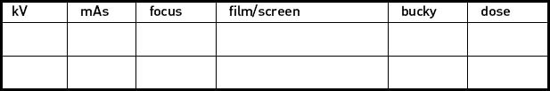
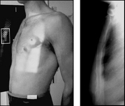
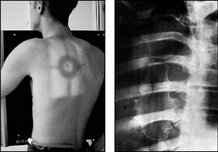
Projection: RIGHT OR LEFT POSTERIOR OBLIQUES

Centring Point: In the mid-clavicular line of the side under examination – at level of the midpoint of the sternal body. Horizontal central ray

image

Projection: LOWER RIBS – AP

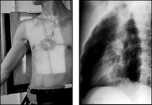
Centring Point: To a point in the midline midway between the xiphisternum and the lower costal margin

Points to consider

Technique

image Trauma – oblique ribs not always necessary – rarely changes management of the patient

image Oblique – exposure on arrested full inspiration

image Oblique – patient erect – better with inspiration

image AP – exposure on arrested expiration to show the maximum number of ribs below the diaphragm

image
  Page 49 

Radiological assessment

image Check each rib for # – it is rare to see displacement due to the numerous attached muscles

image Oblique – ribs away from cassette will be foreshortened

image Oblique – posterior rib articulations seen well on the raised side

image #s of ribs will unite spontaneously – treatment is limited

image Look for metastatic deposits – associated with rib destruction
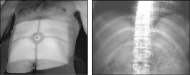
Right or left posterior obliques

Patient supine or erect, facing the X-ray tube

Body rotated approximately 45° onto the affected side

Arms are abducted away from the trunk

Collimation

To include: SUPERIORLY: First rib

INFERIORLY: Diaphragms

LATERALLY: Rib cage

Lower ribs – AP

Patient lies supine on the X-ray table

Shoulders and anterior superior iliac spines (ASIS) equidistant from the table top

Collimation

To include: SUPERIORLY: Diaphragms

INFERIORLY: Lower costal margin

LATERALLY: Rib cage and abdominal wall

  Page 50 

STERNUM

image

Projection: LATERAL

Centring Point: To a point 3 cm below the sternal angle. Horizontal central ray. SID 150 cm

image

Projection: ANTERIOR OBLIQUE (RAO)

Centring Point: To a point 8 cm lateral to the palpable fifth thoracic vertebra on the side furthest from the cassette. Horizontal central ray

Points to consider

Technique

image A grid or bucky is essential for both projections

image Lateral – exposure on arrested full inspiration

image Lateral – greater SID of 150 cm will decrease magnification

image Oblique – patient allowed to breathe gently during exposure

image Right anterior oblique – cardiac shadow will help visualise the sternum

image
  Page 51 

Radiological assessment

image Must include from the sternoclavicular joint to xiphisternum

image Sternum must not be overpenetrated

image Gentle breathing will blur the rib shadows

image Obliquity – the spine and sternum must not be superimposed

image Over-rotation – sternum outside cardiac shadow

Lateral

Patient erect

Median sagittal plane is parallel to the erect bucky

Patient’s shoulders are rotated posteriorly

Arms placed behind the trunk and shoulders and gently pulled back

Collimation

To include: SUPERIORLY: Acromioclavicular joints

INFERIORLY: Xiphisternum

ANTERIORLY: Anterior soft tissues

POSTERIORLY: Posterior sternum

Anterior oblique (RAO)

Patient erect facing the cassette

Trunk is rotated approximately 30° so that the right side of the body is in contact with the erect bucky

Arms are placed around the erect bucky to maintain stability

Collimation

To include: SUPERIORLY: Acromioclavicular joints

INFERIORLY: Xiphisternum

LATERALLY: Costal cartilage

  Page 52