Chapter 5 Urogenital system
This chapter considers the anatomical features and the physiological control of the urinary system with particular reference to water balance and sodium retention. The anatomy of the male and female reproductive systems is discussed and the mutual components of the urogenital system are identified.
The urinary and reproductive systems are often collectively known as the urogenital system as there are a number of shared components between the two.
THE URINARY SYSTEM
The urinary system consists of:
Kidneys
The kidneys filter blood. They lie against the dorsal wall of the abdomen attached by a fibrous covering called the capsule. The right kidney is situated about half its own length in front of the left kidney. The hilus is an indentation through which the renal artery, vein, nerves and ureters enter and leave the kidney. Each kidney has a pad of fat located next to it for protection and as an energy store.
The internal structure of the kidney comprises three distinct areas (Figure 5.1):
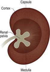
The kidney is surrounded by a dense fibrous connective tissue covering the capsule. The cortex contains the renal corpuscles and convoluted tubules of the nephron. The medulla contains the collecting ducts and pyramids located between them and the loops of Henle of the nephrons. The renal pelvis is a whitish colour and made of dense connective tissue. It is effectively a funnel through which urine collects and drains from the kidney into the ureter.
Functions of the kidneys
The kidneys produce urine from water, salts and wastes that they filter from the blood. By adjusting the amount of water and salts they excrete, the kidneys perform a vital homeostatic service in maintaining the internal chemical balance of the body (Table 5.1). Urine is excreted in a continuous trickle, collecting in the renal pelvis.
| pH | Cat/dog 5–7 |
| Slightly acidic due to carnivorous diet | |
| Horse 7.42–7.45 | |
| Slightly alkaline due to diet | |
| Specific gravity | A measure of density or concentration of urine |
| Cat 1.020–1.040 | |
| Dog 1.016–1.060 | |
| Horse 1.020–1.060 | |
| Protein | Should not appear in a healthy animal |
| Can be present if the urinary tract is damaged, or if there is increased permeability of glomerulus, chronic renal disease and cystitis | |
| Blood | Not normal |
| It can be due to physical damage, contaminant (prostate) or because an animal is in season (bitch) | |
| Bile | Can be due to liver disease or blockage of bile duct |
| Glucose | Present if the renal threshold is exceeded, e.g. in diabetes mellitus |
| Ketones | Produced when fats are oxidised (used for energy) |
| Can appear in diabetes mellitus and in malnutrition cases where fat is used by the body as a food source | |
| Deposits | Calculi, cells, casts of renal tubules may all be present |
Ureters
From the renal pelvis peristaltic contractions move the urine along the ureters. The ureters connect the kidneys to the urinary bladder; the length of ureters varies depending on the species. The ureters have thick walls of smooth muscle and are lined with mucous membrane and transitional epithelium to allow them to expand as the urine passes. They enter the bladder close to its neck on the dorsal aspect where there is a simple flap valve to prevent backflow of urine.
Bladder
The bladder is located in the lower, anterior portion of the pelvic cavity. It is made up of a double layer of smooth muscle and transitional epithelium (that allows stretch) that continues into the urethra. It is covered with peritoneum and its lining is invaginated (folded upon itself) which allows for great expansion. The trigone is the area between the ureteral openings and the neck of the bladder. As the volume of urine held increases, distension of the muscle wall stimulates nerve endings which in turn send impulses to the brain, producing a feeling of fullness. Impulses may then be sent back to stimulate urination. Urination is also known as micturition. Micturition is defined as the expulsion of urine from the bladder.
Urethra
The urethra leads from the bladder to outside the body in males and to the urethral orifice in females (which opens into the genital tract at the junction of the vagina and the vestibule).
The urethra has two sphincters close to where it leaves the bladder:
The urethra is shorter in females than in males. In females it only transports urine but in males it transports semen (sperm plus seminal fluid) and urine. The length of urethra in the male discourages bacterial infection due to its length whereas the female urethra is shorter, leading to a higher incidence of infection. Conversely the length and smaller diameter of the male urethra encourage blockages along its length and particularly at areas where the urethra curves, e.g. the ischial arch. Bladder control depends upon an animal’s learned ability to facilitate or inhibit the reflex action that causes micturition. In dogs and cats this ability is not inherent and requires time to learn.
Nephron
Each kidney contains millions of nephrons that regulate the composition of the blood and excrete waste. A nephron comprises a renal corpuscle and a renal tubule.
Renal corpuscle
Renal tubule
This consists of three main regions:
The proximal convoluted tubule is lined with simple cuboidal or columnar epithelium which has microvilli to increase the surface area for absorption. The loop of Henle is lined with simple squamous epithelium and the distal convoluted tubule is lined with cuboidal epithelium but contains no microvilli. Each distal convoluting tubule empties into a collecting duct, lined with columnar epithelium, with lots of distal tubules which empty into each collecting duct. The collecting ducts drain into the renal pelvis at the apices of the pyramids of the medulla. The calyces are defined as the indentations between the pyramids of the medulla.
Renal filtration and absorption
Figure 5.2 shows absorption in the renal nephron.
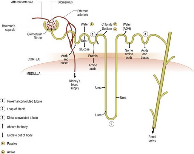
Figure 5.2 Absorption in the renal nephron. A, active; P, passive; 1, proximal convoluted tubule; 2, loop of Henle; 3, distal convoluted tubule.
Note: the renal blood supply is via the renal artery and vein which travel past the glomerulus to the kidney.
Reabsorption
Reabsorption is the passage of substances from the lumen of renal tubules into the renal capillaries to return them to the systemic circulation.
Remember – substances that are reabsorbed in the nephron are taken back into the body, usually via the blood, to be utilised; they are normally substances which are useful.
Water reabsorption
The distal convoluted tubule can be thought of like a tube containing holes that can change their diameter. Under normal circumstances the holes are small and water molecules cannot fit through them back into the blood. However when times are hard and water is scarce, ADH increases the diameter of the holes and they are now big enough for the water molecules to fit through, increasing the amount of water in the blood stream.
Sodium reabsorption
Osmoregulation is an important aspect of homeostasis and the control of water and sodium reabsorption in the body plays an important role in the regulation and balance of body water.
Species variation – urinary tract
The urinary system of the bird has a pair of kidneys, a pair of ureters but no bladder; they excrete nitrogenous waste in the form of uric acid and urates suspended in urinary water. The semisolid fluid leaves the ureters and enters the urodeum of the cloaca, then returns to the colon for more water reabsorption to occur before excretion. Reptile kidneys do not possess loops of Henle and therefore produce very dilute urine; snakes also do not have a bladder.
THE REPRODUCTIVE SYSTEM
Many generalisations can be made for sexually reproducing animals even though the details of the reproductive process vary with species.
The basic components of the male reproductive system are:
The basic components of the female reproductive system are:
Not all male and female animals possess all the modifications. Some simple animals have complex reproductive systems whereas some complex animals have simple reproductive systems. The reproductive systems of the male and female will be considered individually, taking the dog as an example of a mammal.
Male reproductive system
Figure 5.3 shows the male reproductive tract.
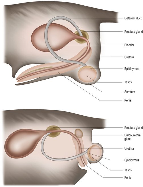
Figure 5.3 Male reproductive tract.
(Reproduced from Aspinall V 2006 The Complete Textbook of Veterinary Nursing. Butterworth Heinemann, London, with permission.)
Functions of the male reproductive system include:
Note: the urethra and penis are common to both the urinary and reproductive tracts.
Testes
These descend from the abdomen through the inguinal ring which then closes as the animal develops. They are developed from undifferentiated gonad tissue in the fetus. When the testes move through the inguinal ring the perineum lining that is pulled through with them develops into the scrotum.
The scrotum has two layers, an inner and an outer layer:
At the same time the blood vessels, nerves and ducts of the glands also descend through the inguinal ring and collectively form the spermatic cord. The testes may remain in the abdomen for various reasons:
Testes normally begin their descent around 8–10 weeks of age and should be palpable within the groin if not actually present in the scrotum. The testes remain relatively small until puberty (but this is dependent on breed and species). Running concurrent with the spermatic cord is a thin strip of muscle originating from the external oblique, called the cremaster muscle. The cremaster muscle retains the ability to move the testes closer to or away from the body to help control the temperature at which the spermatozoa are stored.
In small mammals the inguinal ring remains open and the cremaster muscle can withdraw the testes into the abdomen.
Temperature control of the testes is important as spermatozoa production and storage can be affected by fluctuations. Ideally the temperature of the testes should be lower than body temperature. The blood supply to the testes originates from the abdominal aorta along the spermatic cord to the associated epididymus where it becomes involuted and forms the pampiniform plexus. The pampiniform plexus allows the blood to cool and pulsations to smooth out before entering the gonad, preserving the lower temperature required for optimum sperm production and storage.
Surgical removal of testes is commonly known as castration or orchidectomy. Early castration can prevent the development of the secondary sexual characteristics:
The scrotum comprises the testis and epididymus within the tunica vaginalis.
Internal structure of the testis
The testis is made up of hundreds of convoluted tubules called seminiferous tubules. These are supported by connective tissue and cells. There are two types of cell that line the seminiferous tubules:
The cells located within the connective tissue matrix are called the cells of Leydig. Cells of Leydig are interstitial cells which produce testosterone. Testosterone is thought of as male hormone and is responsible for regulating spermatogenesis and for the development and maintenance of the secondary sexual characteristics. Testosterone has an inhibitory effect on the hypothalamus and subsequently the anterior pituitary gland from producing interstitial cell-stimulating hormone (ICSH) and luteinising hormone (LH) until puberty.
ICSH and LH are responsible for the development of the seminiferous tubules, where the sperm are produced, i.e. no sperm are produced until after puberty.
When puberty is achieved, testosterone no longer inhibits the hypothalamus and the production of ICSH and LH results in the development of the seminiferous tubules and increased testosterone production by the cells of Leydig, i.e. sperm are produced and male characteristics are formed.
Epididymus
One epididymus is located upon each of the testis. It may be palpable during examination of the testis. It consists of many coiled tubules where the spermatozoa are stored. Spermatozoa are transported to the epididymus via the vas efferentia (efferent ducts).
Vas deferens (deferent duct)
The vas deferens lie within the spermatic cord. They transport the sperm from the epididymus through the inguinal ring to the urethra. They empty into the urethra through the tissue of the prostate gland.
Structure of spermatozoa
Sperm comprise a head (acrosome and nucleus), a mid-piece(mitochondria) and a tail (flagella for movement).
Spermatogenesis is the process of sperm production. It is a complicated process with many stages. The age of the animal affects the number of spermatozoa produced, with fertility levels declining as animals age. The size of an animal may also play a role: large dogs produce more sperm than small dogs. High or low temperatures, radiation, drugs, toxins, vitamin A deficiency and limited diets can affect spermatozoa production. Abstinence reduces fertility due to blockage of tubules by old spermatozoa and it is normal practice for two coverings to occur to negate this issue.
Prostate gland
The prostate gland produces a large proportion of the seminal fluid – the fluid that nourishes the sperm and provides it with a transport medium. The ejaculate comprises spermatozoa and seminal fluid. The prostate is a bilobed structure that surrounds the urethra at the level of the pelvis brim. It can often be palpated and may cause problems in older uncastrated animals as it may develop neoplasia which results in an increase in size and excretory problems, as enlargement can cause obstruction to faeces.
Bulbourethral glands
These are only found in certain species, e.g. cat. Their function is to contribute a proportion of the seminal fluid and they are located further down the urethra than the prostate, near to the perineum.
Penis
The penis runs from the ischial arch through the perineum to its conclusion between the hind legs. It differs greatly in structure between species. The distal portion is protected by the prepuce. The prepuce is a sheath or covering constructed from abdominal skin tissue suspended from the ventral abdominal wall. It is lined with mucous epithelium for lubrication and its function is to protect the distal penis or glans penis. It is pushed back to expose the glans penis during coitus. Problems can occur if the prepuce is too small for the erect penis and surgical intervention may be necessary.
Structure of the penis
The penis comprises the following structures:
Two strips of erectile tissue or crura run along the penis from the root to almost the tip. This tissue is known as the corpus cavernosum and houses the urethra in the middle of the two layers. The corpus spongiosum runs beneath the corpus cavernosum and urethra and expands proximally to form the bulb (important to achieve the tie) and the tip or glans penis. At the glans penis the corpus spongiosum completely engulfs the urethra. The os penis is a small bone found within penile tissue in some species, e.g. the dog. It is v-shaped with the urethra running along a groove upon it.
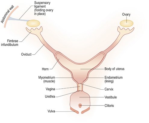
Female reproductive system
The female reproductive system (Figure 5.4) comprises the following:
Ovaries
The ovaries are located in the abdomen, one behind each kidney. They are held in place by the ovarian ligament which attaches the ovary to the kidney capsule. Their blood supply is provided by the ovarian artery developed from the aorta; it is very convoluted and runs alongside the ovarian ligament to the ovary itself. The ovaries lie within a fold of the mesentery called the mesovarium, which has an opening within it to allow the mature ova to depart. This opening is known as the ovarian bursa. When an animal is born it has a definitive number of undeveloped ova within each ovary. Depending upon species ova may be released individually or in a multitude.
A primary ovarian follicle develops into a mature or ripe ovarian follicle or graafian follicle under hormonal influence. The almost ripe follicle bulges out of the surface of the ovary until it is ready to pop and release the ovum (female gamete). The follicle consists of the ovum and a large quantity of fluid. The rupture of the follicle and release of the ovum occur at ovulation. The ovum is ejected and usually caught by the hair-like projections or fimbriae of the infundibulum at the head of the oviduct. The ruptured follicle then develops into the yellow body or corpus luteum (which has a role in pregnancy by producing progesterone).
Oviduct or fallopian tube
The oviduct is a narrow tube that transports the ovum from the ovary to the horn of the uterus. It consists of the infundibulum that encloses the ovary and the tube portion that runs to the uterus.
Uterus (womb)
Most animals are multiparous, i.e. they give birth to many young, including the cat and the dog. These animals have a uterus comprising two uterine horns. The uterine horns lead to the body of the uterus which in turn leads to the cervix or entrance of the uterus. The uterus is located within the broad ligament or mesometrium of the uterus and is dorsal to the abdomen. The exception to this location is when the uterus is enlarged during pregnancy, when the weight of the uterus brings it into a more ventral position.
The uterus consists of layers of smooth muscle, the myometrium and a mucous epithelial lining or endometrium. The cervix is located at the base of the uterus and is a muscular sphincter that closes the uterus except during mating or parturition. The round ligament runs from the ovary through the inguinal ring where it terminates in a pad of fat known as the vaginal process. The round ligament is the female equivalent of the cremaster muscle.
Vagina
The vagina extends from the cervix to the external urethral orifice. It is completely contained within the pelvis. At the end of the vagina the genital tract deviates sharply downwards and passes into the vestibule. The vestibule is utilised by both the urinary and reproductive systems. The muscles in the wall of the vestibule are very strong and help maintain the tie during mating in the dog.
Vulva
The vulva forms the boundary between the genital tract and the outside world. It takes the form of protective folds of skin or vulval lips. Contained within the vulval lips is the female equivalent to the penis, the clitoris. The clitoris is a piece of erectile tissue and lies in the clitoral fossa, a small cavity just inside the base of the vulva. The vulval lips are normally soft but during oestrus become enlarged and turgid.
Reproductive variations
Birds

Reptiles
Fish
Aspinall V. The complete textbook of veterinary nursing. London: Butterworth Heinemann; 2006.
Aspinall V, O’Reilly M. Introduction to veterinary anatomy and physiology. Oxford: Butterworth Heinemann; 2004.
Campbell N, Reece J, Mitchell L. Biology, 5th edn. San Francisco: Benjamin Cummings; 1999.
Colville T, Bassett J M. Clinical anatomy and physiology for veterinary technicians. St Louis: Mosby; 2002.
Dyke K M, Sack W O, Wensing C J G. Text book of veterinary anatomy, 2nd edn. Philadelphia: W B Saunders; 1996.
Green N P O, Stout G W, Taylor D J. Biological science. Cambridge: Cambridge University Press; 1991.
Lane D R, Cooper B, Turner L. BSAVA Textbook of Veterinary Nursing. Oxford: BSAVA; 2007.
Moore M. Manual of veterinary nursing. Oxford: BSAVA; 2000.
Pratt P W. Principles and practice of veterinary technology. Philadelphia: Mosby; 1998.
Roberts M B V. Biology: a functional approach. UK: Nelson, Surrey; 1986.
Simpson G. Practical veterinary nursing, 3rd edn. Oxford: BSAVA; 1994.