CHAPTER 89 Hyperproteinemia
The plasma protein fraction is composed mainly of albumin, globulins, and fibrinogen; fibrinogen is absent in serum as a result of clotting and conversion into fibrin. In some breeds, such as the Greyhound, serum protein concentrations are below the reference ranges for most laboratories (Fayos et al., 2005). Hyperproteinemia is the term given to an absolute or relative increase in the serum or plasma protein concentration. Before further evaluating a cat or dog with hyperproteinemia, the clinician should make sure that the condition is not attributable to a laboratory artifact (e.g., interference of other substances in protein determination), which constitutes one of the most common causes of “hyperproteinemia.” Lipemia and, to a lesser degree, hemolysis typically result in artifactual increases in the plasma or serum protein concentration.
Once true hyperproteinemia has been established, the clinician should determine whether it is relative or absolute. Relative hyperproteinemia is usually accompanied by erythrocytosis and caused by hemoconcentration (i.e., dehydration). However, in an anemic cat or dog, relative hyperproteinemia may be present in association with a normal packed cell volume (i.e., the volume is low but hemoconcentration results in an artifactual increase). The relative proportions (ratio) of albumin and globulin provide considerable information regarding the pathogenesis of hyperproteinemia. This information is usually contained in reports of serum biochemistry profiles from most referral diagnostic laboratories and in-house analyzers. Occasionally only the total serum protein and serum albumin concentrations are reported. In this event, the total globulin concentration can be determined by simply subtracting the albumin concentration from the total protein concentration.
In dogs and cats with relative hyperproteinemia (i.e., hemoconcentration), both the albumin and globulin concentrations are increased above the reference values, whereas in those with absolute hyperproteinemia only the globulin concentration is increased, usually in association with a mild or marked decrease in the albumin concentration. Hyperalbuminemia does not occur because the liver is already at its maximal synthetic capacity. The finding of “hyperalbuminemia” and hyperglobulinemia indicates either the presence of dehydration or a laboratory error. Rehydration results in resolution of relative hyperproteinemia.
When exposed to an electrical field (i.e., protein electrophoresis), the protein molecules migrate according to their shape, charge, and molecular weight. Staining of the electrophoresis gel after migration usually reveals six distinct protein bands: albumin (closer to the anode or negative electrode), α1-globulin, α2-globulin, β1-globulin, β2-globulin, and γ-globulin (closer to the cathode or positive electrode) (Fig. 89-1, A). The albumin fraction is responsible for conferring oncotic properties on body fluids. Acute phase reactants (APRs), also referred to as acute phase proteins, migrate in the α2 and α1 regions, whereas immunoglobulins (Igs) and complement usually migrate in the β and γ regions. Igs migrate in the following order (from anode to cathode and beginning in the α2 region): IgA, IgM, and IgG. By evaluating a protein electrophoretogram, the clinician can gain insight into the pathogenesis of the hyperglobulinemia.
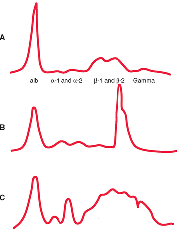
FIG 89-1 A, A normal canine or feline serum protein electrophoretogram. B, Electrophoretogram from a dog with multiple myeloma and a monoclonal gammopathy in the β2γ region. Note the narrow spike approximately the same width as the albumin band. C, Electrophoretogram from a cat with feline infectious peritonitis and a typical polyclonal gammopathy. Note the α2 spike (APRs) and the broad-based βγ
Increased production of globulins occurs in a variety of clinical situations, but mainly in two groups of disorders: inflammatory-infectious and neoplastic. In inflammation and infection the hepatocytes elaborate a variety of globulins, collectively termed APRs, which result in increases in the α2- and α1-globulin fractions. Because the hepatocytes are “reprogrammed” to produce APRs, the albumin production is “switched off,” resulting in hypoalbuminemia. In conjunction with these changes, the immune system produces a variety of immune proteins (mainly Igs), which result in increases in the α2, β or γ regions or a combination of these.
Because the immune system reacts against an organism (e.g., a bacterium) by producing antibodies against each somatic antigen, several clones of lymphocytes–plasma cells are “instructed” to simultaneously produce specific antibody molecules (i.e., each clone is programmed to produce one specific antibody type against a specific antigen). As a consequence, immune stimulation leads to the appearance of a polyclonal band in the β or γ regions or both. This polyclonal band is broad based and irregular and contains most of the Igs and complement generated by the immune cells. A typical inflammatory-infectious electrophoretogram therefore consists of a normal to mildly decreased albumin concentration and hyperglobulinemia resulting from increased α2- (i.e., APR) and βγ-globulins (polyclonal gammopathy) (Fig. 89-1, C).
Typical inflammatory-infectious electrophoretograms are seen in several common disorders, including chronic pyoderma, pyometra, and other chronic suppurative processes; feline infectious peritonitis; feline and canine hemobartonellosis and other hemoparasite infections; canine ehrlichiosis, anaplasmosis, and leishmaniasis; chronic autoimmune disorders (e.g., systemic lupus erythematosus, immune polyarthritis); and some neoplastic diseases, although they are rare (Box 89-1). Polyclonal gammopathies are also common in otherwise healthy old cats.
Monoclonal gammopathies occur when one clone of immune cells produces the same type and subtype of Ig molecule. Because these molecules are identical, they migrate in a narrow band (monoclonal spike, or M-component) located typically in the β or γ regions (Fig. 89-1, B). Monoclonal gammopathies occur in dogs with chronic multiple myeloma, lymphocytic leukemia, and lymphoma. They arealso present in dogs with ehrlichiosis and occasionally in dogs with leishmaniasis and other chronic inflammatory disorders (Box 89-2). In most cats monoclonal gammopathies occur in association with multiple myeloma or lymphoma, but they can occur in cats with feline infectious peritonitis. Occasionally an M-component is detected in an otherwise asymptomatic cat or dog but additional evaluation fails to reveal a source for the monoclonal gammopathy. Although this likely represents the counterpart of human idiopathic monoclonal gammopathy, the patient should be reevaluated frequently for a clinically emerging malignancy. In cats the source of the M-component is usually the spleen, where a neoplastic population of well-differentiated plasma cells is frequently identified in asymptomatic cats with a monoclonal gammopathy.
 BOX 89-2 Diseases Associated with Monoclonal Gammopathies in Dogs and Cats
The treatment of dogs and cats with monoclonal or polyclonal gammopathies is aimed at the primary disease. Refer to specific sections throughout this text for discussion of these treatments.
Breitschwerdt EB, et al. Monoclonal gammopathy associated with naturally occurring canine ehrlichiosis. J Vet Intern Med. 1987;1:2.
Burkhard MJ, et al. Monoclonal gammopathy in a dog with chronic pyoderma. J Vet Intern Med. 1995;9:357.
Dorfman M, et al. Paraproteinemias in small animal medicine. Compend Contin Educ. 1992;14:621.
Fayos M, et al. Serum protein electrophoresis in retired racing Greyhounds. Vet Clin Pathol. 2005;34:397.
Font A, et al. Monoclonal gammopathy in a dog with visceral leishmaniasis. J Vet Intern Med. 1994;8:233.
Forrester SD, et al. Serum hyperviscosity syndrome: its diagnosis and treatment. Vet Med. 1992;87:48.
Harrus S, et al. Serum protein alterations in canine ehrlichiosis. Vet Parasitol. 1996;66:241.
Patel RT, et al. Multiple myeloma in 16 cats: a retrospective study. Vet Clin Pathol. 2005;34:341.
Weiser MG, et al. Granular lymphocytosis and hyperproteinemia in dogs with chronic ehrlichiosis. J Am Anim Hosp Assoc. 1991;27:84.