Chapter 2 Principles and physics of ultrasound imaging
simple terminology definitions
Absorption: This is the major cause of attenuation. Absorption occurs when ultrasound energy is lost to tissues by its conversion to heat. Higher frequency waves undergo greater absorption.
Acoustic impedance: A property of all substances and is equal to the product of the tissue density and the speed of sound. Comparatively speaking, two substances with greater differences in acoustic impedance produce stronger ‘echoes’ or reflected waves than two similar substances. Structures of different acoustic impedance (for example, gallbladder and gallstone) are easier to distinguish from one another than two structures of similar acoustic impedance (for example, liver and kidney).
Acoustic power: The rate of flow of energy through the cross-sectional area of the beam.
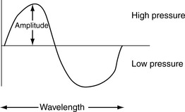
Acoustic waves: These are the vibrations that occur as a result of the rapid forward and reverse vibrations of the transducer, and which result in a number of longitudinal waves being transmitted. The transducer causes molecules in the medium through which it is passing to vibrate in a series of rhythmic, mechanical compressions (high-pressure regions) and rarefactions (low-pressure regions). These vibrations are commonly known as acoustic waves.
Acoustic window: An area of the patient that enhances ultrasound transmission and provides optimal scanning access to the area of interest. To improve image quality, ultrasound transmission should be as uniform as possible and areas which are likely to cause artifacts (such as ribs or bowel gas) should be avoided.
ALARA: An acronym for ‘as low as reasonably achievable’, referring to the principle of keeping power and exposure time to a minimum while acquiring the necessary clinical information.
Amplitude: The height of a wave. The amplitude and intensity of sound represent the energy associated with the sound wave. The greater the amplitude or intensity, the more the energy, and the ‘louder’ the sound. Increasing the acoustic power will increase both the intensity and the amplitude (see Fig. 2.1).
Anechoic: Areas on the image showing no internal echoes, appearing dark or black on the image.
Artifacts: In the context of ultrasound, artifacts are echo signals whose displayed position on the image does not correspond to the actual position of a reflector in the body, or whose displayed brightness is not indicative of the reflecting or scattering properties of the region from which the echo originated. Artifacts are a result of the following programmed machine assumptions:
Attenuation: The process that occurs as a sound wave travels through a medium; it loses energy, and as a result, its intensity and amplitude decrease, and it becomes attenuated. Attenuation is proportional to the frequency of the sound wave and the distance that the wave travels. The higher the frequency and the further the wave travels, the greater the attenuation. Attenuation results from three main effects: absorption, reflection, and scattering.
Axial resolution: This refers to reflectors that lie along the axis of the ultrasound beam. This resolution is dependent upon the pulse length, which is equal to the product of the number of cycles in a pulse and the wavelength. If two reflectors along the axis of the ultrasound beam are separated by a distance longer than half the pulse length, they will appear as two separate reflectors. If the distance between the reflectors is less than the pulse length, they will appear as one reflector. Since the wavelength and frequency are inversely related, axial resolution is improved by increasing the frequency of the transducer. Thus, high-frequency transducers have better axial resolution.
Beam former: Provides pulse delay sequences to individual elements (an element consists of a piezoelectric crystal and its electrical connection) to achieve focusing of the ultrasound beam.
Cavitation: The pressure oscillations produced by sound can create gas bubbles from the air dissolved in tissue fluids. If the oscillations are rapid and intense, they can cause the bubbles to expand, contract, or collapse. The potential for cavitation is related to the acoustic pressure amplitudes produced by the ultrasound system. These amplitudes are reported by the manufacturer in the operator’s manual. Some scanning machines provide continuous assessment of the potential risk of bioeffects due to cavitation, by calculating the mechanical index (MI) for a given transducer in a particular mode during a scan. The MI is inversely proportional to the square root of the frequency; thus, as frequency increases, MI decreases.
Contact coupling: This can be either gel or liquid. Adequate coupling agent is needed to ensure that there is no air between the transducer and the skin.
Coronal plane: Divides the body into anterior and posterior sections, perpendicular to the sagittal.
Depth: The depth range or depth control varies the depth of the patient which is displayed on the image. The optimal depth is dependent upon beam penetration, which is determined by the transducer frequency.
Diffuse reflectors: These are also known as scatterers, and reflect sound in all directions. The brightness is not dependent on the angle of the incident beam.
Doppler shift: This is the phenomenon that occurs when sound is reflected from a moving object and the frequency of the reflected sound changes. The change in frequency is known as the Doppler shift, named after Christian Doppler, the Austrian physicist, who described it in 1842. Analysis of the Doppler shift can be used to determine the speed and direction of blood as it courses through the cardiovascular system. A positive Doppler shift means that the received frequency exceeds the transmitted frequency and that red blood cells are approaching the transducer. A negative Doppler shift means that the received frequency is less than the transmitted frequency and that the red blood cells are moving away from the transducer.
Dynamic range: This allows the range of echoes or shades of gray displayed on the screen to be decreased. This will remove low-level echoes from the display and result in an image with more contrast.
Echo: A sound wave that is reflected from a tissue interface at 90 ° and is received by the ultrasound transducer.
Echogenic: This is an ambiguous term and should be avoided, unless used as a comparative descriptor, e.g. increased echogenicity (meaning increased reflectivity).
Electrical shock: A damaged transducer housing, or a break in the insulation of the transducer cable, can result in a significant electrical shock to the ultrasonographer or to the patient. This bioeffect is easily prevented by regular equipment maintenance and frequent checks of the cable, transducer, and electrical connections.
Focus: To improve the resolution at a given depth, the transducer must be focused in order to narrow the beam width. This can be performed electronically by varying the number of transmitting and receiving elements and by delaying the signal once it is received from some of the elements.
Frequency: The number of cycles of acoustic waves per second. The unit of frequency is the hertz (Hz). One cycle per second is equal to 1 Hz; 106 cycles/second is equal to 1 megahertz (MHz). Audible sound has a frequency between 20 and 20 000 Hz, whereas ultrasound has a frequency greater than 20 000 Hz. Diagnostic ultrasound has a frequency of 2–20 MHz.
Gain: The degree of amplification of the returning echo is called the gain. Echo or signal amplification is necessary because the returning echoes are too weak to be displayed and visualized. Echoes can be strengthened by increasing the intensity of the transmitted signal (that is, increasing the power) or by increasing the amplification of the returning signal (that is, increasing the gain). Mathematically, the gain is the ratio of the signal amplitude input into the amplifier to the signal amplitude output from the amplifier. Gain is commonly expressed in decibels (dB). If the gain is increased too much, then inherent noise within the system will also be amplified, leading to poor image quality.
Heat: Absorption represents the conversion of ultrasound energy to heat. Heating is one of the mechanisms for the production of biological effects by ultrasound and is proportional to the power applied to the beam or pulse and the duration of exposure. Imaging modes using more frequent pulses (higher pulse repetition frequency (PRF)) deliver more energy or power to the patient. A number of criteria are used to indicate the amount of power or acoustical output of scanners. These criteria are specific to the system and to the manufacturer and can be found in the operator’s manual. Some equipment provides continuous assessment of the potential risk of bioeffects due to heating, by calculating the thermal index (TI) for a given transducer in a particular mode during a scan. To reduce bioeffects, the TI and exposure time should be minimized. Obviously, the higher the TI, the lower the exposure time should be.
Hyperechoic: Areas on the image with more reflected echoes (brighter) than surrounding tissue.
Hypoechoic: Areas on the image with fewer reflected echoes (darker) than surrounding tissue.
Image characteristics: Image characteristics are determined by ‘reflectors’ and ‘scatterers’ of the ultrasound beam. The degree to which an area reflects, transmits, and scatters the ultrasound beam determines how brightly it is portrayed on the image.
Impedance matching layers: These layers are interfaced between the crystals and the patient to reduce the acoustical impedance mismatch between the patient and the transducer. A high acoustical impedance mismatch between the patient and the transducer would create a strong reverberation artifact.
Intensity: Defined as the power per unit area and expressed in milliwatts per square centimeter.
Isoechoic: Areas on the image showing a level of reflected echoes similar to that of surrounding tissue.
Lateral resolution: This refers to reflectors that lie perpendicular to the axis of the ultrasound beam. The resolution is related to the beam width; that is, the wider the beam, the poorer the lateral resolution. An ultrasound beam is narrowest at its focal length, and this is where the lateral resolution will be optimum.
Overall gain control: This simple gain control increases amplification of echoes from all depths; it has an effect similar to increasing the power.
Piezoelectric effect: An effect exhibited by certain crystals with piezoelectric properties. The crystal changes shape and vibrates when a voltage is applied to it. These are the primary components of ultrasound transducers. The most common type of piezoelectric material found in ultrasound transducers is lead zirconate titanate (PZT). These specialized crystals enable the transducer to convert electrical energy into acoustic energy during transmission, and to reverse the process (that is, convert acoustical energy into electrical energy) during reception. If piezoelectric materials are heated, they become depolarized and lose their piezoelectric properties. Therefore, ultrasound transducers should not be heat sterilized! In addition they are sensitive to mechanical shock and should not be dropped!
Power: Increasing the output power to the transducer produces high-intensity ultrasound pulses. This increases the amplitude of the electrical signal applied to the transducer, which has the effect of making returning echo signals from all reflectors appear brighter. The disadvantage of increasing the power is that acoustic exposure of the patient increases.
Presets: After a transducer and a type of scan (that is, abdominal, vascular, obstetric, etc.) are selected, the system can be programmed to automatically select certain controls, such as the power or gain controls. This can save time setting up the equipment for each individual patient; however, there are often occasions when the preset is not adequate and the operator therefore needs to have an understanding of the controls in order to optimize the image quality.
Propagation speed: The speed at which sound moves through a medium, and is equal to the product of wavelength and frequency. Generally, the speed depends on tissue density and is lowest in gases, higher in liquids, and highest in solids. The average speed of sound in soft tissue is 1540 m/s.
Pulse-echo principle: This refers to the process which occurs when ultrasound waves encounter an interface between two tissues with different acoustic impedances. Most of the waves are transmitted into the tissue, and part of the beam will be reflected back to the transducer. The electrical signal that is generated from those waves is proportional to the strength of the returning wave. The strength of the returning wave is proportional to the amount of difference in acoustic impedance between the two tissues at the interface. The ultrasound image is formed only by those waves that are reflected back and received by the transducer.
Pulse repetition frequency (PRF): The PRF controls the rate at which pulses of sound waves are produced and transmitted.
Pulse transmitter: Provides electrical signals to excite the piezoelectric crystals. The pulsing signals are applied at a rate known as the pulse repetition frequency (PRF). The PRF varies from 500–12 000/s with the operating mode and other settings on the machine. For example, real-time gray scale imaging requires a PRF of 2000–4000/s, whereas pulsed Doppler requires a PRF of 4000–12 000/s.
Reflection: This occurs when two large structures of significantly different acoustic impedance (such as an organ boundary) form an interface, the interface becomes a reflector, and some of the wave energy is reflected back to the transducer. The energy remaining in the wave (not reflected, but transmitted beyond the interface) is decreased. Reflection occurs when a sound wave strikes an object that is larger than the wavelength.
Refraction: When the beam encounters an interface between two different tissues at an oblique angle, the beam will be deviated as it travels on through the tissue. This is known as refraction. If the angle of incidence is 90 °, no refraction will occur.
Resolution: The ability of an ultrasound system to distinguish two closely spaced reflectors as separate structures is known as resolution.
Sagittal (longitudinal) plane: Divides the body into right and left sections, parallel to the long axis.
Scattering: This occurs when an ultrasound wave strikes a boundary or interface between two small structures, and the wave is scattered in different directions. Scattering occurs when a sound wave strikes an object that is equal to or smaller than the wavelength. Scattering is therefore directly related to the frequency of the wave.
Sound: Energy transmitted as a mechanical, longitudinal wave that requires a medium through which to travel (see Fig. 2.1).
Spatial resolution: The minimum distance between two adjacent features that can be detected by the imaging system.
Specular reflectors: These are strong reflectors and, as a result, the brightness of the echoes in the image is dependent on the angle of the incident beam and the reflector surface.
Temporal resolution: The ability to accurately portray movement occurring within the field of view during real-time imaging.
Texture: In B-mode scanning, the amplitudes of the returning signals are displayed on a gray scale from white (strong echo) to black (no echo perceived) and in-between shades of gray. The arrangement of reflected echoes as ‘dots’ on the image is referred to as the texture.
Time gain compensation (TGC) or depth gain compensation (DGC): This is required in order to compensate for the fact that signals returning from deeper reflectors will be weaker than signals returning from more shallow reflectors, because of attenuation. Increasing TGC amplifies signals more from deeper structures (that is, those that are relatively more delayed – hence, time gain) than it does from shallow structures (that is, less delayed).
Transaxial (transverse) plane: Divides the body into superior and inferior sections, perpendicular to the long axis.
Transducer: A device which converts one form of energy to another. An ultrasound transducer converts electrical energy into sound energy, and vice versa. It contains the piezoelectric crystals which transmit the ultrasound beam and receive the reflected echoes.
Transducer backing material: This material is placed behind the crystals to dampen vibrations from them and shorten the pulse duration.
Transducer housing: This housing consists of durable metal and plastic, enclosing the transducer to protect it from damage.
Transducer orientation: Convention dictates the orientation of the transducer relative to the patient. This results in a display which demonstrates the patient’s head to the left of the screen during a sagittal (or longitudinal) scan, and the patient’s right side on the left of the screen during a transaxial (or transverse) scan. Therefore images are always viewed as if from the patient’s right side during a sagittal scan, and as if from the patient’s feet during a transaxial scan. Transducers have an embedded indicator that must be correctly aligned with the corresponding indicator on the monitor, in order to ensure correct display of the scanning plane.
Ultrasound beam: This is composed of a near zone or Fresnel zone where the beam is cylindrical in shape (the area between the transducer and the focus); a focal zone (the area where the diameter of the sound beam is at a minimum and the image quality is best); and a far zone or Fraunhofer zone where the beam diverges (the area extending beyond the focus).
Wavelength: The length of one cycle, usually measured in mm. As the frequency becomes higher, the wavelength becomes shorter. Conversely, as the frequency becomes lower, the wavelength becomes longer (see Fig. 2.1).