7 The Permanent Mandibular Incisors
The mandibular incisors are four in number. The mandibular central incisors are centered in the mandible, one on either side of the median line, with the mesial surface of each one in contact with the mesial surface of the other. The right and left mandibular lateral or second incisors are distal to the central incisors. They are in contact with the central incisors mesially and with the canines distally.
The mandibular incisors have smaller mesiodistal dimensions than any of the other teeth. The central incisor is somewhat smaller than the lateral incisor, which is the reverse of the situation in the maxilla.
These teeth are similar in form and have smooth crown surfaces that show few traces of developmental lines. Mamelons on the incisal ridges are worn off soon after eruption, if the occlusion is normal, which leaves the incisal ridges smooth and straight (compare Figure 7-9, 7 and 8). The contact areas are near the incisal ridges mesially and distally, and lines drawn through the contact areas are near the same level on both central and lateral incisors; here also the situation is unlike that of the maxillary incisors. The mandibular incisors show uniform development, with few instances of malformations or anomalies (see Figure 7-12).1,2
The anatomical form of these teeth differs entirely from that of the maxillary incisors. The inclination of the crowns differs from the mesial and distal aspects; the labial faces are inclined lingually so that the incisal ridges are lingual to a line bisecting the root. After normal wear has taken place, obliterating the mamelons, the incisal surfaces thus created show a labial inclination when the occlusion has been normal. Note that the incisal surfaces of maxillary incisors have a lingual inclination. With this arrangement, the incisal planes of the mandibular and maxillary incisors are parallel with each other, fitting together during incising action.
Mandibular Central Incisor
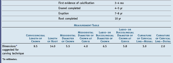
Figures 7-1 through 7-12 illustrate the mandibular central incisor in various aspects. Generally, the mandibular central incisor is the smallest tooth in the dental arches (Table 7-1). The crown has little more than half the mesiodistal diameter of the maxillary central incisor; however, the labiolingual diameter is only about 1 mm less. The lines of greatest masticatory stress are brought to bear on the mandibular incisors in a labiolingual direction, which makes this reinforcement necessary.
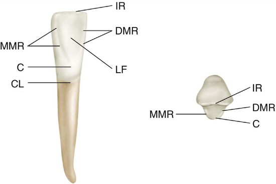
Figure 7-1 Mandibular right central incisor, lingual and incisal aspects. IR, Incisal ridge; DMR, distal marginal ridge; LF, lingual fossa; CL, cervical line; C, cingulum; MMR, mesial marginal ridge.
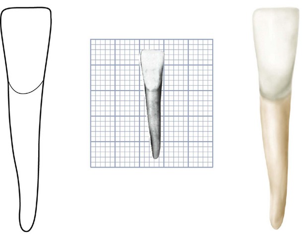
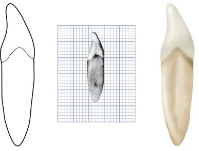
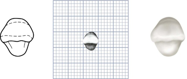
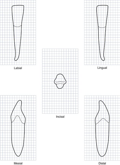
Figure 7-7 Mandibular right central incisor. Graph outlines of five aspects are shown. (Grid = 1 sq mm.)
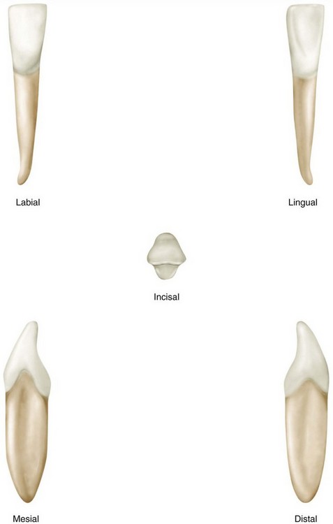
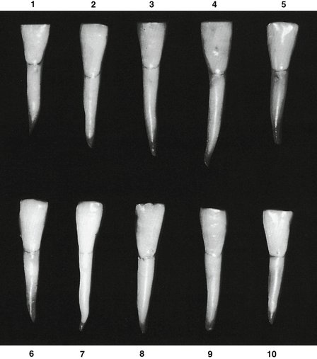
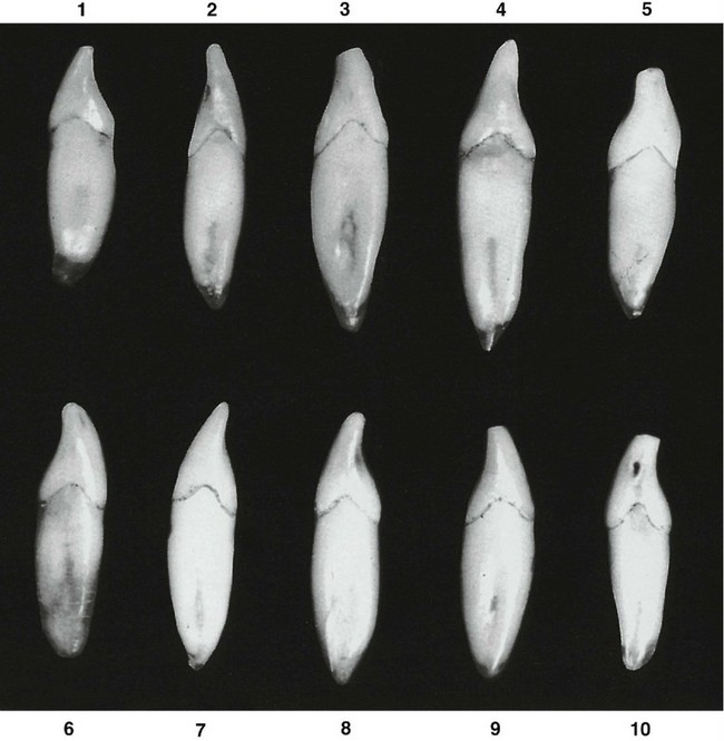
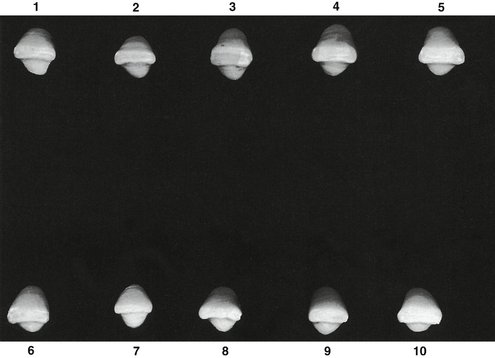
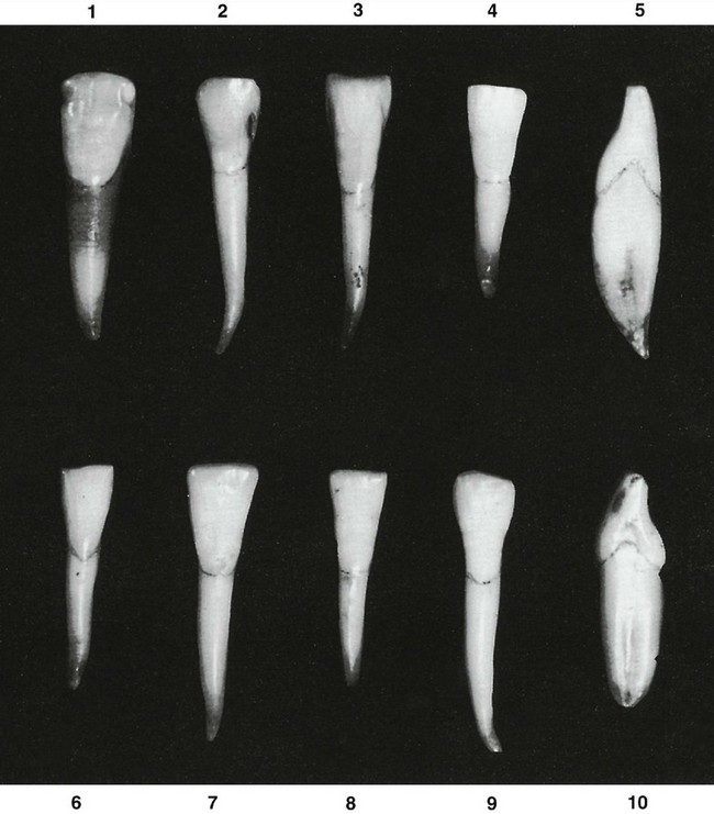
Figure 7-12 Mandibular central incisor. Ten specimens with uncommon variations are shown. 1, Crown and root very broad mesiodistally; malformed enamel at incisal third of crown. 2, Crown wide at incisal third, with short crown; root length extreme. 3, Unusual contours at middle third of crown; cervix narrow. 4, Well-formed crown; short root. 5, No curvature labially at cervical third; extreme labial curvature at root end. 6, Specimen well formed but undersized. 7, Contact areas pointed at incisal edge; crown and root very long. 8, Crown long and narrow; root short. 9, Crown measurement at cervical third same as root; crown and root of extreme length. 10, Crown and root very wide labiolingually; greater curvature than average above cervical line at the cervical third of the crown.
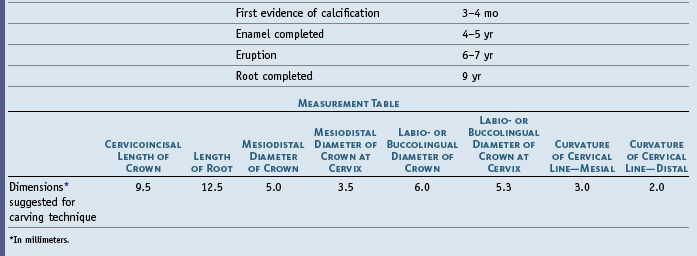
The single root is very narrow mesiodistally and corresponds to the narrowness of the crown, although the root and crown are wide labiolingually. The length of the root is as great as, if not greater than, that of the maxillary central incisor.
DETAILED DESCRIPTION OF THE MANDIBULAR CENTRAL INCISOR FROM ALL ASPECTS
Labial Aspect
The labial aspect of the mandibular central incisor is regular, tapering evenly from the relatively sharp mesial and distal incisal angles to the apical portion of the root (see Figures 7-7 through 7-9). The incisal ridge of the crown is straight and is at approximately a right angle to the long axis of the tooth. Usually, the mesial and distal outlines of the crown make a straight drop downward from the incisal angles to the contact areas, which are incisal to the junction of incisal and middle thirds of the crown. The mesial and distal sides of the crown taper evenly from the contact areas to the narrow cervix.
The mesial and distal root outlines are straight with the mesial and distal outlines of the crown down to the apical portion. The apical third of the root terminates in a small, pointed taper, in most cases curving distally. Sometimes the roots are straight (see Figure 7-9, 2 and 10).
The labial face of the mandibular central incisor crown is ordinarily smooth, with a flattened surface at the incisal third; the middle third is more convex, narrowing down to the convexity of the root at the cervical portion.
Except in newly erupted teeth, central incisors show few traces of developmental lines. The labial surface of the root of the mandibular central incisor is regular and convex.
Lingual Aspect
The lingual surface of the crown is smooth, with very slight concavity at the incisal third between the inconspicuous marginal ridges (see Figures 7-1, 7-3, 7-7, and 7-8). In some instances, the marginal ridges are more prominent near the incisal edges (see Figure 7-11, 2 and 8). In these cases, the concavity between the marginal ridges is more distinct.
The lingual surface becomes flat and then convex as progression is made from the incisal third to the cervical third.
No developmental lines mark the cingulum development on this tooth at the cervical third. No other tooth in the mouth, except the mandibular lateral incisor, shows so few developmental lines and grooves. The outlines and surfaces of the mandibular incisors are regular and symmetrical.
Mesial Aspect
The curvature labially and lingually above the cervical line is less than that found on maxillary incisors (see Figures 7-4, 7-7, 7-8, and 7-10).
The outline of the labial face of the crown is straight above the cervical curvature, sloping rapidly from the crest of curvature to the incisal ridge. The lingual outline of the crown is a straight line inclined labially for a short distance above the smooth convexity of the cingulum; the straight outline joins a concave line at the middle third of the crown, which extends upward to join the rounded outline of a narrow incisal ridge. The incisal ridge is rounded or worn flat, and its center is usually lingual to the center of the root.
The curvature of the cervical line representing the cementoenamel junction (CEJ) on the mesial surface is marked, curving incisally approximately one third the length of the crown.
The root outlines from the mesial aspect are straight with the crown outline from the cervical line, so that the root diameter is uniform through the cervical third and part of the middle third; the outline of the root begins to taper in the middle third area, tapering rapidly in the apical third to either a bluntly rounded or a pointed root end.
The mesial surface of the crown is convex and smooth at the incisal third and becomes broader and flatter at the middle third cervical to the contact area; it then becomes quite flat, with a tendency toward concavity immediately below the middle third of the crown and above the cervical line (see Figure 7-10, 5, 8, and 10).
The mesial surface of the root is flat just below the cervical line. Most of these roots have a broad developmental depression for most of the root length. The depressions usually are deeper at the junction of the middle and apical thirds (see Figure 7-10, 3 and 9).
Distal Aspect
The cervical line representing the CEJ curves incisally about 1 mm less on the distal aspect than on the mesial aspect (see Figures 7-5, 7-7, and 7-8).
The distal surface of the crown and root of the mandibular central incisor is similar to that of the mesial surface. The developmental depression on the distal surface of the root may be more marked, with a deeper, more well-defined developmental groove at its center.
Incisal Aspect
The incisal aspect illustrates the bilateral symmetry of the mandibular central incisor (see Figures 7-1, 7-6, 7-7, 7-8, and 7-11). The mesial half of the crown is almost identical with the distal half.
The incisal edge is almost at right angles to a line bisecting the crown labiolingually. This feature is characteristic of the tooth and serves as a mark of identification in differentiation between mandibular central and lateral incisors (see Mandibular Lateral Incisor later). Note the comparison between the diameter of these crowns labiolingually and their diameters mesiodistally. The labiolingual diameter is always greater.
The labial surface of the crown is wider mesiodistally than the lingual surface. The crown is usually wider labially than lingually at the cervical third, which latter area is represented by a smooth cingulum.
The labial surface of the crown at the incisal third, although rather broad and flat in comparison with the cervical third, has a tendency toward convexity, whereas the lingual surface of the crown at the incisal third has an inclination toward concavity.
When this tooth is posed from the incisal aspect so that the line of vision is on a line with the long axis of the tooth, more of the labial surface may be seen than of the lingual surface.
Mandibular Lateral Incisor
Figures 7-13 through 7-21 illustrate the mandibular lateral incisor in various aspects. The mandibular lateral incisor is the second mandibular tooth from the median line, right or left. It resembles the mandibular central incisor so closely that only a brief description of each aspect of the lateral incisor is necessary. Direct comparison is made with the mandibular central incisor, and the variations are mentioned. The two incisors operate in the dental arch as a team; therefore their functional form is related. As with the mandibular central incisor, the shape of the lateral incisor is uniform compared with that of other teeth. Rarely, it will have a labial and lingual root division in the cervical third. Somewhat more commonly it has two canals in the single root.3


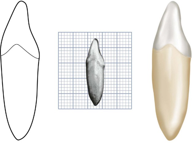
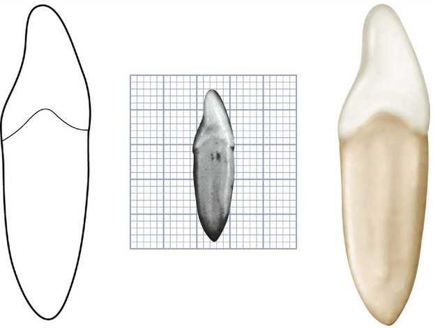
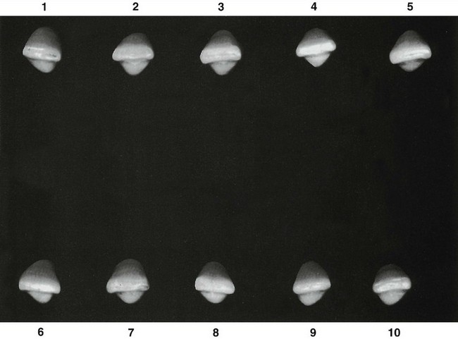
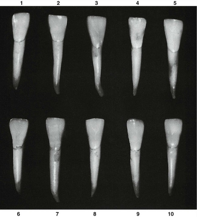
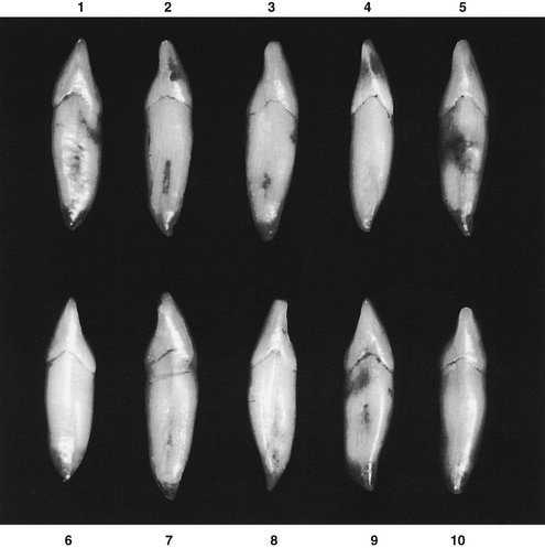
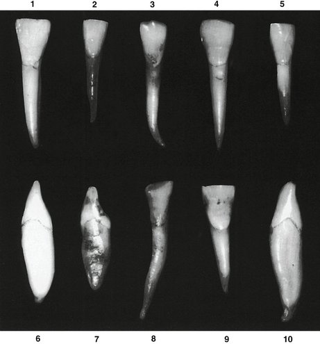
Figure 7-21 Mandibular lateral incisor. Ten specimens with uncommon variations are shown. 1, Tooth very large; cervix constricted in comparison with crown width. 2, Specimen well formed, smaller than average. 3, Root extra long; extreme curvature at apical third; mesial and middle mamelons intact on incisal ridge. 4, Extreme mesiodistal measurement for crown length; contact areas very broad cervicoincisally. 5, Specimen undersized. 6, Incisal ridge thin; little or no curvature at cervical third of crown. 7, Incisal edge labial to center of root; root rounded; cingulum with more curvature above root than average. 8, Malformed crown and root; root with extreme length. 9, Crown very wide; root short. 10, Very slight curvature at cervical third of crown; entire tooth oversized, malformation at root end.
The mandibular lateral incisor is somewhat larger than the mandibular central incisor (compare measurements), but generally speaking, its form closely resembles that of the mandibular central incisor (Table 7-2). Ten specimens with uncommon variations are shown in Figure 7-21.
BRIEF DESCRIPTION OF THE MANDIBULAR LATERAL INCISOR FROM ALL ASPECTS
Labial and Lingual Aspects
The labial and lingual aspects show the added fraction of approximately 1 mm of crown diameter mesiodistally added to the distal half (see Figures 7-13 and 7-14). This, however, is not always true (see Figure 7-19, 3 and 6). The lingual aspect of the mandibular incisors in some Mongoloid groups is marked by a deep but short cervicoincisal groove, which is vulnerable to dental caries.4
Mesial and Distal Aspects
The mesial side of the crown is often longer than the distal side; this causes the incisal ridge, which is straight, to slope downward in a distal direction (see Figure 7-19, 1). The distal contact area is more toward the cervical than the mesial contact area to contact properly the mesial contact area of the mandibular canine.
Except for size, no marked difference is evident between the mesial and distal surfaces of central and lateral incisors (see Figures 7-15, 7-16, and 7-20). Even the curvatures of the cervical line mesially and distally are similar in extent. A tendency exists toward a deeper concavity immediately above the cervical line on the distal surface of the mandibular lateral incisor.
Although the crown of the mandibular lateral incisor is somewhat longer than that of the central incisor (usually a fraction of a millimeter), the root may be considerably longer. Therefore in most instances the tooth is a little larger in all dimensions. The root form is similar to that of the central incisor, including the presence of developmental depressions mesially and distally.
Incisal Aspect
The incisal aspect of the mandibular lateral incisor provides a feature that can usually serve to identify this tooth. The incisal edge is not at approximate right angles to a line bisecting the crown and root labiolingually, as was found when observing the central incisor; the edge follows the curvature of the mandibular dental arch, which gives the crown of the mandibular lateral incisor the appearance of being twisted slightly on its root base (see Figures 7-17 and 7-18). It is interesting to note that the labiolingual root axes of mandibular central and lateral incisors remain almost parallel in the alveolar process, even though the incisal ridges are not directly in line.
1. Carlsen O. Dental morphology. Copenhagen: Munksgaard; 1987.
2. Pindborg JJ. Pathology of the dental tissues. Philadelphia: Saunders; 1970.
3. Woelfel JB, Scheid RC. Dental anatomy: its relevance to dentistry, ed 5. Baltimore: Williams & Wilkins; 1997.
4. Hanihara K. Racial characteristics in the dentition. J Dent Res. 1967;46:293.
 site for additional study resources.