ACUTE DIARRHEA OF ADULT (NONSUCKLING) HORSES

Synopsis

Etiology Salmonella spp., Strongylus spp., cyathostomes, Neorickettsia (Ehrlichia) risticii, Clostridium spp., antibiotic administration, idiopathic

Epidemiology Usually a sporadic disease of young horses, often temporally associated with mild respiratory disease or a stressful event such as transport. Helminthiasis has a seasonal distribution and can occur as a herd problem. N. risticii has a defined geographical distribution

Clinical signs Acute onset of profuse watery diarrhea. Depression, fever, dehydration and anorexia are common

Clinical pathology Leukopenia, hemoconcentration, hyponatremia, hypokalemia or hyperkalemia, metabolic acidosis. IFA or PCR for N. risticii, fecal culture of Salmonella spp. may be positive. Fecal culture for Clostridium spp. and ELISA to demonstrate toxin in feces

Lesions Colitis with or without enteritis

Diagnostic confirmation Cause is frequently not confirmed

Treatment Intense supportive care including maintenance of hydration and correction of acid–base and electrolyte abnormalities. Oxytetracycline for equine monocytic ehrlichiosis. Metronidazole for C. difficile-associated diarrhea

Control None

ETIOLOGY

Causes are as follows:

Salmonellosis: Various Salmonella spp.

Helminthiasis: Strongylus sp., cyathostomes

Equine monocytic ehrlichiosis (Potomac horse fever Equine neorickettsiasis): Neorickettsia risticii

Antibiotic administration: macrolides (lincomycin, tylosin, erythromycin), tetracyclines, ciprofloxacin, trimethoprim–sulfonamide combination, penicillin, aminoglycosides, ceftiofur, and others

Intestinal clostridiosis: C. perfringens (types A and C),1 toxigenic strains of C. difficile2 and possibly Clostridium cadaveris

Aeromonas spp.: Isolated from horses with diarrhea, definitive role as a causative agent has not been demonstrated3

Colitis-X: Idiopathic.

E. coli does not appear to be an important cause of diarrhea in adult horses.4

In most cases (65%) of acute diarrhea in horses the cause is not determined, or if the cause is determined it is frequently at necropsy examination or after the horse has recovered.5

EPIDEMIOLOGY

Occurrence

The syndrome of acute diarrhea occurs worldwide in adult horses of all breeds and both sexes. The pattern of occurrence of the syndrome is dependent on the causative factors, with equine monocytic ehrlichiosis (equine neorickettsiasis), associated with N. risticii, having a geographical distribution and acute cyathostomiasis having a seasonal distribution. Salmonellosis can occur sporadically or as outbreaks in stables, barns and veterinary hospitals. C. difficile enterocolitis is associated with hospitalization and/or antibiotic administration to adult horses.2 The disease also occurs in foals.

Colitis-X is usually a sporadic disease, but multiple cases can occur in a barn or racing stable over a period of weeks and cause considerable economic hardship. With the exception of salmonellosis, equine monocytic ehrlichiosis, strongylosis and colitis-X, the syndrome is characterized by a sporadic distribution. Estimates of incidence, morbidity and mortality are not available for all diseases.

The case fatality rate for the spontaneous disease can be 25–50% even in intensively treated horses.5-8 The case fatality rate is higher for horses with C. difficile-induced diarrhea than for horses with acute diarrhea of other causes2 and for horses with antibiotic-induced diarrhea.8 The prognosis is worse in horses with tachycardia, severe dehydration (PCV > 45% (0.45 L/L)), azotemia, metabolic acidosis, low serum albumin concentration or higher immature neutrophil (band cell) count in peripheral blood.6-8

Risk factors

The risk factors for salmonellosis, equine monocytic ehrlichiosis and strongylosis are addressed under those topics.

Stress

Stressful episodes, such as shipping or racing, hospitalization, surgery, administration of antibiotics or mild respiratory disease, frequently precede the onset of diarrhea.6

Celiotomy

Celiotomy for colic is associated with an incidence of severe diarrhea of up to 27% in surviving horses.9 The risk of diarrhea is greatest in horses with large-colon disease or with enterotomy, but is not influenced by the type of antibiotic administered after surgery.9

Antibiotic administration

Antibiotic administration is associated with acute diarrhea in horses. The macrolide antibiotic lincomycin causes acute, often fatal, disease of horses even when administered at relatively low doses, such as that resulting from horses ingesting medicated pig feed.10 Tetracyclines have been associated with the development of acute diarrhea but, when given intravenously at therapeutic doses (6.6 mg/kg every 12–24 h) are probably no more likely to cause diarrhea than other broad-spectrum antibiotics. Tetracycline contamination of feed causes outbreaks of diarrhea on horse farms.11 Ciprofloxacin might be a cause of diarrhea in horses.12 The combination of trimethoprim and sulfadiazine given orally causes diarrhea in 7% of hospitalized horses, whereas pivampicillin, a prodrug of ampicillin, causes diarrhea in 3%.13 However, horses treated with trimethoprim–sulfadiazine combinations are not at greater risk of developing diarrhea than horses treated with penicillin.14 Almost all adult horses with diarrhea from which C. difficile or its toxin can be isolated were administered antibiotics before onset of diarrhea.15

PATHOGENESIS

Diarrhea is the result of abnormalities in colonic water and electrolyte metabolism. Approximately 90 L of isotonic fluid enters the colon of an adult (450 kg) horse every 24 hours, and any disruption to the normal absorption of this fluid results in increased fecal water and electrolyte excretion. Colitis results from physical, chemical or infectious causes that induce inflammation in the colon. The proximate causes vary with the etiology of the disease. For example, colitis due to infection by toxigenic strains of C. perfringens type C is attributable to binding of beta-2 toxin to colonic mucosa,16 whereas colitis due to salmonellosis is associated with invasion of the organism and loss of colonic mucosa. Colitis is associated with increased production of inflammatory cytokines, including tumor necrosis factor, in the colon,17 and with impaired mucosal absorptive function. Additionally, bacterial toxins and inflammation result in an increase in mucosal permeability with loss of plasma proteins into the colonic lumen and systemic absorption of toxins, including endotoxin. Loss of plasma proteins causes a reduction in plasma colloidal oncotic pressure with subsequent extravasation of water and electrolytes and development of edema and decreased effective intravascular volume (hypovolemia). The effect of the decrease in oncotic pressure becomes most apparent in horses that are treated aggressively with fluids. These horses, which often receive excessive amounts of sodium, rapidly develop edema of the ventral body wall and colon, among other tissues. Loss of other plasma proteins, including antithrombin III, and absorption from the gut of activators of coagulation, fibrinolysis or inflammation, may contribute to the disseminated intravascular coagulation often observed in horses with enterocolitis.

The large volume of diarrhea in horses causes a reduction in body water and electrolyte content. Hypovolemia, hyponatremia, hypochloremia and hypoproteinemia develop. Derangements in acid–base and electrolyte status impair gastrointestinal motility. Hypovolemia impairs perfusion of peripheral tissues, which, combined with absorption of endotoxin through the damaged colonic mucosa, results in toxemia, lactic acidosis and death.

CLINICAL SIGNS

The onset of clinical signs is usually abrupt, although in some horses diarrhea may be presaged for up to several days by inappetence, mild depression and a mild fever. The disease varies in severity from short-lived with mild to moderate diarrhea and minimal systemic signs of disease to a fulminant disease with death in hours. The description here is of the more severe forms of the disease. Once diarrhea occurs there is rapid progression, with some horses dying within 12 hours of initial clinical signs, although most survive at least 24 hours. In a peracute form of the disease horses die, often within 6 hours, before developing diarrhea.

Typically horses are often severely depressed and stand with their heads down. They may play in water, but rarely eat or drink. Horses are usually mildly pyrexic (101.5–103°F, 38.6–39.5°C) but markedly tachycardic (80–100 bpm), tachypneic (30–40 bpm) and dehydrated (8–12%). There is slow capillary refill of mucous membranes, which are usually bright red initially and then become bluish-purple as toxemia and dehydration become severe. The development of a purple line at the gingival margins is a sign of a poor prognosis. Most horses are oliguric.

The diarrhea is profuse and watery. Abdominal pain is usually present but mild; the onset of severe abdominal pain is often associated with necrosis of the large colon or cecum and impending death. Rectal examination reveals large amounts of fluid feces with minimal distension of the large colon.

Complications of acute, severe enterocolitis include laminitis, thrombophlebitis of the jugular veins, thrombosis of vessels including arteries in the limbs.18 renal failure, pulmonary aspergillosis19-21 and necrotizing enterocolitis.22 Laminitis develops within 1–3 days of onset of diarrhea in approximately 10% of cases and can occur in any horse with enterocolitis, but is most common in horses with Potomac horse fever (equine monocytic ehrlichiosis). Thrombophlebitis, which may or may not be septic, usually affects veins, usually jugular, that have or have had catheters placed or are the site of frequent intravenous injections. Thrombosis of the vein can occur several days to a week after removal of the catheter, although most occur while the catheter is in place. Renal failure occurs as a result of the combined insults of hypovolemia, endotoxemia and administration of nephrotoxic drugs, including aminoglycosides and NSAIDs. Pulmonary aspergillosis is usually clinically inapparent.20 Clinically affected horses have rapidly progressive toxemia, respiratory distress, hypoxemia and blood-tinged, frothy nasal exudates. Fatal necrotizing enterocolitis of horses is characterized by a brief course, most horses dying within 48 hours of onset of diarrhea, profound dehydration, electrolyte derangements, severe metabolic acidosis and, terminally, severe abdominal pain.22

Most horses that survive have resolution of diarrhea in about 7 days, although a small but clinically important proportion develop chronic diarrhea.

CLINICAL PATHOLOGY

Hematological examination reveals an increased hematocrit (45–60%), variable changes in plasma protein concentration and neutropenia with a marked left shift. As the disease progresses and horses are treated by intravenous administration of fluids, plasma protein concentrations and plasma oncotic pressure decline. Plasma or serum albumin concentration may be as low as 1.2 g/dL (12 g/L). Changes in coagulation and fibrinolysis are evident as increases in one or more of one-stage prothrombin time, activated partial thromboplastin time and concentration of fibrin degradation products, variable changes in plasma fibrinogen concentration and a reduction in blood platelet concentration.23 Approximately one-third of horses hospitalized for treatment of severe diarrhea have subclinical evidence of disseminated intravascular coagulation,23 which carries a reduced likelihood of recovery.

Serum biochemical analysis usually reveals hyponatremia, hypochloremia, variable changes in serum potassium concentration, hypocalcemia (both concentrations of ionized and total calcium24), azotemia (increased serum urea nitrogen and creatinine concentrations), hyperphosphatemia and increased activities of enzymes indicative of muscle (creatine kinase) or intestinal damage (aspartate aminotransferase and alkaline phosphatase).

Blood gas analysis often reveals a severe metabolic acidosis, and the more negative the base excess the worse the prognosis.7 Interpretation of acid–base status in horses with severe enterocolitis is difficult because of the opposing effects of hypoproteinemia and combination of lactic acidosis and electrolyte loss on blood pH. Hypoproteinemia causes a metabolic alkalosis whereas increases in plasma lactate concentration and hyponatremia cause metabolic acidosis. The presence of hypoproteinemia therefore tends to diminish the effect of lactic acidosis on blood pH, which underestimates the severity of the acidosis. Acid–base status in horses with severe abnormalities in plasma protein concentration should be ascertained by examination of base excess, strong ion gap or strong ion difference.25

Plasma endothelin concentrations are higher in horses with enterocolitis than in normal horses,26 although the clinical significance of this finding is unclear.

Abdominal fluid is usually normal initially but becomes bloody and has an increased white blood cell count and protein concentration if intestinal necrosis occurs.

DIAGNOSTIC CONFIRMATION

This depends on the results of fecal culture for Salmonella sp., fecal examination for helminth eggs or larvae, and indirect fluorescent antibody (IFA) or polymerase chain reaction (PCR) tests for N. risticii. Demonstration of large numbers of salmonellae in feces on multiple fecal samples, or in lymph nodes of horses dying of the disease, is persuasive evidence that the horse had salmonellosis. However, demonstration of low numbers of salmonellae in a single fecal culture is not definitive evidence that Salmonella sp. infection was the cause of the horse’s diarrhea. Fecal examinations for helminth eggs may be negative in cases of acute cyathostomiasis, although large numbers of fourth stage larvae may be present in the feces. Diagnosis of N. risticii infection is based on a positive IFA test. Isolation of Clostridium sp. and demonstration of clostridial enterotoxin in feces of horses with acute diarrhea supports a diagnosis of intestinal clostridiosis, although demonstration of toxin alone is usually considered sufficient evidence for diagnosis.27 Latex agglutination tests are available for the detection of C. perfringens type A and C. difficile toxins.1,28

NECROPSY

There are extensive lesions at necropsy examination, the most dramatic being in the large intestine, especially the cecum and ventral colon. These include hyperemia, extensive petechiation, and edema of the gut wall in the early stages, and later an intense, greenish black, hemorrhagic necrosis. The contents are fluid, often foamy and foul-smelling, and may be bloodstained.

Histological examination demonstrates mucosal necrosis with a fibrinohemorrhagic exudate and extensive inflammation of the mucosa and submucosa.

DIFFERENTIAL DIAGNOSIS

Differential diagnosis list:

Salmonellosis

Equine monocytic ehrlichiosis (equine neorickettsiasis) (Potomac horse fever)

Cyathostomiasis

Antibiotic-induced diarrhea

Clostridium sp. infection (C. difficile)

Colitis-X

Intoxication with inorganic arseni, cantharidin or purgatives such as castor oil

The incipient disease in horses before onset of diarrhea can resemble colon torsion or ischemia of the large colon secondary to verminous arteritis

TREATMENT

Horses with mild disease, those that do not manifest systemic signs of disease, usually recover with symptomatic treatment. However, horses with severe disease require more specific treatment and supportive care, which is often intensive and expensive.

The principles of treatment for horses with acute diarrhea are:

Restoration and maintenance of normal hydration

Correction of electrolyte and acidbase abnormalities

Provision of analgesia

Prophylaxis and treatment of the effects of endotoxemia

Prevention of absorption of toxins

Correction and prevention of disseminated intravascular coagulation.

Restoration of hydration

Restoration of hydration should be considered an emergency procedure in severely affected horses. Fluids should be administered intravenously until hydration is restored, after which hydration can be maintained by either oral (via nasogastric tube) or intravenous administration of fluids. Suitable fluids for restoration of hydration are sodium-rich, isotonic, preferably polyionic, electrolyte solutions such as lactated Ringer’s or Ringer’s solution. Isotonic sodium chloride is also suitable. Isotonic dextrose solutions are not suitable because they do not contain any electrolytes. After correction of dehydration, attention should be paid to sodium balance because administration of excessive quantities of sodium, especially to horses with plasma oncotic pressure that is lower than normal, may cause expansion of the extracellular fluid volume and edema.

Fluid therapy is discussed elsewhere. Maintenance of hydration in severely affected horses can be challenging and is best accomplished by intravenous administration of fluids. Oral administration of fluids to horses with diarrhea, although not providing ideal rehydration or maintenance of hydration, may be effective and less costly than intravenous administration.29-31

Horses that become hypoproteinemic may require transfusions of plasma. Clinical signs indicating the need for transfusion include a persistently elevated heart rate and poor peripheral perfusion in spite of administration of large quantities of fluids. Ventral edema and edema of the head and legs may develop in hypoproteinemic horses. Sufficient plasma should be administered to restore the plasma protein concentration to at least 40 g/L.

Electrolyte and acid–base status

Hyponatremia and hypochloremia will usually be corrected by administration of isotonic, sodium-rich electrolyte solutions such as lactated Ringer’s solution. If this does not occur, then sodium chloride or sodium bicarbonate can be added to the intravenous fluids, or given orally. Hypocalcemia can be corrected by the addition of calcium gluconate (20 mL of 23% calcium gluconate per liter of fluids) to the fluids, provided that the fluids do not contain sodium bicarbonate. The mixture of sodium bicarbonate and calcium gluconate causes calcium to precipitate out of solution. Affected horses have total body potassium depletion,32 even though serum potassium concentrations may be normal or elevated, and maintenance fluids should contain potassium at up to 25 mEq/L. Fluids with high potassium concentration should be administered slowly. Alternatively, potassium chloride can be given orally (50–100 g per 450 kg every 12 h).

The metabolic acidosis in horses with acute diarrhea often resolves either partially or completely when hydration is restored. However, severe acidosis can be treated with intravenous sodium bicarbonate. Oral administration of sodium bicarbonate (100 g per 450 kg every 8–12 h) is often adequate in restoring and maintaining normal acid– base status. The serum sodium concentration should be monitored if large quantities of sodium bicarbonate are administered.

Antimicrobial therapy

Administration of tetracycline to horses with acute diarrhea associated with N. risticii is clearly indicated and is often curative. However, the administration of antimicrobial drugs to horses with acute diarrhea other than that associated with N. risticii is controversial. There is no evidence that administration of antimicrobials improves the prognosis of horses with acute diarrhea.7,33 The concern with antimicrobial administration is that antimicrobials may exacerbate the diarrhea in some cases. Conversely, withholding antimicrobials from severely ill horses with damaged colonic mucosa, and therefore presumably increased risk of bacteremia, is problematic. Regardless, many clinicians chose to treat horses with acute diarrhea with broad-spectrum antibiotics such as the combination of potassium penicillin (20000 IU/kg, intravenously every 6 h) and gentamicin (7 mg/kg intravenously or intramuscularly every 24 h) or trimethoprim and sulfadiazine (30 mg/kg intravenously or orally every 12 h). Metronidazole (15–20 mg/kg orally every 6–12 h) or vancomycin have been recommended for horses with intestinal clostridiosis, although the wisdom of veterinary use of vancomycin, a drug used for the treatment of methicillin-resistant staphylococci in humans, could be questioned.34 In areas in which equine monocytic ehrlichiosis (equine neorickettsiasis) is endemic, all suspected cases should be treated with tetracycline (6.6 mg/kg intravenously every 12 h for 3 d), or another effective antibiotic, pending confirmation of the disease. Isolates of toxigenic C. difficile from horses with diarrhea are almost always susceptible to metronidazole (15–29 mg/kg orally every 6–12 h).15

Prophylaxis and treatment of endotoxemia

Treatment of endolemma has been recently reviewed.35 Administration of plasma from horses hyperimmunized with Salmonella typhimurium or E. coli reduces the severity of clinical signs and shortens the duration of disease in horses with endotoxemia secondary to enterocolitis or colic.36 Polymyxin (5000 IU/kg intravenously every 12 h) attenuates the effect of endotoxin in experimental disease and is used for the prevention and treatment of endotoxemia in hospitalized horses.37 Its efficacy in clinical settings has not been determined. Aspirin (10 mg/kg orally every 48 h) is administered to diminish platelet aggregation around intravenous catheters. Flunixin meglumine (1 mg/kg intravenously every 8–12 h) or phenylbutazone (2.2 mg/kg intravenously every 12 h) is given for analgesia and to prevent endotoxin-induced increases in plasma prostaglandins. Pentoxifylline (8 mg/kg orally every 8 h) is administered for its putative effective in attenuating the effects of endotoxemia. The efficacy of these treatments in a clinical setting and their effect on measures of outcome of disease, such as duration of illness, case fatality rate and incidence of complications, has not been determined, with the exception of hyperimmune plasma or serum.36

Binding of toxins

Smectite or activated charcoal are sometimes administered to horses with acute enterocolitis in an attempt to adsorb toxins, such as those produced by Clostridium spp., and prevent systemic absorption. There is in-vitro evidence that smectite may bind clostridial toxins and endotoxin,38 but evidence of efficacy in vivo is lacking.

Disseminated intravascular coagulation

Prevention and treatment of disseminated intravascular coagulation includes monitoring for changes in variables indicative of coagulation and fibrinolysis including d-dimer concentration, antithrombin III activity, one-stage prothrombin and activated partial thromboplastin times, platelet count and fibrinogen concentration. Plasma can be administered to increase blood antithrombin III activity, often in conjunction with heparin or low-molecular-weight heparin (dalteparin or enoxaparin). Doses of 50U of dalteparin or 40U of enoxaparin per kilogram subcutaneously every 24 hours seem to be adequate for prophylactic anticoagulatory treatment of horses. For treatment of coagulation disorders or for ill horses that are considered to be at high risk of developing thrombotic disease, dosages may need to be increased to 100U of dalteparin or 80U of enoxaparin per kilogram subcutaneously every 24 hours.39

CONTROL

Specific control measures for Salmonella spp. infection, equine monocytic ehrlichiosis and cyathostomiasis are discussed under those headings. The incidence of antibiotic-induced colitis can be reduced by minimizing the frequency with which antibiotics are administered to horses.

REVIEW LITERATURE

Divers TJ. Prevention and treatment of thrombosis, phlebitis, and laminitis in horses with gastrointestinal diseases. Vet Clin North Am Equine Pract. 2003;19:779.

McConnico R. Acute Equine colitis. Compend Contin Educ Pract Vet. 2003;25:623.

REFERENCES

1 Donaldson MT, Palmer JE. J Am Vet Med Assoc. 1999;215:358.

2 Weese JS, et al. Equine Vet J. 2001;33:403.

3 Hathcock TL, et al. J Vet Intern Med. 1999;13:357.

4 Van Duijkeren E, et al. Vet Q. 2000;22:162.

5 Mair TS, et al. Vet Rec. 1990;126:479.

6 Stewart MC, et al. Aust Vet J. 1995;72:41.

7 Staempfli HR, et al. Can Vet J. 1991;32:232.

8 Cohen ND, Woods AM. J Am Vet Med Assoc. 1999;214:382.

9 Cohen ND, et al. J Am Vet Med Assoc. 1996;209:810.

10 Raisbeck MF, et al. J Am Vet Med Assoc. 1981;179:362.

11 Keir AAM, et al. Can Vet J. 1999;40:718.

12 Weese JS, et al. Equine Vet Educ. 2002;14:182.

13 Ensink JM, et al. Vet Rec. 1996;138:253.

14 Wilson DA, et al. J Vet Intern Med. 1996;10:258.

15 Baverud V, et al. Equine Vet J. 2003;35:465.

16 Bacciarini LN, et al. Vet Pathol. 2003;40:376.

17 Davidson AJ, et al. Res Vet Sci. 2002;72:177.

18 Brianceau P, Divers TJ. Equine Vet J. 2001;33:105.

19 Rosenstein DS, Mullaney TP. Equine Vet Educ. 1996;8:200.

20 Sweeney CR, Habecker PL. J Am Vet Med Assoc. 1999;214:808.

21 Slocombe RF, Slauson DO. Vet Pathol. 1988;25:277.

22 Saville WJ, et al. J Vet Intern Med. 1996;10:265.

23 Dolente BA, et al. J Am Vet Med Assoc. 2002;220:1034.

24 Van der Kolk JH, et al. Equine Vet J. 2002;34:528.

25 Constable PD. Vet Clin Pathol. 2000;29:115.

26 Ramaswamy CM, et al. Am J Vet Res. 2002;63:454.

27 Weese JS, et al. Proc Am Assoc Equine Pract. 1999;45:50.

28 Traub-Dargatz JL, Jones RL. Vet Clin North Am Equine Pract. 1993;9:411.

29 Schott HC. Vet J. 1998;155:119.

30 Ecke P, et al. Vet J. 1998;155:161.

31 McGinniss SG, et al. Compend Contin Educ Pract Vet. 1996;18:942.

32 Muylle E, et al. Equine Vet J. 1984;16:450.

33 Van Duijkeren E, et al. Vet Q. 1997;18:153.

34 Jang SS, et al. Clin Infect Dis. 1997;25(Suppl 2):S266.

35 Sykes BW, Furr MO. Aust Vet J. 2005;83:45.

36 Spier SJ, et al. Circ Shock. 1989;28:235.

37 Barton MH, et al. Equine Vet J. 2004;36:397.

38 Weese JS, et al. Equine Vet J. 2003;357:638.

39 Schwarzwald CC, et al. Am J Vet Res. 2002;63:769.

CHRONIC UNDIFFERENTIATED DIARRHEA OF HORSES

Synopsis

Etiology Common sign of many enteric and non-enteric diseases

Epidemiology Sporadic disease of adult horses, except for cyathostomiasis and salmonellosis, which are discussed under those headings

Clinical signs Passage of unformed or liquid feces, either in increased or normal quantities. Weight loss, increased appetite. Otherwise normal physical examination. Rectal examination is usually normal

Lesions Colitis in most cases

Diagnostic confirmation Examination of feces for cyathostome larvae, rectal biopsy demonstrating lymphoma or granulomatous enteritis, Salmonella spp. in rectal mucosal biopsy or feces. Sand in feces or evident on abdominal radiography

Treatment Supportive: anthelmintics, corticosteroids, antidiarrheal preparations

Control As for cyathostomiasis and salmonellosis

ETIOLOGY

Chronic diarrhea is the final common sign of a number of causes of colonic dysfunction in horses. Diseases that cause chronic (more than 2 weeks duration) diarrhea in horses include: cyathostomiasis, chronic idiopathic colitis, salmonellosis, alimentary lymphosarcoma, granulomatous colitis, eosinophilic colitis, ingestion of sand, chronic liver disease, peritonitis, lymphangiectasia and as a sequela to acute diarrhea. Immune deficiency, including variable adult onset B-cell deficiency, may predispose to the disease.1 Brachyspira sp. have been implicated as a cause of chronic diarrhea in horses in Australia and Japan.2,3

There are many causes and their relative importance varies between locations. Even with concerted effort, a definitive antemortem diagnosis is achieved in fewer than 30% of cases.4

EPIDEMIOLOGY

The occurrence is sporadic, with only single cases occurring in a group. Other horses in contact are not affected. The case fatality rate is 35–65%.5 There appears to be no age-related, sex-related or breed-related variation in incidence. Older horses do not appear to be at increased risk of having chronic diarrhea.6 The epidemiology of cyathostomiasis and salmonellosis are discussed under those headings.

PATHOGENESIS

Diarrhea is attributable to colonic dysfunction, which may result in excessive loss of electrolytes in feces and diminished absorption of nutrients from the large colon. Disease of exclusively the small intestine does not cause diarrhea in horses. Protein-losing enteropathy may be present. Colonic dysfunction may be associated with inflammatory or infiltrative lesions of the colon but in many cases an anatomical lesion is not detected. However, the colonic contents of affected horses have a greater fermentative capacity than those of normal horses, suggesting that in some horses the disease is essentially one of abnormal colonic digestion and absorption.7

CLINICAL FINDINGS

The characteristic finding is chronic diarrhea. The feces vary in consistency from thick porridge (oatmeal), through undigested fibers in liquid, to liquid without fiber. The consistency of the feces in an individual horse may vary widely from one day to the next. The duration of the diarrhea is variable but may be lifelong. Death or euthanasia usually results from progressive weight loss. The onset of diarrhea is usually abrupt and may be associated with signs of toxemia and dehydration, as described under Acute diarrhea, above. However, often there is no toxemia or other systemic sign apart from weight loss, and affected horses are bright and alert and have a normal or increased appetite.

Rectal examination usually fails to reveal any abnormalities, although horses with granulomatous enteritis or alimentary lymphosarcoma may have enlarged mesenteric lymph nodes.

Abdominal radiography may reveal the presence of excessive amounts of sand in the large colon.

CLINICAL PATHOLOGY

Hematological examination may reveal a mild neutrophilia and anemia, but these changes are of little use in determining the etiology of the diarrhea

Serum biochemical examination typically demonstrates a mild hypoalbuminemia, hypoglobulinemia, hyponatremia and hypokalemia, but again these changes are not specific for any particular disease

Hypoalbuminemia is consistent with the presence of protein-losing enteropathies such as chronic colitis, alimentary lymphosarcoma, cyathostomiasis and granulomatous colitis

Hyperbilirubinemia and elevated serum concentrations of serum bile acids are suggestive of liver disease

Increases in serum alkaline phosphatase activity, while common, are of no diagnostic utility

Horses with cyathostomiasis may have increased concentrations of beta-globulins, although the sensitivity of this test is low.5

Peritoneal fluid has a neutrophilic leukocytosis and increased (>25 g/L) protein concentration in horses with peritonitis but is normal in most horses with chronic diarrhea, including those with alimentary lymphosarcoma or granulomatous colitis.

Fecal examination of horses with cyathostomiasis may reveal strongyle-type ova or fourth-stage cyathostome larvae. The presence of sand in feces, demonstrated by allowing feces to settle in a transparent rectal glove or similar container, suggests sand accumulation in the colon as a cause of the diarrhea. The presence of protozoa in feces has no diagnostic significance.8 Giardia spp. are commonly found in feces of normal horses of all ages and, despite earlier reports of their presence in feces of horses with diarrhea, they are not associated with disease.9 Coccidiosis is very uncommon in horses, and Eimeria leuckarti is probably not pathogenic.10

Demonstration of Salmonella spp. in feces or rectal mucosal biopsy, either by culture or PCR, is suggestive but not diagnostic of salmonellosis, given the high proportion of normal horses that shed Salmonella spp. in feces. Isolation of Rhodococcus equi from feces of young horses with diarrhea is suggestive of enteric disease associated with that organism.

An abnormal D-xylose, glucose or starch absorption test indicates small-intestinal disease and is suggestive of granulomatous enteritis, although most horses with this disease do not have diarrhea.

Exploratory laparotomy, either ventral midline under general anesthesia or through the left flank under local anesthesia, and intestinal biopsy may demonstrate alimentary lymphosarcoma, granulomatous enteritis, eosinophilic enteritis, chronic colitis and other abdominal disease. Rectal biopsy is less expensive and invasive but has a relatively poor sensitivity, although good specificity for granulomatous enteritis, eosinophilic enteritis and alimentary lymphosarcoma.11

NECROPSY FINDINGS

Necropsy findings are consistent with the underlying disease, although in many cases gross lesions are not evident. The histological changes in some cases are restricted to a mild inflammatory response and may be difficult to correlate with the severity of clinical disease. In some of these cases the diarrhea probably reflects an imbalance in the microflora of the large bowel, and demonstration of a specific etiological agent is an unrealistic goal. Conversely, isolation of Salmonella spp. from the gastrointestinal tract or mesenteric lymph nodes should be interpreted with caution in the absence of histological evidence of salmonellosis.

Because of the wide variety of potential causes of chronic diarrhea of horses it is not possible to list all the samples required to ‘confirm’ a diagnosis. In most instances, formalin-fixed samples from the liver, mesenteric lymph nodes and numerous levels of the gastrointestinal tract comprise the minimum diagnostic material required. Regardless of what other testing is performed, it is prudent to hold back frozen segments of both large and small bowel (with content) in case other tests are deemed necessary.

DIFFERENTIAL DIAGNOSIS

Differential diagnosis list:

Chronic idiopathic colitis

Salmonellosis

Cyathostomiasis

Granulomatous colitis

Sand ingestion12

Lymphosarcoma

Peritonitis

Intestinal lymphangiectasia

Hyperlipemia

Liver disease

Basophilic enteritis

Eosinophilic gastroenteritis.

TREATMENT

The principles of treatment are to deal with the underlying disease, correct fluid and electrolyte disturbances, give symptomatic treatment of diarrhea and provide supportive care. Except in cases of cyathostomiasis or sand accumulation, treatment of horses with chronic diarrhea is frequently unrewarding.

Specific treatments

Cyathostomiasis should be treated with larvicidal doses of anthelmintics such as fenbendazole (50 mg/kg once, or 7.5 mg/kg daily for 3 d), moxidectin (400 μg/kg) or ivermectin (200 μg/kg). Treatment may be unrewarding if there is severe damage to the large colon.

Diarrhea secondary to sand accumulation in the gastrointestinal tract should be treated by preventing the horse from ingesting sand and, although the efficacy is debatable, with psyllium mucilloid (1–2 g/kg orally once daily for 4–5 weeks.

Chronic idiopathic colitis may be treated with corticosteroids (dexamethasone 0.2–0.4 mg/kg once daily) or prednisolone (0.5–1.0 mg/kg once daily) for 3–4 weeks and the dose reduced as clinical signs permit.

Chronic salmonellosis has been treated with enrofloxacin (2.5–5 mg/kg orally every 12 h for 3–4 weeks), sometimes in combination with metronidazole (15–20 mg/kg orally every 6–12 h), but one should be aware of the risk of articular cartilage damage in horses treated with enrofloxacin.

Many diseases commonly associated with chronic diarrhea are not treatable.

Symptomatic and supportive treatments

Symptomatic treatments include metronidazole (7.5–20 mg/kg orally every 6–12 h) or iodochlorhydroxyquin (10–20 mg/kg orally once daily). While some horses have resolution of diarrhea while being treated with these compounds, there is no clear demonstration of their efficacy. Antibiotic administration, other than as described above, does not usually alter the course of the disease. Antidiarrheal preparations such as codeine phosphate, loperamide and bismuth subsalicylate often provide temporary improvement in fecal consistency. Some horses with chronic diarrhea respond to transfaunation, whereby 5–10 L of colonic fluid collected immediately after death from a horse without enteric disease is administered via nasogastric intubation.

Supportive treatment includes provision of supplemental electrolytes, principally sodium, potassium and bicarbonate, as a feed additive. Suitable supplements include some commercial products designed for fluid replacement in diarrhetic calves, or a mixture of potassium chloride (300 g), sodium chloride (400 g) and sodium bicarbonate (300 g). This mixture is isotonic when dissolved at the rate of 90 g/12 L, or can be given orally at the rate of 30–90 g per 400 kg horse every 24 hours. Unsupplemented water should be supplied without restriction and serum electrolyte concentrations should be monitored. Severely affected horses may require intravenous administration of polyionic isotonic electrolyte solutions or plasma.

Nutritional support should include provision of a diet of high-quality roughage and grain. Some trials may be needed to determine the diet that is best for individual horses, but care should be taken that the diet contains adequate energy and is nutritionally balanced. Horses should be fed to attain, and then maintain, an ideal body weight.

Spontaneous recovery does occur, particularly in young horses, and this, and the often lengthy duration (6–12 months) of the illness, make it difficult to decide accurately the value of the treatment.

CONTROL

Control of cyathostomiasis and salmonellosis is discussed under those headings. Diarrhea due to sand accumulation in the colon should be prevented by not feeding horses on the ground and by avoiding grazing of short pastures on sandy soil.

REVIEW LITERATURE

Merritt AM. Chronic diarrhea in horses: a summary. Vet Med. 1994;89:363-367.

REFERENCES

1 MacLeay JM, et al. Vet Immunol Immunopathol. 1997;57:49.

2 Shibahara T, et al. J Vet Med Sci. 2002;64:633.

3 Lester GD. Proc Am Coll Vet Int Med Forum. 2005;23:181.

4 Love S, et al. Vet Rec. 1992;130:217.

5 Mair TS, et al. Equine Vet J. 1993;25:324.

6 Brosnahan MM, Paradis MR. J Am Vet Med Assoc. 2003;223:99.

7 Minder HP, et al. Am J Vet Res. 1980;41:564.

8 Ike K, et al. Jpn J Vet Sci. 1983;45:157.

9 Xiao L, Herd RP. Equine Vet J. 1994;26:14.

10 Taylor MA. Equine Vet J. 1994;26:4.

11 Lindberg R, et al. Equine Vet J. 1996;28:275.

12 Bertone JJ, et al. J Am Vet Med Assoc. 1988;193:1409.

ACUTE DIARRHEA OF SUCKLING FOALS

ETIOLOGY

The causes of diarrhea in suckling foals are listed in Table 5.16. In a large proportion of foals the cause of diarrhea is not determined, in part because the disease is usually sporadic, mild and transient. The more common causes of diarrhea in foals on breeding farms in Britain include Clostridium perfringens, rotavirus, Salmonella, Cryptosporidium sp., and Strongyloides westerii,1-3 although the relative importance of various pathogens varies from year to year, from farm to farm and from region to region.

Table 5.16 Epidemiological and clinical features of suckling foals with diarrhea

Etiological agent or disease Important epidemiological factors Major clinical findings; diagnostic criteria
Idiopathic
Foal heat diarrhea Foals < 2 weeks of age. No systemic signs of disease. Diarrhea is mild and pasty.
No specific diagnostic criteria
Bacterial causes
Septicemia (coliforms, Actinobacillus sp., Salmonella sp., Klebsiella sp. and others) Newborn foal to < 2 weeks of age. Failure of transfer of passive immunity Signs of systemic sepsis in addition to diarrhea. Fever, depression, recumbency, failure to nurse, swollen joints, pneumonia, omphalitis or omphalophlebitis. Blood culture
Salmonella sp. Outbreaks in newborn foals, even those with adequate passive immunity. Mare likely carrier. Hygiene at parturition may prevent disease Acute onset diarrhea, depression, fever, toxemia. Culture of blood and feces
Escherichia coli Not well documented disease in foals (cf. calves and piglets) Nonfetid diarrhea. Culture of feces yields heavy growth of mucoid E. coli (circumstantial evidence only)
Enterococcus (Streptococcus) durans Young foals. Disease is rarely reported Diarrhea. Demonstration of S. durans in feces
Rhodococcus equi Foals 2–5 months of age, some with history of respiratory disease Diarrhea associated with R. equi pneumonia; culture respiratory tract
Clostridium difficile < 2 weeks of age. Colic, fever, ileus, hematochezia, toxemia, depression. Fecal culture and demonstration of toxin in feces
Clostridium perfringens type c Neonatal foals. Sporadic disease to annual outbreaks on breeding farms. Most foals excrete C. perfringens type A, which rarely causes diseases in foals

Colic, fever, ileus, hematochezia, toxemia, depression.

Culture of C. perfringens type C in feces, demonstration of toxin in feces

Lawsonia intracellularis Older suckling foals and weanlings. Sporadic or outbreaks on farms Weight loss, mild to moderate diarrhea, ventral edema, depression, hypoproteinemia. Serology and PCR on feces
Yersinia pseudotuberculosis Suckling foals. Outbreaks on breeding farms Watery diarrhea and suppurative pneumonia. Culture of feces and lesions
Aeromonas hydrophila Reports of disease are uncommon. Uncertain importance Diarrhea. Culture of feces
Viral causes
Rotavirus < 3 months of age. Occurs as outbreaks or endemic disease on farm. Highly contagious Profuse watery diarrhea with variable hypovolemia and depression. Detection of virus in feces by electron microscopy, IFA, ELISA
Adenovirus Immunodeficient foals (Arabians with severe combined immunodeficiency) Diarrhea, depression. May be associated with other diseases including pneumonia. Detection of virus in feces by electron microscopy
Coronavirus

Young foals (age range not well defined).

Apparently rare cause of diarrhea in foals

Diarrhea. Detection of virus in feces by electron microscopy
Parasites
Cryptosporidium sp. Foals of any age. May be spread from other species, including calves and cria

Inapparent infection to fulminant disease with diarrhea, hypovolemia, and collapse. Chronic diarrhea.

Detection of oocysts in feces, IFA

Strongyloides westerii Individual foals. Uncertain importance as a cause of diarrhea Acute to chronic diarrhea. Patent infections evident by fecal examination for parasite eggs
Other
Nutritional Sporadic. Orphan foals fed inappropriate or poor-quality milk replacers. Nursing foals fed inappropriate supplements Mild to moderate chronic diarrhea. Failure to thrive. Feed diet intended for foals (not plant-protein- or bovine-milk-based)
Lactose intolerance Nursing foals Moderate to profuse diarrhea. Historical confirmation of administration of compounds
Overdosing of cathartics (DSS, MgSO4, NaSO4, castor oil) Sporadic. Secondary to viral diarrhea. Occurs only in milk-fed foals Moderate to severe watery, acidic diarrhea. Oral lactose tolerance test or trial administration of lactase with milk feedings
Enema History of administration. Diarrhea short-lived Bright alert and responsive foal with mild to moderate diarrhea. No specific diagnostic tests
Antibiotic-induced Administration of antibiotics Mild to moderate diarrhea. May be associated with Candida sp. or C. difficile. Culture of feces, examination for C. difficile toxin

C. perfringens causes diarrhea in young foals. There are five major types of C. perfringens and, while the organism is clearly associated with disease, a definitive role for each of these types in causing disease has not been established, partly because toxin production for strains isolated from foals with diarrhea has not been routinely documented. However, there is clear evidence that C. perfringens type C causes diarrhea in foals.4 C. perfringens types A, B, D and E might be associated with disease in foals, but definitive proof is lacking. E. coli, an important cause of disease in neonates of other livestock species, does not appear to be an important cause of diarrhea in foals, although some strains are pathogenic.5,6 Similarly, although there are reports of coronavirus causing severe disease in foals, this does not appear to be a common cause of diarrhea in foals.7,8 Candida spp. can cause diarrhea in critically ill foals and those administered antibiotics.9 Yersinia spp. have been associated with diarrhea in foals but do not appear to be a common cause of disease.10 Bacteroides fragilis is an uncommon cause of diarrheal disease in foals. The role of Campylobacter spp. in foal diarrhea, if there is any, is unclear.

EPIDEMIOLOGY

Diarrhea is common in suckling foals worldwide although studies of its incidence, risk factors and outcome are exiguous. Diarrhea affects 21% of foals annually in Texas, being second only to respiratory disease (22%) as a cause of disease.11 The frequency of disease varies with age: 25% of foals 0–7 days of age have diarrhea, compared to 40% and 8% of foals aged 8–31 days and 32–180 days, respectively.11 While a common disease syndrome, diarrhea is not associated with a high death rate (2.6%).11 Results of the Texas study may not be applicable to foals in other regions.

Among the common causes of diarrhea the highest death rates are associated with diarrhea associated with C. perfringens, Salmonella sp. and Cryptosporidium sp.2

Risk factors for development of the diarrhea vary depending on its etiology, but in general the disease is less common in foals born at pasture and at low stocking density.11

Rotavirus diarrhea is often endemic on farms and the disease occurs as outbreaks on successive years. Affected foals range in age from less than 7 days to more than 3 months.

Diarrhea due to Rhodococcus equi occurs in foals with R. equi pneumonia and the disease is endemic on some farms. Not all foals with R. equi pneumonia develop diarrhea. The disease occurs in foals 2–5 months of age.

Salmonellosis also occurs as outbreaks of disease among foals less than 8 days of age on breeding farms and is associated with a carrier status in mares.12

Diarrhea associated with C. perfringens type C occurs in foals less than 10 days of age with most foals being less than 6 days old4 and can occur as a farm problem with multiple foals affected on each of several successive years.13 Farm risk factors include presence of other livestock, stock-horse-type foals, foals born on dirt, and stall or dry lot confinement for the first few days of life.14 C. perfringens type A is excreted in feces of most normal foals, whereas C. perfringens type C is rarely isolated from feces of normal foals.15 Clostridium difficile causes diarrhea in foals not administered antibiotics,16 in contrast to the situation in adult horses, and usually affects foals less than 14 days of age, although foals up to 120 days of age may be affected.17 Failure of transfer of passive immunity is not a risk factor for C. perfringens or C. difficile enteritis in foals.

Lawsonia intracellularis causes mild to moderate diarrhea in older suckling or weaned foals. The disease occurs as outbreaks on breeding farms. There are no recognized foal or farm risk factors.

PATHOGENESIS

The pathogenesis of diarrhea varies somewhat depending on the inciting cause (see appropriate sections of this text for discussion of pathogenesis), although if sufficiently severe all cause excessive loss of fluid and electrolytes in feces and subsequent hypovolemia, electrolyte abnormalities, metabolic acidosis and weakness. Although not demonstrated in foals, diarrhea in calves causes metabolic acidosis through loss of sodium and other cations in feces, which results in a decrease in the strong ion difference in blood, causing acidosis. Bicarbonate loss, per se, is not a cause of the metabolic acidosis, at least in calves. Infectious agents generally cause enteritis, although rotavirus infection is associated with loss of villus cells and subsequent loss of enzyme activity derived from the mature epithelial cell. The loss of enzyme activity, including that of disaccharidases, causes malabsorption of nutrients in milk and other feed. Failure to absorb nutrients in the small intestine causes them to be delivered to the cecum and large intestine where they are fermented. Subsequent reductions in colonic pH and increases in osmotic activity of the colon contents results in excretion of large quantities of fluid and electrolytes. C. difficile and C. perfringens produce enterotoxins that cause damage to intestinal cells and accumulation of hemorrhagic fluid in the intestine.16 L. intracellularis causes an infiltrative and proliferative enteropathy with subsequent protein loss and, possibly, malabsorption.

CLINICAL SIGNS

Clinical signs vary from mild, pasty diarrhea that adheres to the perineum and causes no detectable systemic signs of disease to profuse water diarrhea with rapid development of loss of suckling, depressed mentation, tachycardia, increased skin tent, ileus and recumbency.

Signs of systemic disease include failure to nurse, increased frequency or prolonged duration of recumbency, foals at pasture may fail to follow the mare, fatigue, less frequent urination, production of concentrated urine (urine from normal foals is normally dilute) and weakness. Affected foals often have depressed mentation, tachycardia, fever (depending on the cause of the diarrhea), decreased capillary refill time, dry mucous membranes, increased skin tent and eyes that are retracted into the orbit (consistent with dehydration). Depending on the cause of the diarrhea, foals may have colic, which can range from mild with intermittent flank watching or biting and restlessness, through profound agitation, rolling and dorsal recumbency. Severely affected foals may have seizures as a result of profound hyponatremia.18

Chronic diarrhea and that due to nutritional imbalance or lactose intolerance causes rapid weight loss, failure to thrive, poor hair coat and lethargy. Chronic fecal contamination of the perineum and escutcheon causes excoriation and loss of hair.

Diarrhea associated with foal heat is usually mild and transient and not associated with systemic signs of disease. However, diarrhea due to infectious agents is often severe and accompanied by systemic signs of disease.

Diseases associated with Clostridium sp. are often severe with rapid onset of signs of toxemia, colic, hypovolemia and death. Diarrhea is usually present and is often bloody, although it may be watery and profuse. Severely affected foals may have signs of colic, toxemia and ileus and not develop diarrhea before dying. Salmonellosis can present as septicemia, with subsequent development of diarrhea, although in older foals diarrhea is a common presenting sign.

CLINICAL PATHOLOGY

Diarrhea in foals with systemic signs of disease cause hyponatremia, hyperkalemia, hypochloremia, metabolic acidosis, hypoproteinemia and azotemia. The magnitude of abnormalities varies with the cause of disease and its severity. Hyponatremia may be profound (< 100 mEq/L). Hypoproteinemia may be a result of loss of protein from the inflamed intestine or a reflection of failure of transfer of passive immunity. All young foals with diarrhea should have serum or plasma immunoglobin concentrations measured or some other test for transfer of passive immunity performed.

Viral causes of diarrhea can be diagnosed by examination of feces by electron microscopy. However, more rapid and sufficiently sensitive and specific tests exist for diagnosis of rotaviral disease (ELISA, IFA). Culture of feces will demonstrate Salmonella spp in most cases if they are the cause of disease. Fecal culture yielding C. perfringens or C. difficile is insufficient for diagnosis of clostridial enterocolitis as these organisms can be recovered from normal foals. Confirmation of the diagnosis is achieved by demonstration of clostridial toxins in feces, which can be problematic given that the toxins are very labile.

DIAGNOSTIC CONFIRMATION

For diagnostic criteria for specific diseases, see the appropriate sections in this text.

LESIONS

Lesions associated with diarrhea in foals depend on the inciting cause. Characteristically in severe cases there is enteritis and colitis with ulceration of intestinal mucosa. Foals with rotavirus diarrhea, most of which survive, have flattening of small-intestinal epithelium.

TREATMENT

The principles of treatment are:

Correction and maintenance of hydration, acid–base and electrolyte status

Ensuring adequate transfer of passive immunity

Ensuring adequate nutrition

Preventing complications of disease, including bacteremia.

Correction of hypovolemia and electrolyte abnormalities should follow the general guidelines presented elsewhere in this text. Mildly affected foals, such as those with no systemic signs of disease, might not require administration of fluids orally or parenterally. More severely affected foals might require oral supplementation with balanced, isotonic electrolyte rehydration solutions, such as those marketed for use in calves. The amount and frequency will depend upon the size of the foal, severity of disease and response to treatment. Foals that have clear signs of hypovolemia should be administered fluids intravenously. These fluids should ideally be selected based on the foal’s serum electrolyte concentrations, but in most instances a balanced, polyionic, isotonic fluid such as lactated Ringer’s solution is appropriate. Correction of hyponatremia in some but not all foals requires administration of hypertonic (7%) sodium chloride intravenously. However, rapid correction of hyponatremia, especially if it is long-standing (more than 24 h) might be associated with an increased risk of cerebral demyelination. Correction of hyponatremia will resolve seizure activity.

Correction of acid–base usually occurs with correction of fluid and electrolyte abnormalities. Provision of fluids that are sodium-rich and have a high strong ion gap, for instance lactated Ringer’s solution, will usually correct the metabolic acidosis common in foals with diarrhea. However in some foals the rate of fecal loss of cations including sodium, and perhaps bicarbonate, prevents resolution of metabolic acidosis without administration of sodium bicarbonate. Sodium bicarbonate can be administered intravenously or orally. Oral administration has the advantages that it is convenient and does not require administration of large amounts of fluid or of hypertonic solutions. The dose of sodium bicarbonate can be calculated from the foal’s body weight and base deficit. As a guideline, a 40 kg foal that is not hypovolemic but has continued profuse watery diarrhea and metabolic acidosis should receive 30 g sodium bicarbonate orally every 6 hours. Serum sodium and bicarbonate concentrations should be measured at least daily and doses of sodium bicarbonate should be adjusted on the basis of these values. Overdosing, or continued dosing when diarrhea has resolved, results in hypernatremia and metabolic alkalosis.

Foals with diarrhea should have serum immunoglobulin concentrations measured. Hypogammaglobulinemic foals should be administered plasma intravenously (20–40 mL/kg BW).

Ensuring that foals affected by diarrhea continue to ingest sufficient calories is critical to the foal’s survival. Foals require up to 150 (kcal/kg)/d for growth but can maintain weight on as little as 50 (kcal/kg)/d, especially if the nutrients are provided intravenously. Foals with mild to moderate diarrhea should be permitted to nurse at will. If there is concern that the foal is not nursing sufficiently, a feeding tube can be placed and the foal’s diet supplemented with mare’s milk, milk substitute lactose-free milk. Lactase is sometimes added to the milk on the assumption that enteritis causes lactase deficiency (for details of lactose tolerance testing in foals).

Foals with severe diarrhea benefit from parenteral administration of nutrition and gastrointestinal rest. Feed withholding results in a marked reduction in fecal volume and the extent of electrolyte and acid–base abnormalities. However, it is critical for foal recovery that complete feed withholding be accompanied by partial parenteral nutrition.

Antibiotics are usually administered to foals with severe diarrhea on the presumption that such foals are more likely to have bacteremia. Although there is no evidence that parenteral administration of antibiotics reduces morbidity or case fatality rate, the precaution has merit, as it does in calves.19 Oral administration of antimicrobials to foals with diarrhea is common but is not recommended because of the risk of exacerbating the disease, and unknown efficacy. Foals with suspected clostridial enterocolitis should be administered metronidazole (15–20 mg/kg, intravenously or orally, every 6–12 h).

Drugs that affect gastrointestinal motility, such as loperamide, parasympatholytics and narcotics, have no demonstrated efficacy in reducing morbidity or case fatality rate and their use is not recommended.

CONTROL

Control of foal diarrhea is problematic because it is very common, many cases are mild and transient, a definitive diagnosis is frequently not available in a timely fashion, and it can be associated with a wide variety of infectious and noninfectious agents. Basic principles include ensuring adequate transfer of passive immunity, reducing exposure to pathogens and minimizing the effect of other risk factors.

Of the important causes of disease, in terms of morbidity and case fatality rate, control of diarrhea associated with rotavirus and clostridial species is most important. Control of rotaviral diarrhea is discussed elsewhere. Control of clostridial diarrhea on farms with an endemic problems includes vaccinating of mares, administration of metronidazole to at-risk foals and supplementation of passive immunity with antitoxins to clostridial toxins. Vaccination of mares with toxoids (C. perfringens type c and d toxoid) prepared for use in other species has been practiced, but there are no reports of safety or efficacy. Administration of antitoxin raised against C. perfringens C, D and E may provide protection against the alpha, beta and epsilon toxins that have the potential to affect foals. The antiserum, which is intended for use in ruminants, is administered orally (50–100 mL per foal) soon after birth. The efficacy of this practice has not been determined. Foals at risk may also be administered metronidazole (10 mg/kg every 12 h) for the first 4–5 days of life. Again, the efficacy of this practice has not been determined.

Administration of a probiotic containing Lactobacillus pentosus WE7 did not confer any protection against development of diarrhea in foals, and was associated with an increased risk of clinical disease, including diarrhea.20

REVIEW LITERATURE

Jones RL. Clostridial enterocolitis. Vet Clin North Am Equine Pract. 2000;16:471.

REFERENCES

1 Grinberg A, et al. Vet Rec. 2003;153:628.

2 Netherwood T, et al. Epidemiol Infect. 1996;117:375.

3 Browning GF, et al. Equine Vet J. 1991;23:405.

4 East LM, et al. J Am Vet Med Assoc. 1998;212:1751.

5 Holland RE, et al. Vet Microbiol. 1996;52:249.

6 Holland RE, et al. Vet Microbiol. 1996;48:243.

7 Guy JS, et al. J Clin Microbiol. 2000;38:523.

8 Davis E, et al. J Vet Diagn Invest. 2000;12:153.

9 De Bruijn CM, Wijnberg ID. Vet Rec. 2004;155:26.

10 Czernomysy-Furowicz D. Zentralbl Bakteriol. 1997;286:542-546.

11 Cohen ND. J Am Vet Med Assoc. 1994;204:1644.

12 Walker RL, et al. Vet Diagn Invest. 1991;3:223.

13 MacKay RJ. Compend Contin Educ Pract Vet. 2001;23:280.

14 East LM, et al. Prev Vet Med. 2000;46:61.

15 Tilloston K, et al. J Am Vet Med Assoc. 2002;220:342.

16 Arroyo LG, et al. J Vet Intern Med. 2004;18:734.

17 Magdesian KG, et al. J Am Vet Med Assoc. 2002;220:67.

18 Lakritz J. J Am Vet Med Assoc. 1992;200:1114.

19 Constable PD. J Vet Intern Med. 2004;18:8.

20 Weese JS, Sousseau J. J Am Vet Med Assoc. 2005;226:2031.

INTESTINAL HYPERMOTILITY

A functional increase in intestinal motility seems to be the basis of a number of diseases of animals. Clinically there is some abdominal pain and, on auscultation, an increase in alimentary tract sounds and, in some cases, diarrhea. Affected animals do not usually die and necropsy lesions cannot be defined but it is probable that the classification as it is used here includes many of the diseases often referred to as catarrhal enteritis or indigestion.

The major occurrence of intestinal hypermotility is spasmodic colic of the horse. Other circumstances in which hypermotility and diarrhea occur without evidence of enteritis include allergic and anaphylactic states and a change of feed to lush pasture.

DIETARY DIARRHEA

Dietary diarrhea occurs in all species and all ages but is most common in neonatal animals that ingest too much milk or a diet that is indigestible.

ETIOLOGY

Milk replacers

The use of inferior-quality milk replacers in young calves under 3 weeks of age is one of the commonest causes of dietary diarrhea. The quality of the milk replacer may be affected by the use of skim-milk powder that was heat-denatured during processing, resulting in a decrease in the concentration of noncasein proteins. This results in ineffective clotting in the abomasum and reduced digestibility. The use of excessive quantities of nonmilk carbohydrates and proteins in milk replacers for calves is also associated with a high incidence of diarrhea, loss of weight, emaciation and starvation. The use of large quantities of soybean protein and fish protein concentration in milk replacers for calves will result in chronic diarrhea and poor growth rates.

Most attempts to raise calves on diets based on large amounts of certain soybean products, such as heated soybean flour, have been unsuccessful because the animals developed diarrhea, loss of appetite and weight or inferior growth rate. Preruminant calves develop gastrointestinal hypersensitive responses to certain soybean products because major proteases of the digestive tract do not denature soluble antigenic constituents of the soybean protein.1

Diarrhea of nutritional origin has become one of the most important problems where large numbers of calves are raised under intensive conditions. Because of the relatively high cost of good-quality skim-milk powder, large quantities of both nonmilk proteins and carbohydrates are used in formulating milk replacers. While some calves in these large units can satisfactorily digest the nutrients in these milk replacers, many cannot and this leads to a high incidence of diarrhea and secondary colibacillosis and enteric salmonellosis.

Milk replacers made from bovine milk and milk byproducts used to feed orphan piglets, lambs and foals may cause nutritional diarrhea for the same reasons as given above. In milk-replacer-fed calves, increasing the total daily fluid intake as a percentage of body weight causes a greater incidence of loose feces, dehydration and dullness than lower levels of fluid intake and higher dry matter concentration. This suggests that a greater amount of fluid intake increases the passage rate of dry matter and decreases absorption. The concentration of solids in the liquid diet should range between 10% and 13% and should be offered at 8% of body weight in calves fed milk replacer once daily and allowed free access to calf starter.

Overfeeding of milk

The feeding of excessive amounts of cows’ whole milk to hand-fed calves will result in large amounts of abnormal feces but usually not a profuse watery diarrhea with dehydration and loss of weight. This suggests that simple overfeeding of milk may not be a cause of acute neonatal diarrhea of calves. However, it may predispose to secondary colibacillosis. There is some limited evidence that dietary diarrhea may occur in nursing beef calves ingesting milk that does not clot properly. Only the milk from cows with diarrheic calves showed evidence of impaired clotting in an in-vitro test.

The ingestion of excessive quantities of sows’ milk by piglets at 3 weeks of age is thought to be a contributory cause of 3-week diarrhea of piglets. This may be due to the sow reaching peak production at 3 weeks.

Beef calves sucking high-producing cows grazing on lush pasture are often affected with a mild diarrhea at about 3 weeks of age. The cause is thought to be simple overconsumption of milk. Similarly, vigorous lambs sucking high-producing ewes may develop diarrhea.

Foals commonly have diarrhea at about 9 days of age, which coincides with the foal heat of the mare. It has been thought for many years that the cause was a sudden change in the composition of the mare’s milk but this has not been supported by analyses of mares’ milk at that time. The fecal composition in foal heat diarrhea suggests that the diarrhea is a secretory-type hypersecretion of the small intestine mucosa, which may not be controlled by an immature colon.2

There is considerable interest in the optimal conditions for feeding liquid diets to young calves. The temperature of the liquid when fed, feeding once or twice daily and the amount of dry matter intake can affect the performance of calves. However, there is a range of safety in which the performance of the calves will not be significantly affected if management is good.

Change of diet

Dietary diarrhea also occurs in all species following a sudden change in diet, but particularly in animals at weaning time. This is particularly important in the pig weaned at 3 weeks of age and not adjusted to the postweaning ration. Diarrhea occurs commonly when animals are moved from a dry pasture to a lush pasture and when first introduced to liberal quantities of concentrates containing a large percentage of the common cereal grains.

PATHOGENESIS

Digestion of milk

In calves, the ingestion of excessive quantities of cows’ whole milk after several hours of no intake causes gross distension of the abomasum and possibly of the rumen. Under these conditions, the milk-clotting capacity of the abomasum may be limited, resulting in incomplete clotting. The flow of nutrients from the abomasum is more uniform in calves fed twice daily than once daily, which suggests that twice-daily feeding allows for more effective clotting and digestion.

Under normal conditions, the milk clot forms in the abomasum within minutes after feeding, and the whey moves to the duodenum 5–10 minutes later. The dilution of cows’ whole milk will result in increased clotting time when treated with rennin (chymosin). Overfeeding could result in whole milk or excessive quantities of whey entering the duodenum, which cannot digest whole milk or satisfactorily digest and hydrolyze the substrates in whey. The presence of excessive quantities of such substrate, especially lactose, in the intestinal lumen would serve as a hydragogue and result in a large increase in intestinal fluid, failure of complete absorption and abnormal feces. The speed of drinking is probably also important. Prolongation of drinking time results in dilution of the milk with saliva and the production of a more easily digested milk clot. Failure of the esophageal reflex in pail-fed calves may also be important. The milk enters the rumen, where it undergoes putrefaction.

Milk replacers and diarrhea

The pathogenesis of diarrhea in calves fed inferior-quality milk replacers is well known. In calves fed low-heat-treated skim-milk powder milk replacer, curd formation in the abomasum, compared with no curd formation, slows down the passage of total abomasal content (retained matter from the last feeding, residual matter from the penultimate feeding, saliva, and gastric secretions), dry matter, crude protein and fat from the abomasum to the intestine.3 Heat-denatured skim-milk powder is incompletely clotted in the abomasum, leading to reduced digestibility.

Nonmilk carbohydrates and nonmilk proteins are not well digested by preruminant calves under 3 weeks of age because their amylase, maltase and sucrase activities are insignificant, and their pepsin-HCl activity is not well developed until at least 3 weeks of age. Following the ingestion of these nutrients, there is reduced digestibility, malabsorption and diarrhea. This results in a negative nutrient balance, loss of body weight and gradual starvation, all of which are reversible by the feeding of cows’ whole milk. The digestion of fat is particularly affected, resulting in varying degrees of steatorrhea. Preruminant calves fed milk replacer containing corn oil will have diarrhea and not do well because of inadequate dispersion of the oil.4

The mechanism for the diarrhea, which may occur in all species following a sudden change in diet, is not well understood. However, several days may be necessary for the necessary qualitative and quantitative changes to occur in the digestive enzyme capacity. Not much is known about the development of intestinal enzymes in the fetus and newborn, but this is likely to be of importance in individual animals. In calves, lactase activity is fully developed at birth and in the period between birth and weaning there are significant changes in enzyme activity, some of them influenced by the presence or absence of dietary substances.

In dietary diarrhea, the presence of undigested substrate in the intestine may result in marked changes of the bacterial flora, which may result in excess fermentation of carbohydrates and putrefaction of protein, the products of which accentuate the malabsorption. If enteropathogenic E. coli or Salmonella spp. are present they may colonize, proliferate in large numbers and cause enteric colibacillosis and salmonellosis.

CLINICAL FINDINGS

Nursing beef calves

Dietary diarrhea of beef calves 3 weeks of age on pasture is characterized by the passage of light yellow feces that are foul-smelling and soft. The perineum and tail are usually smudged with feces. The calves are bright and alert and usually recover spontaneously without treatment in a few days.

Hand-fed dairy calves

When overfed on cow’s whole milk these animals are usually dull, anorexic and their feces are voluminous, foul-smelling and contain considerable mucus. The abdomen may be distended because of distension of the abomasum and intestines. Secondary enteric colibacillosis and salmonellosis may occur, resulting in severe dehydration. Most uncomplicated cases will respond to oral fluid therapy and withdrawal from or deprivation of milk.

Milk replacer diarrhea

In calves fed inferior-quality milk replacers, there will be a chronic diarrhea with gradual weight loss. The calves are bright and alert, they usually drink normally, appear distended after drinking and spend considerable time in recumbency. Not uncommonly, many treatments will have been tried unsuccessfully. The diarrhea and weight loss continues and in 2–4 weeks emaciation is evident and death from starvation may occur. Affected calves will often have a depraved appetite and eat bedding and other indigestible materials, which further accentuates the condition. When large numbers of calves are involved, the incidence of enteric colibacillosis and salmonellosis may become high and the case mortality very high. This is a common situation in veal-calf-rearing units.

Alopecia occurs occasionally in calves fed a milk replacer, but the cause is unknown.

CLINICAL PATHOLOGY

Laboratory evaluation of the animals with dietary diarrhea is usually not necessary other than for elimination of other possible causes of the diarrhea. When milk replacers are being used the determination of the rennet-clotting time of the milk replacer compared with whole milk is a useful aid in assessing the quality of the skim-milk powder for calves.

NECROPSY FINDINGS

Emaciation, an absence of body fat, dehydration and serous atrophy are present in calves which have died from diarrhea and starvation while being fed inferior quality milk replacers.

DIFFERENTIAL DIAGNOSIS

Dietary diarrhea occurs following a change in diet, the consumption of too much feed at once, or poor quality feed. There are usually no systemic signs and recovery occurs spontaneously when the dietary abnormality is corrected or the animal adapts to a new diet

Dietary diarrhea must be differentiated from all other common causes of diarrhea in a particular age group within each species

Examination of the recent dietary history and examination of the diet and its components will usually provide the evidence for a dietary diarrhea

TREATMENT

Alter diet of hand-fed calves

In hand-fed calves affected with dietary diarrhea, milk feeding should be stopped and oral electrolyte solutions given for 24 hours. Milk is then gradually reintroduced. If milk replacers are being used their nutrient composition and quality should be examined for evidence of indigestible nutrients. Occasional cases of dietary diarrhea in calves will require intensive fluid therapy and antibacterials orally and parenterally. The feeding practices should be examined and the necessary adjustments made.

The care and management of hand-fed calves to minimize the incidence of dietary diarrhea is an art. Much has been said about the use of slow-flowing nipple bottles and pails to reduce dietary diarrhea but they are not a replacement for good management. Calves that are raised for herd replacements should be fed on whole milk if possible for up to 3 weeks. When large numbers of calves are reared for veal or for feedlots the milk replacer used should be formulated using the highest quality milk and milk byproducts that are economically possible. The more inferior the milk replacer the more impeccable must become the management, which is difficult given today’s labor situation.

Monitor beef calves with dietary diarrhea

Beef calves affected with dietary diarrhea while sucking the cow and running on pasture do not usually require treatment unless complications develop. They must be observed daily for evidence of dullness, anorexia, inactivity and profuse watery diarrhea, at which point they need some medical care.

Muzzle foals

Foals with dietary diarrhea should be muzzled for 12 hours, which may require hand-stripping of the mare to relieve tension in the udder and to prevent engorgement when the foal begins to suck again. Antidiarrheal compounds containing electrolytes, kaolin and pectin with or without antibiotics are used commonly but are probably not any more effective than oral electrolyte solutions for 24 hours.

REFERENCES

1 Lalles JP, Dreau D. Res Vet Sci. 1996;60:111.

2 Masri MD, et al. Equine Vet J. 1986;18:301.

3 Cruywagen CW, et al. J Dairy Sci. 1990;73:1578.

4 Jenkins KJ. J Dairy Sci. 1988;71:3013.

INTESTINAL OR DUODENAL ULCERATION

Intestinal ulceration occurs in animals only as a result of enteritis and clinically with manifestations of enteritis. As far as is known there is no counterpart of the psychosomatic disease that occurs in humans. Ulceration does occur in many specific erosive diseases listed elsewhere, and in salmonellosis and swine fever, but the lesions are present in the terminal part of the ileum, and more commonly in the cecum and colon.

Duodenal ulcers in cattle and horses have a similar epidemiological distribution to gastric ulcers and also resemble them clinically. Occasionally they perforate, causing subacute peritonitis. A perforated duodenal ulcer in a foal is recorded as causing acute, fatal peritonitis manifested by pain, dyspnea and vomiting. Moderate to severe ulceration of the mucosa of the cecum and colon is described in phenylbutazone toxicity in ponies. The dose rate of phenylbutazone was 12 mg/kg BW per day for 8 days. There is significant hypoproteinemia due to protein loss from the gut. A similar hypoproteinemia has been produced in Thoroughbred horses, but there was no clinical illness.

DIVERTICULITIS AND ILEITIS OF PIGS (PROLIFERATIVE ILEITIS)

In this disease there is thickening of the wall of the ileum, particularly in the terminal portion, so that the intestine becomes thick and rigid. There is a close clinical similarity to Crohn’s disease in humans and the etiology of both conditions is obscure. Familial predisposition is probable in humans and has been suggested in pigs.

The signs are those of acute peritonitis due to ulceration and, sometimes, perforation of the affected ileum. Illness occurs suddenly with loss of appetite, excessive thirst, dullness and disinclination to rise. The temperature is subnormal, the respiration is distressed and there is a bluish discoloration of the skin. Death occurs in 24–36 hours. Acute cases occur in young pigs up to 3 months of age, and chronic cases, due to ulceration and chronic peritonitis, in the 7–8-month age group.

At necropsy there may be diffuse peritonitis due to leakage of alimentary tract contents through perforating ileal ulcers. Gross thickening of the ileal wall with nodular proliferation of the ileal mucosa and enlargement of the mesenteric lymph nodes are common accompaniments. Although the macroscopic findings are similar to those of Crohn’s disease in man, the histopathological findings differ markedly. There is an obvious and significant protein loss through the intestinal lesion and a marked hypoproteinemia.

RECTAL PROLAPSE

Prolapse of the rectum occurs commonly in pigs, is an occasional occurrence in cattle and is rarely seen in the other species. In a prospective study of rectal prolapse in a commercial swine herd, 1% of the pigs prolapsed between 12 and 28 weeks of age, with a peak incidence occurring at 14–16 weeks of age.1 Prolapse rates were highest during the winter and autumn months. Other risk factors included:

Male – relative risk 2.3

Birth weight less than 1000 g – relative risk 3.4

A particular Yorkshire boar – relative risk 2.8

Dams of litter number 1 – relative risk 14.9; number 2 – relative risk 8.2; number 3 – relative risk 9.8.

There was no evidence to support the hypothesis that diarrhea and coughing are factors associated with a risk of prolapse. Feeding rations with lysine concentrations in excess of the requirements is considered a risk factor for rectal prolapse in swine.2

The common causes include enteritis with profuse diarrhea, violent straining such as occurs in coccidiosis in young cattle, in rabies sometimes, in spinal cord abscess and also when the pelvic organs are engorged. The use of estrogens as a growth stimulant and access to estrogenic fungal toxins predispose to rectal prolapse for this reason. It has been suggested that mycotoxins in swine rations are a cause of rectal prolapse but there is insufficient evidence to make such a claim.

Treatment is surgical.3

REFERENCES

1 Gardner IA, et al. Vet Rec. 1988;123:222.

2 Amass SF, et al. Vet Rec. 1995;137:519.

3 Douglas RGA. Vet Rec. 1985;117:129.

RECTAL STRICTURE

There are two notable occurrences: as part of an inherited rectovaginal constriction in Jersey cattle and a syndrome of acquired rectal stricture that occurs in feeder pigs at about 2–3 months of age. Although the latter is generally classed as a sequel to enteric salmonellosis associated with Salmonella typhimurium, it has been suggested that there is an inherited component in the etiology. The presumed pathogenesis is that a prolonged enterocolitis with ulcerative proctitis results in an annular cicatrization of the rectal wall 2–5 cm anterior to the anorectal junction. This results in colonic dilatation and compression atrophy of the abdominal and thoracic viscera. Clinically there is progressive abdominal distension, inappetence, emaciation, dehydration and watery to pasty feces. The stricture of the rectum can be palpated on digital examination of the rectum. Most affected pigs die or are destroyed but a surgical technique for relief of the condition is described. Some pigs with incomplete strictures are unaffected clinically. The disease can be reproduced experimentally with S. typhimurium or the surgical manipulation of the rectal arterial blood supply, resulting in ischemic ulcerative proctitis.

At necropsy there is a low-grade peritonitis and dilatation of the colon, and sometimes the terminal ileum also. A stricture is present 2–5 cm from the anus, and may be so severe that it exists as a scirrhous cord with or without a narrow luminal remnant in the center. Histologically there is necrotic debris and granulation tissue at the site of the stricture.

Congenital defects of the alimentary tract

HARELIP AND CLEFT PALATE

Harelip may be unilateral or bilateral and may involve only the lip or extend to the nostril. It may be associated with cleft palate and cause dysphagia and nasal regurgitation of milk and food, and a risk of inhalation pneumonia. It may be inherited or result from poisoning of lambs with Veratrum californicum. Cleft palate is difficult to correct surgically, especially in foals, in which it is a common congenital defect. Cleft palate (palatoschisis) is a common inherited defect in calves and is described under that heading.

ATRESIA OF THE SALIVARY DUCTS

Congenital atresia of salivary ducts usually results in distension of the gland followed by atrophy. Rarely the gland may continue secreting, resulting in a gross distension of the duct.

AGNATHIA, MICROGNATHIA AND BRACHYGNATHIA

These are variations of a developmental deficiency of the mandible, relatively common in sheep. The mandible and its associated structures are partially or completely absent. Single cases of a similar defect, combined with cleft palate, are recorded in calves.1

Brachygnathia is an abnormal shortening of the mandible, resulting in malocclusion of the maxillary and mandibular dental arcades and creating the appearance of a maxillary overbite.2 It is considered to be a congenital abnormality but may be acquired within the first few months of life. The incisive malocclusion is of little consequence to the nursing foal but can affect the ability to prehend and masticate as the animal matures. It is not known to spontaneously regress and surgical intervention is necessary to correct the malocclusion.

The cause may be genetic or environmental. Some reports indicate a genetic influence but the mode of inheritance is controversial. One report suggests that brachygnathia in Angus calves was transmitted by a single autosomal recessive gene but such mode of inheritance has not been supported in other studies.2 In a series of 20 horses with brachygnathia the amount of disparity between the mandible and premaxilla varied between 0.75 and 3.0 cm. Surgical correction of the abnormality resulted in improved incisive occlusion. Complete correction of the malocclusion was more likely to occur if foals were treated before 6 months of age.

PERSISTENCE OF THE RIGHT AORTIC ARCH

Persistence of the right aortic arch as a fibrous band may occlude the esophagus and cause signs of obstruction, particularly chronic bloat in young calves.

CHOANAL ATRESIA

Failure of the bucconasal membrane to rupture during fetal life prevents the animal breathing through the nostrils. The membrane separates the alimentary tract and the nasal cavities in the pharynx. It is incompatible with life in foals and lambs, the two species in which it is identified.3 The defect is usually bilateral; a unilateral lesion is tolerable. Surgical correction is likely to be only partially effective.

CONGENITAL ATRESIA OF THE INTESTINE AND ANUS

Congenital intestinal atresia is characterized by the complete closure of some segment of the intestinal tract. Intestinal atresia has been reported in calves, lambs, foals and piglets and the affected newborn usually dies of autointoxication within a few days of birth. The incidence of intestinal atresia in 31 Irish dairy herds monitored over 1 year was 0.3% of all calves born.4

ATRESIA OF THE ANUS

This is recorded as a congenital defect in pigs, sheep and calves.5 Its occurrence is usually sporadic and no genetic or management factors can be indicated as causes. In other circumstances the occurrence can be suggestive of conditioning by inheritance, or be at such a rate as to suggest some environmental cause. Atresia of the ileum and colon is probably conditioned by inheritance in Swedish Highland cattle. Congenital atresia of the intestine can be differentiated from retention of meconium in foals, and rarely calves, by the passage of some fecal color in the latter. Affected animals die at about 7–19 days of age unless the defect is corrected surgically. The intestine is grossly distended by then and the abdomen is obviously swollen as a result. There is marked absence of feces. When the rectal lumen is quite close to the perineum, surgical intervention is easy and the results, in terms of salvaging the animals for meat production, are good. These animals can usually be identified by the way in which the rectal distension bulges in the perineum where the anus should be; pressure on the abdomen provokes a tensing or further distension of this bulge. Other signs include tenesmus with anal pumping and inability to pass a proctoscope or other instrument.

INTESTINAL ATRESIAS

Intestinal atresias have been classified into type I – membrane atresia caused by a diaphragm or membrane; type II – cord atresia caused by blind ends joined by a small cord of fibrous or muscular tissue or both, with or without mesentery; and type III – blind-end atresia, caused by absence of a segment of the intestine, with disconnected blind ends and a gap in the mesentery, and often a short small intestine.6

ATRESIA OF THE TERMINAL COLON

This occurs in foals,7 especially those of the Overoo breed; the ileum and colon are affected in calves8 and the small intestine in lambs. Atresia coli has been reported in Holstein, Ayrshire, Shorthorn, Simmental, Hereford, Angus and Maine Anjou breeds and in crossbred cattle. In one dairy herd over a 10-year period, the overall incidence of atresia coli in calves was 0.76%.9 All the affected calves were related to one another, some were inbred and the frequency was higher in males than females. Some affected calves were aborted or born dead at term. More calves were born with atresia coli from dams in which pregnancy was diagnosed prior to 41 days of gestation than from dams diagnosed as pregnant at a later date.

It is suggested that atresia coli in calves has an inherited basis and that affected calves are homozygous recessives for the defective allele for atresia coli. This is supported by planned matings between putative carrier sires and putative carrier dams.10 The estimated minimum gene frequency of atresia coli in cattle is 0.026 and it is thought that the defective allele for atresia coli is at high frequency in Holstein cattle in the USA. It is also plausible that early pregnancy diagnosis by palpating the amniotic sac before 40 days of gestation may be a contributing factor, but it is not essential for all cases.11 Intestinal atresia can be produced experimentally by terminating the mesenteric blood supply to some parts of the intestine during development.

In atresia coli, the abdomen may be grossly distended before birth when the defect is in the small intestine and the distension may interfere with normal parturition. In defects of the large intestine, distension usually occurs after birth. In these the anus is normal and the part of the intestine caudal to the obstructed section may be normal or absent. The principal clinical findings are depression, anorexia and abdominal distension. Frequently the owner has not seen the calf pass meconium or feces. Thick mucus may be passed through the anus if it is patent or through the vagina in heifers with concomitant rectovaginal fistula. In many cases the animal has not sucked since the first day and 5–6-day-old animals are very weak and recumbent. The intestine may rupture and acute diffuse peritonitis develop. Intestinal segmental atresia has been produced experimentally by occluding the blood supply to the intestine in fetal lambs. In one large series of congenital defects in calves the most common site of atresia was the mid-portion of the spiral loop of colon.12 The passage of a rectal tube or the infusion of barium and radiography may assist in the detection of atresia of the intestine. There are usually large quantities of thick tenacious mucus in the rectum with no evidence of meconium or feces. In the latter case only exploratory laparotomy can reveal the extent and nature of the defect.13 The differential diagnosis of atresia coli in calves includes acute intestinal obstructions such as volvulus and intussusception, diffuse peritonitis and septicemia. The presence of feces in the rectum rules out the presence of atresia coli.

Surgical repair appears to be a satisfactory outcome in 30–50% of cases.14 In a series of intestinal atresia in calves admitted to a veterinary teaching hospital over a period of 10 years, the survival rate was influenced by the atretic segments affected.15 In a series of 58 cases of intestinal atresia in calves, seven of 18 cases corrected surgically made a satisfactory recovery; the remaining 40 calves were euthanized for different reasons.16

The incidence of atresia coli in foals has been reported at 0.44% of foals under 2 weeks of age admitted to veterinary teaching hospitals over a period of 27 years.17 Clinical findings included progressive abdominal distension, colic, lack of feces and lack of response to enemas. A neutropenia may reflect the presence of toxemia. The large transverse and/or small colon is commonly involved. Agenesis of the mesocolon in a 1-month-old foal with colic has been described.18 The prognosis for most cases is grave and surgical correction is usually unsuccessful.

The common causes of colic in newborn foals include ileus with or without gas distension, intussusception, diaphragmatic hernia, gastroduodenal ulcers, necrotizing enterocolitis, small and large intestinal strangulation, large intestine displacement, intraluminal obstruction other than meconium, ruptured bladder and congenital abnormalities of the gastrointestinal tract.

MULTIPLE ORGAN DEFECTS

In many animals the congenital defects of the intestine are accompanied by defects in other organs,8,12 especially the lower urinary tract, so that reparative surgery is not possible. For example multiple gut and urogenital defects are recorded in one calf19 and gut defects plus defects of the pancreas and gallbladder in another.20

Congenital constriction of the anus and vagina is an inherited defect of Jersey cattle and is recorded under that heading. The defect may be combined with rectovaginal fistula manifested by the passage of feces via the vulva or penile urethra.21

REVIEW LITERATURE

Syed M, Shanks RD. Cornell Vet. 1993;83:261.

REFERENCES

1 Griffith JW, et al. J Comp Pathol. 1987;97:95.

2 Gift LJ, et al. J Am Vet Med Assoc. 1992;200:715.

3 Crouch GM, et al. Compend Contin Educ. 1983;5:S706.

4 Mee JF. Irish Vet J. 1994;47:63.

5 Cho DY, Taylor HW. Cornell Vet. 1986;76:11.

6 Young RL, et al. Equine Vet J. 1992;24:60.

7 Vanderfecht SL, et al. Vet Pathol. 1983;20:65.

8 Anderson WI, et al. Cornell Vet. 1987;97:119.

9 Syed M, Shanks RD. J Dairy Sci. 1992;75:1357.

10 Syed M, Shanks RD. J Dairy Sci. 1992;75:1105.

11 Syed M, Shanks RD. Cornell Vet. 1993;83:261.

12 Ducharme NG, et al. Can Vet J. 1988;29:818.

13 Constable PD, et al. J Am Vet Med Assoc. 1989;195:118.

14 Smith DF, et al. J Am Vet Med Assoc. 1991;199:1185.

15 Dryfuss DJ, Tulleners EP. J Am Vet Med Assoc. 1989;195:508.

16 Martens A, et al. Vet Rec. 1995;136:141.

17 Nappert G, et al. Equine Vet J Suppl. 1992;13:57.

18 Steinhaut M, et al. Vet Rec. 1991;129:54.

19 Dunham BM, et al. Vet Pathol. 1989;26:94.

20 Kramme PM. Vet Pathol. 1989;26:346.

21 Kingston RS, Park RD. Equine Pract. 1982;4:32.

Neoplasms of the alimentary tract

MOUTH

Oral neoplasms in ruminants, other than viral papillomas, may be associated with heavy bracken intake. The tumors are usually squamous cell carcinomas arising from the gums and cause interference with mastication. They occur most commonly in aged animals and probably arise from alveolar epithelium after periodontitis has caused chronic hyperplasia. Sporadic occurrences of other tumors, e.g. adenocarcinoma, cause obvious local swelling and dysphagia.

PHARYNX AND ESOPHAGUS

Papillomas sometimes involve the pharynx, esophagus, esophageal groove and reticulum and cause chronic ruminal tympany in cattle. A high incidence of malignant neoplasia affecting the pharynx, esophagus and rumen has been recorded in one area in South Africa. The tumors were multicentric in origin and showed evidence of malignancy on histological examination. The clinical disease was chronic and confined to adult animals with persistent, moderate tympany of the rumen and progressive emaciation as typical signs. A similar occurrence has been recorded in cattle in western Scotland and related to the long-term consumption of bracken. The tumors were squamous cell carcinoma in the pharynx and dorsal esophagus. The principal clinical abnormality was difficulty in eating and swallowing. Many of the carcinomas arise in pre-existing papillomas, which are associated with a virus infection. The carcinomas occur only in cattle more than 6 years of age.

STOMACH AND RUMEN

Squamous cell carcinomas occasionally develop in the mouth and stomach of horses and the rumen of cattle. In the stomach of the horse, they occur in the cardiac portion and may cause obscure indigestion syndromes, lack of appetite, weight loss, anemia, obstruction of the lower esophagus,1 dysphagia, colic and occasionally chronic diarrhea. Or a tumor may ulcerate to terminate with perforation of the stomach wall and the development of peritonitis. Metastases may spread to abdominal and thoracic cavities with fluid accumulating there. Subcutaneous edema is a common accompanying sign. There may also be pleural effusion due to metastases in the pleura.2 Metastases in the female genital tract have also been noted. Most affected animals are euthanized because of anorexia and chronic weight loss. Large masses of metastatic tumor tissue may be palpable on rectal examination. In such cases an examination of paracentesis fluid sample cells should be valuable.

Lymphosarcoma in horses is often manifested by chronic diarrhea due to massive infiltration of the intestinal wall. There is severe weight loss, even in the absence of diarrhea in some cases, usually a large appetite and often severe ascites, and anasarca and sometimes colic. The same signs are recorded in a case of mesothelioma in a horse. The oral glucose absorption test is abnormal with a poor absorption response. Rectal examination may reveal large masses of hard nodular tissue and hematological examination may be of assistance in diagnosis. Paracentesis and examination of cells in the fluid for the presence of mitotic figures is an essential part of an examination in suspected cases of neoplasia in the abdominal cavity. Nasal fibergastroscopy is an obvious technique for visualizing this tumor but suffers the limitation that standard instruments are not long enough.3 The course of this disease in horses is very variable, with the period of illness lasting from 3 weeks to 3 months.

Ruminal tumors may obstruct the cardia and cause chronic tympany. In lymphomatosis of cattle, there is frequently gross involvement in the abomasal wall causing persistent diarrhea. Ulceration, hemorrhage and pyloric obstruction may also occur.

INTESTINES

A higher than normal rate of occurrence of carcinoma of the small intestine has been recorded in sheep in Iceland, Norway4 and New Zealand and in cows only in New Zealand.5 A series of intestinal carcinomas is also recorded in Europe, and another series in Australia.6 The tumors in the Australian series were located at abattoirs and were causing intestinal stenosis. Metastasis to regional lymph nodes occurred readily. In New Zealand there appeared to be a much higher prevalence in British-breed ewes (0.9–0.15%) compared to Merino and Corriedale ewes (0.2–0.4%), and significantly higher tumor rates were observed in sheep that had been pastured on foodstuffs sprayed recently with phenoxy or picolinic acid herbicides.7 The use of the herbicides 2,4-D, 2,4,5-T, MCPA, piclorum and clopyralid has been associated with an increased incidence of these tumors. A higher prevalence in sheep kept at higher stocking rates was also suggested.

Occasional tumors of the intestine are recorded in abattoir findings but they can cause clinical signs such as chronic bloat and intermittent diarrhea8 in cattle, persistent colic due to partial intestinal obstruction in horses9 and anorexia and a distended abdomen in sheep.10 A series of cases of lymphoma in horses were characterized by malabsorption without diarrhea but with anemia in some.11

Occasional tumors recorded as causing colic in horses include an intramural ganglioneuroma occluding the jejunum,12 an intraluminal leiomyoma causing an intussusception of the small colon,13 a granulosa cell tumor of an ovary causing external pressure and occlusion of a small colon.14 A juvenile granulosa cell tumor in a weanling filly caused a fatal volvulus and severe continuous colic.15 Anorexia, weight loss, abdominal distension, constant chewing and swallowing movements are the prominent signs in gastric leiomyoma16 and squamous cell carcinoma.17 Metastases in the peritoneal cavity are palpable in some cases. Leiomyosarcomas have caused chronic intermittent colic due to constriction of the duodenum and partial intestinal obstruction.18 A colonic adenocarcinoma has caused weight loss, intermittent colic, poor appetite and scant feces and a mass palpable in the abdomen.19

Tumors of the anus are rare: a mucoepidermoid carcinoma is recorded in a goat20 but most tumors of the perineal area are anogenital papillomata.

REFERENCES

1 Tennant B, et al. Equine Vet J. 1982;14:238.

2 Wrigley RR, et al. Equine Vet J. 1981;13:99.

3 Keirn DP, et al. J Am Vet Med Assoc. 1982;180:940.

4 Ulvund M. NZ Vet J. 1983;31:177.

5 Johnstone AC, et al. NZ Vet J. 1983;31:147.

6 Ross AD. Aust Vet J. 1980;56:25.

7 Newell KW, et al. Lancet. 1984;2:1301.

8 Cho DY, Archibald LF. Vet Pathol. 1985;22:639.

9 Wright JA, Edwards GB. Equine Vet J. 1984;16:136.

10 Anderson BC. J Am Vet Med Assoc. 1983;183:1467.

11 Platt H. J Comp Pathol. 1987;97:1.

12 Allen D, et al. Cornell Vet. 1989;79:133.

13 Mair TS, et al. Vet Rec. 1992;132:403.

14 Wilson DA, et al. J Am Vet Med Assoc. 1989;194:681.

15 Hultgren BD, et al. J Comp Pathol. 1987;97:137.

16 Boy MG, et al. J Am Vet Med Assoc. 1992;200:1363.

17 Olsen SN. Vet Rec. 1992;131:170.

18 Mair TS, et al. J Comp Pathol. 1990;102:119.

19 Rottman JB, et al. J Am Vet Med Assoc. 1991;198:657.

20 Turk JR, et al. Vet Pathol. 1984;21:364.

Diseases of the peritoneum

PERITONITIS

Inflammation of the peritoneum is accompanied by abdominal pain, fever, toxemia and a reduction in the amount of feces. Symptoms vary in degree with the severity and extent of the peritonitis.

ETIOLOGY

Peritonitis may occur as a primary disease or secondarily as part of an etiologically specific disease. As a primary disease it results most commonly from injury of the serosal surfaces of the alimentary tract within the abdomen, allowing gastrointestinal contents to enter the peritoneal cavity. Less commonly there is perforation of the abdominal wall from the exterior from traumatic injury, perforation of the reproductive tract, or the introduction of pathogens or irritating substances as result of injections into the peritoneal cavity or exploratory laparotomy. Some of the more common individual causes are as follows.

Cattle

Traumatic reticuloperitonitis

Secondary to ruminal trocarization

Perforation or leakage of abomasal ulcer

Concurrent abomasal displacement and perforating ulcer1

Necrosis and rupture of abomasal wall after abomasal volvulus

Rumenitis of cattle subsequent to acute carbohydrate indigestion

Complication of caesarean section

Rupture of vagina in young heifers during violent coitus with a young, active bull

Deposition of semen into the peritoneal cavity by any means

Injection of sterile hypertonic solutions, e.g. calcium preparations for milk fever. The chemical peritonitis that results may lead to formation of constrictive adhesions between loops of the coiled colon

Transection of small intestine that becomes pinched between the uterus and the pelvic cavity at parturition

Intraperitoneal injection of nonsterile solutions

Spontaneous uterine rupture during parturition, or during manual correction of dystocia

Sadistic rupture of vagina

Spontaneous rupture of rectum at calving2

As part of specific diseases such as tuberculosis.

Horses

Peritonitis in horses is usually secondary to infectious, chemical, or parasitic peritoneal injuries, and can be a major complication after abdominal surgery.3

Rupture of dorsal sac of cecum or colon4 at foaling, usually related to a large meal given just beforehand

Cecal rupture in foals subjected to anesthesia and gastric endoscopy5

Administration of NSAIDs causing cecal stasis and dilatation and eventually perforation6

Rectal rupture or tear during rectal examination, predisposed to by inflammation of mucosa and overenthusiasm by the operator; this subject is presented separately under the heading of rectal tear

Extension from a retroperitoneal infection, e.g. Streptococcus equi after an attack of strangles, Rhodococcus equi in foals under 1 year of age, both probably assisted by migration of Strongylus vulgaris larvae

Gastric erosion or rupture related to ulceration associated with larvae of Gasterophilus or Habronema spp.

Colonic perforation associated with aberrant migration of Gasterophilus intestinalis7

Leakage from a cecal perforation apparently associated with a heavy infestation of Anoplocephala perfoliata tapeworms

Spontaneous gastric rupture

Actinobacillus equuli infection by unknown means.8,9 Septicemia and peritonitis due to A. equuli infection in an adult horse has been described.10

Pigs

Ileal perforation in regional ileitis

Glasser’s disease associated with Haemophilus suis.

Sheep

Spread from intestinal wall abscess following infestation with Esophagostomum sp. larvae

Serositis-arthritis associated with Mycoplasma sp.

Goats

Serositis-arthritis associated with Mycoplasma sp.

All species

Traumatic perforation from the exterior of the abdominal wall by horn gore, stake wound

Faulty asepsis at laparotomy, peritoneal injection, trocarization for tympany of rumen or cecum

Leakage through wall of infarcted gut segment

Spread from subperitoneal sites in spleen, liver, umbilical vessels.

PATHOGENESIS

At least six factors account for the clinical findings and the various consequences of peritonitis. They are toxemia or septicemia, shock and hemorrhage, abdominal pain, paralytic ileus, accumulation of fluid exudate and the development of adhesions.

Toxemia and septicemia

Toxins produced by bacteria and by the breakdown of tissue are absorbed readily through the peritoneum. The resulting toxemia is the most important factor in the production of clinical illness and its severity is usually governed by the size of the area of peritoneum involved. In acute diffuse peritonitis, the toxemia is profound; in local inflammation, it is negligible. The type of infection present is obviously important because of variations between bacteria in their virulence and toxin production.

With rupture of the alimentary tract wall and the spillage of a large quantity of gut contents into the peritoneal cavity, some acute peritonitis does develop, but death is usually too sudden, within 2–3 hours in horses, for more than an early lesion to develop. These animals die of endotoxic shock due to absorption of toxins from the gut contents. In acute diffuse peritonitis due solely to bacterial contamination from the gut, the reaction depends on the bacteria that gain entry and the capacity of the omentum to deal with the peritonitis, and the amount of body movement that the animal has to perform. Cows that suffer penetration of the reticular wall at calving have lowered immunological competence, a greater than normal negative pressure in the peritoneal cavity, are invaded by F. necrophorum, Corynebacterium spp. and E. coli, and are required to walk to the milking parlor, to the feed supply and so on. They are likely to develop a massive diffuse purulent peritonitis and a profound toxemia and die within 24 hours. By contrast, horses that develop acute peritonitis due to streptococci or A. equuli show little toxemia and manifest only abdominal pain due to the inflammatory reaction of the peritoneum.

Shock and hemorrhage

The shock caused by sudden deposition of gut contents, or infected uterine contents, into the peritoneal cavity, plus the hemorrhage resulting from the rupture, may be significant contributors to the common fatal outcome when an infected viscus ruptures. Following rupture of the uterus in cows, the shock and hemorrhage may be minor and peritonitis may not develop if the uterine contents are not contaminated. Failure of the uterus to heal or be repaired may be followed by peritonitis several days later.

Abdominal pain

Abdominal pain is a variable sign in peritonitis. In acute, diffuse peritonitis, the toxemia may be sufficiently severe to depress the response of the animal to pain stimuli, but in less severe cases the animal usually adopts an arched-back posture and shows evidence of pain on palpation of the abdominal wall. Inflammation of the serous surfaces of the peritoneum causes pain, which may be severe enough to result in rigidity of the abdominal wall and the assumption of an abnormal humped-up posture.

Paralytic ileus

Paralytic ileus occurs as a result of reflex inhibition of alimentary tract tone and movement in acute peritonitis. It is also an important sequel to intestinal obstruction and to traumatic abdominal surgery, in which much handling of viscera is unavoidable. Rarely, it arises because of ganglionitis and a loss of neural control of peristalsis, similar to the idiopathic intestinal pseudo-obstruction of humans.11 The net effect is functional obstruction of the intestine, which, if persistent, will increase the likelihood of death. The end result is a complete absence of defecation, often with no feces present in the rectum.

Accumulation of fluid exudate

Accumulation of large quantities of inflammatory exudate in the peritoneal cavity may cause visible abdominal distension and, if severe enough, interfere with respiration by obstruction of diaphragmatic movement. It is a comparatively rare occurrence but needs to be considered in the differential diagnosis of abdominal distension.

Adhesions

Trauma to the peritoneum results in a serosanguineous exudate, which contains two closely bound proteins: fibrinogen and plasminogen. Fibrinogen is converted by thrombin to fibrin, forming an early fibrinous adhesion. Plasminogen may be converted by plasminogen activators to plasmin, a specific fibrinolytic enzyme favoring lysis of the early adhesion. Peritoneal mesothelial cells are a source of plasminogen activators and each species of domestic animal has its own baseline peritoneal plasminogen activity. Cattle have a high capacity to respond to trauma with fibrin deposition.6 Intra-abdominal fibrin deposition and adhesion formation is the most important factor in localizing peritonitis after peritoneal trauma from penetrating foreign bodies or abomasal ulcers. However, these adhesions can cause mechanical or functional intestinal obstruction.

In chronic peritonitis, the formation of adhesions is more important than either of the two preceding pathogenetic mechanisms. Adhesions are an essential part of the healing process and are important to localize the inflammation to a particular segment of the peritoneum. If this healing process is developing satisfactorily and the signs of peritonitis are diminishing, it is a common experience to find that vigorous exercise causes breakdown of the adhesions, spread of the peritonitis and return of the clinical signs. Thus, a cow treated conservatively for traumatic reticuloperitonitis by immobilization may show an excellent recovery by the third day but, if allowed to go out to pasture at this time, may suffer an acute relapse. The secondary adverse effects of adhesions may cause partial or complete obstruction of the intestine or stomach, or by fixation to the body wall interfere with normal gut motility. Adhesions are important in the pathogenesis of vagus indigestion in cattle.

CLINICAL FINDINGS

Peritonitis is common in cattle, less common in horses and rarely, if ever, identified clinically in sheep, pigs or goats. There are general signs applicable to all species and most forms of the disease in a general way. In addition, there are special findings peculiar to individual species and to various forms of the disease.

Acute and subacute peritonitis

Inappetence and anorexia

Inappetence occurs in less severe and chronic cases, and complete anorexia in acute diffuse peritonitis.

Toxemia and fever

Toxemia, usually with a fever, is often present but the severity varies depending on the area of peritoneum involved, the identity of the pathogens and the amount of tissue injury. For example, in cattle with acute local peritonitis the temperature will be elevated (39.5°C; 103°F) for the first 24–36 hours, but then return to normal even though the animal may still be partly or completely anorexic. A high fever (up to 41.5°C; 106°F) suggests an acute diffuse peritonitis, but in the terminal stages the temperature usually falls to subnormal. It is most noteworthy that a normal temperature does not preclude the presence of peritonitis. In horses with peritonitis, the temperature will usually exceed 38.5°C but the fever may be intermittent.12 There is usually a moderate increase in heart and respiratory rates, the latter contributed to by the relative fixation of the abdominal wall because of pain. In some cases there is spontaneous grunting at the end of each expiratory movement.

Feces

The amount and composition of feces is usually abnormal. The transit time of ingesta through the alimentary tract is increased and the dry matter content of the feces increases. The amount of feces is reduced, although in the early stages there may be transient period of increased frequency of passage of small volumes of soft feces, which may give the false impression of increased fecal output. In some horses with peritonitis, periods of diarrhea may occur but the feces are usually reduced in amount.12 Feces may be completely absent for periods of up to 3 days, even in animals that recover, and the rectum may be so dry and tacky, because of the presence of small amounts of tenacious mucus, that it is difficult to do a rectal examination. This may suggest a complete intestinal obstruction.

In pastured cattle with peritonitis the feces are characteristically scant, dark and like small fecal balls accompanied by thick, jelly-like mucus. The feces may alternatively have a thick, sludge-like consistency, be tenacious and difficult to remove from a rubber glove, and have a foul smell.

Alimentary tract stasis

As well as absence of feces, there are other indicators of intestinal stasis. In cows with acute peritonitis ruminal contractions are reduced or absent; in chronic peritonitis the contractions may be present but are weaker than normal. In the horse, intestinal stasis is evidenced by an absence or reduction of typical intestinal peristaltic sounds on auscultation, although the tinkling sounds of paralytic ileus may be audible. It is very important to differentiate the two.

Abdominal pain evidenced by posture and movement

In cattle with acute peritonitis there is a disinclination to move, disinclination to lie down, lying down with great care and grunting with pain. The posture includes a characteristically arched back, the gait is shuffling and cautious, with the back held rigid and arched. Grunting at each step and when feces or urine are passed is common, and when urine is eventually passed it is usually in a very large volume. Sudden movements are avoided and there is an absence of kicking or bellowing or licking the coat.

In horses these overt signs of peritonitis that characterize the condition in cattle are uncommon, which makes the diagnosis difficult. In the horse peritonitis is often manifested as an episode of abdominal pain including flank watching, kicking at the belly and going down and rolling, which suggests colic caused by intestinal obstruction.8,11

In a series of 51 cases of peritonitis associated with A. equuli in horses, most had tachycardia, increased respiratory rates, fever and reduced intestinal borborygmi.9 Affected horses were depressed, lethargic and inapparent. Mild to moderate abdominal pain was manifested as reluctance to move, pawing on the ground, lying down or splinting of the abdominal musculature. The onset of clinical signs was acute (<24 h) in 30 horses, 1–4 days in 8 horses, or longer and associated with weight loss in 3 horses. In 10 horses, there was no record of the duration of clinical signs.

Abdominal pain as evidenced by deep palpation

In cattle, deep firm palpation of the abdominal wall elicits an easily recognized pain response. It may be possible to elicit pain over the entire abdominal wall if the peritonitis is widespread. If it is localized the response may be detectable over only a very small area. Increased tenseness of the abdominal wall is not usually detectable in the cow, although it is responsible for the characteristic arched-back posture and apparent gauntness of the abdomen, because the wall is already tightly stretched anyway.

Several methods are used to elicit a grunt in cattle with abdominal pain. In average-sized cows with acute local peritonitis (most commonly traumatic reticuloperitonitis), while listening over the trachea with a stethoscope, a controlled upward push with the closed fist of the ventral body wall caudal to the xiphoid sternum is most successful. In large bulls, especially if the peritonitis is subsiding, it may be difficult to elicit a grunt with this method. In these cases, the best technique is to use a heavy pole held horizontally under the area immediately caudal to the xiphoid sternum to provide a sharp lift given by assistants holding the pole on either side. Pinching of the withers while auscultating over the trachea is also used and with some clinical experience is highly reliable.

In horses with acute or subacute peritonitis, it is usually easy to elicit a pain response manifested by the animal lifting its leg and turning its head with anger when its lower flank is firmly lifted, but not punched. The abdominal wall also feels tense if it is lifted firmly with the heel of the hand. In all cases of peritonitis in all species a pain response is always much more evident in the early stages of the disease and severe chronic peritonitis can be present without pain being detected on palpation.

Rectal examination

The general absence of feces is characteristic. In cattle, it may be possible to palpate slightly distended, saggy, thick-walled loops of intestine in some cases. Also, it may be possible to feel fibrinous adhesions separating as the intestines are manipulated. Adhesions are not often palpable and their absence should not be interpreted as precluding the presence of peritonitis. Only adhesions in the caudal part of the abdomen may be palpable. Tough, fibrous adhesions may be present in long-standing cases. In horses, there are no specific rectal findings, other than a reduced fecal output, to indicate the presence of peritonitis. Distension of segments of both the small and large intestines may provide indirect evidence of paralytic ileus. However, there is a lack of clarity as to what can be felt in chronic cases because of the presence of fibrin deposits and thickening of the peritoneum. There may also be more than usual pain when an inflamed area is palpated or a mesenteric band or adhesion is manipulated.

In rupture of the rectum associated with a difficult dystocia, the rupture is usually easily palpable rectally in the ventral aspect of the rectum deep in the abdomen.2 Distended loops of intestine may become entrapped in the rectal tear.

Peracute diffuse peritonitis

In those cases in which profound toxemia occurs, especially in cows immediately after calving or when rupture of the alimentary tract occurs, the syndrome is quite different. There is severe weakness, depression and circulatory failure. The animal is recumbent and often unable to rise, depressed almost to the point of coma, has a subnormal temperature of 37–37.5°C (99–100°F), a high heart rate (110–120/min) and a weak pulse. No abdominal pain is evidenced spontaneously or on palpation of the abdominal wall. In mares that rupture the dorsal sac of the cecum during foaling, the owner observes that the mare has been straining and getting results when suddenly she stops making violent muscular contractions, and progress towards expelling the foal ceases.13 Moderate abdominal pain followed by shock are characteristic developments. Death follows 4–15 hours after the rupture.

The outcome in cases of acute, diffuse peritonitis varies with the severity. Peracute cases accompanied by severe toxemia usually die within 24–48 hours. The more common, less severe cases may be fatal in 4–7 days, but adequate treatment may result in recovery in about the same length of time.

In a series of 31 cases of generalized peritonitis in cattle most cases occurred peripartum.14 The most consistent clinical findings were depression, anorexia, decreased fecal output and varying degrees of dehydration. The duration of illness ranged from 1–90 days with a median of 4 days. In 19 animals, the duration of clinical disease was less than 1 week and in 12 cases the duration of illness was more than 1 week. All animals died or were euthanized.

Chronic peritonitis

Cattle

The development of adhesions, which interfere with normal alimentary tract movements, and gradual spread of infection as adhesions break down combine to produce a chronic syndrome of indigestion and toxemia that is punctuated by short, recurrent attacks of more severe illness. The adhesions may be detectable on rectal examination but they are usually situated in the anterior abdomen and are impalpable. If partial intestinal obstruction occurs, the bouts of pain are usually accompanied by a marked increase in alimentary tract sounds and palpable distension of intestinal loops with gas and fluid. The course in chronic peritonitis may be several weeks and the prognosis is not favorable because of the presence of physical lesions caused by scar tissue and adhesions. In some cases there is marked abdominal distension with many liters of turbid-infected fluid present. This may be restricted in its location to the omental bursa.15 Detection of fluid in the peritoneal cavity of a cow is not easy because of the fluid nature of the ruminal contents. Results obtained by testing for a fluid wave should be interpreted cautiously. Collection of fluid by paracentesis abdominis is the critical test.

Horses

Horses with chronic peritonitis usually have a history of ill-thrift for a period of several weeks. Weight loss is severe and there are usually intermittent episodes of abdominal pain suggesting intestinal colic. Gut sounds are greatly diminished or absent, and subcutaneous edema of the ventral abdominal wall occurs in some cases. There may also be a contiguous pleurisy. Identification of the cause of the colic depends on the examination of a sample of peritoneal fluid.

Diagnostic medical imaging

In cattle with traumatic reticuloperitonitis, inflammatory fibrinous changes, and abscesses can be imaged16 (see also Ch. 6).

In cattle, standing reticular radiography is a useful aid for the diagnosis and management of traumatic reticuloperitonitis.5 It can accurately detect the presence of a foreign body and in most instances if that foreign body is perforating the reticular wall.

CLINICAL PATHOLOGY

Hematology

The total and differential leukocyte count is a useful aid in the diagnosis of peritonitis and in assessing its severity. In acute diffuse peritonitis with toxemia there is usually a leukopenia, neutropenia and a marked increase in immature neutrophils (a degenerative left shift). There is ‘toxic’ granulation of neutrophils. In less severe forms of acute peritonitis of a few days’ duration there may be a leukocytosis due to a neutrophilia with the appearance of immature neutrophils. In acute local peritonitis, commonly seen in acute traumatic reticuloperitonitis in cattle, there is commonly a normal total leukocyte count, or a slight increase, with regenerative left shift. In chronic peritonitis, depending on the extent of the lesion (diffuse or local), the total and differential leukocyte count may be normal, or there may be a leukocytosis with a marked neutrophilia and occasionally an increase in the total numbers of lymphocytes and monocytes. The plasma fibrinogen levels in cattle, in general, tend to increase as the severity of acute peritonitis increases and may be a useful adjunct to the cell counts for assessing severity.5

In horses with peritonitis associated with A. equuli, there was hemoconcentration, hypoproteinemia and a neutrophilia count with a left shift.

Abdominocentesis and peritoneal fluid

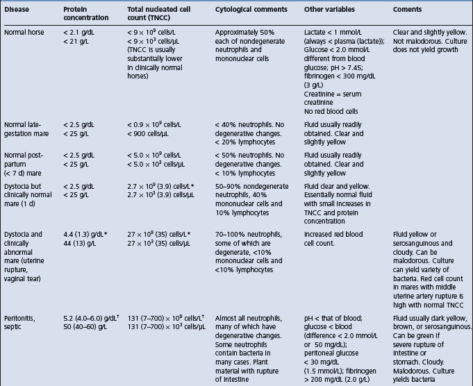
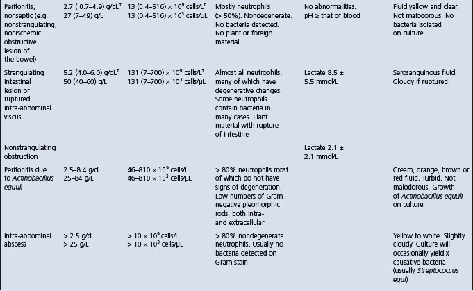
Examination of peritoneal fluid obtained by paracentesis is a valuable aid in the diagnosis of peritonitis and in assessing its severity. It may also provide an indication of the kind of antibacterial treatment required. The values in healthy horses, and horses with various intestinal or peritoneal diseases are provided in Table 5.2. The maximum peritoneal fluid nucleated cell counts in healthy foals is much lower than reported maximum values for adult horses17 and similarly for calves. Particular attention should be paid to:

The ease of collection of the sample as a guide to the amount of fluid present

Whether it is bloodstained, indicating damage to a wall of the viscus

The presence of feed or fecal material, indicating intestinal ischemic necrosis or rupture

Whether it clots and has a high protein content, indicating inflammation rather than simple transudation

The number and kinds of leukocytes present, as an indication of the presence of inflammation, and also its duration

Microbiological examination.

Table 5.2 Characteristics of equine peritoneal fluid in selected diseases of horses

image

image

When these results are available they should be interpreted in conjunction with the history, clinical signs and other results, including hematology, serum chemistry and possibly radiology. In particular, it must be noted that failure to obtain a sample does not preclude a possible diagnosis of peritonitis.

Interpretation of peritoneal fluid is also influenced by simple manipulation of the abdominal viscera and the response is greater than that following opening and closing of the abdomen without manipulation of the viscera. Surgical manipulation results in a significant and rapid postoperative peritoneal inflammatory reaction.11

In peritonitis in horses associated with A. equuli, the peritoneal fluid was turbid and had an abnormal color in 98% of cases. The protein content was elevated above normal in 50 samples (range 25–84 g/L, mean 44 g/L, normal < 20 g/L). Total nucleated cell count was elevated in all samples (range 46–810 × 109 cells/L, mean 230 × 109 cells/L, normal < 10 × 109 cells/L). A nucleated cell count above 100 × 109 cells/L, was present in 88% of animals.9 Pleomorphic Gram-negative rods were seen on cytology in 53% of samples, and a positive culture of A equuli was obtained in 72% of samples.

Experimentally, resection and anastomosis of the small colon in healthy horses causes a different inflammatory response than does manipulation. Absolute values in the peritoneal fluid for cell count, total protein and differential count are inadequate to differentiate between a normal surgical reaction and a postoperative infection. Cytological examination of peritoneal fluid is necessary to demonstrate degenerative cell changes and the presence of bacteria and ingesta. The peripheral leukon and fibrinogen concentration should always be compared with the peritoneal fluid for evidence of postsurgical infection. The nucleated cell and red blood counts of peritoneal fluid are commonly elevated for several days in horses following open castration.15 These elevated counts may be mistaken for peritonitis.

Septic peritonitis in the horse

Diagnosis of septic peritonitis is routinely made on the basis of physical examination and hematologic findings, and peritoneal fluid analysis.18 After abdominal surgery, differentiation between septic peritonitis and other postoperative complications can be difficult using physical and hematological findings alone. As a result of the exploratory process itself, diagnosis of septic peritonitis is often complicated in horses after surgery because the total nucleated cell count and protein concentration in the peritoneal fluid are often high. Consequently, identification of bacteria on cytological evaluation or isolation of bacteria from peritoneal fluid is a more definitive indicator of septic peritonitis, but sometimes there are false-negative results. Although bacterial cultures are considered the standard criterion for the diagnosis of sepsis, positive results may not always be obtained and results may be delayed by a minimum of 24 hours for aerobic organisms and up to 10–14 days for anaerobic organisms. Thus ancillary tests such as pH, glucose concentrations and lactate dehydrogenase (LDH) activity in equine pleural and synovial fluid have been used to detect sepsis with the potential advantages of speed, ease of measurement and lower cost relative to bacterial cultures.18

Horses with septic peritonitis have significantly lower peritoneal fluid pH and glucose concentrations than horses with nonseptic peritonitis and healthy horses.18 Compared with other tests, serum-to-peritoneal fluid glucose concentration differences of more than 50 mg/dL had the highest diagnostic use for detection of septic peritonitis. Peritoneal fluid pH below 7.3, glucose concentration below 30 mg/dL and fibrinogen concentration above 200 mg/dL were also highly indicative of septic peritonitis.

NECROPSY FINDINGS

In acute diffuse peritonitis, the entire peritoneum is involved but the most severe lesions are usually in the ventral abdomen. Gross hemorrhage into the subserosa, exudation and fibrin deposits in the peritoneal cavity and fresh adhesions that are easily broken down are present. In less acute cases, the exudate is purulent and may be less fluid, often forming a thick, cheesy covering over most of the viscera. In cattle, F. necrophorum and Actinomyces (Corynebacterium) pyogenes are often present in large numbers and produce a typical, nauseating odor. Acute local peritonitis and chronic peritonitis are not usually fatal and the lesions are discovered only if the animal dies of intercurrent disease such as traumatic pericarditis or intestinal obstruction.

DIAGNOSIS

The diagnosis of peritonitis can be difficult because the predominant clinical findings are often common to other diseases. The clinical features that are the most reliable as indicators of peritonitis are:

Abnormal feces – in amount and composition

Alimentary tract stasis based on auscultation and evaluation of the passage of feces

Abdominal pain evinced as a groan with each respiration or on light or deep percussion of the abdomen

Abnormality of intestines on rectal palpation

Fibrinous or fibrous adhesions on rectal palpation

Abnormal peritoneal fluid with an increased leukocyte count collected by paracentesis

A normal or low blood leukocyte count with a degenerative left shift

The peritonitis may be chemical, so that, although microbiological examination usually yields positive results, these are not essential to a diagnosis of peritonitis.

PROGNOSIS

Case fatality rate in horses

Peritonitis in the horse is a potentially life-threatening disease that must be treated promptly and aggressively.20 Therapy must be aimed at reducing systemic shock and hypovolemia, correction of the primary cause, antibiotic therapy, and abdominal drainage and lavage. The reported case fatality rates for peritonitis in horses range from 30–67%. In a series of 67 cases of peritonitis in horses, of those which developed peritonitis after abdominal surgery the case fatality was 56%.3 Peritonitis not associated with intestinal rupture or abdominal surgery had a lower case fatality rate of 43%. Horses that died had higher heart rates, red blood cell count, serum creatinine concentration, PCV and anion gap; lower venous blood pH; and a greater number of bacterial species cultured from the peritoneal fluid compared with survivors. Those that died were more likely to have clinical evidence of abdominal pain, shock and bacteria in the peritoneal fluid.

DIFFERENTIAL DIAGNOSIS

The diseases which could be considered in the differential diagnosis of peritonitis are as follows.

Cattle

Acute local peritonitis– Traumatic reticuloperitonitis, acute intestinal obstruction, splenic or hepatic abscess, simple indigestion, abomasal displacement (right and left), postpartum metritis, ketosis

Acute diffuse peritonitis – Parturient paresis, coliform mastitis (peracute form), acute carbohydrate indigestion, perforation of or rupture at abomasal ulcer, acute intestinal obstruction, uterine rupture, postpartum metritis

Chronic peritonitis – Vagus indigestion, lipomatosis or extensive fat necrosis of the mesentery and omentum, persistent minor leakage from an intestinal lesion, large accumulations of fluid as in ascites, rupture of bladder, chronic pneumonia and chronic toxemias due to a great variety of causes

Ascites associated most commonly with primary or secondary cardiac disease, cor pulmonale with chronic pneumonia, endocarditis, thrombosis of the caudal vena cava, and diffuse abdominal epithelioid mesothelioma19

Horses

Acute and subacute peritonitis– Acute intestinal obstruction and thromboembolic colic

Chronic peritonitis – Repeated overeating causing colic, internal abdominal abscess (retroperitoneal or mesenteric abscess) may be classified as chronic peritonitis but is dealt with separately under the heading of retroperitoneal abscess. Horses with both intra-abdominal neoplasms and abscesses will have clinical findings including anorexia, weight loss, fever, colic and depression.13 Both groups may also have peritoneal fluid that can be classified as an exudate

Pigs, sheep and goats

Peritonitis is not usually diagnosed antemortem in these species.

TREATMENT

The specific cause must be treated in each case and the treatments used are described under the specific diseases listed above. An exploratory laparotomy may be indicated to determine the cause of the peritonitis and to effect repair. The literature on the treatment of peritonitis in horses has been reviewed.20

Antimicrobials

Broad-spectrum antimicrobials given intravenously or intramuscularly are indicated for the infection and toxemia. However, there are no published reports of clinical trials to evaluate the effectiveness of various antimicrobials for the treatment of peritonitis in cattle or horses. Thus the recommendations are empirical. In general, peritonitis in cattle is commonly treated with any of the broad-spectrum antimicrobials, with the choice dependent on ease of administration and drug withdrawal times necessary in lactating dairy cattle. Treatment for traumatic reticuloperitonitis has commonly been restricted to the use of antimicrobials; supportive therapy has not been indicated with the exception of diffuse peritonitis.

Peritonitis in horses associated with abdominal surgery or rupture of the gastrointestinal tract is likely be accompanied by a mixed flora of bacteria, and broad-spectrum antimicrobials are necessary. They must be given at doses high enough to achieve high blood and tissue levels and maintained daily until recovery has occurred. In a series of cases of peritonitis in horses, the most commonly used antimicrobials were gentamicin at 2.2–3.3 mg/kg BW intravenously every 8–12 hours; penicillin at 22000 IU/kg BW intravenously or intramuscularly every 6–12 hours. Metronidazole given orally at 15–25 mg/kg BW has also been used in horses with peritonitis.3

Horses with peritonitis associated with A. equuli respond quickly to treatment with penicillin at 20 mg/kg BW intramuscularly twice daily for 5 days to 2 weeks.9 Most isolates of the organism are sensitive to penicillin but some are resistant and gentamicin sulfate at 6.6 mg/kg BW intravenously once daily for 5 days to 2 weeks in combination with the penicillin has also been used successfully.9 In a series of 51 cases in horses, the recovery rate following treatment with penicillin and gentamicin and supportive therapy was 100%.9 Most horses responded favorably within 48 hours following commencement of treatment.

Administration of antimicrobials into the peritoneal cavity has been attempted on the basis that higher levels of the drug may be achieved at the site of the inflammation. However, there is no scientific evidence that it is superior to daily parenteral administration and there is some danger of causing adhesions and subsequent intestinal obstruction.

Fluid and electrolytes

Intensive intravenous fluid and electrolyte therapy is a vital part of treatment of peritonitis when accompanied by severe toxemia and shock, especially during the first 24–72 hours following abdominal surgery in the horse. It is continued until recovery is apparent and the animal is drinking water voluntarily; water can then be supplemented with electrolytes. (See Ch. 2 for details of fluid and electrolyte therapy for the treatment of dehydration and toxemia.)

Nonsteroidal anti-inflammatory drugs

Flunixin meglumine is recommended at 0.25–1.1 mg/kg BW intravenously every 8–12 hours when the peritonitis is accompanied by shock. However, no information is available on efficacy.

Lavage

Peritoneal lavage with large volumes of fluid containing antimicrobials is rational and has been attempted when large quantities of exudate are present. However, it is not easy to maintain the patency of drains, especially in cattle. Also, peritoneum is highly susceptible to inflammation and chemical peritonitis is common following the introduction of certain materials into the peritoneal cavity. Peritoneal lavage of ponies with saline and antimicrobials induces a mild, transient inflammatory response with minimal change visible at necropsy.21 Solutions containing povidone-iodine induced chemical peritonitis, which was severe when 10% povidone-iodine solution was used. A 3% solution also causes peritonitis and the use of these solutions is not recommended. Extreme caution is required when foreign materials are introduced into the cavity in order to avoid exacerbating the existing inflammation. The peritoneum is also a very vascular organ and toxic material is rapidly absorbed from it.

An active intra-abdominal drain has been used successfully to treat abdominal contamination in horses.22 Closed-suction abdominal drains were placed, mostly under general anesthesia. Abdominal lavage was done every 4–12 hours and about 83% of the peritoneal lavage solution was retrieved.

Prevention of adhesions

No attempt is made to prevent the development of adhesions.

REVIEW LITERATURE

Dyson S. Review of 30 cases of peritonitis in the horse. Equine Vet J. 1983;15:25-30.

Davis JL. Treatment of peritonitis. Vet Clin North Am Equine Pract. 2003;19:765-778.

REFERENCES

1 Cable CS, et al. J Am Vet Med Assoc. 1998;212:1442.

2 Tyler JW, et al. Vet Rec. 1998;143:280.

3 Hawkins JF, et al. J Am Vet Med Assoc. 1993;203:284.

4 Platt H. J Comp Pathol. 1983;93:343.

5 Edwards JF, Ruoff WW. J Am Vet Med Assoc. 1991;198:1421.

6 Ross MW, et al. J Am Vet Med Assoc. 1985;187:249.

7 Lapointe JM, et al. Vet Pathol. 2003;40:338.

8 Fubini SL, et al. J Am Vet Med Assoc. 1990;197:1060.

9 Matthews S, et al. Aust Vet J. 2001;79:536.

10 Patterson-Kane JC, et al. Vet Pathol. 2001;38:230.

11 Hanson RR, et al. Am J Vet Res. 1992;53:216.

12 Mair TS, et al. Vet Rec. 1990;126:567.

13 Zicker SC, et al. J Am Vet Med Assoc. 1990;196:1130.

14 Ebeid M, Rings DM. Bovine Pract. 1999;33:144.

15 Schumacher J, et al. J Vet Intern Med. 1988;2:22.

16 Braun U. Vet J. 2003;166:112.

17 Grinden CB, et al. Equine Vet J. 1990;22:359.

18 Van Hoogmoed L, et al. J Am Vet Med Assoc. 1999;214:1032.

19 Milne MH, et al. Vet Rec. 2001;148:341.

20 Davis JL. Vet Clin North Am Equine Pract. 2003;19:765.

21 Schneider RK, et al. Am J Vet Res. 1988;49:889.

22 Nieto JE, et al. Vet Surg. 2003;32:1.

RECTAL TEARS

Iatrogenic tears of the equine rectum are a serious problem in equine practice. They are a leading cause of malpractice suits for the veterinarian, comprising approximately 7% of insurance claims against veterinarians in equine practice in the USA,1 and can be a large economic loss for the owner. Occurrence of rectal tears is often an emotionally charged event because they are unexpected and they usually occur in otherwise healthy horses being subjected to routine rectal examination. Prompt diagnosis and vigorous treatment, along with frank disclosure of the event to the horse’s owner or handler, is essential in increasing the likelihood of a good outcome both for the horse and for the veterinarian–client relationship.

ETIOLOGY

The etiology of rectal tears is usually readily apparent, with the vast majority of rectal tears in horses being iatrogenic. Iatrogenic rupture occurs during rectal examination by veterinarians or laypersons for reproductive management (brood mares), or examination of other intra-abdominal structures, for example during evaluation of a horse with colic.2 Spontaneous or non-iatrogenic rupture can occur associated with infarctive lesions of the distal small colon or rectum, injuries during parturition or coitus, and malicious trauma caused by insertion of foreign objects by attendants.3 It is important that rectal tears should not be assumed to be iatrogenic until a thorough evaluation of the animal and the history has been performed.

EPIDEMIOLOGY

Risk factors for rectal tears in horses have not been well quantified but include:

Age – young animals appear to be at increased risk, perhaps because they are smaller and less accepting of rectal palpation

Sex – stallions and geldings have a smaller pelvic inlet than do mares and appear to have a rectum of smaller diameter than mares, thus increasing the risk of tension on the wall of the rectum, with subsequent tearing

Breed – Arabian horses appear to be at increased risk of iatrogenic rectal tears

Size – smaller animals can be at increased risk

Inadequate restraint – horses must be adequately restrained for rectal examination (see Prevention, below)

Inadequate preparation of the rectum – the rectum and distal small colon should be emptied of feces before an examination of the reproductive organs or gastrointestinal tract is performed

The experience of the examiner is not a factor in the risk of rectal tears in horses4

The use of ultrasonographic probes per rectum does not appear to increase the risk of rectal tears.4

The case fatality rate varies depending on the type of tear (see Clinical signs, below). Horses with grade I or II tears almost all survive, whereas the survival rate for horses with grade III tears treated appropriately is 60–70%.5 Almost all horses with Grade IV rectal tears die.

PATHOGENESIS

Rectal tears occur in horses because the rectum of the horse is so sensitive and fragile and powerful contractions occur during rectal palpation. In contrast, the bovine rectum is relatively durable and, while often traumatized, is rarely ruptured. Tears occur because of excessive tension on the rectal wall. This usually occurs in horses by peristalsis and contraction of the rectum over the examiner’s hand, with splitting of the rectum often occurring over the back (knuckles) of the hand.

Complete rupture of the peritoneal portion of the rectum results in fecal contamination of the abdomen and rapid onset of septic peritonitis and death. Tears in the nonperitoneal portion of the rectum (that is, caudal to the peritoneal reflection) cause perirectal cellulites and abscessation.

CLINICAL SIGNS

The prominent clinical sign of the occurrence of a rectal tear is the presence of blood on the rectal sleeve of the examiner. Slight blood staining of mucus or lubricant is usually not associated with rectal tears (although this should be verified by repeat examination) whereas the presence of frank hemorrhage on the sleeve is usually indicative of a rectal tear. The rectum in an adult, 450 kg horse, is approximately 30 cm long and is partially within the abdomen, where it is covered by peritoneum, and partially in the pelvic canal, where it is not surrounded by peritoneum but is supported by thick connective tissue and muscle. The peritoneal portion of the rectum is supported dorsally by the mesorectum (mesocolon). Most iatrogenic rectal tears in horses occur within 25–30 cm of the anus, but can occur up to 60 cm from the anus, in the peritoneal portion of the rectum. The tears are almost always in the dorsal or dorsolateral wall and are longitudinal (parallel to the long axis of the rectum). It is speculated that the dorsal wall of the rectum is weaker than other segments because it is not covered by serosa, and blood vessels perforate the muscularis layers, thereby weakening it.5

Rectal tears in the horse have been classified according to the layers of the rectal wall disrupted. The classification is also a useful guide to the clinical signs to be expected and the treatment that is indicated (see under ‘Treatment’ for management of each grade of tear):

Grade I – Disruption of the mucosa only, or the mucosa and submucosa. There are usually no clinical signs other than some blood on the examiner’s sleeve. Most of these injuries occur to the mucosa of the ventral aspect of the rectum5

Grade II – Disruption of the muscular layer of the rectal wall with the mucosal and serosal surfaces intact. This is a rarely recognized form of tear. There are minimal clinical signs

Grade IIIa – Tear includes mucosa, submucosa and muscularis but the serosal surface is intact. This degree of tear usually causes septic peritonitis. If the tear is caudal to the peritoneal reflection the pelvic fascia becomes infected, but the infection may remain contained within it for 7–10 days, forming a local cellulitis or abscess. During this period, the horse is likely to be affected by mild chronic peritonitis, with mild abdominal pain, fever and mild toxemia. At the end of this time, the infection can erode through the peritoneum and cause an acute, severe, diffuse peritonitis, or rupture through the perianal tissue causing a fistula

Grade IIIb – Tear is on the dorsal wall and includes the mucosa, submucosa and muscularis. Because there is no serosa at this position, the tear extends into the mesocolon. There is usually septic peritonitis

Grade IV – Complete rupture with leakage of fecal material into the peritoneal space. Clinical signs of septic peritonitis are severe and death is inevitable.

Horses with a rectal tear will not display any immediate signs of discomfort. However, if there is grade III or grade IV tear, the horse will have signs of septic peritonitis, including elevated heart and respiratory rates, sweating, colic, increased capillary refill time and discolored mucus membranes, within 1–2 hours.

CLINICAL PATHOLOGY

Hematological and serum biochemical changes in horses with grade III and grade IV tears are consistent with acute septic peritonitis. These changes include leukopenia and neutropenia, increased band cell count, elevated hematocrit and total protein concentration initially, after which serum total protein concentration can decline as protein leaks into the abdomen. Peritoneal fluid has a high white blood cell count and protein concentration. Cytological examination reveals the presence of degenerate neutrophils, intra- and extracellular bacteria and plant material.

PROGNOSIS

The prognosis depends of the size, grade and location of the tear and the time between occurrence and treatment. All horses with grade I or II lesions survive, approximately 60–70% of horses with grade III lesions survive, and almost all horses with grade IV lesions die.5

TREATMENT

If the person doing the rectal examination feels the mucosa tear, if there is blood on the rectal sleeve, or if a horse that has had a rectal examination up to 2 hours previously starts to sweat and manifest abdominal pain, a rectal tear should be suspected. A thorough examination should be conducted immediately but great care is necessary to avoid damaging the rectum further. The principles of care are to: verify the presence of a tear, determine its severity, prevent leakage of fecal material into the peritoneum or tissues surrounding the tear, treat for septic peritonitis, prevent extension of the tear and provide pain relief.

Immediate care6

If a rectal tear is suspected the horse should be appropriately restrained and examined immediately. There should be no delay in conducting this examination. The client should be informed of the concern about a rectal tear. First aid measures taken at the time of a grade III or IV tear can have a marked influence on the outcome.2,5 Horses with grade III or IV rectal tears should receive first aid treatment and then be referred for further evaluation and treatment.

The existence of a tear should be determined and its severity assessed. This is best achieved by sedating the horse, providing local analgesia of the rectal mucosa and anus, and careful manual and visual examination of the rectal mucosa. Sedation can be achieved by administration of adrenergic agonists (xylazine, romifidine, detomidine) with or without a narcotic drug (butorphanol, meperidine, pethidine, morphine). Analgesia of the rectum and anus can be induced by epidural anesthesia (lidocaine or xylazine) or local application of lidocaine gel or lidocaine enema (10–15 mL of 2% lidocaine in 50–60 mL of water infused into the rectum). Peristalsis can be reduced by administration of hyoscine (N-butylscopolammonium bromide, 0.3 mg/kg intravenously).

Manual or visual examination of the rectum can then be performed. Manual examination is performed after generous lubrication of the anus and examiner’s hand and arm. Some authorities prefer to use bare hands, rather than gloves or a rectal sleeve, for this examination because of the decreased sensitivity when wearing gloves. However, one should be aware of the health risks to the examiner of not using barrier protection (gloves) during a rectal examination. The rectum should be evacuated of feces and a careful and thorough digital examination should be performed. If a tear is detected, the position, distance from the anus, length and depth of the tear should be determined. Gentle digital examination should be used to determine the number of layers involved and if there is rupture of the rectum and communication with the peritoneal space.

Alternatively, the rectum can be examined visually through a mare vaginal speculum, or using an endoscope. Both these approaches are likely to minimize the risk of further damage to the rectum. These examinations can be impaired by the presence of fecal material.

If a grade III or IV rectal tear is detected, then the horse should be administered broad-spectrum antibiotics (penicillin, aminoglycoside and possibly metronidazole) and NSAIDs, and referred for further evaluation. Some, but not all, authorities recommend placement of a rectal pack to prevent further contamination of the rectal tear. This is formed from a 3 inch (7.5 cm) stockinette into which is inserted a roll of cotton (approximately 250 g) The roll is moistened with povidone-iodine solution, lubricated and inserted into the rectum in the region of the tear. Epidural anesthesia will prevent expulsion of the roll in the short term.

Prompt referral and care is essential for maximizing the likelihood of a good outcome in horses with grade III and IV tears.

Grade I and grade II tears

Treatment of these tears is medical. Horses should be administered broad-spectrum antibiotics and feces should be softened by the administration of mineral oil. These wounds heal in 7–10 days.

Grade III tears

Both medical and surgical treatments are effective in approximately 60–70% of cases of grade III tears.7-9 The choice of treatment depends on the expertise and experience of the attending clinician and financial constraints imposed by the horse’s owner. Surgical treatment includes direct repair of the tear (for those lesions that can be readily exposed via the anus), placement of a rectal sheath by ventral laparotomy and placement of a loop colostomy. Surgical repair is in addition to aggressive treatment of peritonitis.

Medical treatment includes administration of broad-spectrum antibiotics (such as penicillin, aminoglycoside and metronidazole), anti-endotoxin drugs (such as hyperimmune serum or polymyxin sulfate), NSAIDs, crystalloid fluids, colloidal fluids (hetastarch, plasma) and heparin, and other care. Peritoneal lavage might be indicated. Manual evacuation of the rectum at frequent intervals (every 1–2 hours for 72 hours and then 4–6 times daily for a further 7 days) was suggested to improve the prognosis,9 although others caution against manual evacuation of the rectum because of the risk of worsening the tear.8

Grade IV tears

Tears of this severity require immediate surgical intervention to minimize fecal contamination of the peritoneum. However, the grave prognosis and high cost of treatment, and poor success of surgical intervention in these cases, means that most horses are euthanized. If surgical care is attempted, there should also be aggressive medical treatment of the peritonitis.

PREVENTION

As noted above, rectal tears can occur during examination by even the most experienced operators. Ideally, the owner should be informed of the risks of rectal palpation and explicit consent to perform the examination should be obtained. This is especially important for animals that are at increased risk of rectal tears.

The examination should be performed only when there is a clear clinical reason for performing a rectal examination, when the animal is a suitable candidate for rectal examination, and when the animal can be adequately restrained to permit a thorough examination to be performed in relative safety for both the examiner and the animal.

The examiner should proceed cautiously with the examination. The gloved hand and arm of the examiner should be well lubricated with a water-based lubricant. The anus should be gently dilated by using fingers shaped into a cone. Feces should be evacuated from the rectum such that the rectum is empty to the most cranial extent of the region to be examined. If the horse is anxious and straining, or if there is excessive peristalsis, then the animal should be sedated and antiperistaltic drugs (such as hyoscine) should be administered. The examination should be halted if the horse begins to struggle or resist the examination excessively. Application of a nose twitch often facilitates the examination.

During the examination care should be exercised not to resist peristaltic waves – the hand should be withdrawn in front of these advancing waves and reinserted as peristalsis passes. The fingers should not be opened widely during the examination and care should be taken not to put excessive pressure on a small region of rectum, such as might occur when trying to grasp an ovary or loop of distended intestine.

A rectal tear in a horse is a common cause of a malpractice suit and the veterinarian involved with the case is advised to recommend to the owner that a second opinion be solicited from another veterinarian in order to minimize any misunderstanding.

REFERENCES

1 Blikslager AT, Mansmann RA. Compend Contin Educ Pract Vet. 1996;18:1140.

2 Watkins JP, et al. Equine Vet J. 1989;21:186.

3 Guglick MA, et al. J Am Vet Med Assoc. 1996;209:1125.

4 Sloet van Oldruitenborgh-Oosterbaan MM, et al. Tijdschr Diergeneeskd. 2004;129:624.

5 Eastman TG, et al. Equine Vet Educ. 2000;12:263.

6 Sayegh AI, et al. Compend Contin Educ Pract Vet. 1996;18:1131.

7 Alexander GR, Gibson KT. Aust Vet J. 2002;80:137.

8 Mair TS. Equine Vet J Suppl. 2000;32:104.

9 Katz LM, Ragle CA. J Am Vet Med Assoc. 1999;215:1473.

RETROPERITONEAL ABSCESS (INTERNAL ABDOMINAL ABSCESS, CHRONIC PERITONITIS, OMENTAL BURSITIS)

A recognized form of chronic or rarely intermittent colic is associated with an abscess in the abdominal cavity. The abscesses are usually retroperitoneal, sometimes involving the omental bursa, and chronic leakage from them into the peritoneal cavity causes chronic or recurrent peritonitis. Complete recovery is difficult to effect and there is a high failure rate in treatment. These abscesses result from any of the following:

Infection of a verminous aneurysm, especially in young horses

Post-strangles infection localizing anywhere, but particularly in pre-existing lesions such as verminous aneurysms

Minor perforations of intestinal wall allowing minimal leakage of intestinal contents so that omental plugging is possible

Erosion through a gastric granuloma associated with Habronema sp. or a squamous cell carcinoma of stomach wall

In mares, development of an abscess in the pelvic fascia commonly results after tearing of the rectal wall during pregnancy diagnosis.

Clinical findings suggestive of the disease include persistent or intermittent chronic colic and weight loss. A fever is common and varying degrees of anorexia are typical. In cases with a concurrent chronic peritonitis or an omental bursitis the amount of inflammatory exudate may be large enough to cause abdominal distension. When the abscess is perirectal and in the pelvic fascia there may be straining and constipation due to voluntary retention of feces.

On rectal examination it may be possible to feel an abscess, or adhesions to one. They are often multiple and quite large and adherent to one another, so that tight bands of mesentery can be felt that will lead the hand to the site of the abscess. Pain is usually elicited by rectal palpation of the infected sites and by firm palpation of the external abdominal wall. Ultrasonography through the abdominal wall has been used to locate large retroperitoneal abscesses in a foal.

The hemogram, especially in acute cases, is characterized by a neutrophilia, which may be as high as 30000/μL with a left shift. Chronic anemia due to bone marrow depression may occur and increased plasma fibrinogen and hypoalbuminemia occur. Abdominocentesis may yield turbid fluid with a protein content greater than 2.5 g/dL and an increase in leukocytes. If culture is possible the causative bacteria are usually S. equi, S. zooepidemicus, C. equi, Corynebacterium pseudotuberculosis or mixed infections if there has been intestinal leakage. It is common, even when there is an active infection in a retroperitoneal abscess, to fail to grow bacteria from a peritoneal effusion.

Intra-abdominal abscesses must be differentiated from abdominal neoplasms in the horse.1 Anorexia, weight loss, fever, colic and depression are common to both syndromes. The laboratory findings in both groups are similar but cytological examination of the peritoneal fluid may yield an accurate diagnosis in the case of neoplasms.1

Leakages from stomach wall may result in adhesions to the spleen and development of splenic abscesses. In these animals a sharp pain response can be elicited on firm palpation of the abdomen in the left flank just behind the last rib. Abscesses in liver are not so easily located. Abscesses in pelvic fascia are usually not very discrete but are instantly noticeable on inserting the hand into the rectum.

TREATMENT

Treatment with broad-spectrum antimicrobials is indicated and the initial response is good but often transitory if the usual course of treatment is only 3–5 days’ duration. The prognosis is usually tentative because of the difficulty of completely eliminating the infection. Treatment must be continued for at least 2 weeks and in some cases for a period of 2 to even 4–5 months. Surgical treatment may be possible but is usually ineffectual because of the deformity of the area by adhesions and the usual outcome of tearing the intestine and spillage into the peritoneal cavity while attempting to exteriorize the lesion.

REFERENCE

1 Zicker SC, et al. J Am Vet Med Assoc. 1990;196:1130.

ABDOMINAL FAT NECROSIS (LIPOMATOSIS)

The hard masses of necrotic fat that occur relatively commonly in the peritoneal cavity of adult cattle, especially the Channel Island breeds and possibly Aberdeen Angus, are commonly mistaken for a developing fetus and can cause intestinal obstruction. The latter usually develops slowly, resulting in the appearance of attacks of moderate abdominal pain and the passage of small amounts of feces. Many cases are detected during routine rectal examination of normal animals. The lipomatous masses are located in the small omentum, large omentum and mesentery in cattle and more diffusely to other parts of the body in sheep and goats.1 The composition of the fatty deposits is identical with the fat of normal cows and there is no suggestion that the disease is neoplastic. Sporadic cases are most common but there are reports of a herd prevalence as high as 67%.2 The cause is unknown but there appears to be a relation between such high prevalence and the grazing of tall fescue grass,2 and an inherited predisposition is suggested. The rate of occurrence increases with age, the peak occurrence being at 7 years of age. It has been suggested that excessive fattiness of abdominal adipose tissue may predispose cattle to fat necrosis.3 An unusual form of the disease with many lesions in subcutaneous sites has been recorded in Holstein–Friesian cattle and is regarded as being inherited. There is no treatment and affected animals should be salvaged. A generalized steatitis has been reported in pony foals.

Pedunculated lipomas provide a special problem especially in older horses. Their pedicles may be 20–30 cm long and during periods of active gut motility these pedicles can become tied around a loop of intestine anywhere from the pylorus to the rectum. At the pylorus they cause acute intestinal obstruction with gastric dilatation. At the rectum they cause subacute colic and a characteristic inability to enter the rectum with the hand. This is accompanied by a folded coning-down of the mucosa, not unlike that in a torsion of the uterus. Early diagnosis and surgical intervention can produce a resolution but delay is disastrous because the blood supply is always compromised: it is always a loop and its blood supply that are strangulated. The pedicle is always tied in a very tight knot.

REFERENCES

1 Xu LR. Acta Vet Zootechnol Sinica. 1986;17:113.

2 Stuedemann JA, et al. Am J Vet Res. 1985;46:1990.

3 Katamoto H, et al. Res Vet Sci. 1996;61:214.

TUMORS OF PERITONEUM

Disseminated peritoneal leiomyomatosis has been reported occurring in a mature Quarter horse.1 Clinical findings included inappetence, weight loss, intermittent fever, chronic abdominal pain and enlargement of the abdomen. Rectal examination revealed a prominent, firm, smooth-walled mass in the ventral aspect of the abdomen. Transabdominal ultrasonography was used to detect the mass, which was a friable, polycystic structure occupying a large portion of the abdominal cavity and weighing 34 kg. The mass was removed and recovery was complete.

Mesothelioma has been reported in cattle, predominantly in the peritoneal cavity, but mesothelioma can also occur in the pleural cavity and the vagina of adult cattle. The cause of mesothelioma in cattle is unknown but pleural mesothelioma in humans is associated with asbestos exposure. One report suggested that the frequency of diagnosis in cattle is increasing.2 All ages of cattle can be affected with peritoneal mesothelioma, but affected animals are typically young, with fetal and neonatal cases also being reported.3,4 Calves and adult cattle most frequently present with moderate abdominal distension.5 Other presenting signs include scrotal edema in intact males5 and ventral pitting edema. Occasionally, small 2–20 mm, well demarcated ‘bumps’ can be felt on all serosal surfaces during palpation per rectum in adult cattle. Peritoneal fluid is easily obtained by ventral abdominal paracentesis and has the characteristics of a modified transudate with a moderate to marked increase in phagocytically active mesothelial cells. Definitive diagnosis is made during a right-sided exploratory laparotomy, where numerous raised, white, well demarcated masses are palpated on all serosal surfaces, with copious abdominal fluid being present. Biopsy of these masses and microscopic examination confirms the presumptive diagnosis of mesothelioma. Extensive peritoneal mesothelioma is fatal and there is no known treatment. All cases reported to date are sporadic and there is no apparent association with asbestos or other toxic agent in cattle.

REFERENCES

1 Johnson PJ, et al. J Am Vet Med Assoc. 1994;205:725.

2 Pizarro M, et al. J Vet Med A. 1992;39:476.

3 Klopfer U, et al. Zentralbl Vet Med B. 1983;30:785.

4 Anderson BC, et al. Vet Med. 1984;79:395.

5 Wolfe DF, et al. J Am Vet Med Assoc. 1991;199:486.