Control of bovine mastitis
Improvement in udder health has been a major initiative of the dairy industry for over 40 years. The thrust of these efforts has been on the implementation and use of management techniques to limit the spread of major mastitis pathogens, thereby reducing the quarter infection rate. Detailed mastitis control strategies have been outlined and promoted by the National Institute for Research in Dairying (NIRD) and the National Mastitis Council (NMC). With proper implementation, these programs result in a dramatic decrease in the prevalence of common contagious mastitis pathogens. Herds that have successfully implemented a comprehensive mastitis control program also need to develop strategies to control infection with environmental organisms, as well as utilizing an effective monitoring system for new infections. Achievement of excellent udder health for the production of high-quality milk is a realistic and important goal for all aspects of the dairy industry.
The adoption of effective mastitis control programs has often been less than desirable, even with extensive research validation of the recommended control practices and with major extension efforts at both national and local levels. The reasons for this slow adoption of proven mastitis control strategies are not well documented, even though producers look to the veterinary profession for information on mastitis and its control.1 If significant progress is to be made towards a general improvement in udder health status, veterinarians must become more active in organizing, conducting and monitoring comprehensive mastitis control programs.
Veterinarians usually become involved in mastitis control in one of the following circumstances:
• The herd is experiencing a higher than normal incidence of clinical cases
• The milk processing plant reports a higher than permissible total bacterial count or bulk tank milk SCC
• A farmer who is not carrying out the standard program of postmilking teat dipping and dry period treatment asks for advice – either as a single mastitis control program or, more probably, as part of a herd health management program.
The procedure is the same in all these situations, any variation being in terms of speed and intensity. It consists of an assessment of the herd’s mastitis status and the implementation of a recommended mastitis control program.
Udder health improvement
The benefit of an integrated mastitis program is improved udder health; this improvement is progressive and can usually be observed within a few years after implementation at the herd level. In a closed, monitored herd applying the control measures there can be a reduction in the annual incidence of clinical mastitis from 130 cases/100 cows to a level of 35–40 cases/100 cows.2 In the UK it is estimated that, since 1970, farms that have followed the recommended control procedures have reduced the average annual incidence of cases of clinical mastitis from 135 to 40 cases/100 cows each year, while the percentage of quarters remaining uninfected for a whole year has increased from 65% to 80% of the total quarters.3 The control program has been most effective in reducing mastitis due to the contagious pathogens such as S. aureus, S. agalactiae, and S. dysgalactiae. These are now responsible for one-third or less of all clinical cases of mastitis compared to 30 years ago. This represents an annual reduction from about 90 to 12 cases/100 cows. The percentage of infected cows declined from 22% to 12% and the percentage of infected quarters from 7% to 3%. Methods now exist to control contagious pathogens and reduce the bulk tank milk SCCs to below 400000 cells/mL. With good management, the incidence of clinical mastitis can be kept low (7–21 cases/100 cows/year with dairy herds averaging 45 cows/herd) by culling any cow with chronic or recurrent mastitis and by paying great attention to housing and management standards.4-6
While the rate of contagious mastitis has been decreased with the program, the rate of infections and the incidence of mastitis associated with environmental pathogens such as S. uberis and the coliform bacteria has not decreased. Approximately 65% of clinical cases are now due to environmental pathogens. Organisms prevalent in the cow’s environment currently cause the most costly types of mastitis in the USA.7
Economic benefits, incentives, and penalties
Mastitis is one of the most costly diseases in dairy herds. Some surveys indicate that the cost incurred by producers because of clinical mastitis is much higher than the cost of prevention.7 An integrated mastitis control program has always been an excellent investment for the dairy farmer,3 with a revenue to cost ratio of approximately 6:1,3 most of the additional revenue being due to increased milk production.
Differential payments to farmers for milk quality are also an economic incentive to adopt a control program.6 The widespread adoption of bulk tank milk SCCs as a measure of milk quality, and the adoption of payment schemes of increasing severity, has stimulated farmers to reduce their cell count. Many milk marketing cooperatives have established both penalty and incentive programs based on bulk tank milk SCC and total bacterial counts as global measures of milk quality.
Requirements
The requirements for a successful mastitis control program include a willing farmer, a capable diagnostic laboratory, an enthusiastic and knowledgeable veterinarian, a record keeping system, adequate milking machinery and adequate housing facilities.
The farmer must have health and production goals and be willing to achieve them by making a commitment to invest the resources to control mastitis. Wide variations in the costs of controlling and monitoring mastitis in herds are evident because of lack of client compliance with accepted recommendations for mastitis control.8 There are also variations in the level of mastitis control procedures adopted by producers, which affect the success of a program. A survey of a large number of dairy herds in Ohio revealed that most herd managers teat dipped all cows after milking and used dry cow therapy.9 However, other mastitis control practices were not as widely adopted. In addition, those herds classified as negative for contagious pathogens used predipping compared with herds classified positive for contagious pathogens, which did not. Predipping was also associated with a number of management factors, including having a clean and dry environment for the cows, cows with clean udders and teats, and minimal use of water in the milking parlor. A survey of Ontario dairy farms indicated that only one-third of producers used all five of the recommended mastitis control practices.10 Lack of adoption may result from a lack of awareness of the economic returns from a complete program, adoption of a new practice only in response to a problem, or competition for liquid financial resources from other aspects of the enterprise.
The veterinarian must be knowledgeable about all aspects of mastitis and be willing to invest the time and effort required to provide sound advice based on the health and production information obtained from monitoring the herd. Some surveys of veterinarians indicate wide variations in their clinical approach to cases of mastitis in the field.11 Many factors of importance for the clinical diagnosis of mastitis were not considered.
A data recording system that records all the udder health and production data, and the milk quality of each cow and the herd on a regular basis is a vital requirement. A diagnostic laboratory or milk recording agency that provides regular SCCs of individual cows is necessary to monitor udder health. The milking machine and the housing facilities must be adequate for the size of the herd. The animal attendants must be aware of the health and production goals of the herd and adhere to the principles of mastitis control.
OPTIONS IN THE CONTROL OF MASTITIS
The broad options for control are either eradication or decreasing the infection rate, and either legislative control or implementing a voluntary program.
Eradication
Complete eradication of bovine mastitis from a herd or geographical region is not a practicable target in most circumstances. The exception is mastitis due to Streptococcus agalactiae, which can be eradicated from individual herds by a blitz technique. The difficulty in attempting to eradicate mastitis is that the contagious causes of mastitis, S. agalactiae and Staphylococcus aureus, are so contagious, and the sources of infection so widespread, that adequate quarantine would be very difficult to maintain. In the case of S. aureus there is the additional difficulty of eliminating the infection from its intracellular sites in mammary tissue. The environmental infections, especially Escherichia coli, pose an even greater problem. They are so ubiquitous that reinfection would be almost immediate in cows housed in economically practicable surroundings.
Decreasing the infection rate
This is a practicable proposition, the degree of limitation being dependent on the need to maintain cost-effectiveness. One of the virtues deriving from this necessity is the concept that subclinical mastitis causes a continuous low-level leukocytosis in the milk that acts as a protective mechanism against other infetions. Present-day knowledge about immunity in the mammary gland suggests that control programs that reduce milk SCCs to unrealistically low concentrations may reduce the gland’s resistance to clinical mastitis. Correspondingly, the complete elimination of common udder pathogens such as S. agalactiae and S. aureus is thought to increase the susceptibility of the udder to environmental pathogens, especially the coliforms. Another relevant example is the commonly encountered minor pathogen Corynebacterium bovis, which may be a significant microbial agent in maintaining the resistance of udders. The mastitic effect of this organism is too low to warrant action against it but the infection rate with major pathogens is significantly lower in quarters that harbor it than in those that do not.12 An intensive program to disinfect udders could well eliminate C. bovis and increase susceptibility to other pathogens. There is good field evidence to suggest that this is in fact happening in North America and the UK. C. bovis is likely to be more important where cows are housed, or confined in straw yards, and therefore more exposed to teat contamination with coliforms. The criticism relates to the increasing numbers of clinical cases, often associated with Gram-negative organisms, that occur in herds that have achieved very low levels of udder infection, as indicated by very low bulk milk cell counts. The question of whether it is better practice to maintain some level of bacterial infection with innocuous organisms in the udder as a protection against more damaging pathogens, rather than to attempt complete bacterial sterilization, is still unresolved. For the time being it is generally agreed that decreasing the infection rate is the appropriate target.
Legislative control
Mastitis does not lend itself to eradication (as set out under Eradication above) and so legislative control of the disease is not attempted.
A voluntary program
Most of what is done in mastitis control is by voluntary involvement by producers in programs aimed at reducing the incidence of mastitis and maintaining the infection rate at a low level. The justification for control of the disease is purely economic, and a control program must therefore be based on its applicability on each individual farm. Area or national control can only be in the form of providing incentives by educational and laboratory assistance to individual farmers who wish to participate. The value of a mastitis awareness program, and the part played by the two-way flow of information between farmers and the program operators is most apparent when an area campaign is conducted by a government or industrial sponsor.6 Once a control program is in place it is customary for milk processors, aided in some places by government agencies, to encourage participation by paying incentives for bulk tank milk with low SCCs or bacteria counts, or refusing to accept milk for processing or, in some cases, refusing to transport milk that does not satisfy statutory requirements. This could be the first step in incorporating the program into planned health and production programs that promote mastitis control and maintenance of milk production at financially optimal levels. Mastitis infections in beef cattle herds are currently at too low a level for a mastitis control program to be financially advantageous.
PRINCIPLES OF CONTROLLING BOVINE MASTITIS
Dynamics of infection
The principles of a bovine mastitis control program are based on changing the dynamics of infection, which are as follows:13
• Prevalence of infection is a function of the rate of new infection minus the rate of elimination
• Rate of new infection is a function of the level of exposure times the number of susceptible quarters
• Rate of elimination is a function of the number of infections times the efficacy of treatment plus spontaneous cure.
Successful control occurs when the level of infection is held low or is decreased, either by preventing new infections or eliminating existing infections.
The dynamics are not, however, so simple in reality. They vary with the susceptibility of the individual animal, which changes with age and stage of lactation and is season-dependent. The dynamics may vary with the pathogens involved, and the relative importance between herds can be very considerable and also vary with time. The duration of infection may be extremely different for different pathogens. E. coli causes mild to severe acute clinical disease but usually self-eliminates quickly; it is rarely found in subclinical infections. S. agalactiae and S. aureus are very persistent, and S. aureus responds poorly to treatment. The rate of elimination and the persistency of these pathogens are highly variable. Similarly, there can be large variations in the rate of new infections, which is very much related to the identifiable risk factors, including rate of teat contamination, mechanisms aiding teat penetration, and effectiveness of establishment and growth of bacteria in the mammary gland.
The success of a control program can be measured by the decrease in level of infection and the speed with which this is achieved. The farmer must be able to appreciate progress within a year in order to remain enthusiastic about application of the methods. The level of infection can be controlled significantly by lowering the rate of new infections, but the speed of change very much depends on the duration of the infection and is thus related more to the rate of elimination. No control procedures are available to prevent all new infections and only culling of chronically infected cows is absolutely successful in eliminating infections. Control schemes therefore require both prevention and elimination to give optimal effect, and that optimum will vary with each pathogen.
The specific components of a mastitis control program must be devised to fulfill three basic principles, which are to eliminate existing infections, prevent new infections and monitor udder health.
1. ELIMINATE EXISTING INFECTIONS
The control program must reduce the duration of infection in the cows. Antimicrobial therapy during the dry period is the best method of achieving this objective. Treatment during lactation can be useful to eliminate some existing infections, depending on the causative agent. Culling of chronic cases that are not eliminated with dry period treatment is also used to remove the most persistent existing infections. Further study needs to focus on development of treatment protocols and on cowside identification of the causative bacterial agent.
2. PREVENT NEW INFECTIONS
The control program must reduce the rate at which new infections occur. The dipping of all teats in an effective teat dip after each milking is the best method of reducing the new infection rate. Insuring that the milking machine is functioning properly and used correctly will result in less spread of infection. The dry period is the time of greatest risk of new infection, and blanket dry cow therapy or application of an internal teat sealant is efficacious in preventing new infections during the dry period. Environmental and nutritional management have also become important for the prevention of new infections. Specific recommendations for methods of reducing new infection rate depend upon the predominant pathogen in the herd.
3. MONITOR UDDER HEALTH STATUS
An ongoing program to monitor the udder health status of individual cows as well as the herd is needed to evaluate the effectiveness of the control efforts. Monitoring methods should also assist with specific decision-making, such as optimized treatment protocols or culling. In the five point mastitis control programs recommended by the NIRD and the NMC, monitoring was not emphasized. As udder health status improves, and as milk quality premiums and penalty programs become meaningful, there is a need to continuously monitor udder health.
MASTITIS CONTROL PROGRAMS
A major step forward in mastitis control occurred in 1970 with the publication of the results of controlled field studies carried out by the NIRD.13 The five-point control plan was based on attacking the key areas in the dynamic processes of mastitis and the individual components of the plan were evaluated as efficacious by field testing in dairy herds. Its success has been well documented. The five-point plan has been highly successful for the control of contagious mastitis but is not adequate for environmental mastitis. The plan depends heavily on the motivation, education and financial commitment of the milkers and the herd owner to achieve good and consistent results.
The five-point mastitis control program is as follows:
1. Udder hygiene and proper milking methods
2. Proper installation, function, and maintenance of milking equipment
3. Dry cow management and therapy
4. Appropriate therapy of mastitis cases during lactation
5. Culling of chronically infected cows.
Five additional management practices are recommended to make a ten-point mastitis control program, which includes emphasis on an appropriate environment, particularly for the control of environmental mastitis, and the keeping of records, monitoring udder health and setting goals for udder health status.
6. enance of an appropriate environment
8. Monitoring udder health status
The ten-point mastitis control program satisfies the basic needs of the farmers, an essential prerequisite in the implementation of a voluntary program. The program is profitable, within the scope of the producer’s technical skill and understanding, capable of being introduced into current management systems, and encourages farmers to continue the program by rapidly reducing the occurrence of clinical mastitis and the rejection of milk by milk processors on the grounds of quality.
The components of the recommended ten-point mastitis control program are the same for all situations. The exact level of severity at which it will be implemented depends on its cost-effectiveness; higher milk and cattle prices will justify higher financial inputs. The program has the virtues of simplicity, profitability and widespread applicability, and most countries with a significant dairy industry have devised their own variant of it to suit their own local needs, especially the targets of freedom from infection and other quality-control criteria. The ten-point program was designed primarily for the control of the common contagious mastitis pathogens and may encounter difficulties unless measures to control the environmental infections receive special attention.
THE TEN-POINT MASTITIS CONTROL PROGRAM
1. UDDER HYGIENE AND PROPER MILKING METHODS
The principles of a proper milking procedure include:
These principles are important for controlling the spread of contagious pathogens and for preventing new intramammary infections associated with environmental organisms. There is much farm-to-farm and region-to-region variation in how these milking procedures are applied. Milking methods are often taught to milkers by observation of the current methods used on the farm, and milkers are seldom objectively evaluated, especially in family farm operations with only one or two farm employees.
Several important steps are necessary in establishing a milking management routine, including the following.
Establish and maintain a regular milking schedule in a stress-free environment
A management routine using twice-daily milking should strive for a 12-hour interval. In the same way, an 8-hour interval between milkings is necessary for thrice-daily milking. The milking schedule is obviously less important with robotic milking. Consistency is as important as maintaining these exact intervals. Any influence that may add stress to the milking environment is to be avoided. For example, harsh crowd gates, rough handling, barking dogs and people shouting can be associated with epinephrine release, which will counteract the effect of oxytocin for efficient milk letdown.
Insure that teats are clean and dry prior to milking
The major objective of premilking udder preparation and teat sanitation is to reduce the microbial population of teat skin, and particularly at the teat end. The aim of these techniques is to minimize the probability of new intramammary infection and have good milking performance. Milking time hygiene is extremely important because of the potential interaction between milking machine function and the microflora of teat skin. The incidence of intramammary infection is highly correlated with the number of mastitis pathogens on the teat end at milking.
Premilking cow preparation
Premilking cow preparation is a step in milking management where there is considerable variability between what is recommended and what is actually practiced. The goal is to milk clean and dry teats. Current recommended procedures for premilking udder preparation range from waterhose washing and manual drying of teats, to washing teats with a paper towel wetted in warm sanitized solution plus drying with a single service paper towel, to the use of premilking teat dipping in germicide plus paper towel drying. The additional step of premilking teat disinfection (predipping) has been incorporated as part of the milking routine on many dairy farms. It is argued that manual teat washing improves stimulation and the release of oxytocin for milk letdown, in addition to cleaning debris from the teats and teat ends. However, with properly functioning milking equipment, there is little evidence that the manual massage is necessary for good milk letdown. In milking parlors where hand-held spray washers are used, it is important to avoid wetting the udder. Excessive water use can lead to bacterial contamination of the teat cups, and to an increase in the incidence of mastitis. In addition to individual paper towels, the use of latex gloves is also recommended in order to minimize cow-to-cow contamination.
Udder hygiene score
An udder hygiene scoring system has been developed, with the udder being viewed from behind. Score 1 is an udder free of dirt, score 2 has 2–10% of the surface area dirty, score 3 has 10–30% of the surface area covered with dirt, score 4 has more than 30% of the surface area covered with caked on dirt.14 A hygiene scoring system is repeatable and easy to use, but only hygiene scores for the udder and hind limbs were associated with cow composite milk SCCs.15
Premilking teat disinfection
Premilking teat disinfection, more commonly referred to as predipping, is used by some dairy producers as a component of a mastitis control program. Premilking teat disinfection with chlorhexidine in association with good udder preparation and postmilking teat disinfection can further reduce the occurrence of new intramammary infections during lactation.16 The use of a 0.25% iodine premilking teat disinfectant is more effective against major pathogens than postmilking disinfection only.17 The use of predipping is increasing as the predominant cause of mastitis shifts from contagious pathogens to environmental pathogens. Controlled studies on the effectiveness of predipping indicate significant merit in the use of iodine predipping for the reduction of udder infections due to environmental pathogens.18,19 Some studies found that premilking teat dipping with 0.25% iodophor did not reduce the incidence of clinical mastitis due to environmental pathogens19 and the use of 0.5% iodophor plus good udder preparation did not affect the prevalence of infection of coagulase-negative Staphylococcus spp., but the rate of clinical mastitis in the control group was 1.38 cases per 1000 cow-days compared to 1.06 cases per 1000 cow-days in the predipped group.20 The benefit–cost ratio of 0.37 indicated that the benefit of reduced incidence of clinical cases of mastitis did not justify the added expense required to predip the herd.
Although premilking teat dipping with iodine-based sanitizers may play a role in reducing new intramammary infections, there are some precautions that should be taken. The major concern is the potential for increased iodine residues in milk. Predipping with either 0.5% or 1% iodophor does not significantly increase milk iodine residues if a paper towel is used to dry the teats.19 Without drying, iodine residues are significantly increased. In addition, predipping in combination with postmilking teat disinfection may increase the potential for residues.21
Implementation of predipping into the cow preparation methods may require significant management changes, such as the drying of teats. Some of the improvement in udder health associated with the implementation of a predipping program may be attributable simply to the milking of clean, dry teats. Prior to the commencement of predipping, attaching the unit to wet or dirty teats may have been common. Whatever management methods are adopted on a particular farm, premilking hygiene and udder preparation can have a significant effect on milk bacterial counts, and on the incidence of mastitis.20 The overall objective is to have clean and dry teats ready for attachment of the milking unit.
Check foremilk and udder for mastitis
Early clinical mastitis can be detected by physical examination of the udder for swelling, heat or pain, and by using a strip cup or black plate to examine foremilk from each quarter of each cow prior to every milking. This step has been a standard NMC management recommendation but the supporting evidence has been inconsistent. The rate of implementation of foremilk stripping is widely variable and depends upon the management system used being used more commonly in milking parlor situations.
Checking foremilk has three major advantages:
• Detection of clinical mastitis (such as clotty, stringy or watery milk), as early as possible. Detection of abnormalities is enhanced if the milk is evaluated against a dark surface such as a black strip plate
• Forestripping theoretically aids in preventing new infections of the mammary gland by flushing pathogens from the teat streak canal prior to milking. Bacterial colonization of the teat canal may not represent a problem until the organisms gain access to the teat sinus beyond the rosette of Furstenburg
• Stimulation of the milk letdown process. This could be helpful in systems where minimal cow preparation is used, such as a premilking program consisting of only a dry wipe.
In tie-stall barns, a strip cup is necessary to avoid contaminating the stall bedding or the cow herself. In milking parlors, it is common to use the concrete floor surface for detection of abnormalities in the milk. In either case it is important to recognize the potential for cow-to-cow transmission of pathogens by milk contact from one teat to another. For this reason, forestripping is often implemented prior to the predipping or udder washing step.
Attach the milking unit properly
The milking unit teat cups should be carefully attached to the udder within 90 seconds of starting udder preparation. The milk letdown process that follows the release of oxytocin after udder stimulation is at maximum for 3–5 minutes. Some effects of the oxytocin may last up to 8 minutes. It is important to use this physiological event to its maximum for the most efficient removal of the milk. The proper timing of milking unit attachment has been shown to shorten milk-out time and increase lactation productivity. However, consistency in the time interval from stimulation to attachment of the unit is as important as the exact time.
When attaching the teat cups, it is imperative to minimize the amount of air drawn into the system. Excessive air inlet could result in vacuum fluctuations, which may predispose to milk aerosol impacts of the teat end and machine-induced infections.
The machine position and support should be adjusted as necessary during milking. This will insure that quarters milk out properly. The milking unit should hang on the cow as straight and level as possible. Improperly adjusted support could contribute to uneven milk-out, and to an unbalanced udder on some cows; in addition, there is an increased probability of liner slips and squawking, which in turn will increase the risk of new intramammary infections. The mechanics and importance of liner slips will be discussed with milking machine function later in this chapter.
The use of proper milking machine attachment and adjustment methods affects the number of milker units that can be efficiently handled per person. With a tie-stall barn pipeline milking system it is recommended that a maximum of three units per person be used. It is unlikely that producers who milk with more than three units in a tie-stall barn are using appropriate cow preparation and milking machine attachment methods.
Minimize machine stripping and avoid liner slips
The majority of milking-machine-induced intramammary infections occur near the end of milking. Liner slips occur with a greater frequency near the end of milking. During a liner slip, air sneaks in between the teat and liner (heard as a squawk), increasing the potential for small droplets of contaminated milk to be propelled backwards against the end of the other teats (teat end impacts). Over a sustained period of time, liner slips and milk impacts may result in an intramammary infection.
Machine stripping is the act of putting hand pressure on the milker unit at the end of milking, for the purpose of removing extra milk. Machine stripping is habit forming, and will eventually lead to increased milking time. It also increases the risk of squawking, liner slips and milk impacts.
Avoid overmilking or removing the unit under vacuum
As soon as a cow is milked out, the vacuum to the milker unit should be shut off and the teat cups should be removed. The milker unit should gently ‘fall off’ the teats, causing no irritation. Removing the unit under vacuum will cause milk and air to impact on to the teat ends. Overmilking should be avoided to prevent teat end irritation. The unit should be removed as soon as the first quarter is milked out. The risk of liner slip is also increased during overmilking but there is little evidence that overmilking will result in an increased rate of intramammary infection, unless liner slips and teat end impacts occur. The practice of removing teat cups individually is also discouraged.
Use an effective and safe postmilking teat germicide after every milking
Teat dipping or spraying with a germicidal solution immediately after every milking is an effective milking management practice to reduce the rate of new intramammary infections. Postmilking teat antisepsis is regarded as the single most effective mastitis control practice in lactating dairy cows.
Teat dipping is a simple, effective and economical means to reduce bacterial populations on teat skin. There is general agreement that the numbers and types of bacteria on teat skin have a direct relationship to the incidence and types of intramammary infections that develop in a herd. An effective teat dip, correctly used, will reduce the incidence of new udder infections by 50–90%.
There are several major classes of postmilking teat sanitizer, and many available products within each class. The classes of product vary widely in their composition, formulation and mode of action. Each product should be evaluated for its safety, efficacy, advantages and disadvantages. The most commonly used teat dips in the USA and Canada fall into several major classes.
Iodophor formulations
Iodophor teat dips have been used extensively and marketed in a variety of formulations, ranging from 0.1–1.0% available iodine. The safety and efficacy of these products are well established.21
Chlorhexidine
Chlorhexidine teat dips are also widely used and effective for reducing new infections.22 They are more efficacious in the presence of organic material than other classes of product. Chlorhexidines have a broad spectrum of antimicrobial activity and excellent persistence on teat skin.18 Commercial preparations are formulated with a dye to make the product visible, and with glycerine to minimize teat skin irritation.
Linear dodecyl benzene sulfonic acid products
Linear dodecyl benzene sulfonic acid (LDBSA) teat dips contain an organic acid and are formulated with emollients. They are generally nonstaining, tolerant of organic matter and less irritating than most other products; their efficacy against major mastitis organisms is well established.
Quaternary ammonium compounds
A variety of quaternary ammonium chemicals, in combination with lanolin or glycerine, are available as teat dip germicides and are safe and effective. They are readily broken down in the environment and depend heavily upon proper formulation for effectiveness.
Sodium hypochlorite
Many dairy farmers prepare their own teat dip by dilution of commercial laundry bleach to a final concentration of 4% sodium hypochlorite. It is effective and extremely low-cost. However, these dips are not government approved, have a strongly disagreeable odor, and can be inactivated by organic material. There is also a risk of mixing errors, resulting in the potential for irritation of teats and milkers’ hands.
External teat sealants (barrier teat dips)
A goal that has yet to be achieved is development of a barrier teat dip that provides an effective teat sealant for use in lactating cows and withstands environmental contamination but is easily removed with minimal premilking udder preparation. Latex and acrylic latex-based products have been developed to act as a physical barrier to the entrance of mastitis pathogens into the udder. These products were aimed at the prevention of coliform mastitis. However, it has proved to be difficult to remove the residual product from teats. Furthermore, the barrier product alone is not intended to be effective against other major mastitis organisms.
External teat sealants have been formulated in combination with disinfectants to provide protection as both a barrier and a germicide. A postmilking teat disinfectant containing 0.64% sodium hypochlorite in a gel formulation was an effective and safe teat dip preparation.23 However, in experimental studies, barrier teat dips were no more efficacious in preventing new intramammary infections due to S. aureus and S. agalactiae than no teat dip or the use of a nonbarrier product.24 In contrast to their current use in lactating dairy cows, external and internal teat sealants are being increasingly applied at dry-off (see Dry cow management and therapy, below).
Selection and use of teat disinfectants
With the extensive array of commercially available postmilking teat germicidal preparations, producers need some guidelines in order to make an appropriate selection for use on their farms. Manufacturers of teat dips should provide the producer with documentation of the efficacy and safety of each product. In the USA, teat dip products must be listed with the Food and Drug Administration (FDA). The FDA regulates teat dips for compliance with label accuracy and manufacturing quality, but efficacy data is not required for registration. In Canada, teat dips must be approved by the Bureau of Veterinary Drugs. This approval process requires extensive data on human safety, animal safety and the efficacy of each new teat dip submission. Standard protocols have been endorsed for the evaluation of teat dip efficacy under conditions of experimental challenge with mastitis pathogens, as well as under conditions of natural exposure in commercial dairy herds.
In the USA, iodine-based teat dips are the most commonly used product for postmilking disinfection, and an iodine-based teat dip in 10% glycerin is generally regarded as the gold standard teat dip against which all other teat dips are compared. Dairy producers should request information on effectiveness when selecting a teat dip. Veterinarians should assist producers with interpretation of the data. There is no evidence that changing teat dips on a regular basis is necessary to prevent the development of resistant mastitis bacteria. Monitoring several measures of udder health status will signal the need for a change in teat dip product. The teat dip selected must be compatible with other chemical preparations used in the milking management system.
Postmilking teat dips can be applied by dipping or spraying. In North America, dipping has been the most popular method. However, with the increase in herd size and parlor automation, there is an increase in the use of teat spraying. Spray and dip application of the same product result in equal efficacy, when done appropriately; however, it is easier to do a bad job of teat coverage with spraying than dipping. Under field conditions, the effectiveness of either method will depend upon adequate coverage of each teat. A general recommendation is that as much of each teat should be covered as is possible, and no less than the lower half.
Teat dips should be stored in a cool dry place and not allowed to freeze. Contamination should be prevented, and expiry dates observed. For economic reasons, producers are tempted to dilute commercially available products; however, their effectiveness and safety may not be maintained. At the end of milking, unused teat dip solution should NOT be poured back into the original container. Dipping devices should be cleaned regularly.
In cold weather conditions, precautions should be taken with respect to teat dipping. Dipped teats should be allowed to dry before cows are exposed to cold and windy conditions. This will minimize the occurrence of frostbite of wet teats.
Establish milking order and segregation programs
In herds with a significant prevalence of contagious pathogens, such as S. aureus, establishing a specific milking order may be helpful to limit the rate of new infections. This is a popular veterinary recommendation that is difficult to implement because it usually requires massive disruption of the milking procedure. In general, first-lactation heifers and fresh cows should be milked first. Cows with high SCCs, chronic clinical mastitis and current clinical cases should be milked last. The maintenance and management of both SCCs and clinical mastitis records becomes important to make milking order programs work.
In larger herds, cows are usually grouped according to stage of lactation and production level. For nutritional management reasons, it is often suggested to have high-, medium- and low-production groups. In herds with a high prevalence of S. aureus mastitis, it has been suggested that the problem of spread would be stopped by simply isolating infected cows and milking them last. In theory, segregation combined with culling and effective dry cow management should allow the prevalence of S. aureus to approach zero. However, the change in prevalence of S. aureus infection in unsegregated herds compared with herds using a segregated program indicates no significant difference.25 A more significant decrease in prevalence of S. aureus mastitis was found in herds that gave priority to a full milking hygiene program, in combination with dry cow therapy and culling. Segregation is not a simple, stand-alone solution to contagious mastitis problems.
Disinfect teat liners
Disinfection of the milking machine teat cup liners between cows has the potential of limiting the spread of contagious organisms from cow to cow since bacterial populations in liners can be greatly reduced by sanitization. However, there is considerably less documentation that flushing liners will result in major reductions in contagious mastitis problems.26
In tie-stall milking barns, liner disinfection is a laborious process that involves dipping the claw in a series of solutions. Liners must be put through a rinse, a disinfectant and another rinse to remove the germicide. The solutions should be kept hot and replaced when they become overly contaminated. Only two liners can be dipped at one time if the milk hose remains connected to the pipeline, in order to avoid an air lock in the claw, which will reduce the disinfection process. However, if the milk hose is disconnected from the milk pipeline, then all four liners can be dipped at one time. Even with these limitations, dairy herds with intensive management, utilizing individual cow SCCs and culture information, can effectively use liner sanitization to limit the spread of contagious pathogens. Electric hot pails are commercially available in order to maintain the disinfection solution at a sterilization temperature.
In large milking parlor operations, automatic backflushing of milking units between cows is commercially available but expensive to install. In conjunction with automatic take-offs, the claw is flushed with rinse water, followed by disinfectant, and again rinsed, immediately after the unit detaches from a cow. An alternative procedure (cluster dunking) involves back flushing the milking units with water until a clear stream is obtained and then dunking the milking units in a bucket containing disinfectant while avoiding trapping of air in the dunking process. Large numbers of pathogens can be removed from teat cups by the backflushing process but documented reductions in the new intramammary infection rate are not available. For instance, backflushing decreased the numbers of staphylococci and Gram-negative bacteria on liners by 98.5% and 99.5%, respectively, caused a small decrease in the number of new infections by C. bovis, but had no effect on the incidence of new infections by staphylococci, streptococci or coliforms.27 Until backflushing has been demonstrated to decrease the new infection rate, the procedure cannot be a routine recommendation.
2. PROPER INSTALLATION, FUNCTION AND MAINTENANCE OF MILKING EQUIPMENT
The milking machine plays an integral role in the efficiency of the operation of a dairy farm, and it has direct contact with teat tissue. It must perform properly and consistently, twice or three times a day (or much more frequently in robotic milkers), day after day, year after year. For these reasons, it is important that the milking system is installed according to approved guidelines. Regularly scheduled maintenance should be carried out, and machine function should be evaluated by periodic analysis of the system. All persons in the milking management process should thoroughly understand the basic components, function and operation of the milking equipment. They should also be aware of the significance of regular equipment maintenance and of the importance of good milking techniques.
Milking system function and objectives
The milking system performs several basic functions to achieve its objectives. These are:
• Causing milk to flow from the teat by exposing the teat ends to a partial vacuum
• Massaging the teat in an effort to relieve the effects of a continuous milking vacuum
• Protecting the milk from contamination while it is transported to a storage device, which cools and stores the milk until it can be transported to the processing plant.
Components of a milking system
In order to carry out the basic functions and to achieve the objective of efficient removal of the milk with minimal opportunity for intramammary infection, milking and milk handling equipment requires three basic components. These are:
Considerable engineering expertise goes into the proper design, installation and function of milking equipment. For the purposes of understanding the basic principles of machine milking, a brief description of these three components will be provided.
The vacuum system
Vacuum pump
The function of milking equipment depends upon the creation of a partial vacuum. A vacuum pump is used to continuously remove some of the air from the various lines in the milking system. The amount of air removed determines the system vacuum level, which is important for proper function. The vacuum level is monitored using a gauge which is read in either kilopascals (kPa), millimeters of mercury (mmHg) or inches of mercury (in.Hg). If one-half of the air is removed from the system, the vacuum gauge will read 50 kPa (15 in.Hg) vacuum. Vacuum pumps are rated on the basis of the volume of air they can move when the intake vacuum is at 50.7 kPa (15.0 in.Hg). Cubic feet per minute (CFM) is the standard air flow measurement used. The CFM rating of a vacuum pump determines the number of milking units that can be used on the system. For example, in order to operate six units, the minimum vacuum pump capacity is 52 CFM.
Vacuum reserve tank
Since the vacuum pump continuously removes a constant amount of air from the system, a vacuum reserve tank is placed between the pump and the vacuum supply line. The purpose of this tank is to provide a common site for connecting the vacuum header lines and to provide a reserve of vacuum to help buffer the sudden admission of air into the system. For example, when a milking unit falls off a cow, there should be enough reserve vacuum to maintain the system function. The amount of reserve vacuum needed in a system is a function of pump capacity, pump performance, regulator operation and the degree of system leakage. Vacuum reserve tanks are usually constructed of PVC plastic and should not be less than 75 L capacity.
Vacuum regulator
A vacuum regulator or controller is an important component of the vacuum system. The function of the regulator is to keep the vacuum of the milking system at a preset level by responding to changing air admissions into the system. The regulator should be located in proximity to, or directly on, the vacuum reserve tank. The regulator should be sensitive, for a rapid response to changes in vacuum. Servo-diaphragm regulators are the most sensitive style available, and are highly recommended. An increase in vacuum pump capacity cannot compensate for poor regulator function. Likewise, a sensitive regulator cannot compensate for a deficiency in pump capacity. The two components must work well together.
It is recommended that two vacuum gauges be installed in the system, for the purpose of monitoring the system vacuum. One gauge should be located on the milking vacuum supply line, near the regulator. A second gauge is best situated at the far end of the vacuum pulsation line. A portable mercury manometer should be used on a regular basis to calibrate the accuracy of the system vacuum gauges, as well as to make adjustments to the vacuum regulator. The preferred vacuum system installation consists of two header lines from the vacuum reserve tank, continuing to form a completely looped pulsation line. The recommended vacuum lines are 76 mm diameter PVC pipe, adequately supported, slightly sloped in the direction of air flow, and with automatic drain valves. This line allows for attachment of the milking unit pulsators.
Pulsation system
A properly functioning pulsation system is critical to teat and udder health. The pulsator causes the chamber between the teat cup shell and the liner to alternate regularly from vacuum to air source. Pulsators are either electromagnetic or pneumatic. In an electromagnetic system, all pulsators function together off an electrical signal. An electronic control circuit turns current on and off to the electromagnet. Pneumatic pulsators run off the vacuum system, and use air to move a plunger or slide valve to cover and uncover the air passage, producing the pulsating action.
An understanding of the dynamics within the teat cup and the characteristics of pulsation is crucial to insuring that the objectives of mechanical milking are achieved. The chamber between the teat cup shell and the liner is regularly subjected to a vacuum source, whereas the inside of the liner is under stable milking vacuum at all times. The pulsation cycle involves a milk phase and a rest or massage phase. When air is admitted between the shell and the liner, the liner collapses around the cow’s teat. The collapsed liner has a massaging action on the teat; this is called the rest or massage phase. Milk does not flow from the teat during this phase. When the pulsator opens, the space between the liner and the shell is exposed to system vacuum. This creates equal pressure on both sides of the liner, causing it to open. The cow’s teat end is now exposed to milking vacuum. This vacuum, in combination with the internal pressure of milk letdown within the cow’s udder, causes milk to be drawn out through the teat streak canal. This component of pulsation is called the milk phase. The process of milking involves repeatedly opening (milk phase) and closing (rest phase) the teat cup liner.
The pulsation cycle is measured by the time, in seconds, for the completion of one milk phase and one rest phase. The pulsation rate refers to the number of cycles completed by a pulsator in one minute. Pulsation rates range from 45–60 cycles per minute. The pulsation ratio is the length of time in each cycle that the pulsator is in its milk phase compared to its rest phase. A common pulsation ratio is 60:40, indicating that in each pulsation cycle the teat cup chamber will be milking 60% of the time and massaging the teat 40% of the time. Wide pulsation ratios can speed up milking time but can put undue stress on the teats and teat ends from insufficient rest, predisposing to new intramammary infections.
Pulsation phase refers to the method of pulsation for the whole milking unit, and is either simultaneous or alternating. In simultaneous pulsation all four teat cups milk at the same time and rest at the same time. With alternating pulsation, two teat cups milk while two teat cups rest, then alternate to complete the pulsation cycle. The alternating action may be from side to side, or from front to rear. Alternating pulsation has several advantages. It allows a more uniform milk flow into and out of the claw, which helps to minimize flooding of the claw, which can result in fluctuations in teat end vacuum. In addition, front/rear alternating pulsation allows for a wider pulsation ratio on the rear quarters, which encourages a more uniform and timely milk-out of all four quarters. For alternating pulsation systems with two different ratios, care must be taken to insure that air hoses are not reversed when attached to the claw.
Electromagnetic pulsators are unaffected by environmental temperature and can function at a constant preset pulsation rate and ratio. Pneumatic pulsators can be greatly affected by changes in temperature and system vacuum. They require more maintenance and constant checking of the settings. Thus, electromagnetic pulsators using alternating pulsation are most commonly recommended, particularly for high-producing cows with fast milk letdown.
If a teat cup is not positioned properly on a teat, the liner may slip down the teat and produce a ‘squawking’ sound. As this is happening, air is entering around the teat into the liner. The entrance of this air changes the system of stable milking vacuum within the claw and the other teat cups. These changes lead to droplets of milk being driven in a reverse direction back at the teat ends of the other teats. These are referred to as ‘milk impacts’. Repeated teat end milk impacts, particularly with milk contaminated by mastitis pathogens, can result in new intramammary infections.
The milk transport system
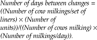
Milking parlors and stanchion barn pipelines have similar systems for transporting milk from the cow to the bulk tank. The components of the transport system will be described in the direction of milk flow. The rubber or silicone insert in each teat cup is referred to as a liner or inflation. The liner should milk cows safely, with a minimal number of squawks from downward slippage, and without the teat cup crawl action of riding up on the teats to the base of the udder. Liner performance depends upon many interrelated characteristics of the milking system. Narrow bore liners are recommended. Liners must be compatible with the teat cup shell. The most important management consideration with respect to teat cup liners is to insure regular replacement, as recommended by the manufacturer. As a general guideline, natural rubber liners last 500–700 cow milkings, synthetic rubber liners 1000–1200 cow milkings and silicone liners 5000–10000 cow milkings. The desired milking inflation replacement interval (in days) can be calculated using the following formula:
Other rubber parts of the unit, such as the short air tubes on the claw, should be constantly checked for cracks or signs of wear. These problems could seriously affect air flow and liner pulsation. Proper storage in dark, cool conditions, as well as the correct use of cleaners and sanitizers, can affect the life of rubber parts.
The milk claw
The milk claw is an important component of the milking unit. The claw is the collection point for milk from the four teat cups and should have adequate capacity to handle peak milk flow without flooding. Each claw should have a means of shutting off the vacuum to the teat end, so that the unit is not removed under vacuum. Most claws have an air vent in the upper half to allow a predetermined quantity of air into the unit to facilitate milk flow away from the cow and into the pipeline. Claws should routinely be inspected for cleanliness, plugged air vents and dented liner connectors.
A long milk hose is used to carry milk from the claw to the pipeline. The hoses can be made of plastic, rubber or silicone. They should be as short as possible, with an appropriate hose hanger. If the milk hose is crimped or allowed to loop, milk flows will be interrupted, which leads to irregular fluctuations in teat-end vacuum. The milk hose should attach to an inlet located in the top third of the milk pipeline, at the eleven or twelve o’clock position. Inlets should be self-draining, self-closing and should not cause milk flow restrictions that would result in irregular teat-end vacuum fluctuations.
The milk pipeline
The milk pipeline serves two important functions: transporting milk from the cow to the receiver jar and carrying air flow to provide milking vacuum to the teat end. Either glass or stainless steel can be used for milk pipeline construction. The milk line should form a complete circuit and must be rigidly supported from the floor in order to maintain the appropriate slope. It is generally recommended that milk lines be installed as low as is practical. In milking parlors, low pipelines are installed below udder level. In stall barns, high pipelines are used, but they should be no higher than 2 m above the cow platform. Milk moves by gravity through the pipeline to the receiver jar. The milk line must be self-draining and should have a continuous slope from the high point toward the milk receiver jar. The correct slope is important for the movement of milk and air during milking, as well as for proper cleaning of the system. In the construction of new tie-stall barns, it is recommended that the foundation, floor and gutter be sloped towards the milk house end. This will help to minimize pipeline height and to insure that line slope will facilitate drainage during milking and washing.
Line diameter is another important feature of milk pipeline design. In addition to line slope and the level of herd production, pipeline diameter will determine the number of milking units that a system can handle. Too many units will lead to milk line flooding and a reduction in air flow rate. Slugs of milk moving through the line is an obvious sign of milk line flooding. This problem will have a negative impact on milking time, herd production and udder health. The recommended minimum pipeline diameter is 51 mm (2.0 in.). At this pipeline size, high-producing herds should not use more than three milking units per pipeline slope. Thus, larger pipeline sizes are often recommended for new installations. Pipeline couplers or welds must prevent air leakage into the system.
Milk should flow into the receiver jar in a continuous, unimpeded fashion. When sufficient milk has accumulated, an electronic probe triggers the milk pump to transfer milk from the receiver jar to the bulk tank. A milk filter is inserted into the transfer system, as a mechanism to remove coarse impurities that may have entered the line. The receiver jar is connected to the main vacuum supply. A device called a sanitary trap is used to separate the ‘air only’ portion of the milking system from the ‘milk handling’ side of the system. The sanitary trap is designed to protect the vacuum supply from potential damage caused by the chemical cleaning and sanitizing solutions used to clean milk pipelines.
A milking system should have the capability of measuring the amount of milk from each cow. In older milking parlor systems, weight jars were often used for this purpose. They allowed for a quick visual means of monitoring individual cow production at each milking, as well as providing vacuum stability to the cow. However, they were expensive and represented a challenge to clean. More recently, milk metering systems have been developed that give an electronic digital readout of the milk volume produced at each parlor station. These systems can often be adopted to automatic data recording in an on-farm computer system. In stall barn pipeline installations, several types of mechanical milk meter are in use. It is important that any metering system not be restrictive to the flow of air and milk. These restrictions can cause a drop in teat-end vacuum and the occurrence of irregular teat end vacuum fluctuations. Increased milking time, incomplete milk-out and new intramammary infections can result.
Bulk milk tank
The bulk milk tank is the vessel used to cool and store raw milk until it is picked up by the bulk milk transport truck. All tanks must be of an approved sanitary design and construction. They must be of sufficient capacity to cool and store up to 3 days of milk production. The cooling capabilities of bulk milk tanks are clearly specified. Appropriate cleaning and sanitizing procedures for bulk tanks are critical, in order to prevent bacterial growth and contamination of raw milk.
Relationship of milking equipment to udder health
The milking machine can influence new intramammary infection rates in several ways:
• The milking machine may be a carrier of mastitis pathogens from one cow to the next
• The milking machine may serve as a pathway of cross-infection within cows
• Malfunctioning or improperly used equipment may result in failure to relieve congestion in teat tissue. Eventually, teat end damage and intramammary infection can occur
• Abrupt loss of milking vacuum may create changes in air movement of sufficient force to move pathogens past the streak canal defenses. This phenomenon, known as the impact mechanism, was described earlier.
The pathogenesis of new infections related to machine milking probably involves all four of these factors.26 However, even though the milking system becomes the focus of many herd udder health investigations, there is little evidence that machine factors are of primary importance in most problem herds. It has been difficult to link milking machine factors and prevalence of herd infection. Mastitis has been difficult to produce experimentally by altering machine function.28
A great deal of research has been directed towards the identification of machine factors related to mastitis. Although many problems are identified, the only factors consistently associated with udder health problems are pulsation failure and the impact mechanism.
Appropriate pulsation is important for sufficient teat end massage. Although continuous vacuum will remove milk from cows’ teats, eventually it will result in excessive congestion, edema and teat end damage. An adequate compressive load by the liner on the teat tissue is necessary to relieve the congestion. Mechanical failure of the pulsator, shortness of the liner barrel and a too-short liner rest phase are the most common examples of pulsation problems. The impact mechanism results from an abrupt loss of milking vacuum. Poor liner design has been shown to increase the frequency of liner slips. During a liner slip, a reverse pressure gradient occurs across the streak canal of the other three teats. Liner design has been shown to be very important in reducing the amount of slippage.29 In combination with liner slips, the vacuum fluctuations that result from pulsation problems can lead to new intramammary infection.
Even with the myriad of potential machine problems, milking equipment is not usually the major risk factor for poor udder health.
Maintenance and evaluation of milking equipment
The most important aspect of udder health management related to milking equipment is the establishment of an appropriate evaluation, maintenance and service schedule. Farm personnel should incorporate an inspection of the equipment into their regular milking process. Many of the problems discussed in conjunction with the description of milking system components can be discovered during this daily inspection. In addition, the producer should have milking equipment serviced on a regular basis. Items such as the vacuum pump, regulator, pulsators and sanitary trap would be included in this check list. Also included in this inspection will be regular changing of the teat cup liners and other rubber parts. It is common for equipment dealers to schedule a regular visit to each farm client for the purpose of conducting this periodic maintenance schedule and for dispensing chemical cleaners and disinfectants used in the udder health management program.
A complete milking system analysis should be conducted on a regularly scheduled basis. This regular analysis is perhaps just as important as the initial design and installation of the system. Many dairy cattle specialists believe that a regular independent analysis will insure proper equipment function. Milking system analysis can be conducted by equipment dealers, government extension staff, veterinarians or independent technicians. All these individuals need the appropriate knowledge and training. It is essential to use some type of systematic milking system analysis worksheet to record various performance measurements and to identify components requiring service or upgrading. A complete system analysis should be conducted at least once a year, and records should be kept for future reference.
3. DRY COW MANAGEMENT AND THERAPY
The proper management of dry cows and late-gestation heifers is an important component of a mastitis control program. The dry period offers a valuable opportunity to improve udder health while cows are not lactating. However, the beginning and the end of the dry period represent periods of increased risk of infection.30 The objective of udder health management during the dry period is to minimize the number of infected quarters at calving. Two of the three major principles of udder health management must be met in order to achieve this objective. Infections present at the time of drying off should be eliminated and the rate of new intramammary infections during the dry period must be minimized. Thus dry cow therapy has a dual role in eliminating existing infections and preventing new infections during the dry period, and has been widely adopted by dairy farmers. If these two principles are followed, udders will be free of infection at calving and can be expected to produce a maximum amount of low-cell-count milk in the subsequent lactation. Intramammary administration of long-acting antimicrobial agents to all cows at drying off remains a routine recommendation.
Epidemiology of intramammary infection during the dry period
The development of effective udder health management strategies for the dry period requires an understanding of the epidemiology of intramammary infections in dry cows. This in turn requires an understanding of the incidence of new infections during the dry period and the types of pathogen involved. Risk factors that affect the susceptibility of dry cows should also be understood.
Incidence of new infections
The rate of new intramammary infections is significantly higher in the dry period than during lactation.30 The greatest increase in susceptibility is during the first 3 weeks of the dry period. In this period, the new infection rate is many times higher than during the preceding lactation as a whole. A second period of heightened susceptibility occurs just prior to parturition. The reported rates of new intramammary infection in the dry period vary widely. Reasons for these differences include the diagnostic criteria used and the types of organism considered to be major pathogens. There are also important herd-level effects, such as the prevalence of existing infections at drying off and the method of dry-off. The average rate of new infections in untreated dry cows is expected to be between 8% and 12% of quarters.30
Types of pathogen causing new infections during the dry period
Contagious pathogens are transmitted among cows and quarters in association with the milking process. Environmental pathogens are primarily contracted from contamination with organisms in manure and bedding. Teat skin opportunistic pathogens are present on the teat, particularly the teat end. Contagious, environmental and teat skin opportunistic pathogens need to be considered in designing mastitis control schemes for the dry period.
Exposure to environmental pathogens is likely to continue throughout the dry period; thus prevention of new dry period infection with environmental agents represents a considerably greater challenge.30 Herds that have implemented a basic mastitis control program still need to be aware of the importance of preventing environmental infections in the dry period. There are different rates of infection by the various environmental agents as the dry period progresses. For example, infections with environmental streptococcal species, Klebsiella spp. and Enterobacter spp. occur more frequently early in the dry period. On the other hand, E. coli infections tend to occur immediately before calving. Dry cow management strategies need to account for the risk of infection during the entire period from last milking until the next calving.
Risk factors that affect susceptibility in dry cows
Several factors contribute to the variation in susceptibility during the dry period. These factors include the following.
Teat end protection
The cessation of routine milking-time hygienic practices such as teat dipping allows bacterial subpopulations on teat skin to increase in number and diversity. S. aureus numbers are high immediately after drying off and environmental pathogens are more prevalent on teat skin late in the dry period and at calving time.30 Teat end lesions increase the likelihood of intramammary infections during the dry period. A plausible mechanism to explain this association is that teat end lesions increase the surface area available for bacterial colonization while presenting a variety of environmental niches. For instance, quarters with cracked teat ends were 1.7 times more likely to develop a new intramammary infection during the dry period than unaffected quarters.31
The streak canal of the teat is more penetrable by bacteria during the early dry period. The keratin plug in the streak canal must form early and completely in the early dry period in order to prevent penetration and growth of bacteria and decrease the incidence of new intramammary infections. However, this natural internal teat sealant does not form in some cows, and delay in formation is common. For instance, in cows in New Zealand, 45% of teats are open on day 7 of the dry period, and 25% are still open on day 35 of the dry period.32 Similar results were obtained in North American dairy cows.31 Quarters that remain open during the dry period are 1.8 times more likely to develop a new intramammary infection than quarters that have developed an effective keratin plug.31 Internal and external teat sealants are discussed further later in this chapter.
Swelling of the mammary gland, an increasing volume of secretion and leaking colostrum contribute to the high risk of new infection during the prepartum period.
Resistance mechanisms within the mammary gland
Throughout the dry period there are marked changes in the composition of mammary gland secretions and in the concentration of protective factors such as leukocytes, immunoglobulins and lactoferrin. These changes probably influence the variation in susceptibility to both environmental and contagious pathogens.
Substantial evidence exists that innate and acquired defense mechanisms are lowest from 3 weeks precalving to 3 weeks postcalving.33 This lowered responsiveness includes aspects of systemic and mammary gland immunity that may account, in part, for the increased incidence of peripartum disease. Polymorphonuclear neutrophil function is impaired during the peripartum period and may contribute to the increased incidence of mastitis following calving. Diminished lymphocyte responsiveness around calving has also been observed.33 The role of the cow in effectively transferring antibodies and cells to the mammary gland prior to parturition to insure high-quality colostrum is also an important function, and this may be affected by prepartum vaccination schedules and the ability of the animal to respond effectively.
Milk production at dry off
A high level of milk production at dry off increases the incidence of new intramammary infections at calving.34 It is reasonable to assume that high milk production at dry-off will produce a higher intramammary pressure, thereby increasing the likelihood of an open streak canal early in the dry period. High milk production at dry-off will also decrease the concentration of protective fractions such as phagocytic cells, immunoglobulin and lactoferrin,35,36 thereby decreasing resistance within the mammary gland. The finding that cows leaking milk following dry-off are four times more likely to develop clinical mastitis in the dry period37 supports the concept that increased milk production at dry-off increases the rate of new intramammary infections.
Method of drying off
The industry standard method for cessation of lactation (drying off) is abrupt cessation of milking, whereby milking stops on the day scheduled for dry-off (all cows are usually scheduled to ‘go dry’ on the same day each week) in order to facilitate administration of dry cow intramammary antibiotics, vaccinations and vitamin E/selenium injections. Abrupt cessation is associated with a higher new intramammary infection rate in the dry period compared to intermittent cessation,38,39 although the increase in prevalence is most evident in cows that are not dry treated. The best approach to dry off cows may therefore be intermittent milking, although additional studies are required before the industry standard is altered. In particular, the method of drying off is probably less important with blanket dry cow therapy.
Parity
Older cows are more likely to develop new intramammary infections during the dry period. This increased predilection may be due to increased milk production at dry-off, increased prevalence of abnormal teat placement (increasing exposure of the teat end to pathogens) or increased prevalence of open streak canals because older cows have higher milk production.
Risk factors that affect susceptibility in heifers
An increased risk for intramammary infection in the preparturient period in heifers is associated with the presence of S. aureus or M. bovis in the herd, calving in summer, high herd bulk tank milk SCCs, poor fly control, mastitic milk fed to calves and contact with adult cows.40 Other risk factors are increased age at first calving, prepartum milk leakage,41 blood in milk42 and udder edema.43
Udder health management strategies for dry cows
Antimicrobial therapy (dry cow therapy)
Antimicrobial therapy at the end of lactation (dry cow therapy) has been one of the key steps in mastitis control programs and has become the most effective and widely used control method for dry cows. The efficacy and advantages of antimicrobial therapy are well known. The use of effective dry cow products results in 70–98% elimination of existing infections. However, elimination of S. aureus is less successful. Dry cow therapy also reduces the incidence of new intramammary infections by approximately 80%.44
Long-acting antimicrobial preparations have been formulated to eliminate existing infections and to prevent new infections. These preparations include benzathine cephapirin, benzathine cloxacillin and sustained-release formulations of erythromycin, novobiocin and penicillin. The withholding period for milk from animals treated with these dry cow formulations ranges from 30–42 days after treatment. It is important that the label directions be followed carefully for the recommended dosage level, required withdrawal period, storage guidelines and expiry dates. A general recommendation is that dry cow treatment should never be administered within 1 month of the expected calving date. Single-dose syringe preparations of dry cow antibiotic treatment are recommended. The risk of contamination by environmental bacteria and yeast is much higher for multiple-dose bottles than for single-dose syringes. If bulk containers are used, great attention should be paid to maintaining sterility.
The use of long-acting and short-acting antimicrobial intramammary infusions have been compared.45 In some cases, short-acting antimicrobial agents were more effective than long-acting ones in eliminating infections due to S. aureus or treating cows infected with major pathogens diagnosed twice before drying off. Intramammary infusion of cephapirin sodium 15 days prepartum in heifers was effective in reducing intramammary infections during late gestation and reduced the occurrence of residues in milk during early lactation.46 The milk of heifers that calve less than 15 days after treatment may contain antimicrobial residues.
Intramammary infusion is a widely used and highly recommended procedure for mastitis therapy; however, there is a potential for the introduction of pathogens during the infusion process. Insanitary infusion practices can introduce antibiotic-resistant environmental organisms into the udder. Infection with opportunistic microorganisms, such as yeast or Nocardia spp., may cause more extensive udder damage than the original organism for which treatment was being administered. Adequate teat end preparation and careful dry cow treatment procedures can reduce this risk. Dry cow treatment procedures should be carried out as follows:
• Milk out the udder completely
• Immediately following teat cup removal, dip all teats in an effective teat dip
• Allow the teat dip to dry. If necessary, remove excess dip from teat ends with a clean single-service paper towel
• Disinfect each teat end by scrubbing for a few seconds with a separate alcohol-soaked cotton swab. Start with the teats on the far side of the udder and work to the near side
• Infuse each quarter with a single-dose syringe of a recommended dry cow treatment. Start with the teats on the near side of the udder. Use the partial insertion method of administration into the teat streak canal. Preferably, a modified infusion cannula should be provided with the treatment product
• Dip all teats in an effective teat dip immediately following treatment.
The necessity of using appropriate dry cow treatment procedures cannot be overemphasized. An increased incidence of Nocardia spp. mastitis has been associated with blanket dry cow therapy, especially neomycin-containing products. However, Nocardia spp. were not found as a contaminant of the suspected products. Teat end preparation by scrubbing with an alcohol-soaked cotton swab was protective against the occurrence of Nocardia spp. infection when teats were experimentally contaminated with organisms immediately before drying off.47 Most commercial dry cow treatment products provide individually wrapped alcohol-soaked cotton swabs for use with each syringe. The use of good teat end preparation prior to intramammary infusion needs to be continually emphasized.
The method of intramammary infusion may be important. Partial insertion of the infusion cannula (up to 4 mm) results in fewer new intramammary infections and improved cure rates. The improvement with a short cannula is attributed to fewer organisms being delivered beyond the streak canal and decreased physical trauma to the streak canal. In addition, antimicrobial agents that are deposited within the streak canal should control local infections. Modified infusion cannulas for the convenient use of a partial insertion method of administration are now available for commercial dry cow products.
Another approach to preventing the problems associated with intramammary infusion would be the development of an effective systemically administered dry cow treatment. Preliminary results have indicated improved efficacy against S. aureus infections using a systemically administered quinoline antibiotic (norfloxacin nicotinate).
Blanket versus selective dry cow therapy
Three strategies for intramammary antimicrobial treatment of dry cows are available,48 although the current recommendation for all herds is blanket therapy:
• Blanket therapy (treat all quarters of all cows)
• Selective cow therapy (treat all quarters of any cow infected in one or more quarters)
Although blanket dry cow therapy is a cornerstone of any mastitis control program, there is some controversy concerning the need to treat all quarters of all cows (blanket therapy) or only those quarters or cows requiring treatment. The controversy has gained momentum as the implementation of udder health management practices has reduced the prevalence of infection. The major reasons for selective therapy are:
• To avoid the elimination of minor pathogens, which may make cows more susceptible to environmental agents
• To reduce the expense of treatment
• To address increasing consumer concern regarding the routine administration of antibiotics to food-producing animals
• To avoid the possible emergence of antibiotic-resistant organisms.30
Each of these reasons should be carefully considered in making a decision between blanket and selective dry cow therapy. Selective dry cow therapy is preferable provided that an accurate, practical and inexpensive method for selecting infected cows is available.48 This is the major problem with selective dry cow therapy – in most herds the sensitivity and specificity of the test used for selection is not adequate.
As general udder health improves and bulk tank milk SCC remains low, producers question the need to continue dry treatment on all cows and are attracted by a potential reduction in costs for the purchase of dry cow treatment. However, selective therapy requires a decision as to which cows or quarters are to be treated. The sensitivity and specificity of currently available screening tests are inadequate as a basis for decisions concerning selective therapy. The history of the number of episodes of clinical mastitis, individual cow composite SCC during lactation and at dry off, CMT results during lactation or at dry-off and even bacteriological culture towards the end of lactation all result in leaving some infected cows untreated, but conversely result in the treatment of many uninfected cows. An important requirement for large-scale implementation of selective dry cow therapy is the development of a cheap, practical, sensitive and specific test to identify infected cows. The failure to prevent new intramammary infections during the dry period with the selective approach must also be considered. New infections in the dry period will become increasingly important as contagious pathogens are eliminated from herds. Finally, blanket dry cow therapy reduces new infection rates for quarters from approximately 14% to 7%. The increase in milk production alone resulting from prevention of these new infections provides enough return to offset the cost of treatment for all cows.
Information presently available indicates that the general recommendation should be for routine treatment of all quarters of all cows at the time of drying off (blanket dry cow therapy). There is a need to identify important management practices to limit new infections in untreated dry cows and to develop new screening tests to determine which cows should be treated. New environmental management methods and modern information processing capabilities may lead to the development of better selective dry cow treatment programs. These may include the administration of ancillary therapeutic agents. For instance, the intramammary infusion of recombinant bovine interleukin-2 along with cephapirin sodium at drying off marginally increased the cure rate of intramammary infections associated with S. aureus, but not other pathogens, during the dry period compared with the administration of cephapirin only.29 Interleukin did not affect the incidence of new intramammary infections for any pathogen group. However, the intramammary infusion of interleukin at drying off was associated with an increased incidence of abortion in dairy cows 3–7 days after the infusion.
Factors affecting the success of antimicrobial treatment of dry cows
Despite blanket dry cow therapy, some cows calve with infected quarters and some with clinical mastitis. Several risk factors affecting the results of dry cow treatment have been evaluated.49,50 Some of these factors are:
• Number of quarters infected. With S. aureus infections, there is a significant decrease in cure rate as the number of quarters infected per cow increases. Quarters from cows with either three or four of their quarters infected have a very poor cure rate
• Age of the cow. As the age of the cow increases, the probability of S. aureus infections being cured by dry cow therapy decreases
• Somatic cell count before drying off. The cure rate of S. aureus-infected quarters diminishes as the SCC prior to treatment increases. Controlling for age and number of quarters infected, there was a significantly lower cure rate in quarters with an SCC of more than 1 000 000 cells/mL49
• Herd of origin. There is a distinct herd effect on the success of dry cow therapy. The cure rate of S. aureus has been shown to be higher in herds with good hygiene and with a low prevalence of S. aureus infections at drying off.49
There is considerable potential in using individual cow and herd-level information to predict the likelihood of a cure with dry cow therapy. For example, an older cow with three quarters infected with S. aureus and a persistently high SCC has a low probability of a cure. Continued development of information management systems to assist with therapy and culling decisions will clarify the expectations of dry cow treatment.
Persistent S. aureus infections represent only one of the shortcomings of antibiotic treatment for dry cows. Most dry cow products are formulated for efficacy against Gram-positive cocci. These antibiotics are of limited usefulness against Gram-negative bacteria. In other words, new coliform infections would not be prevented by this therapy. Even though dry cow products are formulated for sustained activity, the provision of adequate protection during the critical prepartum period is questionable. The persistence of effective levels of antimicrobial agents has been evaluated for various dry cow treatments;51 very few products have persistent activity until the time of calving.
Vaccination of the dry cow
Immunization and immunotherapy for the control and prevention of mastitis have been active areas of research.52 Effective vaccines would have to eliminate chronic intramammary infections, prevent new intramammary infections or decrease the incidence or severity of clinical mastitis. Currently available mastitis vaccines may reduce the incidence and severity of clinical mastitis but have not eliminated chronic intramammary infections or prevented new intramammary infections. The inability of vaccines to prevent infection may be due to the wide variety of pathogens, inadequate specific antibodies or the failure of antibodies to enter the mammary gland prior to infection. Currently available vaccines should be used as adjuncts to other more effective control strategies.
Vaccines have been developed to reduce the incidence and severity of clinical mastitis associated with Gram-negative pathogens.53 R-mutant bacteria have an exposed inner wall structure (core lipopolysaccharide antigens) that is highly uniform, even among diverse and distantly related Gram-negative bacteria. Vaccines containing killed R-mutant bacteria provide broad-spectrum immunity against a wide variety of unrelated Gram-negative bacteria. The most commonly used coliform mastitis vaccines are the Rc-mutant E. coli O111:B4, known as the J5 vaccine, and the Re-mutant Salmonella typhimurium; both of which are commercially available in the USA.52 R-mutant vaccines have been efficacious in the reduction of the incidence and severity of clinical mastitis due to Gram-negative bacteria. More than 50% of large (> 200 cows) dairy herds and more than 25% of all dairy herds in the USA are using core lipopolysaccharide antigen vaccines.54 No protection is provided against environmental streptococci and staphylococci, or the contagious pathogens.
No effective vaccines are currently available for the control of mastitis due to S. aureus, S. agalactiae, environmental streptococci, and M. bovis.
The use of recombinant bovine cytokines as adjuvants to enhance specific immunity in the mammary gland of cows after primary immunization against indicate an enhancement of specific immunity in the mammary gland, which may be effective in mastitis immunization protocols.
Management of the environment for dry cows
Dry cows should be provided with an environment that is as clean and dry as possible. If this is not feasible in confinement housing, it is probably better to maintain dry cows on pasture. Variations in the load of coliforms and environmental streptococci in the environment are important predictors of new infection rates. Minimizing the exposure to environmental bacteria will reduce the new infection rate. However, some pasture conditions promote the crowding of cows under shade trees. In hot, humid and muddy conditions, heavy contamination of such a small area can result in a significant risk of new environmental infections in the dry period. In good weather, it is ideal to hold parturient cows in a clean, grassy area where they can be observed and assisted if necessary.
In confinement housing systems for dry cows, it is important to provide adequate space, ventilation, bedding and lighting to insure cleanliness and comfort. Maternity (calving) stalls should be bedded with clean straw, sawdust or shavings. Other important procedures for managing the environment for dry cows include adopting an effective fly control program. Clipping the hair on the udders, flanks and inside the hind legs will help reduce contamination. The words clean, dry, cold and comfortable summarize the ideal environment for dry cows. The words clean and dry also summarize the goal for the teat before attachment of the inflation during milking.
Nutritional management of dry cows
A nutritionally balanced dry cow feeding program is important to insure udder health. A role has been suggested for specific nutritional factors in resistance to mastitis, especially over the dry period. Adequate levels of vitamin E and selenium in dry cow rations appear to be important for udder health at calving and in early lactation.55 This effect may be mediated through enhanced resistance mechanisms. Other vitamins and minerals may be important in udder health, but their role is less well substantiated.
Nutritional management of dry cows is also important for reducing the risk of milk fever, which is an important predisposing factor to mastitis in fresh cows. Appropriate body condition can be achieved by good nutritional management in late lactation. The association between body condition, energy metabolism and udder health needs further clarification.
Internal teat sealants
As discussed previously in risk factors for infection in the dry period, the keratin plug is a natural internal teat sealant that provides an effective barrier to new intramammary infections. High milk production at dry-off increases the likelihood that the streak canal remains open and presumably compromises formation of the keratin plug, thereby increasing the risk of intramammary infection.34
A recent promising development in mastitis control has been exogenous internal teat sealants that are applied at dry off. The teat sealant product most extensively evaluated contains a heavy inorganic salt (bismuth subnitrate) in a paraffin wax base; this product does not have antibacterial properties but acts as a physical barrier to ascending intramammary infections. A New Zealand study involving 528 cows indicated that the internal teat sealant was retained for dry period lengths of more than 100 days, while producing a ten-fold decrease in the new intramammary infection rate, which was similar to that produced by dry cow intramammary infusion alone.56 In contrast, a UK study involving more than 1000 cows indicated that bismuth subnitrate teat sealant alone decreased the number of new intramammary infections to a great extent than intramammary dry cow infusion alone.57 The UK study had a greater incidence of new intramammary infections due to Gram-negative bacteria than the New Zealand study. A US study involving 437 cows found that an internal teat sealant at dry-off decreased new intramammary infections by 30% in the dry period and treated cows were 33% less likely to have a clinical mastitis episode between dry-off and the first 60 days of the subsequent lactation58 compared to untreated quarters. The addition of an antimicrobial agent is a logical addition to the bismuth subnitrate internal teat sealant and has been a routine addition to the teat sealant for some years in Ireland.
Bismuth subnitrate internal teat sealants clearly show promise for the prevention of new intramammary infections during the dry period. However, because bismuth subnitrate teat sealants do not eliminate existing intramammary infections, and an accurate method for determining the infection status of a quarter is unavailable (the exception being milk culture), the recommended application of internal teat sealants requires combined application with intramammary dry cow therapy. This combined therapy may be uneconomic. Widespread adoption of internal teat sealants will require an accurate test for determining intramammary infection status at dry-off. Finally, administration of internal teat sealants alone obviously requires proper aseptic technique.
External teat sealants
A longer-standing approach to providing a physical barrier to ascending infections is the use of external teat sealants, which were originally developed for use in lactating cows. The major problem with external teat sealants is the duration of adherence, which is too long for lactating cow use and too short for dry cows. Teat end lesions and teat length influence the adherence of external teat sealants.31 Widespread adoption of external teat sealants will require a product that provides prolonged protection but is easily removed at calving.
Teat disinfection
Postmilking teat disinfection is a very effective means of reducing new infections in lactating cows. However, the efficacy of teat disinfection in decreasing the incidence of new intramammary infections in the dry period has been discouraging. Daily teat dipping for the first week of the dry period is not effective in reducing S. uberis infections. The lack of efficacy of teat disinfection needs to be contrasted to the efficacy of internal teat sealants.
Intramammary devices
Intramammary devices have been developed for use in preventing new infections in both lactating and dry cows. However, there is conflicting evidence as to the reduction in infection rate in quarters fitted with these devices, and such devices are no longer being investigated. The incidence of clinical mastitis may be less in cows fitted with these devices compared to control cows but the prevalence of subclinical infection is unaffected.59 Intramammary devices induce a significant increase in postmilking SCC compared to control cows, and test-day SCCs may be higher than in control cows.
4. APPROPRIATE THERAPY OF MASTITIS DURING LACTATION
The early recognition and treatment of clinical cases remains an important part of a mastitis control program. Improvements in understanding of the epidemiology, pathophysiology, and response to therapy of various mastitis pathogens have clarified the role of intramammary and parenteral antimicrobial agents for of the treatment of clinical and subclinical mastitis during lactation. This is covered extensively earlier in this chapter.
5. CULLING CHRONICALLY INFECTED COWS
The final step of the five-point mastitis control program is the selective removal of cows with chronic intramammary infection from the herd. Most producers have interpreted this recommendation to mean that cows with recurrent episodes of clinical mastitis should be eliminated. For example, some herds have established that cows having three or more clinical cases of mastitis in a lactation will be culled (the popular ‘three strikes and out’ approach). However, very little research has been conducted to determine the effect of various culling strategies on herd udder health status, and on the incidence of clinical cases.
Nevertheless, culling chronically infected cows meets one of the three guiding principles of mastitis control, namely the elimination of existing intramammary infections. Through the use of available monitoring techniques and the establishment of a defined culling program, a valuable opportunity exists to improve udder health by culling.
In general, a record of chronic mastitis and severe fibrosis detected on deep udder palpation should be the basis of a recommendation to cull. Culling is an effective and documented mastitis control measure for some specific mastitis pathogens. For example, the removal of infected cows is a key element of the recommended mastitis control program for herds with a high prevalence of S. aureus infection. Removal of cows found to be infected with S. aureus accounted for more than 80% of the costs involved in the control program. Culling is also important in the control of other mastitis pathogens that respond poorly to antimicrobial therapy. Herds with mastitis cases associated with M. bovis, Nocardia spp., and P. aeruginosa should be aware of the benefits of culling infected cows.
A dairy herd culling program should be based upon consideration of the net present value of each cow in the herd as compared to the value of a replacement heifer.60 The net present value depends on the age of the cow, her potential for milk production, the stage of lactation and her pregnancy status. Factors that determine the likelihood of treatment success, such as pathogen and duration of mastitis, as well as the cost of treatment, also need to be considered in calculating the net present value of the cow with mastitis. After consideration of the relative importance of udder health in the overall herd health management program, additional economic pressure may be applied to cows with a specific udder health status. For example, if a S. aureus control program is a major priority in the health management program, additional economic pressure should be applied in removal decisions of cows with known S. aureus infections. As health management data collection and analysis improve in sophistication, decision analysis methods and expert computer models will be used to provide this information automatically.
Biosecurity for herd replacements
Replacement animals may be purchased to increase herd size or to maintain cow numbers following culling. Biosecurity measures must be used to insure that herd replacements are not infected with contagious mastitis pathogens (specifically S. aureus, S. agalactiae, M. bovis). However, an economic analysis of the different components of a biosecurity program has not been performed, and it is likely that some components of currently used programs are not cost-effective.
An optimal biosecurity program includes knowing the herd of origin, knowing the cows and protecting the home herd.
Know the cows
When purchasing single or small groups of animals the following prepurchase procedures are recommended:
• SCC and clinical mastitis records for each cow to be purchased
• Results of bacteriological culturing of quarter milk samples from each cow on arrival (if lactating) or at calving (if late-gestation) for S. aureus, S. agalactiae and M. bovis. In general, the sensitivity of a single milk culture to detect the presence of intramammary infections due to S. agalactiae is approximately 95%, for S. aureus it ranges from 30–86%, and for M. bovis it is 24%
• A physical examination of each cow, including udder, milk quality and teat ends.
Protect the home herd
Consider all purchased animals as potential health risks to the home herd by doing the following:
• Maintain all newly purchased animals in separate or isolated facilities until diagnostic tests for udder health have been completed and there is no evidence of infection that may spread to the rest of the herd (usually < 14 d quarantine)
• Evaluate all herd replacements for evidence of antimicrobial residues in milk
• Milk all purchased animals last or with separate milking equipment until it is determined that they are free of infection
• Obtain results of bacteriological culturing of bulk tank milk samples or string samples for S. aureus, S. agalactiae and M. bovis; culturing should be done on more than one occasion because the sensitivity of bulk tank milk culturing is not 100%, and is less than 50% for M. bovis.
6. MAINTENANCE OF AN APPROPRIATE ENVIRONMENT
The multifactorial nature of mastitis has been emphasized throughout this chapter. Intramammary infection results from a complex interaction between the cow, the mastitis pathogens and the environment. Thus, the control of unfavorable environmental influences is extremely important in dairy herd udder health management programs.
Intramammary infection involves exposure of the teat surface to potentially pathogenic microorganisms, entry of the pathogens into the gland via the teat duct and establishment of the pathogens in the mammary tissue, producing an inflammatory response. Many environmental factors can influence this process of exposure, bacterial entry and establishment of infection. For example, the type of bedding and manure management can have a great impact on the contamination of teat skin with microorganisms. Housing design can have an impact on the prevalence of teat injuries, which will influence intramammary invasion by mastitis pathogens. Extreme climatic conditions, poor nutritional management and cow stocking densities will influence the immune system and the establishment of intramammary infection. A comprehensive udder health management program should involve steps to minimize the detrimental influences of the environment.
Global environmental influences
Worldwide, there are major differences in dairy herd health and production systems. For example, the type of animal used, economics of production, climatic conditions, housing structures and management methods are widely variable. These differences greatly affect the interaction of cows with their environment, even though the predominant causative organisms are the same under different systems. Thus, there are major variations in the relative incidences of different pathogens, and in the importance of various approaches to mastitis control.
Classification of environmental influences
The influences of the total environment can be divided into:
• The external environment. All aspects of the environment outside the housing facilities make up the external environment. This includes the regional differences in climate, geography and agricultural tradition. There are also local factors within a region that can have an important influence. These local factors include the topographical features of the land, natural shelters from the climate and the availability of pasture
• The internal environment. All environmental conditions inside the cow buildings make up the internal environment. The general internal environment includes the type of housing system, temperature, humidity and air quality. There are also specific internal environmental influences such as stall design, type of bedding, nutritional management and manure disposal. The milking environment has a major influence through the equipment, cow preparation methods and approach to general hygiene.
External environmental influences on mastitis control
There is minimal evidence that external environmental factors directly influence the incidence of mastitis; however, the external environment determines the way in which cows are housed, fed and milked. Through these associations, the external environment can be an important risk factor in problems with udder health in dairy herds.
Regional environment
The climate and geographical features of a region have implications for the prevalence of mastitis. The ambient temperatures and amount of rainfall often determine the types of housing and nutritional program used. Extremely hot or cold conditions interact with other predisposing management factors. In areas prone to severe rainstorms, teats may be exposed to wet or muddy conditions. The soil type, cropping policy and presence of other industries can also have an indirect impact on the prevalence of mastitis; for example, regions suitable for growing cereal grains will commonly use straw as a bedding material, which may favor the growth of environmental streptococcal organisms. In contrast, dairying areas close to the forestry industries may favor sawdust or shavings as bedding. The use of these materials may influence the incidence of coliform mastitis.
The socioeconomic structure and agricultural policy of a particular region can affect management factors known to influence udder health. These factors determine herd size and labor costs. Large herd sizes necessitated by economic conditions will dictate the housing, feeding and milking management practices employed. More recently, regional policy towards regulation of bulk tank milk SCC levels has had a profound impact on udder health status.61
Local environment
The local environmental factors such as the topography of the land, the presence of natural shelters and the type of pasture grown are thought to influence udder health status. However, direct scientific evidence is lacking. One important exception is ‘summer mastitis’, which affects nonlactating heifers and cows. This udder infection with A. pyogenes is greatly affected by the local environment, probably through the propagation of insects important in its transmission. Protected pastures increase insect populations and can result in a high incidence of infection.
Internal environmental influences on mastitis control
The incidence of new intramammary infections can be greatly affected by the management and facilities used in confinement dairying systems. General aspects of the internal environment exert their influence on all cows in the herd, such as the type of housing and milking system. Tie-stall barns pose different environmental stresses from a free-stall system. The air quality and noise levels can have an impact on animal health. The nutrient content of component feedstuffs can affect disease resistance.
Specific internal environmental factors exert their influence on an individual cow basis. For example, the stall design and tying system affect individual cows differently. Many epidemiological studies have revealed interactions between udder disease and internal environmental conditions. Most of these studies relate to European tie-stall and seasonal grazing systems. However, the results are generally relevant to most housing and management systems. Some of the most important general and specific influences of the internal environment are as follows.
Housing
Housing factors account for a great deal of the variation in udder health status between herds. In both tie-stall and free-stall barns, short and narrow stalls are associated with increased incidence of teat tramps and mastitis. An appropriate partition between stalls is beneficial. Stanchions or neckstraps with chains can restrict the movement of the cow and increase the risk of teat injury. This occurs especially as cows are rising. In addition, the use of electrical cowtrainers has been associated with an increase in the rate of subclinical mastitis. The use of adequate amounts of a good bedding material will reduce mastitis incidence in both tie-stall and free-stall housing systems. Even though there are reports of specific bedding materials being associated with certain mastitis problems, the use of adequate amounts of properly maintained bedding is beneficial. Straw, shavings, sawdust, sand, shredded newspaper and other cushion systems have all been used effectively.
The climate and air quality maintained within a building can have a major influence on udder health. Draughty conditions, high relative humidity and marked changes in indoor temperature over a 24-hour period are factors that contribute to higher mastitis rates. Adaptation to adverse internal environmental conditions may cause stress, which can reduce the cow’s defense mechanisms. Indoor climate, especially temperature and humidity, can also account for differences in the concentration of pathogenic organisms to which cows are exposed.
Nutritional management
A complex relationship exists between the quantity and quality of feed and udder health status. Improper nutritional management can result in an increase of new intramammary infections, the exacerbation of pre-existing chronic infections, and an increase in clinical mastitis. Several mechanisms have been suggested for this association. An improper anion to cation balance in the dry cow ration is a predisposing factor for periparturient hypocalcemia, which in turn increases the risk of new intramammary infections. Feeding programs that result in excessively fat or abnormally thin cows may affect resistance to disease. Also, there is some evidence that feeds high in estrogenic substances may be detrimental to udder health status.
The dietary concentration of some vitamins and minerals may have an important relationship to udder health. Studies have shown that intramammary infection is related to plasma concentrations of vitamin E and blood concentrations of selenium.55 Dietary supplementation of vitamin E and selenium improved the natural resistance of the mammary gland to infection. Associations between udder health and the levels of vitamin A, beta-carotene, zinc and other nutrients have been proposed but are not well documented.
Management approach
Cow supervision, decision-making and general animal care by dairy herd managers may be important epidemiological factors in the relationship between environment and udder health. For example, lack of consistency in the performance and timing of various herd activities results in decreased udder health status. Irregular intervals between milkings should be avoided.
General hygiene
Even outside the milking environment, general hygiene can greatly influence the exposure of the udder and teats to pathogenic bacteria. The degree of hygiene achieved is directly related to the type of housing, the amount of bedding and the efficiency of manure removal. Worldwide trends towards increasing herd sizes and decreasing labor force necessitate more emphasis on the importance of cow hygiene.
Use of recombinant bovine somatotropin
It has been suggested that the use of bovine somatotropin may increase the incidence of clinical mastitis by an indirect mechanism that acts through increased milk production. Controlled field studies have shown that the use of bovine somatotropin is not associated with an increase in the incidence of clinical mastitis, milk discarded because of therapy for clinical mastitis, or culling for mastitis.62
Environmental control in an udder health management program
There is a strong association between herd udder health status and the number of stress factors operating within the herd. It has been proposed that mastitis occurs when stress factors exceed the cow’s ability to adapt. It follows that a major objective of an udder health management program should be to limit the number and severity of environmental stress factors.
Veterinarians responsible for udder health management programs should have a good understanding of the importance of environmental management. The three major objectives of environmental control for improvement of udder health are:
• To prevent contamination of the teat end
• To prevent invasion of mastitis pathogens
• To prevent pathogens from establishing in the mammary gland.
The important steps in achieving these three objectives have been discussed. For example, an adequate housing system and manure handling are important to limit bacterial contamination of the teats. The prevention of environmentally induced teat injuries will aid in preventing invasion of pathogens into the gland. The producer’s approach to cow management, control of the nutritional program and insuring that the internal environment is appropriate, will all greatly improve host defense mechanisms and prevent intramammary pathogens from establishing within the gland.
7. GOOD RECORD KEEPING
Good record keeping involves the collection of useful data to monitor performance, calculation of appropriate indices and decision-making based on comparison to target levels. For acceptable performance the monitoring process is repeated and the cycle continues. If performance is not acceptable, further evaluation and analysis is carried out and a plan of action is instituted. Once again, the monitoring process carries on and the cycle continues. For many of the health management programs in food animal practice, a limiting factor is the availability of accurate and objective data. With respect to udder health, data have been readily available. Bulk tank milk SCC, individual cow composite SCC and bacteriological culture results are all accessible and useful. These data provide the information necessary to monitor udder health status and to make specific health management decisions. However, problems can still exist. Herds with a low prevalence of infection and very low bulk tank milk SCC can still have a high incidence of environmental infections and clinical mastitis cases. Thus, an important step in an effective udder health management program is the maintenance and use of mastitis records. The increasing adoption of computerized dairy health management record systems provides an opportunity to make effective use of clinical mastitis episode and therapy data. Even without a computerized system, manual records for clinical mastitis are easy to implement and use.
Objectives and uses of clinical mastitis records
The objective of maintaining computerized or manual records of clinical mastitis episodes is to complete the decision-making capabilities of a mastitis control program. The availability of this information will allow completion of the health management cycle over the entire spectrum of herd udder health situations.
There are several important uses of clinical mastitis records:
• To assess the risk factors associated with clinical mastitis episodes
• To evaluate lactational and dry cow therapy programs
• To provide information useful in the evaluation of net present value of individual cows for the purposes of culling decisions. Without the ready availability of accurate data surrounding mastitis events, decisions associated with therapy, culling and the removal of risk factors are difficult to make.
Recording clinical mastitis data
There is a limited amount of specific data necessary to make effective use of mastitis records as a health management tool. The cow identification, date of the clinical episode occurrence, type of therapy used and the date that milk withholding will be complete are the essential pieces of information. If a manual record system is used, it will be important to add the lactation number, the date of calving and the most recent test date production. A standard form that calculates the distribution of clinical episodes by lactation number and by stage of lactation is desirable. These are the same distributions often provided with an individual cow SCC report. A record form that allows the recording of treatment used, the number of days treated, milk withholding periods and an estimate of the costs associated with clinical mastitis has been described.63,64
Using clinical mastitis monitoring systems
Since clinical mastitis is a common event in dairy production, it is ironic that these records have not traditionally been kept. The key to overcoming this hurdle is the regular use of this information for health management decisions. Some of these uses and decisions are as follows.
Cow versus herd clinical mastitis problems
Calculation of the percentage of cows affected in the herd and the average number of clinical episodes per affected cow will aid in determining if the clinical mastitis is more of an individual cow problem or a herd-level issue.65
Probability of recurrence of clinical mastitis in the same lactation
The number of animals with repeat cases of mastitis divided by the total number of clinical cases gives an estimate of the likelihood of recurrence. The same calculation can be made for specific parity groups. This information can be useful to characterize the problem, and for culling decisions.
Stage of lactation and seasonal profile
If clinical mastitis data is collected consistently over a considerable period of time, potentially useful problem-solving information can be derived. For example, calculating days in lactation at first occurrence can help to identify specific risk factors for new intramammary infections. There is a higher proportion of clinical occurrences during the first few weeks after calving. However, analysis of clinical mastitis records might yield a different stage-of-lactation profile. In these cases, the evaluation of potential nutritional, environmental or other stress factors would be indicated. There may be different immediate and long-term solutions that should be implemented.
Analysis of clinical mastitis records over several years may identify a significant seasonal pattern, such as the documented seasonal pattern for bulk tank milk SCC data and antibiotic residue violations. Action may be necessary to deal with seasonal environment and housing problems that impact upon new infection rates.
Days of discarded milk
It is very common for producers to have an aggressive attitude towards the treatment of clinical mastitis. This approach may result in huge economic losses if waste milk is not fed to calves. These losses are largely the result of discarded milk during the clearance of antibiotic residues from treated cows. Calculating the days of discarded milk may suggest that the mastitis therapy program during lactation should be evaluated. Establishment of an appropriate treatment program and careful selection of cows for therapy might significantly decrease the need for discarding milk. In addition, cows that are responsible for a large percentage of the discarded milk should be identified for selective removal. The calculations from mastitis records that can help to clarify these issues include:
8. MONITORING UDDER HEALTH STATUS
An important step missing from early mastitis control programs was the monitoring of udder health status. Although intuitively it appears necessary to chart the progress of any program, it is only quite recently that monitoring has been included as an integral component of udder health management. Monitoring is now recognized as the third key principle of mastitis control. The development of objective, inexpensive and efficient methods of monitoring udder health has made it much easier to complete the health management cycle for this component of herd programs.
The implementation of SCC measurement on bulk tank milk and on individual cow samples has been widespread throughout the major dairy regions of the world. Since SCC is objective and standardized, it can be used to evaluate the progress of regional control programs. This has allowed rapid improvement in udder health compared to most of the other components of dairy health management programs. Regional authorities have established new regulatory limits and targets for milk quality performance.
Implementing an effective system of monitoring udder health involves:
This discussion will emphasize the use of monitoring methods for decision-making and problem-solving in udder health management programs. Monitoring of udder health should be done at the herd level and individual cow level.
Monitoring udder health at the herd level
The monitoring of bulk tank milk provides the best method to evaluate the overall udder health status of dairy herds and the effectiveness of mastitis control programs. Herd-level monitors of udder health include bulk tank milk SCC, bulk tank milk bacteriological culture and herd summaries of individual cow SCC data. Analysis of clinical mastitis records is also useful for monitoring udder health at the herd level.
Bulk tank milk somatic cell counts
Most milk marketing organizations and regional authorities regularly measure SCC on bulk tank milk as a monitor of the milk quality and udder health status of each herd. Many of these agencies use bulk tank milk SCC for penalty deductions or incentive payments. Improvement in bulk tank milk SCC is associated with improvement in other measures of milk quality such as bacterial counts, inhibitor test violations and milk freezing point.66 Countries and regions set milk quality targets using this SCC data, with milk being rejected from processing plants when the bulk tank milk SCC exceeds 400000–1 000000 cells/mL, depending on the country.
Several management practices are associated with low, medium and high SCC in bulk tank milk.67 Postmilking teat disinfection and dry cow therapy are most frequently associated with herds with a low bulk tank milk SCC. In herds with a low bulk milk SCC, more attention is given to hygiene than in herds with a medium or high bulk tank milk SCC. Cubicles, drinking buckets and cows are cleaner in herds with a low bulk tank milk SCC. Cleaner calving pens and cubicles for herds with low bulk tank milk SCC coincide with the observations that bedding for lactating cows and in maternity pens is drier for herds with a low bulk tank milk SCC. In herds with a high bulk tank milk SCC, a higher percentage of cows are culled because of a high SCC.
The incidence of clinical mastitis in dairy herds may not be different among those with low, medium and high bulk tank milk SCC.68 However, clinical mastitis associated with Gram-negative pathogens such as E. coli, Klebsiella spp., or Pseudomonas spp. occurs more commonly in herds with a low bulk tank milk SCC. Clinical mastitis associated with S. aureus, S. dysgalactiae, and S. agalactiae occurs more often in herds with a high bulk tank milk SCC. Systemic signs of illness associated with clinical mastitis occur more often in herds with a low bulk tank milk SCC. In herds with a high bulk tank milk SCC, more cows with a high milk SCC were culled. In herds with a low bulk tank milk SCC, more cows were culled for teat lesions, milkability, udder shape, fertility and character than in herds with a high bulk tank milk SCC. In herds with a low bulk tank milk SCC, cows were culled more for export and production reasons.
Herd average of weighted individual cow somatic cell count
The arithmetic mean of individual cow SCC values, weighted by the cow’s milk production, is also a good measure of the general udder health status of the herd. It should be noted that the high degree of variability of SCC measurements makes it inappropriate to compare this mean directly with the bulk tank milk SCC.
Other herd-level somatic cell count monitors
There are several other calculations using individual cow SCC data that are useful for monitoring herd udder health. In general, these indices attempt to use mathematical calculations to reduce the impact of individual cows and to measure the change over time. These summaries are used to assist producers in the use of individual cow SCC information at the herd level. These indices include the following.
Herd average somatic cell count linear score
The use of linear score can simplify SCC interpretation and buffer the effects of individual cows with very high values. Thus, the herd average of SCC linear score is a very useful monitor of herd udder health status. A realistic goal for most dairy herds is an average linear score of less than 3.0, equivalent to fewer than 100000 cells/mL. It is not correct to estimate herd production loss from the average linear score using the individual cow linear score–production loss relationship developed for bulk tank milk SCC.69
Percentage of herd over somatic cell count threshold
Interpretation of SCC and linear score requires the choice of a threshold value for classification of cows as positive and negative. The threshold value used ranges from 200000–400000 cells/mL. A useful herd goal for subclinical mastitis is to have less than 15% of cows with SCC values greater than 200000–250000 cells/mL (prevalence). A second goal is to have fewer than 5% of cows developing new subclinical infections each month (incidence).
Percentage of herd changing somatic cell count to over threshold
Most uses of SCC data focus upon the determination of current udder health status. In other words, SCC is used as an estimate of the prevalence of existing infections in the herd; however, an important objective of a mastitis control program is to minimize the number of new intramammary infections. The change in the SCC of individual cows from month to month can be used as an estimate of the rate of new infections, and the use of SCC data in this way has been evaluated.70 Using SCC changes from month to month as a test for the rate of new infections has low sensitivity and high specificity. More research is needed on the usefulness of SCC to monitor the occurrence of new infections.
A popular way to represent these data graphically is to plot the SCC value (or linear score) for the current month on the y axis and the SCC value (or linear score) for the preceding month on the x axis. This graphing arrangement is preferred because the current SCC value is dependent, in part, on the previous SCC value. Using this graphical approach, individual SCC values in the upper left quadrant are new infections, values in the upper right quadrant are persistent infections and values in the lower right quadrant are resolved infections.
Bacteriological culture of bulk milk
Although SCC is widely used for monitoring udder health status in dairy herds, decision-making often requires information about the prevalence of specific pathogens. With the regular collection of bulk tank milk samples for the purposes of quality monitoring programs, culturing of bulk tank milk is an attractive alternative to culturing milk from individual cows. Bulk tank milk culture has been formally evaluated as a mastitis screening test.71 For the major mastitis pathogens, bulk milk culture had a low sensitivity. Even in herds infected with S. agalactiae, repeated bulk milk cultures were necessary to detect positive herds. Mastitis pathogens of greatest interest are contagious pathogens, such as S. agalactiae, S. aureus, M. bovis, and C. bovis.
The standard plate count (plate loop count) provides an estimate of the total numbers of aerobic bacteria in bulk tank milk and is an important measure of milk quality and udder health. It is most commonly used to evaluate the efficiency of cleaning the milking system. A standard plate count of less than 10000 cfu/mL can be achieved on most farms and less than 5000 cfu/mL should be the goal. Standard plate counts higher than 10000 cfu/mL indicate milking of cows with dirty teats or mastitis, poorly sanitized milking equipment or delayed cooling of milk in the bulk tank. Many herds routinely have bacteria counts of 1000 cfu/mL or less. Total bacterial counts have some value as an early warning system because up to 50% of violations of the standard are associated with mastitis-related bacteria. For example, bacterial counts in the milk of acute clinical cases may be as high as 10000000 cfu/mL. Milk from subclinically infected quarters may contain 1000–10000 cfu/mL, and normal quarters yield less than 1000 cfu/mL. In nonmastitic cows, higher counts, up to six times higher, are seen in housed cows than in pastured cows.
The preliminary incubation count is an estimate of the total number of cold-loving bacteria. As such, the preliminary incubation count provides an index of milk production on the farm. A preliminary incubation count below 50000 cfu/mL can be achieved on most farms, with less than 10000 cfu/mL being the goal. The preliminary incubation count should be less than 3–6 × the standard plate count.
Herd-level measures of clinical mastitis
The incidence of clinical mastitis, calculated as cases per 100 cows per year, can provide a rough assessment of new intramammary infections.72 The goal is less than two new cases per 100 cows each month (equivalent to < 24% of cows affected each year). Calculation of the total treatment days can provide a herd-level assessment of the approach to therapy of clinical mastitis, as well as an estimate of the economic losses.
In a random sample of dairy herds in the Netherlands, the following risk factors were associated with a higher incidence of clinical mastitis:73 one or more cows leaking milk, one or more cows with trampled teats, no disinfection of the maternity area after calving, consistent use of postmilking teat disinfection, Red and White cattle as the predominant breed, and an annual bulk tank milk SCC of less than 150000 cells/mL. Factors associated with a higher rate of clinical mastitis caused by E. coli included cows with trampled teats, no disinfection of the maternity area after calving, consistent use of postmilking teat disinfection, use of a thick layer of bedding in the stall and the stripping of foremilk before cluster attachment. Factors associated with a higher rate of clinical mastitis caused by S. aureus included Red and White cattle as the predominant breed, cows with trampled teats, stripping of foremilk before cluster attachment, no regular disinfection of the stall, no regular replacement of stall bedding and an annual bulk tank milk SCC of less than 150000 cells/mL.73 Teat disinfection appeared to increase the incidence of clinical mastitis associated with E. coli, which may be explained by the higher incidence around calving and during early lactation, when the resistance of cows is low combined with an increase in the numbers of environmental bacteria associated with maternity pens.
Monitoring udder health of individual cows
Earlier in this chapter, one direct (culture) and several indirect tests for intramammary infection were described. Currently, four methods are widely used to detect subclinical mastitis: culture of composite or quarter samples, SCC values of composite or quarter samples, and CMT and electrical conductivity of quarter samples. Currently, cow level data (culture or SCC) are the most commonly used of these four monitoring tools, but the usefulness of these tests varies depending upon their cost, sensitivity, specificity, convenience and availability.
Bacteriological culture of milk
Culture of aseptically collected milk samples has been a cornerstone of mastitis control programs. Extensive diagnostic laboratory systems have been developed for the culture of milk. For many years, milk bacteriology has been recognized as the gold standard of mastitis diagnostic tests. The sensitivity and specificity of milk bacteriology are now being examined and, as the costs of laboratory procedures have risen, there is an increasing need to justify diagnostic expenses. However, there is still an important need for information concerning the predominant types of mastitis organism active in a herd.
Several schemes have been proposed for obtaining a pathogen profile of the mastitis pathogens in a herd. The following suggestions are offered as the most appropriate times to collect samples for milk bacteriology:
• Pretreatment milk samples from clinical cases. Samples should be frozen, collected at a herd visit, and submitted for culture
• Cows that have an increased SCC. At each scheduled herd visit, each cow that has increased in SCC over a preset threshold is sampled and the sample is submitted for culture
• A composite milk sample from each lactating cow in the herd. This whole-herd culture would be conducted annually, or more frequently depending upon the herd situation. This method is most appropriate for herds having problems with contagious pathogens, but the economics of this approach have not been determined
• Culture of cows at a specific management event, for example milk culture at drying off and at the first milking after calving, can be useful for assessment of the dry cow management program.
The cost-effectiveness of any one or a combination of these methods of obtaining a bacteriological profile of a herd’s milk will depend on the current situation in the herd. For the vast majority of dairy herds, the routine culture of cows for subclinical mastitis diagnosis is not cost-effective. In fact, herds with a low bulk tank milk SCC and a low incidence of clinical mastitis episodes can conduct an efficient udder health management program without culturing milk. It is very wise, however, to collect pretreatment milk samples from clinical mastitis cases. These samples can be frozen without significant alterations of the culture results for most pathogens.74 The samples are collected at a scheduled herd visit and submitted to the laboratory. In most herds, this approach will give a meaningful bacteriological profile of the herd and assist in assessment of the treatment protocol.
C. bovis is not a common cause of clinical mastitis on most farms but is frequently found in random milk samples. Because C. bovis is highly infectious and susceptible to teat disinfection, it has been suggested that C. bovis prevalence could be used as an indicator of teat-dipping efficiency in a herd, either of the intensity of the dipping or of the efficacy of the dip. The fact that C. bovis is limited in its colonization to the streak canal makes it valuable as a monitor of teat disinfection.
Somatic cell counts
Several management decisions can be based on individual cow composite SCC. Before any decisions can be made using SCC, criteria must be established to categorize cows based on their SCC results. This involves establishing threshold values or other criteria. The recommended threshold is 250000 cells/mL in herds with a low prevalence (<5%) of subclinical mastitis, providing a sensitivity of 0.55 and specificity of 0.96. In comparison, the recommended threshold is 200000 cells/mL in herds with a high prevalence (40%) of subclinical mastitis, providing an apparent sensitivity of 0.73–0.89 and a specificity of 0.86. A clinically more appropriate approach is to calculate likelihood ratios using test sensitivity and specificity, estimated herd prevalence and a spreadsheet. Cows are identified for further investigation based on their SCC, using three different methods:
• Change in SCC to over the threshold. A cow with a marked increase in SCC from one month to the next would be identified as potentially being infected
• Persistently elevated SCC. Cows with a persistently elevated SCC month after month would be identified for management intervention. The lactation average linear score and the lifetime linear score are also useful in this respect. This information is especially useful if dry period therapy has already been unsuccessful for the cow in question
• Percentage contribution to herd average. An estimate of the percentage of the SCC in bulk tank milk contributed by each problem cow can be calculated using individual cow test-day milk weights and SCC data. It should be noted that cows with high milk production and intermediate SCC levels can make a significantly higher contribution to SCC than some cows with a very high SCC but low production. It is not uncommon for a few problem cows to be responsible for more than 50% of the cells in the bulk tank, particularly in small herds. In most circumstances, the cows with the highest percentage contribution merit immediate action.
With these methods of identifying individual cows based on SCC results, several udder health management decisions can be made, including:
• Selection of cows for milk bacteriological culture. The importance of having a good bacteriological profile of the intramammary infections in the herd has been emphasized. Several lactation events are suggested as useful times to collect milk for bacteriology. One such event is a clinical mastitis episode. Selection of cows for culture can also be based on an elevated SCC
• Selection of cows for dry cow treatment. Blanket dry cow therapy is currently recommended for most herds. However, herds that use selective dry cow therapy need a suitable screening test in order to make therapy decisions; such a test is currently unavailable, although individual cow SCC may be of some help in this decision-making process. Cows with a very high SCC have significantly lower cure rates after dry cow treatment than cows that are infected but have a lower SCC. The SCC can be used as a general indicator of the expected success of dry cow treatment49
• Treatment during lactation. The development of a treatment protocol and the criteria for selecting cows to treat during lactation are presented above. Treatment on the basis of a change in individual cow SCC from month to month is not economically justifiable. Many other factors need to be considered for a cost-effective treatment decision, and SCC could be one of these criteria
• Evaluation of the response to treatment. Individual cow SCC data can be used as a preliminary evaluation of the mastitis therapy program. With good records on clinical cases and the treatment administered, individual cow SCC data in the months following treatment can be used as a general indicator of the response to therapy. Spontaneous cures and new infections will confound this evaluation but with data from multiple farms and over a considerable time period a low-cost preliminary evaluation can be achieved
• Culling decision. The lifetime average SCC or linear score of an individual cow is useful additional information in making specific culling decisions.69 In conjunction with milk culture results, the SCC data are useful to help establish a cow’s net present value. An elevated SCC month after month serves to emphasize that culling is the only method of elimination of some chronic cases of mastitis. Removal of these cows eliminates a source of infection for the rest of the herd, as well as assisting in general improvement in the quality of the bulk tank milk
• Alter the milking order. It is generally recommended that infected cows be milked last, although this is often impractical for freestall and pasture-based dairy enterprises. Individual cow SCC can be used to establish a milking order in tie-stall barns. Alternatively, some large herds establish a special milking string for infected cows. These milking order and segregation programs can be based upon SCC results, but cows with an elevated SCC but no intramammary infection may be incorrectly classified. In segregation programs these false-positive cows may be at increased risk
• Management procedures to limit the effect of individual cows. There is some evidence that machine disinfection after milking infected cows may limit the spread of contagious pathogens. In an intensively managed tie-stall herd, it is possible to manually disinfect the milking unit between cows. In order to maximize the efficiency of this labor-intensive step, individual cow SCC can be used to identify the cows after which machine disinfection would be useful. Another management method involves using the milk from specific cows for feeding calves. In situations where there are significant financial incentives for low SCC bulk milk, removal of the milk from one or two cows can have an impact on the amount of premium received. Individual cow SCC values can be used to identify specific cows that should be eliminated from the bulk tank. Precautions need to be taken to prevent intersucking between calves receiving this high SCC milk
• Use of individual cow SCC in economic decision analysis. The relationship between individual cow SCC and milk production losses has been well established. SCC values can be used to estimate the economic losses from subclinical mastitis. This information may be extremely useful in calculating the potential economic benefit of implementing a new udder health management strategy.
Problem-solving using individual cow somatic cell counts
A simple approach to problem-solving involves defining the problem by answering the questions: who, when, where and what is involved in the situation. Individual cow SCCs provide an inexpensive consistent source of information to answer these questions. This process is completed by dividing the herd into subgroups and calculating the percentage of cows with SCCs over a threshold (250000 cells/mL) in each group.
• Who is affected? The herd can be subdivided based upon several defining characteristics. These include production level, genetic factors (sire) and other characteristics such as having a previous clinical mastitis episode. A gradual increase in the proportion of elevated SCC would normally occur as lactation number increases. Thus, a higher percentage of older cows are expected to have elevated SCCs than first- and second-lactation animals. A high proportion of elevated SCC values in heifers would suggest a problem in the replacement program or a breakdown of hygiene in the immediate periparturient period for first-calf heifers. A markedly elevated percentage of high-SCC cows in older animals suggests that infections have become chronic and that the culling strategy should be re-evaluated
• When does high SCC occur? It is appropriate to examine SCC distributions according to stage of lactation and season of the year. Normally there is a gradual increase in the prevalence of elevated counts as the lactation progresses. If the prevalence of cows with elevated SCCs is high in early lactation, it suggests a problem with dry cow management or with new infections occurring around the time of calving. If the distribution of cows over threshold shows a dramatic increase during lactation, cow-to-cow transmission of contagious organisms is suspected. Measures of new infection rate are also helpful in solving these problems. The percentage of cows over threshold in the herd can be charted over time. It is expected that this indicator will indicate the same seasonal trends as found in bulk tank milk SCC in the population. For example, the percentage of the herd over threshold should be highest in the fall and lowest in the spring. An increase in this index in the spring would contradict the population trend and should be investigated
• Where are the affected cows located? The distribution of cows with elevated SCC according to their location in the tie-stall barn, in milking strings or according to milking order may provide evidence for some risk factors for new infections. A mastitis problem due to environmental pathogens in a free-stall operation can be difficult to solve. Calculating the percentage of cows with a SCC greater than 250 000 cells/mL for each milking group can help to determine where the problem is most severe. If a specific milking order is followed, as is the case in most tie-stall systems, the distribution of cows with elevated counts according to milking order can demonstrate weaknesses in milking hygiene
• What is the problem and why has it occurred? The information obtained by answering the questions who, when and where in the problem-solving process can go a long way toward defining what the problem is. Prevalence distributions can be combined with the incidence of clinical mastitis, information from milk cultures and an estimate of the financial losses, in order to complete the picture. Subsequently, specific solutions will be aimed at why this problem might exist.
With the development of computerized dairy health management records systems, the epidemiological analysis of udder health information can be greatly simplified. Ultimately, specific risk factors would be automatically tested for statistical significance. In addition, the relative importance of many potential risk factors would be evaluated.
9. PERIODIC REVIEW OF THE UDDER HEALTH MANAGEMENT PROGRAM
Many aspects of mastitis control, such as milking management and therapy of clinical cases, become routine practices. However, changes continue to occur in the udder health status of the herd, environmental conditions and available technology. With these changes, the current udder health management program may no longer be appropriate. New employees may be introduced and it is possible that various steps are not being appropriately implemented. In some dairy herds, management practices are passed on from previous generations without critical examination. Mastitis results from a continually evolving relationship between microorganisms, the cow and the environment. Any program intended to limit problems from these relationships needs to be re-evaluated on a regular basis.
An effective udder health management program should undergo regular periodic review. This process of appraisal should involve the producer and the herd veterinarian, although input may be sought from various farm management advisors. The review should be objective and thorough, but simple and easy to conduct. The use of a standard investigation form structured on the 10 steps of the mastitis control program is recommended. The same standard form can be used for the investigation of problem herds.
10. SETTING GOALS FOR UDDER HEALTH STATUS
The establishment of realistic targets of performance for various udder health parameters is the final component in an udder health management program. These goals are important to determine whether there have been shortfalls in the milk quality and udder health performance. The goals should be realistic and achievable, as well as having economic significance. In addition, the targets must be easily measured and should be accepted by all members of the farm management and labor team.
The setting of appropriate goals for mastitis control efforts is crucial for completion of the health management cycle. In some cases, the target will be the industry reference value, however in most situations it will be a farm-specific level of performance.
Relationship of udder health to productivity and profitability
Mastitis is generally considered to be the most costly disease facing the dairy industry. The reduced profitability is due to two major factors:
• Reduced milk production associated with subclinical mastitis accounts for approximately 70% of the economic loss
• The treatment costs, culling and reduced productivity associated with clinical mastitis are responsible for the remaining losses.
Production losses from increased somatic cell count
It is well accepted that milk production decreases as SCC increases, but the relationship between SCC and milk production is curvilinear for individual cattle but approximates a straight line when a logarithmic transformation (such as linear score) is used. Estimates of the milk production losses range from 3–6% with each one unit increase of linear score above 3. The loss in first-lactation heifers is greater than that in older cows. A general rule of thumb would suggest that there is 5% loss for each unit of linear score increase above a linear score of 3.
There is also a relationship between bulk tank milk SCC and milk production in that a linear decrease in herd milk production with an increase in bulk tank milk SCC. Estimates of the production loss range from 1.5–3.0% for each increase of 100000 cells/mL over a baseline of 150000 cells/mL. Using an average of these estimates, daily milk and dollar losses can be calculated from bulk tank milk SCC and herd production levels.
Clinical mastitis and lost productivity
Economic losses associated with the treatment of clinical mastitis arise from the cost of drugs, veterinary services and milk discarded. In addition, decreased milk production, premature culling and replacement heifer costs are also significant. However, more than 80% of the loss attributed to a clinical episode involves the discarding of nonsaleable milk and decreased milk production.
ASSESSMENT OF THE COST-EFFECTIVENESS OF MASTITIS CONTROL
Dairy producers look to their veterinarian for information and services related to mastitis and its control. With this motivation, veterinarians should be able to implement comprehensive udder health management programs on the majority of dairy farms. In order to achieve a high rate of implementation of mastitis control strategies, it may be necessary to demonstrate the cost:benefit ratio of the suggested practices before they are adopted. A reassessment of their impact over time may also be useful.
Mastitis control is feasible, practical, and cost-effective. The economics of the efforts of a mastitis control program in a herd can be estimated. There are several steps necessary to complete this assessment.
1. Programs for monitoring udder health and for establishing achievable goals must be in place for an economic assessment
2. The losses resulting from mastitis must be quantified. The amount of reduced milk production resulting from increased SCC is calculated. In addition, costs associated with the discarded milk and treatment of clinical cases must be estimated
3. The udder health management program to be implemented must be described. An accounting system should be established to calculate the costs associated with this program
4. Using an estimate of the potential loss for a hypothetical herd with no mastitis control efforts, the profitability of the herd’s current udder health management program is determined
5. By estimating the costs of implementing new udder health management measures, the remaining potential profits from mastitis control can be calculated.
This economic assessment is done using a computer spreadsheet program. With such a computer program, veterinarians can simply and rapidly input actual values for a particular farm, and assess the economic circumstances. The impact of each element of control can be considered from the point of view of a cost–benefit ratio. The results of economic assessment will vary widely from farm to farm, but usually the following conclusions are made:
• Mastitis will remain a costly disease, even with implementation of properly applied, effective control programs
• Loss of milk production attributable to subclinical infection will remain a major cause of economic loss due to mastitis in most herds
• Proper application of simple, inexpensive mastitis control procedures will have a significant impact on profitability, and will bring higher returns on investment.
Dinsmore RP. Biosecurity for mammary diseases in dairy cattle. Vet Clin North Am Food Anim Pract. 2002;18:115-131.
Dingwell RT, Kelton DF, Leslie KE. Management of the dry cow in control of peripartum disease and mastitis. Vet Clin North Am Food Anim Pract. 2003;19:235-265.
Jayarao BM, Wolgang DR. Bulk tank milk analysis: a useful tool for improving milk quality and herd udder health. Vet Clin North Am Food Anim Pract. 2003;19:75-92.
Pyörälä S. Indicators of inflammation in the diagnosis of mastitis. Vet Res. 2003;34:565-578.
Ruegg PL. Investigation of mastitis problems on farms. Vet Clin North Am Food Anim Pract. 2003;19:47-74.
Schukken YH, et al. Monitoring udder health and milk quality using somatic cell counts. Vet Res. 2003;34:579-596.
Oliver SP, Gillespie BE, Headrick SJ, Lewis MJ, Dowlen HH. Prevalence, risk factors, and strategies for controlling mastitis in heifers during the periparturient period. Int J Appl Res Vet Med. 2005;3:150-162.
1 Brown WB, et al. Prev Vet Med. 1988;5:197. 213, 225
2 Hillerton JE, et al. J Dairy Res. 1995;62:39.
3 Beck HS, et al. J Dairy Res. 1992;59:449.
4 Booth JM. In: Proceedings of the XIXth World Buiatrics Congress, Edinburgh, July 8–12 1996, vol 1:292.
5 Berry EA. Vet Rec. 1994;135:479.
6 Saran A. Israel J Vet Med. 1996;51:93.
7 Miller GY, et al. J Am Vet Med Assoc. 1993;202:1230.
8 Morin DE, et al. J Am Vet Med Assoc. 1993;202:540.
9 Sischo WM, et al. J Am Vet Med Assoc. 1993;202:595.
10 Howard WH, et al. Can J Agric Econ. 1991;39:299.
11 Ekman T, et al. Acta Vet Scand. 1994;35:329.
12 Lam TJGM, et al. Am J Vet Res. 1997;58:17.
13 Hillerton JE. Progress in dairy science. Wallingford, Oxfordshire, UK: CAB International, 1996;171.
14 Schreiner DA, Ruegg PL. J Dairy Sci. 2003;86:3460.
15 Reneau JK, et al. J Am Vet Med Assoc. 2005;227:1297.
16 Oliver SP, et al. J Food Prot. 1994;57:614.
17 Oliver SP, et al. J Food Prot. 1993;56:852.
18 Dreschler PA, et al. J Dairy Sci. 1993;76:2783.
19 Hillerton JE, et al. J Dairy Res. 1993;60:31.
20 Ruegg PL, Dohoo IR. Aust Vet J. 1997;38:632.
21 Boddie RL, Nickerson SC. J Dairy Sci. 1997;80:1846.
22 Boddie RL, et al. J Dairy Sci. 1997;80:2809.
23 Boddie RL, et al. J Dairy Sci. 1994;77:3192.
24 Nickerson SC, Boddie RL. J Dairy Sci. 1995;78:2496.
25 Fox LK, Hancock DD. J Dairy Sci. 1989;72:540.
26 Spencer SB. J Dairy Sci. 1989;72:1907.
27 Smith TW, et al. J Dairy Sci. 1985;68:424.
28 Olney GR, Mitchell RK. J Dairy Res. 1983;50:141.
29 Erskine RJ, et al. J Dairy Sci. 1998;81:107.
30 Eberhart RJ. J Dairy Sci. 1986;69:1721.
31 Dingwell RT, et al. Vet Clin North Am Food Anim Pract. 2003;19:235.
32 Williamson JH, et al. N Z Vet J. 1995;43:228.
33 Mallard BA, et al. J Dairy Sci. 1998;81:585.
34 Rajala-Schultz PJ, et al. J Dairy Sci. 2005;88:577.
35 Bushe T, Oliver SP. J Dairy Sci. 1987;70:696.
36 Paape MJ, et al. J Dairy Sci. 1992;75:1849.
37 Schukken YH, et al. J Dairy Sci. 1993;76:2925.
38 Oliver SP, et al. In: Proceedings of the International Mastitis Symposium, Indianapolis, IN, 1990:92.
39 Oliver J, et al. J Dairy Res. 1956;23:204.
40 Oliver SP, et al. Int J Appl Res Vet Med. 2005;3:150.
41 Waage S, et al. J Dairy Sci. 1998;81:1275.
42 Waage S, et al. J Dairy Sci. 2001;84:393.
43 Roberson JR, et al. J Dairy Sci. 1998;81:687.
44 Berry EA, Hillerton JE. J Dairy Sci. 2002;85:112.
45 Osteras O, et al. J Vet Med B. 1994;41:529.
46 Oliver SP, et al. J Vet Med B. 1997;44:213.
47 Leslie KE, et al. In: Proceedings of the 31st National Mastitis Council Meeting, 1992:234.
48 Browning JW, et al. Aust Vet J. 1994;71:179.
49 Sol J, et al. In: Proceedings of the International Mastitis Symposium, Indianapolis, IN, 1990:118.
50 Deluyker HA, et al. J Dairy Sci. 2005;88:604.
51 Oliver SP, et al. Prev Vet Med. 1990;9:301.
52 Sordillo LM, et al. J Dairy Sci. 1997;80:1851.
53 Cassell BG. J Dairy Sci. 1994;77:2130.
54 Morin DE, et al. J Am Vet Med Assoc. 1998;212:1423.
55 Erskine RJ. Vet Clin North Am Food Anim Pract. 1993;9:551.
56 Woolford MW, et al. N Z Vet J. 1998;46:12.
57 Huxley JH, et al. J Dairy Sci. 2002;85:551.
58 Godden S, et al. J Dairy Sci. 2003;86:3899.
59 Goodger WJ, et al. J Am Vet Med Assoc. 1993;202:1966.
60 Stott AW, Kennedy JOS. Vet Rec. 1993;133:495.
61 Leslie KE, et al. In: Proceedings of the XIXth World Buiatrics Congress, Edinburgh, July 8–12 1996, vol 1:326.
62 Judge LJ, et al. J Dairy Sci. 1997;80:3212.
63 Reneau JK, Packard VS. Dairy Food Env Sanit. 1991;11:4.
64 Allore HG, et al. J Dairy Sci. 1995;78:1382.
65 Morse D. In: Proceedings of the Annual Meeting of the National Mastitis Council 1991:31.
66 Schukken YH, et al. In: Proceedings of the Annual Meeting of the National Mastitis Council 1992:63.
67 Barkema HW, et al. J Dairy Sci. 1998:1917.
68 Barkema HW, et al. J Dairy Sci. 1998;81:411.
69 Fetrow J, et al. J Dairy Sci. 1988;71:257.
70 Dohoo IR, Leslie KE. Prev Vet Med. 1991;10:225.
71 Godkin MA, Leslie KE. Aust Vet J. 1993;34:601.
72 Reneau JK. Compend Contin Educ Pract Vet. 1993;15:497.