Miscellaneous abnormalities of the teats and udder
Several diseases are characterized clinically by lesions of the skin of the teats and udder. These diseases are most common in dairy cattle and are of economic importance because teat lesions cause pain and discomfort during milking, and udder edema and udder rot are very common in heifers at calving.
The skin of the wall of the teat and the skin surrounding the teat canal orifice must be inspected closely in order to observe lesions and palpated in order to detect lesions covered by scabs. It may be necessary to superficially irrigate and gently wash teat lesions with warm 0.9% NaCl solution in order to see the morphology and spatial arrangement of the lesions. The entire skin of the cranial, lateral and posterior aspects of the udder should be examined by inspection and palpation. Lesions may be restricted to the lateral aspects of the udder and teats, as in photosensitization, or completely surround the teats, as in pseudocowpox.
In North America, the most common viral diseases of the teats of cattle, which result in vesicles or erosion of the teats, include pseudocowpox and bovine herpes mammillitis, with vesicular stomatitis occurring occasionally. The vesicular diseases of the teats are particularly important because they require differentiation from the exotic vesicular diseases such as foot-and-mouth disease. The appearances of the lesions of each of these diseases are similar, which makes clinical diagnosis difficult. However, in most cases, the morphological and epidemiological differences in the lesions in groups of animals aid in the diagnosis.
In pigs, necrosis of the skin of the teats of newborn piglets may occur in outbreak form. Abrasion of the nipples of baby pigs raised on rough nonslip concrete may be observed as acute lesions or be apparent only when the piglets mature and are found to have deficient teat numbers, as described under agalactia.
The skin of the mammary gland and teats of lactating ewes may be affected by the lesions of contagious ecthyma, which are transmitted from the lips of suckling lambs. Ulcerative dermatosis of the teats in lactating ewes has lesions similar to those of herpes mammillitis in cows. It is a disease of housed ewes and may be initiated by bedding on infected straw. Mastitis and teat deformity are common sequels. The etiology varies from Staphylococcus aureus to coagulase-negative staphylococci or Pasteurella spp.1
Traumatic injuries to teats are very common and range from superficial lacerations to deep lacerations into the teat cistern with the release of milk through the wound. Accidental trampling of a teat by a cow may cause amputation of the teat.
Chapping and cracking of the skin of the teats is common in dairy and beef cattle. The cracks in the skin are often linear and multiple and are painful when palpated or when the milking machine teat cups are applied to the affected teats. Cracks of the skin of the teats initiated by milking machine action can be aggravated by environmental factors to create chapping of the teats. The condition is common when adverse weather conditions follow turn-out in spring. Linear lesions appear on the teat wall near the teat–udder junction and extend transversely around the teat. The addition of 10% glycerin to the teat dip provides an excellent method of improving teat skin condition.
Frostbite of teats occurs in dairy cows housed outdoors during severely cold weather without adequate bedding. The skin of the teats is cold, necrotic and oozes serum. Usually the front teats are more severely affected than the rear teats because the latter are less exposed to adverse ambient temperatures.
Teat end lesions are common in dairy cattle.2 Lesions include teat canal eversion, teat canal prolapse, prolapse of the meatus, eversion of the meatus and teat orifice erosion. Limited information is presently available as to the mechanism of development for these lesions and their clinical significance; one study found no association between the presence or absence of a teat end lesion and intramammary infection.2
It is normal to see a 2 mm wide white ring around each teat orifice of machine milked cows. The first stage of a teat orifice abnormality occurs when this ring undergoes hypertrophy, keratinization, and radial cracking. Progression leads to increased hypertrophy, secondary bacterial infection, scab formation, eversion of the distal teat canal and eventually teat orifice erosion. Improper milking machine function can produce teat orifice abnormalities. Excessive or fluctuating vacuum levels, faulty teat cup liners, incorrect pulsation ratios and other faults attributed to inadequate maintenance and careless use of milking machines have been shown to cause teat injury. A high milking vacuum combined with a relatively low pulsation chamber vacuum can result in bruising and hemorrhage of the teat end teat wall by the slapping action of the liner.
Black spot (black pox) is a sporadic lesion of the teat tip characterized by a deep, crater-shaped ulcer with a black spot in the center. Lesions occurring at the ends of the teats commonly involve the teat sphincter. This abnormality is caused in most cases by excessive vacuum pressure or overmilking in teats that are naturally firm and have pointed ends. There is no specific bacteriology, although Fusobacterium necrophorum is commonly present and S. aureus is frequently isolated from the lesions. The latter occur only on the teats and take the form of deep, crater-shaped ulcers with raised edges and a black spot in the center. The lesions are confined almost entirely to the tip of the teat, usually invade the sphincter and are responsible for a great deal of mastitis. Lesser lesions of teat sphincters are listed under vacuum pressure in bovine mastitis control.
The lesions are painful, leading to kicking by the cows, sometimes repeated kicking off of the teat cups, and to blockage of the sphincter. Arcanobacterium pyogenes mastitis is a common sequel.3 The lesions are poorly responsive to treatment even if the machine error is corrected.
Treatment of black spot is usually by topical application of ointments: Whitfield’s, 10% salicylic acid, 5% sulfathiazole and 5% salicylic acid, 5% copper sulfate are all recommended. An iodophor ointment, or iodophor teat dip with 35% added glycerol, is also effective but treatment needs to be thorough and repeated and milking machine errors need to be corrected.
Thelitis or inflammation of the tissues of the teat wall leading to gangrene is a common complication of gangrenous mastitis, most commonly associated with peracute S. aureus mastitis. The skin of the teats is cold, edematous and oozes serum. The subcutaneous aspects are commonly distended with gas. The skin is commonly dark-red to purple-black. Sloughing of the skin may be evident.
Inflammation of the wall of the teat (thelitis) is a nonspecific lesion usually associated with traumatic injury to the lining of the teat cistern. The wall of the cistern is thickened, hardened, painful and, in chronic lesions, irregular in its internal lining. The lesion can be felt as a dense, vertical cord in the center of the teat tip. The lesions have historically been intractable to treatment, which usually consists of intramammary antibiotics and refraining from milking. The recent application of teat endoscopy has assisted in identifying cattle that are most likely to respond to medical or surgical treatment.
Enzootic nodular thelitis of alpine cows in Switzerland4 is characterized by nodular lesions in the teat wall. The lesions are multicentric nodules containing atypical mycobacteria. A similar disease in French cattle is characterized by tuberculoid granulomas on the teats and lower udder that contain acid-fast mycobacteria, including Mycobacterium terrae.5
Corpora amylacea are inert concretions of amyloid that may become calcified and detached from the mammary tissue so that they cause blockage of the teat canal and cessation of milk flow. They are formed as the result of stasis due to blocked mammary tissue ducts and resorption of the milk fluids.
Papillomatosis of the teats is caused by bovine papillomavirus and is characterized clinically by small, white, slightly elevated nodules of 0.3 cm diameter or elongated tags 1 cm long that are removable by traction.
Pseudocowpox is characterized clinically by painful localized edema and erythema with a thin film of exudate over the edematous area. Vesicle formation is uncommon. Within 48 hours of onset of signs, a small orange papule develops, shortly followed by the formation of an elevated, small, dark-red scab. The edges of the lesion then extend and the center becomes umbilicated; at 1 week the lesion measures approximately 1 cm in diameter. By 10 days the central scab tends to desquamate, leaving a slightly raised circinate scab commonly termed a ‘ring’ or ‘horseshoe’ scab. One teat may have several such lesions, which coalesce to form linear scabs. The majority of lesions desquamate by 6 weeks without leaving scars, although occasionally animals develop chronic infection.
Photosensitization of the teats (photosensitive thelitis) is a local manifestation of generalized photosensitization but occasionally photosensitive thelitis is the first clinical abnormality detected by the producer. There is a characteristic erythema and hardness of the unpigmented or white parts of the lateral aspects of each teat. The medial aspect is soft and cool. The teats are also painful and in the early stages apparently irritable, because affected cows will also stand in ponds or waterholes in such a way that the teats are immersed, and then rock backwards and forwards. They will also brush the sides of the udder with the hind feet in a way that could suggest the stamping movements of abdominal pain. In cases where the photosensitization is related to the induction of parturition by the administration of corticosteroids, the skin lesions are usually restricted to the teats. In cases due to other causes there are usually obvious lesions of photosensitive dermatitis on the dorsal aspects of the body but confined to the white parts.
Thermal burns of the skin of the udder and teats may occur in mature cattle exposed to grass fires. The hairs of the udder and base of the teats are singed black. Thermal injury to the skin varies from marked erythema of the teats to blistering and necrosis and weeping of serum.
Bovine herpes mammillitis is characterized by the formation of variable-sized vesicles, severe edema and erythema of the teat with subsequent erosion of the teat epithelium. The vesicles rupture within 24 hours, and copious serous fluid often exudes from the dermis. Fever and depression are common, particularly in young animals. Scabs form over the lesions by the fourth day and the epithelium is re-established under the scab by the third week, although the trauma of milking may delay healing, especially when secondary infection occurs. Scar formation on recovery is uncommon. Lesions may be present on several teats and the base of the udder.
The epidemiology of infection within a herd is consistent with the presence of carrier animals, which may shed virus during times of stress, particularly in the periparturient period. There is a seasonal incidence of clinical disease that has been related to the activity and presence of insect vectors.6 Experimental studies indicate that transmission requires virus inoculation at or below the level of the stratum germinativum of the teat or udder skin, and therefore trauma associated with milking, teat cracks or biting flies (such as Stomoxys calcitrans) is a requirement for infection to be transferred.7
Diagnosis is made on the basis of clinical signs in multiple animals. Virus may be isolated from aspirating the fluid from vesicles before they rupture. Serology has been performed in order to identify a three- to fourfold rising titer but is not widely available and many animals seroconvert early in the disease process.8
Treatment is general supportive, including the application of topical antiseptics. Cattle that develop a thickened teat due to secondary bacterial infections are likely to develop clinical mastitis and treatment success of affected quarters is poor. Standard control measures for mastitis should be implemented, including isolation of clinically active cases. There is one report that suggested iodine-based teat dips are virucidal9 and therefore may be of assistance in decreasing transmission.
Udder impetigo associated with S. aureus is characterized by small, 2–4 mm diameter pustules at the base of the teats that may spread to involve the entire teat and the skin of the udder. This disease is of importance because of the discomfort it causes, its common association with staphylococcal mastitis, its not uncommon spread to milkers’ hands and the frequency with which it is mistaken for cowpox. The lesions are usually small pustules (2–4 mm diameter) but in occasional animals they extend to the subcutaneous tissue and appear as furuncles or boils. The commonest site is the hairless skin at the base of the teats, but the lesions may spread from here on to the teats and over the udder generally. Spread in the herd appears to occur during milking and a large proportion of a herd may become affected over a relatively long period. The institution of suitable sanitation procedures, such as dipping teats after milking, washing of udders before milking and treatment of individual lesions with a suitable antiseptic ointment, as described under the control of mastitis, usually stops further spread. An ancillary measure is to vaccinate all cows in the herd with an autogenous bacterin produced from the S. aureus that is always present. Good immunity is produced for about 6 months but the disease recurs unless satisfactory sanitation measures are introduced.
What appears to have been an exaggerated form of this disease has been reported in a recently gathered herd of cows. It is assumed that the cows were very susceptible and that the S. aureus present was very virulent. In addition to the signs described above, there was a high prevalence of clinical mastitis and a generalized exudative epidermitis reaching from the escutcheon to the thighs.
Sores of bovine teat skin in Norway, characterized by the presence of S. aureus and referred to as ‘bovine teat skin summer sore’ are thought to be caused by cutaneous invasion by Stephanofilaria spp. nematodes. The differential diagnosis of discrete lesions on bovine teat skin is dealt with in the subject of cowpox.
Udder rot (udder cleft dermatitis, flexural seborrhea) of cattle occurs most commonly in dairy heifers that have calved recently. Lesions are present in three locations: on the caudodorsolateral aspect of the udder where it comes into contact with the medial aspect of the thigh, between the halves of the udder (udder cleft dermatitis) or on the ventral midline immediately cranial to the udder. Lesions are usually detected during foot trimming or milking. Udder edema is believed to play an important role in development of lesions on the caudodorsolateral aspect of the udder, because these occur most commonly in periparturient dairy heifers. In contrast, udder cleft dermatitis occurs most commonly in older dairy cattle and the etiology remains uncertain.10
In lesions of all three anatomical sites, there is severe inflammation and a profuse outpouring of sebum. Extensive skin necrosis may develop, characterized by a prominent odor of decay. The irritation and pain of the caudodorsolateral lesion may cause the animal to appear lame when walking, and the animal may attempt to lick the affected part. Shedding of the oily, malodorous skin leaves a raw surface beneath, which heals in 3–4 weeks. Some animals with lesions on the caudodorsolateral aspect of the udder benefit from resolution of udder edema and mechanical debridement using a towel drawn repeatedly across the inguinal area. In advanced cases, a soft tissue curette is used to facilitate debridement of necrotic material. The efficacy of topical treatment is unknown. Lesions in the other two sites are usually asymptomatic.
Blood in the milk is usually an indication of a rupture of a blood vessel in the gland by direct trauma (such as getting caught on top of a wooden fence or the result of a kick) or more commonly by capillary bleeding in heifers with udder edema. Although in the latter circumstance the bleeding usually ceases in 2–3 days, it may persist beyond this period and render the milk unfit for human consumption. The discoloration varies from a pale pink to a dark chocolate brown and may still be present 7–8 days after parturition. Rarely, the blood loss may be sufficiently severe to require treatment for hemorrhagic shock (see Ch. 2).11 Treatment is often requested, although the cow is clinically normal in all other respects. Intravenous administration of calcium borogluconate or parenteral coagulants is widely practiced but efficacy studies are lacking and it is difficult to believe that either treatment has therapeutic value. Difficulty may be experienced in milking the clots out of the teats, but they will usually pass easily if they are broken up by compressing them inside the teat. The presence of bloodstained milk in all four quarters at times other than immediately postpartum should arouse suspicion of leptospirosis or diseases in which extensive capillary damage occurs. Cases of blood in the milk are usually sporadic in occurrence but there are records of herds with over 50% of cows affected. No clotting defects were evident.
Edema of the udder at parturition is physiological but it may be sufficiently severe to cause edema of the belly, udder, and teats in cows and mares. In most cases the edema disappears within a day or two of calving, but if it is extensive and persistent it may interfere with sucking and milking. A 10-level scale of severity has been devised and could be applied in assessing the effects of treatment (Table 15.6).12 Edema is a prominent sign in inherited rectovaginal constriction of Jersey cows, and is described under that heading.
Table 15.6 Scale used in rating udder edema
Score | Definition |
---|---|
0 | No edema apparent |
1 | Edema in the base of the udder around one or two quarters |
2 | Edema in the base of the udder around two or three quarters |
3 | Edema covering the lower half of the udder |
4 | Edema beginning to show in the midline and umbilicus |
5 | Extensive fluid accumulation along the midline and umbilicus |
6 | Edema covering entire udder. |
Median suspensory ligament crease has disappeared | |
7 | Midline fluid accumulation extended to the brisket |
8 | Midline fluid accumulation extended dorsally. The subcutaneous abdominal vein is indistinguishable |
9 | Fluid accumulation extended to the thighs |
10 | Severe edema. Marked fluid accumulation in the vulva. Edema extensive in all of the areas mentioned above |
Source: from Tucker WB et al. J Dairy Sci 1992; 75:2382.
Udder edema is most severe in periparturient heifers, and the mechanism for its development is not well understood. Hypoproteinemia is not a precursor of udder edema. It is a common recommendation that the amount of grain fed in the last few weeks of pregnancy be limited, and there is evidence that heavy grain feeding predisposes to the condition, at least in heifers. High sodium or potassium intakes increase the incidence and severity of udder edema,13 especially in housed cattle; the disease often disappears when cows are turned out to pasture. The tendency for udder edema may be heritable in some herds and selection against bulls that sire edematous daughters is thought to be worthwhile.14 Such a tendency could be mediated through a complex interaction between sex steroids, which are thought to play a role in the etiology.15 There is also a reduction in blood flow through, and an increase in blood pressure in, the superficial epigastric or milk veins of cows with chronic edema, through an unidentified mechanism.16
A mild form of udder edema is the presence of a hard localized plaque along the ventral abdomen immediately cranial to the udder after parturition in heifers. This is common and relatively innocuous but may interfere with milking or ventral abdominal surgical repair of a left displaced abomasum. If the mild udder edema occurs repeatedly over a number of lactations it may result in permanent thickening of the skin (scleroderma) of the lateral aspect of the udder.17 Hot fomentations, massage and the application of liniments are of value in reducing the hardness and swelling. A chronic form of the disease is recorded from New Zealand but no credible etiological agent has been proposed.
If udder edema is severe, one or more of the following empirical treatments is recommended. Milking should be started some days before parturition, but colostrum from heifers should be discarded as it is likely to be of poor quality. After parturition, frequent milking and the use of diuretic agents is recommended. Corticosteroids appear to exert no beneficial effect. Acetazolamide (1–2 g twice daily orally or parenterally for 1–6 d) gives excellent results in a high proportion of cases, the edema often disappearing within 24 hours. Chlorothiazide (2 g twice daily orally or 0.5 g twice daily by intravenous or intramuscular injection, each for 3–4 d) is also effective. Furosemide is the most potent diuretic agent and should be administered parenterally (1 mg/kg BW, intramuscularly or intravenously; 5 mg/kg BW orally) in severe cases of udder edema,18-20 but prolonged use can result in hypokalemia, hypochloremia and metabolic alkalosis. The use of diuretics before calving may be dangerous if considerable fluid is lost. When there is a herd problem, detection of the cause is often difficult.
An outbreak of udder edema in ewes has been recorded.21 Affected animals were afebrile, bright and clinically normal except for the udder, which within 24 hours of lambing was white, cool, and firm, with edema. The milk was normal grossly and laboratory tests detected no abnormalities. Most ewes recovered within 5–10 days of lambing.
Hard udder or indurative mastitis in goats is described under the heading of caprine arthritis–encephalitis, and that in ewes under maedi.
Rupture of the suspensory ligaments occurs most commonly in adult cows and develops gradually over a number of years. The cause is thought to be severe udder edema at calving, with excessive weight on the udder causing breakdown of the udder attachments, particularly the median suspensory ligament. The result is that the teats on affected cows are not vertically aligned but point more laterally. When rupture of the suspensory ligament occurs acutely, just before or after parturition, the udder drops markedly and is swollen and hard, the teats point laterally and serum oozes through the skin. Severe edema occurs at the base of the udder. The condition may be confused with gangrenous mastitis or abdominal rupture due to hydrops allantois on cursory examination. Partial relief may be obtained with a suspensory apparatus but complete recovery does not occur.
The most important cause of agalactia in farm animals is mastitis–metritis– agalactia (MMA) in sows. The general principles that apply there apply also to the less common cases of agalactia that occur in all species. There is partial or complete absence of milk flow, which may affect one or more mammary glands. The condition is of major importance in gilts and sows, although it occurs occasionally in cattle. The importance of the disease in gilts and sows derives from the fact that piglets are very susceptible to hypoglycemia. The condition may be due to failure of letdown or absence of milk secretion.
The causes of failure of letdown include painful conditions of the teat, sharp teeth in the piglets, inverted nipples that interfere with sucking, primary failure of milk ejection, especially in gilts, and excessive engorgement and edema of the udder. In many sows the major disturbance seems to be hysteria, which is readily cured by the use of tranquilizing drugs. Treatment of the primary condition and the parenteral administration of oxytocin, repeated if necessary, is usually adequate.
Ergotism may be a specific cause of agalactia in sows and has been recorded in animals fed on bullrush millet infested with ergot.
Apparent hormonal defects do occur, particularly in cattle. Sporadic cases occur in which cows calve normally and have a normal udder full of milk but fail to let it down when stimulated in the normal way. A single injection of oxytocin is often sufficient to start the lactation. In rare cases repeated injections at successive milkings are required. There is one report of a number of cows in a herd being affected.22 The cows were under severe stress for a number of reasons and had depressed serum cortisol levels. In heifers and gilts there may be complete absence of mammary development and, in such cases, no treatment is likely to be of value. In animals that have lactated normally after previous parturitions, the parenteral administration of chorionic gonadotrophin has been recommended but often produces no apparent improvement.
Mares grazing fescue may fail to lactate after parturition because of inhibition of prolactin release.
This is a herd syndrome in which the milk yield falls precipitately without there being any clinical evidence of disease, especially mastitis, or obvious deprivation of food or water. Heat stress (particularly the combination of heat and humidity), summer fescue toxicosis and leptospirosis due to Leptospira hardjo are among the more common causes.
Free electrical current is common in dairies, especially recently built ones. The problem is most common when a herd moves into a new shed but it also occurs with alterations to electrical equipment and wiring or with ordinary wear and tear to it. The stray current is present in the metallic part of the building construction, much of which is interconnected. Cows are very sensitive to even small amperages and are highly susceptible because they make good, often wet contact with the metal and with wet concrete on the floor. People working in the dairy are not likely to notice the electrical contact because they are usually wearing rubber boots. The voltage present would be too low to be of much interest to the local power authority, and an independent technician may be necessary to carry out the examination, which should be carried out while the milking machine is working. The effects of free electricity in the milking shed may be:
• Fatal electrocution, stunning causing unconsciousness, frantic kicking and bellowing, all manifested when the animal contacts the electrified metal, as set down under the heading of electrocution
• Restlessness, frequent urination, defecation, failure to let milk down, in tie-stalls frequent lapping at the water bowl but refusing to drink. The abnormal behavior may be apparent only when the cow is in a particular position or posture
• Startled, alert appearance with anxiety, baulking, refusal to enter the milking parlor
• Failure of letdown leads to lower milk production and recrudescence of existing subclinical mastitis leading to appearance of clinical signs.
In spite of the many field observations of these abnormalities experimental application of AC current up to 8 mA causes changes in behavior but not in milk yield or letdown.23
Recommended guidelines for a diagnosis of free electricity problems are set out in Table 15.7.24 For simplicity it can be assumed that cows will behave abnormally if the free voltage exceeds 1 V AC, although 2 V and a current of 3.6–4.9 mA does not reduce milk production.25 A safer threshold is 0.35 V AC as a maximum.26 A proper voltmeter is necessary to make a diagnosis and in most circumstances a qualified electrician is necessary for the exercise.
Table 15.7 Diagnosis of free electricity problems
Normal | 0–0.50 |
Suspicious | 0.50–0.75 |
Mild reactions | 0.75–1.50 |
Strong reactions | 1.50–3.50 |
Critical reactions | 3.50–5.0 |
Life at risk | 5.0 |
The development of voltages in the metalwork of the milking shed can arise from many factors. Obvious short circuits from faulty wiring are the least common cause. Most cases are due to accumulation of relatively low voltages because of increased resistance in the earth or ground system, thus neutral to earth voltages. Reasons for the accumulation include a poor earthing or grounding system, grounding rods that are too short to reach the water table, insufficient grounding rods, or dry seasons lowering the water table. The problem may be intermittent and even seasonal, depending on climatic conditions that facilitate the passage of current through the cow as an alternative grounding system.
Neoplasms of the bovine udder are particularly rare and have moderate malignancy.27 A fibrosarcoma originating within or close to a mammary gland has been observed to have high malignancy.28 Primary teat fibroma and fibrosarcoma in heifers have been satisfactorily treated by surgical excision.29 Malignant mammary carcinoma occurs occasionally in mares.30 A mammary adenocarcinoma and ovarian granulosa cell tumor have been recorded in an aged Toggenburg goat.31 Neoplasms of the skin of the udder may spread to involve mammary tissue.
The commonest neoplasm of cows’ teats is viral papillomatosis. It is esthetically unattractive and may play a part in harboring mastitis organisms on the teat skin. It is dealt with in detail under the heading of papillomatosis. Similar papillomata occur on the teat and udder skin of lactating Saanen goats. Rarely, these lesions may develop a squamous-cell carcinoma lesion of low malignancy.32
The common sporadic defects in cows are supernumerary teats, fused teats with two teat canals opening into one teat sinus, hypomastia, absence of a teat canal and sinus33 and absence of a connection between the teat sinus and the udder sinus; in sows insufficient and inverted teats are the common errors. Supernumerary teats are common (up to 33%) in Simmental and Brown Swiss heifers and are removed surgically.34 A high prevalence of defects is recorded in Murrah buffaloes35 and inheritance of hypomastia, rudimentary teats and angulation of teats is suspected in cows.36
Traditionally sows are required to have at least 12 functional teats37 and sows deficient in this regard are likely to be culled. Reasons for the deficit include inherited shortage (teat number is highly heritable), misplaced teats (usually too far posteriorly to be accessible to the piglets) or unevenly placed, inverted teats, either congenital or acquired as a result of injury, vestigial nipples that do not acquire a lumen, cistern or gland, and normal-sized teats that are occluded. Inverted teats in sows may be so because they lack a teat upturn, the teat duct opening directly into the mammary gland cistern.38
1 Gunning RF, Davies CHV. Vet Rec. 1989;125:490.
2 Sieber RL, Farnsworth RJ. J Am Vet Med Assoc. 1981;178:1263.
3 Ogden NH. Vet Rec. 1986;118:674.
4 Rusch P, et al. Schweiz Arch Tierheilkd. 1984;126:467.
5 Thorel MT, et al. Vet Dermatol. 1990;1:165.
6 Gibbs EPJ, et al. Res Vet Sci. 1973;14:139.
7 Gibbs EPJ, et al. Res Vet Sci. 1973;14:145.
8 O’Connor M, et al. Ir Vet J. 1994;47:168.
9 Martin WB, James ZH. Vet Rec. 1969;78:100.
10 Warnick LD, et al. J Am Vet Med Assoc. 2002;221:273.
11 Eddy RG, Clark PJ. Vet Rec. 1982;110:482.
12 Tucker WB, et al. J Dairy Sci. 1992;75:2382.
13 Nestor KE, et al. J Dairy Sci. 1988;71:366.
14 Dentine MR, et al. J Dairy Sci. 1983;66:2391.
15 Malvern PV, et al. J Dairy Sci. 1983;66:246.
16 Al-Ani FK, et al. Vet Rec. 1985;116:156.
17 Nicholls TJ, Rubira RJ. Aust Vet J. 1981;57:54.
18 Vestweber JGA. Am J Vet Res. 1987;48:673.
19 Vestweber JGA, et al. Am J Vet Res. 1989;50:1323.
20 Grunder HD, Bruning G. Berl Munch Tierarztl Wochenschr. 1966;5:81.
21 Grant DA. Vet Rec. 1996;138:168.
22 McCaughan CJ, Malecki JC. Aust Vet J. 1981;57:203.
23 Drenkard DVH, et al. J Dairy Sci. 1985;68:2694.
24 Williams GFH, et al. Proceedings of the 20th Annual Meeting of the National Mastitis Council 1981:13, 18, 25, 31, 36.
25 Southwick LH, et al. J Am Vet Med Assoc. 1992;201:441.
26 Appleman RD, Gustafson RJ. J Dairy Sci. 1985;68:1554.
27 Beamer PD, Simon J. Vet Pathol. 1983;20:509.
28 Orr JP. Can J Comp Med. 1984;48:219.
29 Ford TS, et al. J Am Vet Med Assoc. 1989;195:238.
30 Munson L. J Am Vet Med Assoc. 1987;191:71.
31 Cooke MM, Merrall M. N Z Vet J. 1992;40:31.
32 Ficken MD, Andrews JJ. J Am Vet Med Assoc. 1983;183:467.
33 Duraes MC, et al. J Dairy Sci. 1982;65:1804.
34 Von Steiger HU, Grunenfelder R. Simmentaler Fleckhvieh. 1988;5:86.
35 Rao AVN, Murthy TS. Indian Vet Med J. 1983;7:217.
36 Crinion RAP. Ir Vet J. 1984;38:116.
Mastitis–metritis–agalactia syndrome in sows
The mastitis–metritis–agalactia (MMA) syndrome (also called toxemic agalactia, farrowing fever, lactation failure, periparturient hypogalactia syndrome (PHS), or postpartum dysgalactia syndrome (PPDS)) occurs in sows between 12 and 48 hours (sometimes 72 h) after farrowing and is characterized clinically by anorexia, lethargy, restlessness, lack of interest in the piglets, fever, swelling of the mammary glands and agalactia. Most affected animals respond to therapy within 12–24 hours. Pathologically, there are varying degrees of mastitis. In some sows the level of oxytocin may be half the level in unaffected sows. The disease is of major economic importance when outbreaks occur because the inadequate milk production leads to high piglet mortality from starvation and secondary infectious diseases. In cases of subclinical MMA there is often a failure to achieve weaning weights (< 4 kg at 24 d). The term mastitis– metritis–agalactia was originally developed to describe sows with agalactia that had swollen udders, assumed to be due to mastitis, and the appearance of a vulval discharge, assumed to be due to metritis. Necropsy of spontaneously occurring cases has frequently confirmed the presence of mastitis but the incidence of metritis has been insignificant.
The prevalence of the condition appears to have reduced recently with the increased attention to hygiene in the farrowing house and the use of more porous and less traumatic floorings. When it does occur it can be quite common, with up to 11–58% of the sows being affected.1 A recent case definition2 suggests that the pathognomonic signs are poor piglet growth and sow rectal temperatures greater than 39.5°C.
The etiology is unclear. Several different cause-and-effect relationships have been proposed, based on clinical and epidemiological observations, but only infectious mastitis has been substantiated. The list of proposed causes includes infectious mastitis, metritis, overfeeding during pregnancy, nutritional deficiencies, constipation and endocrine dysfunction. The composite view of this is that there are potential sources of bacterial infection with subsequent endotoxin absorption that lead to the subsequent systemic signs. There is a very considerable farm effect in the appearance of the condition as sow care and management are so important.
A major problem in the determination of the etiology is the difficulty of being precise in the description of the clinical findings of the abnormal mammary glands of affected sows. The common clinical findings are:
There is considerable overlap in the clinical findings from one affected sow to another but the lesion present in the mammary glands may vary from uncomplicated physiological congestion and edema to severe necrotizing mastitis.
Ringarp3 published the classic work on this disease based on 1180 cases of postparturient illness in sows in which agalactia was present. At least five causes of agalactia or hypogalactia were recognized which are as follows (the incidence of each group as a percentage of the total cases is given in brackets):
• Eclampsia (0.6%), usually of older sows, responding to calcium and magnesium therapy
• Failure of milk ejection reflex (3.3%), affecting primarily first-litter gilts and usually treated satisfactorily with oxytocin
• Mammary hypoplasia (1.5%) in gilts, resulting in deficient milk secretion
• Primary agalactia (6%), in which reduced milk supply is the only abnormality
• Toxic agalactia (88.6%), the most important numerically and economically. It is characterized by anorexia, depression, fever, swelling of the mammary glands and a course of 2–4 days. Mastitis was commonly present but there was no evidence of metritis.
Infectious mastitis is suggested as a major cause in many clinicopathological investigations and there is a greater incidence of intramammary infection in affected sows compared to normal sows. Peracute mastitis in sows is readily recognized as a clinical entity but less severe infections may result in small foci of inflammation within the gland that cannot be detected on clinical examination.
Escherichia coli and Klebsiella pneumoniae have been recovered from the mammary glands of naturally affected cases and both bacterial species are associated with histopathological changes of mastitis. Experimental intramammary inoculation of sows with field isolates of E. coli and K. pneumoniae has resulted in cases of lactation failure and mastitis that closely resemble naturally occurring cases. Unfortunately, they cannot always be demonstrated in the plasma of affected sows and neither can endotoxin. Streptococcus spp. and Staphylococcus spp. have also been isolated, but these are frequently isolated from healthy glands unassociated with pathological changes. It is unlikely that Mycoplasma spp. are important.
Coliforms are the most significant pathogens isolated from sows with mastitis. Pathological examination of affected sows that were euthanized within 3 days after parturition revealed the presence of varying degrees of mastitis, and E. coli and Klebsiella spp. were the most common organisms recovered. A recent study has shown that E. coli strains from mastitis in sows are highly variable in serotype, biochemical profile, virulence factors and random amplified polymorphic DNA (RAPD) type. No relationship between serotypes, virulence factors and RAPD types was found.4 Toxic agalactia can be produced experimentally by the introduction of E. coli endotoxin into the mammary gland of sows at parturition. The clinical, hematological, and serum biochemical changes are similar to those that occur in naturally occurring cases of toxic agalactia. E. coli endotoxin acting at the level of the hypothalamus can suppress prolactin release, which results in a pronounced decline in milk production. Experimental Klebsiella mastitis in sows is an excellent model for the study of toxic agalactia due to infectious mastitis.
Agalactia may also be the result of a deficiency of prolactin. Prolactin levels may be dramatically reduced by even the smallest amounts of endotoxin. Any factor that interferes with the release of prostaglandin from the uterus may affect the increase in prolactin that must occur to stimulate lactogenesis immediately prior to parturition.5
In summary, field observations have suggested many different causes and predisposing factors, including infectious mastitis, nutritional disturbances, metabolic disorders and the stress of farrowing in total confinement in a crate. Based on the examination of spontaneously occurring cases, infectious mastitis appears to be a major cause. Both prolactin and oxytocin release can be stopped by stressors and toxins from bacteria such as E. coli.
The disease occurs most commonly in sows at farrowing or within the first 48 hours after parturition. It is most common in sows that farrow in crates indoors and only occasionally occurs in sows farrowing outdoors, which may be a reflection of the greater number of pigs raised in confinement. A peak incidence during the summer months has also been observed. The disease will often occur in one batch and then disappear again for months, which suggests that some unidentified factor may be affecting a whole group.
Morbidity and mortality data are not readily available nor precise because of the difficulty of making a reliable clinical diagnosis. Epidemiological observations indicate that the risk of sows developing toxic mastitis increases with increasing age up to the third or fourth litter. The population incidence of toxic agalactia ranges from 4–10% of all farrowings while the herd incidence may vary from 0–100%. Some surveys report an overall incidence of 6.9% of all farrowings with a range from 1.1% to 37.2%. The incidence rates in Swedish herds ranges from 5.5% in small herds to 10.3% in large herds.6 In Denmark, an incidence rate of 9.5% from a total of 80 000 farrowings, independent of herd size, has been reported.6 In a survey of 70 herds in Norway over a period of 2 years, the incidence rate was about 17.5%7 and there was no consistent relationship between disease incidence and herd size.8,9 Sporadic outbreaks of the disease may occur in which almost all sows farrowing over a period of several weeks or a few months may be affected and then suddenly no further cases develop for no apparent reason.
The fatality rate is usually less than 2%, but piglet losses due to starvation and crushing may be as high as 80%. It is not a major cause of sow mortality.10 The disease does not usually recur in the same animal, which may suggest that immunity develops and possibly that affected sows should not necessarily be culled because of the disease.
The risk factors that have been proposed based on field observations include overfeeding during pregnancy, a drastic change of feed at farrowing, insufficient time for the sow to adjust to the farrowing crate after being transferred from the gestation unit and constipation of the sow at farrowing.
The initiating factors have not been identified. The incidence of the disease may also be higher in sows with larger litters than sows in the same herd that remain healthy, and in those with a higher number of stillbirths and pigs found dead after birth.6
Each section of the mammary gland of the sow is divided into a separate anterior and posterior section, each with its own teat cistern and teat canal. In a sow with 14 teats there are 28 potential portals of entry for environmental infectious agents and perhaps it is little wonder that mastitis should occur commonly immediately after parturition when the teat canals have become patent. Bacteria in the gut and in endometritis have been proposed as a source of endotoxin, particularly as beta-hemolytic streptococci and coliforms have been associated with the condition.
Some clinicopathological examinations of affected sows have revealed the presence of a slightly enlarged, flaccid uterus from which coliform and streptococcal organisms can be recovered. However, pathological evidence of metritis in affected sows is uncommon; the organisms that can be recovered are commonly present in the reproductive tract of normal sows after parturition and their recovery from vaginal mucus is difficult to interpret.
Constipation of sows at farrowing time has been suggested as a cause but has not been substantiated. However, clinical and pathological examinations of both spontaneously occurring cases of agalactia and experimental agalactia induced by the introduction of E. coli endotoxin into the mammary gland have been unable to support the observation of constipation. Both sick and normal sows defecate less frequently from 1 day before farrowing until 2 days later. There is no difference in the weight of feces in the terminal colon and rectum between sick and normal sows. Low exercise has also been suggested as a cause and this also contributes to constipation. The role of water intake or lack of it and stress or disturbance during parturition has also not been investigated.
The nursing behavior of the sow and the sucking behavior of the piglets may provide an explanation for the pathogenesis and clinical findings of some cases of agalactia in sows. Successful ejection of milk by the sow is dependent on proper stimulation of the sow’s udder by the piglets followed by a complex response by the sow. A period of time ranging from 15–45 minutes must elapse from the last successful milk ejection to the next. Failure of milk ejection may occur in up to 27% of sows that attempt to suckle their piglets within 40 minutes after the previous milk ejection. The failure of milk ejection in sows within the first few crucial adjustment days after farrowing might possibly contribute to the cause of mastitis and engorgement of the mammary glands. The possible causes of failure of milk ejection, even when a suitable interval has elapsed since the previous milk ejection, include:
The disease occurs under management, environmental and sanitation conditions ranging from very poor to excellent; however, the possible relationship between the level of bacterial contamination in the farrowing barn and on the skin of the sow and the incidence of the disease has apparently not been examined. Dirty conditions greatly increase the bacterial contamination of the udder.
Digestive disturbances and certain feeding practices have been associated with the disease. Sows that have been on high-level feeding during pregnancy appear to be susceptible to the disease, especially if they are subjected to a change of feed immediately prior to parturition. Also, any management practice that results in a marked change in feed intake at or near farrowing may appear to precipitate the disease. A sudden change of feed severe enough to result in gastrointestinal stasis has been used to reproduce the condition experimentally.
The effects of different feed allowances during late pregnancy may affect the incidence rate of the disease. Feeding sows during the last 15 days of gestation a diet at a level of 3.4 kg daily compared to 1.0 kg daily resulted in an incidence rate of 26.6% and 14.0%, respectively.6 The explanation for the effects of feeding is unknown. It has been proposed that intense feeding may promote toxin production in the alimentary tract but how this is related to mastitis is unknown. Another hypothesis suggests that increased feeding in late gestation may intensify the initiation of lactation and result in udder engorgement and increased susceptibility to intramammary infection.6 A further suggestion is that moldy food may play a part, but this has never been proved.
The clinical status of the mammary glands, the bacteriological findings and the total cell count and its percentage of polymorphonuclear leukocytes and pH in colostrum and milk secretion during the first 3 weeks of lactation of sows on high or low feeding regimes during late pregnancy have been examined.11 E. coli infection was present in 80% of the sows affected with toxic agalactia and 30% of the healthy sows. The E. coli were eliminated at between 3 and 8 days of lactation and were not isolated from sows examined at the time of weaning.12 The different feeding regimes did not influence the total cell count, the polymorphonuclear cells or the pH in milk from bacteriologically negative glands or glands with E. coli mastitis. The two feeding regimes had no influence on total cell count, the percentage of polymorphonuclear cells or the pH of colostrum and milk of healthy sows.13
The pathogenesis of infectious mastitis due to E. coli or Klebsiella spp. is probably similar to that of bovine mastitis in which the infection gains entry through the teat canal and invades the mammary tissue causing mastitis. Endotoxemia occurs accounting for the fever initially, and the depression, anorexia and agalactia, even in glands that are unaffected.14 The lipopolysaccharide endotoxins acting at the level of the hypothalamus and hypophysis suppress the release of prolactin which results in a marked decline in milk production.15,16 The endotoxin may also have a direct inhibitory effect on the mammary gland. There is a higher prevalence of bacterial endotoxin in the blood of affected sows compared to control animals. The endotoxin can be detected in the blood of about 33% of sows affected with coliform mastitis.14 However, the oral administration of endotoxin daily to prepubertal gilts did not result in any clinical abnormalities.17 Experimentally, mastitis can be produced in sows by contamination of the skin of the teats with K. pneumoniae either shortly before or after parturition. The clinical signs are similar to those described for MMA; mastitis is present in more than 50% of the mammary gland subsections and a marked leukopenia and degenerative left shift occurs. A total of 120 organisms is sufficient to produce the mastitis when the organisms are inoculated into the teats. In recent experimental infections with E. coli it was shown that the time of infection of the mammary gland relative to parturition and the number of circulating neutrophils at the time of infection influenced the development of clinical coliform mastitis in the sow.18 Similarly parturition allows the penetration of vaginal organisms further up the reproductive tract and the absorption of endotoxin reduces F2α in the uterus and this stimulates prolactin, which may contribute to the hypogalactia and agalactia.
If noninfectious acute painful swelling of the mammary glands accompanied by agalactia occurs in the sow as a result of the possible noninfectious factors that were described earlier, the pathogenesis is unclear. It is difficult to synthesize a pathophysiological mechanism that would explain how stress, overfeeding, changes in diet or constipation could result in acute swelling of the mammary gland in sows.
Sometimes there may be a delay in parturition of more than 5 hours. The sow is usually normal, with a normal milk flow, for the first 12–18 hours after farrowing. Normally, the sow will suckle her piglets for about 20 seconds once an hour. One of the first indications of the disease is the failure of the sow to suckle her piglets. She is uninterested in the piglets, generally lies in sternal recumbency and is unresponsive to their squealing and sucking demands. Litters of affected sows are more noisy and are generally scattered around the pen searching for an alternative food supply. Such piglets may drink surface water or urine in the pen and infectious diarrhea may occur. If sucking is permitted, it does not progress from the vigorous nosing phase to the quiet letdown stage, and it is accompanied by much teat-to-teat movement by the piglets. Many piglets may die from starvation and hypoglycemia. A failure to grow at more than 105 g/d is a sure sign of piglet problems. Some sows are initially restless and stand up and lie down frequently, which contributes to a high mortality from crushing and trampling.19,20
Affected sows do not eat, drink very little and are generally lethargic. The body temperature is usually elevated and ranges from 39.5–41°C (103.1–105°F), especially if there is mastitis. Mild elevations in body temperatures of sows in the first 2 days after parturition are difficult to interpret because a slight elevation occurs in normal healthy sows.21 This is known as uncomplicated farrowing fever. However, temperatures above 40°C (104°F) are usually associated with acute mastitis that requires treatment. One detailed investigation of the disease in Sweden concluded that 78% of sows with a temperature exceeding 39.5°C had clinical evidence of mastitis. It is suggested that a temperature of 39.4°C at 12–18 hours after farrowing is an appropriate threshold at which to give preventive treatment for the disease. The heart and respiratory rates are usually increased.
Initial temperatures greater than 40.5°C (104.9°F) are usually followed by severe illness and toxemia. Normally, the sows get better within 3 days, but not always if the temperature is very high.
The characteristic findings are present in the mammary glands and consist of varying degrees of swelling and inflammation. In most cases, several sections are affected, which results in the appearance of diffuse involvement of the entire udder. Individual sections are enlarged, warm and painful, and may feel ‘meaty’ and lack the resilience of normal mammary tissue. There may be extensive subcutaneous edema around and between each section, which results in a ridge of edema on the lateral aspects of the udder extending for its entire length. The skin overlying the sections is usually reddened and is easily blanched by finger pressure. The teats are usually empty and may be slightly edematous. A few drops of milk may be expressed out of some teats after gentle massage of the section or the administration of oxytocin but rarely can a normal stream of milk be obtained. In severe cases of mastitis the milk contains flakes and pus or is watery.
The feces are usually scant and drier than normal but whether or not constipation is present in most cases is uncertain. The inappetence and anorexia and failure to drink normally could account for the reduced volume of feces. Constipation with impaction of the rectum with large quantities of feces is uncommon in sows and when it does occur as the only abnormality it has little effect on appetite and milk production.
A vaginal discharge is normal following parturition, and normal sows frequently expel up to 50 mL of a viscid, nonodorous, clear mucus that contains variable amounts of white material within the first 3 days following farrowing. Tenacious strands of this discharge may also be observed within the vagina. The presence of this discharge has been misleading and has been interpreted as evidence of the presence of metritis. Necropsy examination after euthanasia of affected sows has failed to reveal evidence of significant metritis. The clinical diagnosis of metritis in sows is difficult but generally large quantities of dark-brown, foul-smelling fluid are expelled several times daily, accompanied by severe toxemia. This is uncommon in sows. Diagnosis is usually on clinical signs.
The number of somatic cells in the milk from sows with mastitis will range from 2–20 × 109/mL compared to the normal of less than 2 × 10⁹/mL. Significant numbers of bacteria are present in the milk of more than 80% of sows with toxic agalactia. Milk obtained for laboratory examination and culture should be taken after thorough cleaning and disinfection of the teats to minimize contamination by skin flora. However, because mastitis may be present in only one or a few of the mammary gland subsections in the sow and because it is often impossible to clinically identify affected subsections and distinguish them from unaffected adjacent glands, which may be swollen and agalactic because of continuous swelling, a valid assessment of intramammary infection is not possible unless milk samples are obtained from each subsection. Subclinical mastitis may not be easy to detect with cells not reaching 2 × 10⁹/mL but 75% may be polymorphs. Normally, milk is around 1 × 10⁹/mL.
Some hematological and biochemical changes are present in affected sows but may not be marked enough to be a routine reliable diagnostic aid. In severe cases of infectious mastitis, a marked leukopenia with a degenerative left shift is common. In moderate cases there is a leukocytosis and a regenerative left shift. The serum biochemical changes that occur in naturally occurring cases and in the experimental disease are recorded. The plasma cortisol levels are commonly elevated, which may be due to a combination of the stress of parturition and infectious mastitis. The plasma protein-to-fibrinogen ratio is lower than normal and the plasma fibrinogen levels are commonly increased in severe cases that occur 8–16 hours after parturition.
Neither lesions in the udder nor the reproductive tract are consistent. If they are found, the most important lesions are in the mammary gland. There may be extensive edema and some slight hemorrhage of the subcutaneous tissue. Grossly, on cross-section of the mammary tissue there is focal to diffuse reddening and often only one subsection of a mammary gland may be affected. Histologically, the mastitis may be focal or diffuse in distribution and the intensity of the lesion varies from a mild catarrhal inflammation to a severe purulent and necrotizing mastitis usually involving more than 50% of all the mammary glands. There are no significant lesions of the uterus when compared with the state of the uterus in normal healthy sows immediately after parturition. The adrenal gland is enlarged and heavier than normal, presumably due to adrenocortical hyperactivity. In a series of spontaneous cases, E. coli and Klebsiella spp. were most commonly isolated from the mammary tissues. The abscesses of the mammary glands of sows examined at slaughter are not sequelae to coliform mastitis but rather probably due to injuries and secondary infection.
The characteristic clinical findings in toxic mastitis and agalactia are a sudden onset of anorexia and lack of interest in the piglets, acute swelling of the mammary gland, hypogalactia or agalactia, a moderate fever and a course of about 2 days. The mammary secretion from mastitic glands may be watery or thickened and contain pus, and the cell count will be increased up to 20 × 109/mL.
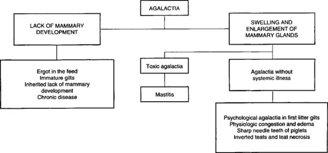
The acute swelling and agalactia of infectious mastitis must be differentiated from other non-infectious causes of acute swelling or ‘caking’ of the mammary glands, which also results in agalactia as follows:
• Agalactia due to a failure in milk letdown is most common in first-litter gilts and is characterized by a fullness of the mammary glands but an inability of the gilt to suckle her piglets in spite of her grunting at them. The gilt is usually bright and alert and systemically normal. The response to oxytocin is dramatic and repeat treatment is rarely necessary
• Farrowing fever is characterized clinically by loss of appetite, inactivity and a body temperature of 39.3–39.9°C (102.7–103.8°F) with minimal detectable changes of the mammary gland
• Parturient psychosis of sows is characterized by aggressive and nervous behavior of the sow after the piglets are born. The sow does not call the piglets and does not allow them to suck. When the piglets approach the sow’s head, she will back away, snap and make noisy staccato nasal expirations. Some sows will bite and kill their piglets. The mammary gland is usually full of milk but the sow will not let it down. Ataractic drugs and/or short-term general anesthesia are indicated and the response is usually excellent. Some sows need repeated tranquilization or sedation for the first few days until the maternal–neonatal bond is established
• Other causes of agalactia accompanied by enlargement of the mammary gland include inherited inverted teats and blind teats due to necrosis of the teats occurring when the gilt was a piglet. These are readily obvious on clinical examination. The sharp needle teeth of piglets may cause the sow to refuse to suckle her piglets. The sow attempts to suckle but leaps up suddenly, grunting and snapping at the piglets. The piglets squeal and fight to retain a teat, thus causing more damage to the teats, which is obvious on clinical examination. Other causes of agalactia accompanied by systemic illness include retained piglets and infectious disease such as outbreaks of transmissible gastroenteritis and erysipelas. The common causes of agalactia in pigs where there is lack of mammary development include ergotism, immature gilts and inherited lack of mammary development. These are set out in Figure 15.1.
Most affected sows will recover within 24–48 hours if treated with a combination of antimicrobials, oxytocin and anti-inflammatory agents. The treatment should begin when the temperature reaches 39.4°C.22
Antimicrobials are indicated in most cases because infectious mastitis and metritis are the two most common causes of the disease. The choice is generally determined by previous experience in the herd or region but broad-spectrum antimicrobials are indicated because E. coli and Klebsiella spp. are the most common pathogens involved. They should be given daily for at least 3 days.23 Usually ampicillin, tetracyclines, trimethoprim– sulphonamide, or enrofloxacin is used.
As soon as possible after the disease is recognized every effort must be made to restore normal mammary function through the use of oxytocin and warm water massaging of the affected mammary glands.
Oxytocin 30–40 U intramuscularly or 20–30 U intravenously, is given, frequently, to promote the letdown of milk. If there is a beneficial response the piglets should be placed on the sow if she is willing to allow them to suck. This will assist in promoting milk flow. Massage of the mammary glands with towels soaked in warm water and hand milking for 10–15 minutes every few hours may assist in reducing the swelling and inflammation and promote the flow of milk. It will also relieve the pain and encourage the sow to suckle her piglets. Intramuscular injections of oxytocin may be repeated every hour, along with massaging of the glands with warm water. Failure of milk letdown or a low response following the use of oxytocin may be due to a reduced sensitivity of the sow to oxytocin during the first week of lactation. In the normal, healthy sow the peak response to oxytocin occurs in the second week of lactation and gradually decreases to a low response at the eighth week.
The use of a long-acting carba oxytocin analog is being explored as a possible substitute for oxytocin. Oxytocin has an effect for about 14 minutes, while the analog has an effect for about 6 hours. Preliminary results of its use in agalactic sows indicate superior results compared to oxytocin.
Anti-inflammatory agents are in common use for their anti-inflammatory effect but are rarely used on their own; flunixin meglumine has been shown to be beneficial and ketoprofen to alleviate pyrexia and endotoxemia. Recently, meloxicam and oxytocin were shown to reduce mortality compared to flunixin only.24 Plasma cortisol levels are increased in the experimental disease and for this reason may be contraindicated. However, field reports suggest that their use along with antimicrobials and oxytocin provides a better response than when they are not used. Corticosteroids used alone do not appear to prevent the disease or enhance recovery. To be effective they must be used in combination with antimicrobials and oxytocin. Dexamethasone at the rate of 20 mg intramuscularly daily for 3 days for sows weighing 150–200 kg has been recommended.
The hypoglycemic piglets must be given a supply of milk and/or balanced electrolytes and dextrose until the milk flow of the sow is resumed, which may take 2–4 days, and most important of all must be kept warm until body reserves are re-established. Piglets should receive 300–500 mL of milk per day divided into hourly doses of 40–50 mL given through a 12–14 French plastic tube passed orally into the stomach. A solution of balanced electrolytes containing 5% glucose can also be given for 1–2 days if a supply of cows’ milk is not available. Condensed canned milk diluted with water 1:1 is a satisfactory and readily available supply of milk. In severe cases where the return to milk production and flow are unlikely, the piglets should be fostered on to other sows. If these are unavailable, the use of milk substitute fortified with porcine gammaglobulin is recommended to prevent the common enteric diseases. This is discussed under colibacillosis. Many more piglets are treated for diarrhea when the sows are treated for MMA, perhaps up to 19% compared to up to 9% normally.1
It has been difficult to develop a rational approach to control because the disease has been considered to be a complex syndrome caused by several different factors. However, the control of infectious mastitis would seem to be of major importance. The routine use of antibiotics and oxytocin without indication does not appear to be helpful.24 Farrowing crates should be vacated, cleaned, disinfected and left vacant for a few days before pregnant sows are transferred from the dry sow barn and placed in the crates. Pregnant sows should be washed with soap and water before being placed in the crate. Farrowing crates must be kept clean and hosed down if necessary, particularly a few days before and after farrowing to minimize the level of intramammary infection. In problem herds, it may be necessary to wash and disinfect the skin over the mammary glands immediately after farrowing. All in/all out in the farrowing area with proper cleaning and disinfection facilitated by batch farrowing will reduce the disease. An opportunity for exercise will help, as under outdoor conditions (sows in paddocks) the condition is rare.
To minimize the stress to the sow of adjusting to the farrowing crate and the farrowing facilities, the sow should be placed in the crate at least 1 week before the expected date of farrowing.
The nature and composition of the diet fed to the sow while in the farrowing crate should not be changed. In order to minimize the risk of toxic agalactia, it is recommended that the daily feed allowance be related to body condition score. It may be necessary to reduce the feed to 1 kg/day (from 100 d gestation) before farrowing.25 The daily intake (compared to the intake during the dry period) may be increased on the day after the sow has farrowed and in increments thereafter as the stage of lactation proceeds. The inclusion of bran at the rate of one-third to one-half of the total diet for 2 days before and after farrowing has been recommended to prevent constipation. In some herds the use of lucerne meal or other vegetable protein at the rate of 15% of the diet may help control the disease. However, under intensified conditions it may be impractical to prepare and provide these special diets on a regular basis. While field observations suggest that a bulky diet at the time of farrowing will minimize the incidence of toxic agalactia, there is little scientific evidence to support the practice.
Antimicrobial agents used prophylactically have apparently been success-ful in controlling some outbreaks. A trimethoprim–sulfadimidine and sulfathiazole combination at 15 mg/kg in feed from day 112 of gestation to day 1 after farrowing may reduce the prevalence. Using oxytocin early may also help. In a recent study where E. coli, streptococci and staphylococci were the most cultured pathogens, marbofloxacin(10% solution) was found to be superior to amoxicillin. All E. coli, were susceptible to the former but 32% were resistant to the latter antibiotic.
The use of prostaglandins for the induction of parturition in sows has not been associated with a marked consistent change in the incidence of the disease. Some field trials have shown a reduction, while others have had no effect.
Smith BP. Pathogenesis and therapeutic management of lactation failure in periparturient sows. Compend Contin Educ Pract Vet. 1985;9:S523-S532.
Liptrap RM. Lactational failure. Vet Annu. 1987;47:134-138.
Martineau GP, et al. Mastitis, metritis and agalactia. Vet Clin North Am Food Anim Pract. 1992;8:661-684.
1 Thorup F. Proceedings of the 16th IPVS Congress, Melbourne 2000:97.
2 Klopfenstein C, et al. Proceedings of the 15th IPVS Congress, Birmingham. 1998;2:230.
3 Ringarp N. Acta Agric Scand Suppl. 1960:7.
4 Ramsoota P, et al. Acta Vet Scand. 2000;41:249.
5 Peter AT, et al. Res Vet Sci. 1985;39:222.
6 Persson A, et al. Acta Vet Scand. 1989;30:9.
7 Lingaas F, Ronningen K. Acta Vet Scand. 1991;32:89.
8 Lingaas F. Acta Vet Scand. 1991;32:97.
9 Lingaas F. Acta Vet Scand. 1991;32:107. 115
10 Chagnon M, et al. Can J Vet Res. 1991;55:180.
11 Persson A, et al. Acta Vet Scand. 1996;37:293.
12 Persson A. J Vet Med A. 1997;44:143.
13 Persson A, et al. Acta Vet Scand. 1996;37:279.
14 Pejsak Z, Tarasiuk K. Theriogenology. 1989;32:335.
15 Smith BB, Wagner WC. Am J Vet Res. 1985;46:175.
16 Smith BB. Compend Contin Educ Pract Vet. 1985;9:S523.
17 Madec F, et al. Rec Med Vet Ec Alfort. 1992;168:341.
18 Magnusson U, et al. J Vet Med B. 2001;48:501.
19 Backstrom L, et al. J Am Vet Med Assoc. 1984;185:70.
20 Delgado J, Jones JET. Aust Vet J. 1981;137:639-643.
21 King GJ, et al. Can Vet J. 1972;13:72.
22 Martineau GP, et al. Vet Clin North Am Food Anim Pract. 1992;8:661.
23 Rose M, et al. Proceedings of the 14th IPVS Congress, Bologna 1996:317.
24 Hirsch AC, et al. J Vet Pharmacol Ther. 2003;26:355.
Mastitis of sheep
Most cases (80–90%) of clinical mastitis are due to Staphylococcus aureus, the remainder to Streptococcus agalactiae and M. haemolytica, with rare occurrences of Escherichia coli and Histophilus somni. (formerly Histophilus ovis). Coagulase-negative staphylococci are a major cause of subclinical mastitis in sheep.1 Sheep at pasture are the only species other than dairy cattle in which outbreaks of mastitis due to S. aureus and M. haemolytica occur. A small percentage of cases are associated with Clostridium perfringens A, Pseudomonas spp., or Corynebacterium pseudotuberculosis. Acholeplasma oculi is pathogenic and can cause mastitis and agalactia.2 Mycoplasma serogroup II is pathogenic in sheep when inoculated experimentally.3
Clinical mastitis in ewes occurs about equally immediately after weaning or close to parturition. In pastured ewes the prevalence is low and due usually to M. haemolytica or Staphylococcus spp. In housed ewes many cases are due to teat injury, especially when the sheep are housed on abrasive floors, such as expanded metal.
Up to 10% of ewes used for milk production have subclinical mastitis.4 Clinical mastitis in pastured ewes averages only about 2% per year, but mastitis causes up to 10% of all ewe deaths.
The forms of loss in milk sheep are the same as those for dairy cattle. In meat and fiber sheep the losses take the form of deaths, due usually to gangrenous mastitis, and to decreased growth in the neonates.5 Where sucking lambs have access to supplemental feed the effect of subclinical mastitis on lamb performance is negligible.6
Although in most European countries sheep are used for mutton and wool production, in Greece nearly all ewes are milked commercially for the production of Greek feta cheese. Any factor that adversely affects the quantity and quality of ovine milk is of great financial interest. The prevalence of subclinical mastitis in sheep in Greece varies between flocks from 29–43%.7 Coagulase-negative staphylococci and S. aureus were isolated in 44% and 33% of the positive milk samples, respectively.
The most prevalent mastitis pathogen of the ewe is S. aureus.8 The incidence of clinical mastitis may be as high as 20%; the mortality rate varies between 25% and 50%, and affected quarters in surviving ewes are usually destroyed. Chronic mastitis can result in a 25–30% reduction in milk yield from affected udder halves.9 The disease can be a very important one in those countries in which ewes’ milk is a staple article of diet. The disease is probably spread from infected bedding grounds, the infection gaining entry through teat injuries caused by sucking lambs.
Other staphylococcal mastitides in ewes include Staphylococcus epidermidis; many clinically normal quarters show a high rate of infection with coagulase-negative staphylococcus.10 Experimental infection with Staphylococcus chromogenes causes clinical mastitis, Staphylococcus simulans causes subclinical mastitis and Staphylococcus xylosus causes a transient increase in the SCC.11
The disease can be reproduced by the introduction of S. agalactiae into the mammary glands and occurs naturally in sheep used as milking animals. The infection originates from an infected udder and is transmitted to the teat skin of other females by milking machine liners, milkers’ hands, wash cloths and any other material that can act as an inert carrier. Also listed as occasional causes are Streptococcus dysgalactiae and Streptococcus uberis.
Peracute, gangrenous mastitis associated with Mannheimia spp. is the common mastitis of ewes. M. haemolytica can be isolated from affected quarters and the disease can be reproduced experimentally12,13 by the intramammary infusion of cultures of the organism. S. aureus, Arcanobacterium pyogenes, and streptococci are often present as secondary invaders.
Mannheimia mastitis occurs sporadically in the western USA, Australia, and Europe in ewes kept under systems of husbandry varying from open mountain pasture to enclosed barns. Mastitis is most common in ewes suckling large lambs 2–3 months old. Infection is thought to occur through injuries to teats, perhaps caused by overvigorous sucking by big lambs. Mannheimia mastitis occurrence is not related to hygiene, many outbreaks occurring in sheep at range but, because of the sheep’s habit of sleeping at night on often used bedding grounds, it is possible that transmission occurs by contact with infected soil or bedding.
In milking ewes clinical mastitis is similar to that in cows, with acute and subacute forms manifested by swelling of the gland and wateriness and clots in the milk. Most clinical cases seen by the veterinarian are in brood ewes, and take the form of gangrenous mastitis, affecting one or both halves.
In sheep there is a strong similarity between this form of mastitis and that associated with M. haemolytica. They are both peracute, gangrenous infections. The ewe is usually recumbent and profoundly toxic, and the affected gland and the surrounding area of belly wall are blue-green in color and cold to the touch. A few drops of clear, bloodstained liquid is all that can be expressed from the udder. A fatal clinical course of 1–2 days is usual.
An acute systemic disturbance, with a high fever (40–42°C, 105–107°F), anorexia, dyspnea and profound toxemia, accompanies acute swelling of the gland and severe lameness on the affected side. This lameness is an important early sign and is useful in locating affected animals in a group. The udder is at first hot, swollen and painful and the milk watery, but within 24 hours the quarter is discolored blue-black and cold, with a sharp line of demarcation from normal tissue. The secretion is watery and red and contains clots. The temperature subsides in 2–4 days, the secretion dries up entirely and the animal either dies of toxemia in 3–7 days or survives with sloughing of a gangrenous portion of the udder, followed by the development of abscesses and the continual draining of pus. Usually only one side is affected. Cases of pneumonia due to the same organism may occur in lambs in flocks where ewes are affected.
Clostridium perfringens A is a rare and highly fatal cause of acute mastitis in ewes. Clinical signs of infection are principally hemolytic and are characterized by hemoglobinuria, jaundice and anemia, plus fever, anorexia and recumbency. The affected quarter is swollen, painful and hot and contains watery, brown, flocculent secretion.14
Suppurative lesions associated with Corynebacterium pseudotuberculosis are found commonly in ovine mammary glands, but they usually involve only the supramammary lymph nodes and are not true mastitis, although the function of the mammary gland may be lost when the infection has spread from the lymph node to mammary tissue.
Determination of the SCC is useful for the prediction of mammary gland infection in sheep.7 Bacteriologically negative ewe’s milk has 2–3% somatic cells, 10–17% lymphocytes, 10–35% neutrophils and 45–85% macrophages.17 In milking ewes milked by machine, SCCs and CMT scores resemble those in dairy cows.18 However, a universally accepted value has not yet been set for the ewe. The SCC in normal ewes’ milk may range from 500000–1 000000 cells/mL. However, more than 95% of milk samples from normal ewes have a total content of less than 500000 cells/mL. In one survey of milk samples from sheep, mean SCC from mastitis-negative samples was 1500000 cells/mL.8 All mastitis-positive samples had somatic cells in excess of 2000000 cells/mL and it is suggested that the threshold level for subclinical mastitis in ewes should be close to 1 500000 cells/mL.8 The CMT is a reliable indicator of the SCC of ewes’ milk and of the level of infection, and it, the SCC and the NAGase all correlate well with the microbiological findings in mastitic quarters. Infection with Maedi–Visna virus does not appear to alter milk SCC in the ewe.
The gross appearance of the affected glands varies with the agent involved and the duration of the process.19 In general, the swollen, hemorrhagic, and/or gangrenous nature of fatal acute ovine mastitis is glaringly obvious. A purulent exudate is sometimes present, especially in the case of chronic C. pseudotuberculosis infection.
Bacteriology – chilled mammary gland for aerobic culture; anaerobic culture if Clostridium sp. is suspected.
Mannheimia mastitis is peracute and resembles mastitis associated with S. aureus. A similar disease in ewes has been ascribed to Actinobacillus lignieresii. Suppurative mastitis associated with C. pseudotuberculosis is chronic in type and no systemic signs occur. Differentiation from clostridial mastitis is also necessary.
Broad-spectrum parenteral and intramammary antimicrobial agents are effective. Although ewes probably require smaller doses of intramammary infusions than cows it is customary to use ordinary cow-type mammary infusion treatments; this may, however, result in a much longer period during which the milk in the quarter has a higher level of antibiotic than is desirable.20 The treatment of ewes with peracute gangrenous mastitis is as unsatisfactory in terms of results as in cows. Systemic treatment is necessary and requires larger doses than normal to achieve significant levels in the mammary secretion.
Removal of sources of infection in sheep flocks necessitates culling some ewes with affected udders but even rigid culling usually fails to completely eradicate the disease.
Control programs for milking sheep flocks could easily be arranged by adapting the one described for cows. Dry period treatment with intramammary infusions of cephapirin benzathine is being used;21 infusion of both halves greatly reduces the subsequent new infection rate.22 For suckling ewes in flocks with a bad history of mastitis another recommended prophylaxis is the infusion of each half with sodium cloxacillin at weaning.
It is possible that vaccination could be an effective method of control for staphylococcal mastitis. A bacterin toxoid has proved moderately effective in reducing the incidence of the disease. Two injections of the vaccine are necessary. Vaccination against staphylococcal mastitis in ewes has been on trial for some time. The latest vaccine, consisting of killed S. aureus cells, toxoided staphylococcal beta-hemolysin plus dextran sulfate as an adjuvant, appears to provide some protection against experimental infection.23 Prophylactic infusion of each half of the udder within a few days of weaning, using half of a tube of dry cow treatment of penicillin and streptomycin, has also given good results. The frequent changing of pasture areas and culling of affected ewes should also help to control the spread of infection.
Polyvalent hyperimmune serum and a formolized vaccine have been shown to be of value in prophylaxis. An autogenous vaccine of killed M. haemolytica24 appears to confer excellent immunity and should be effective in a flock where the disease is occurring.
1 Burriel AR. Vet Rec. 1997;140:419.
2 Sadhana OP, et al. Aust Vet J. 1993;70:227.
3 Kumar D, et al. Aust Vet J. 1993;70:70.
4 Watkins GH, et al. Br Vet J. 1991;147:413.
5 Fthenakis GC, Jones JET. Aust Vet J. 1990;146:43.
6 Keisler DH, et al. J Anim Sci. 1992;70:1677.
7 Stefanakis A, et al. Anim Sci. 1995;61:69.
8 Mavrogenis AP, et al. Small Rumin Res. 1995;17:79.
9 Deutz A, et al. Wien Tierarztl Monatsschr. 1995;82:346.
10 Watson DL, et al. Aust Vet J. 1990;67:6.
11 Fthenakis GC, Jones JET. J Comp Pathol. 1990;102:211.
12 El-Masannat ETS, et al. J Comp Pathol. 1991;105:455.
13 Watkins JH, Jones JET. J Comp Pathol. 1992;106:9.
14 McDonnell AM, Holmes LA. Aust Vet J. 1990;146:380.
15 Bachh AS, Pathak RC. Am J Anim Sci. 1986;56:391.
16 Rapoport E, Bar-Moshe B. Israel J Vet Med. 1986;42:203.
17 Lee CS, et al. J Dairy Res. 1981;48:225.
18 Peris C, et al. J Dairy Sci. 1991;74:1553.
19 Sulaiman MY, Al-Sadhi HI. Prev Vet Med. 1992;13:299.
20 Buswell JF, Barber DML. Aust Vet J. 1989;145:552.
21 Ahmed G, et al. Sheep Res J. 1992;8:30.
22 Hueston WD, et al. J Am Vet Med Assoc. 1989;194:1041.
Mastitis of goats
Goats are much less frequently affected by contagious mastitis than cattle, but mastitis is an important sign in many of the infectious diseases associated with Mycoplasma agalactiae and Mycoplasma mycoides var. mycoides. Details of the other infections encountered are fragmentary and inconsistent; coliform organisms, for example, are listed as not occurring and as being most common. Coagulase-negative staphylococci are a common finding in clinically normal halves1 and appear to cause persistent infection.2
In some surveys, Staphylococcus aureus and Escherichia coli are the most commonly isolated pathogens from mastitic goats.3 Other infections are Pseudomonas spp., Staphylococcus hyicus (much less pathogenic than S. aureus),4 Streptococcus dysgalactiae, Streptococcus pyogenes, Streptococcus intermedius, Arcanobacterium pyogenes, Bacillus coagulans, and Bacillus licheniformis.5 Klebsiella pneumoniae, Corynebacterium pseudotuberculosis, M. haemolytica, and Actinobacillus equuli have also been isolated from mastitic goats.3 Nocardia asteroides causes a systemic reaction and granulomatous lesions in the udder and lungs.6
The prevalence of infection in goats ranges from 10%3 to 30%.7 Prevalence of infection in different herds may range from 7–34% of glands and 17–44% of goats.
This is the most common cause of mastitis in goats. In the experimentally produced disease associated with S. aureus in goats the pathogenesis is very similar to that in the cow except that there is a marked tendency for the staphylococci to invade and persist in foci in the interacinar tissue. As in cattle, some staphylococci in goats’ milk produce enterotoxins and the toxic shock syndrome toxin and are likely to cause food poisoning in humans. Latex agglutination tests are available for the identification of the enterotoxins.8
Goats are uniformly susceptible to Streptococcus agalactiae; mastitis associated with it does occur but to a lesser extent than in cattle. In flocks of milking goats the infection is passed from infected quarters to others by means of the milker’s hands, the teat cups of milking machines and wash cloths used to disinfect the udder before milking. Streptococcus zooepidemicus causes chronic suppurative mastitis in does, and artificially induced infections with S. dysgalactiae are indistinguishable from mastitis associated with S. agalactiae. The pathogenesis is probably similar in all streptococcal mastitides.
Experimental pseudomonas mastitis in goats is acute, with extensive necrosis and fatal septicemia.
Summer mastitis associated with A. pyogenes has been produced experimentally in goats with udder lesions typical of acute suppurative mastitis. Nonlactating goats developed a severe mastitis, lactating animals only a moderate one.
Mastitis in goats is associated with an organism tentatively identified as M. haemolytica. Yersinia pseudotuberculosis has caused mastitis in an aborting goat doe that probably experienced a bout of systemic yersiniosis. The infection would have had zoonotic implications.9 Granulomatous lesions in the mammary glands and in internal organs have been observed in goats experimentally infected with Cryptococcus neoformans.
Clinical mastitis in goats is similar to that in cattle, subclinical, chronic, acute and peracute gangrenous forms occurring. Particular care is needed in the clinical examination of goat’s milk because of its apparent normality when there are severe inflammatory changes in the udder.
Somatic cell counts in milk of goats are higher than in cattle or sheep but vary widely.10 The counts increase with stage of lactation with or without intramammary infection.11 Lower mature equivalent milk production and increased parity are also associated with an increase. Parity of milking does does not affect SCC, standard plate count and major milk components.12 The count is highest in lactating goats during October, December and January.11 Much of the variation was not due to intramammary infection. Noninfected goats may have a SCC of more than 1 000000 cells/mL. These variations make their value as a guide to diagnosis in this species controversial.13 A physiological threshold of 500 000 cells/mL has been suggested;14 however, a count of more than 1 000000 cells/mL can be regarded as positive for mastitis.15 Some observations indicate that the most discriminating threshold for diagnosis of infection is 800000 cells/mL.2 The Fossomatic instruments and infrared milk analyzers must be calibrated with goat milk standards for more reliable and accurate analysis of milk.16
In staphylococcal mastitis infected halves have higher NAGase and CMT tests than normal halves but they and the lactate dehydrogenase and antitrypsin tests give very variable results and are not considered to be reliable. Two of the difficulties encountered are that the efficiency of the tests is not so good in the colostral period, and that halves adjoining affected halves give higher than normal results.17
Treatment and control of mastitis techniques to be used in goat does can be adapted from those used for cattle, with the details of dry period and lactational treatments supplied by a laboratory culture. If the cow dose rate is used, retention of the antibiotic in the udder will be prolonged and the withholding period will need to be increased. Antibiotic residue tests for screening bovine milk adequately identify goat’s milk that is free of antibiotic residues.18 Vaccination against S. agalactiae in goats causes a rise in serum antibodies that may provide a degree of immunity. Intravenous flunixin meglumine (two doses at 2.2 mg/kg BW 8 hours) was a more effective antipyretic agent in goats with experimentally induced coliform mastitis than intravenous dexamethasone (0.44 mg/kg BW once).19
1 Ryan DP, Greenwood PL. Aust Vet J. 1990;67:362.
2 Lerondelle C, et al. Small Rumin Res. 1992;8:129.
3 Ameh JA, et al. Prev Vet Med. 1993;17:41.
4 Maisi P, Riipinen I. Aust Vet J. 1991;147:126.
5 Kalogridou-Vassiliadou D. Small Rumin Res. 1991;197:203.
6 Megid J, et al. Arch Bras Med Vet Zootec. 1990;42:545.
7 Contreras A, et al. Small Rumin Res. 1995;17:71.
8 Kariuki AH. Israel J Vet Med. 1991;46:89.
9 Jones TO. Vet Rec. 1982;110:231.
10 Zeng SS, Escobar EN. Small Rumin Res. 1996;19:169.
11 Wilson DJ, et al. Small Rumin Res. 1995;16:165.
12 Zeng SS, Escobar EN. Small Rumin Res. 1995;17:269.
13 Siddique IH, et al. Vet Med. 1988;83:87.
14 Conteras A, et al. Small Rumin Res. 1996;21:259.
15 Kalogridou-Vassiliadou D, et al. J Dairy Res. 1992;59:21.
16 Zeng SS. Small Rumin Res. 1996;21:221.
17 Maisi P. Small Rumin Res. 1990;3:493.
Mastitis of mares
Mastitis in mares is rare. Corynebacterium pseudotuberculosis, Pseudomonas aeruginosa, Streptococcus zooepidemicus, Streptococcus equi, Streptococcus pyogenes, Staphylococcus aureus, Escherichia coli, Klebsiella spp., and Neisseria spp. are all recorded.1,2 Beta-hemolytic streptococci have been found in the milk of many normal, just-foaled mares.3
Clinical cases occur at any time during the lactation and many occur in nonlactating mares. Many mares with typical signs of severe swelling and soreness of the udder, but without abnormal milk, are first observed when a sick foal has not sucked for 24 hours. In streptococcal mastitis there may be severe local pain and moderate systemic signs. In most cases both halves are affected. Gangrenous mastitis similar to that in cows occurs occasionally.
Severe cases, sometimes accompanied by fever, depression, and anorexia, show swelling, pain, and heat in the affected half, ventral edema and clots in the milk; the mare is lame in the leg on the affected side. Gangrene and sloughing of the ventral floor of a gland may occur.
Because of the high frequency of Gram-negative bacteria as causative agents in mares, treatment should include a broad-spectrum antibiotic in an intramammary infusion plus intensive parenteral antibacterial treatment. Trimethoprim– sulfonamide preparations are generally satisfactory. Hot packs and frequent milking are also recommended.