A major problem which large animal clinicians commonly encounter is a group of cattle that are affected with an acute respiratory disease of uncertain diagnosis.
Acute undifferentiated bovine respiratory disease (UBRD) is characterized clinically by dyspnea, coughing, nasal discharge, varying degrees of depression, inappetence to anorexia, a fever ranging from 40–41°C, evidence of pneumonia on auscultation of the lungs, and a variable response to treatment. Some unexpected deaths may have occurred as the initial indication of the problem.
In most cases, pneumonia is the cause of the disease but determining the etiology is the formidable diagnostic problem. If lesions typical of any of the common diseases of the respiratory tract of cattle can be recognized clinically, like those of infectious bovine rhinotracheitis, then on a clinical basis a specific diagnosis can be made.
The affected group may be recently weaned beef calves in a farm feedlot; weaned beef calves or yearlings that have recently arrived in a feedlot; cattle that have been in the feedlot for varying periods of time; young growing cattle on summer pasture or mature cows that have recently been placed on a lush pasture; yearling or mature lactating dairy cattle; or a group of veal calves.
The morbidity rate can range from 10–50% depending on the age of animals affected, the immune status of the animals, the nature of the stressors involved and the nature of the disease. In a study of mortality ratios among US feedlots over a 5-year period (1994–1999), the relative risk of death attributable to respiratory tract diseases increased during most years of the study.1 Cattle entering the feedlots during 1999 had a significantly increased risk (relative risk, 1.46) of dying of respiratory tract diseases, compared with cattle entering in 1994. Respiratory tract diseases accounted for 57.1% of all deaths. Dairy cattle had a significantly increased risk of death of any cause, compared with beef steers, and beef heifers had a significantly increased risk of dying of respiratory tract diseases compared with steers.
In general, bovine respiratory disease accounts for 65–79% of the morbidity and 44–72% of mortality in feedlot cattle. In addition to direct costs associated with death loss and medical treatment, losses attributable to decreased growth performance associated with the incidence of bovine respiratory disease may also occur. Cattle with detectable lung lesions at slaughter had a reduced (0.08 kg/d) growth rate compared with cattle without lesions. Also, 68% of cattle with lung lesions at slaughter were never treated for bovine respiratory disease, suggesting that current methods of diagnosing bovine respiratory disease based on visual appraisal by feedlot pen riders may not always effectively identify sick animals. In general, medical treatment is more effective the earlier in the process it can be initiated. The limitations of identifying clinically affected animals that need therapy was a major factor in the development of metaphylactic use of antimicrobials.2
The primary goal of the clinician is to make the most accurate clinical diagnosis as rapidly as possible, based on the clinical and epidemiological findings that are identifiable on the farm, preferably when examining the animals on the first visit. Giving a prognosis and the formulation of rational and economic treatment that will minimize morbidity and mortality are the next goals. The outbreak may involve a herd of lactating dairy cows, and the selection of antimicrobials used can have a major impact on the length of time the milk from treated cows must be discarded. In any group situation, mass medication of each in-contact animal is a major consideration that will increase costs and must be balanced against the economic losses that might occur if all animals are not treated prophylactically. The clinical management of the outbreak, which includes treatment of the obvious cases and the prevention of new cases if possible, is dependent in part on the diagnosis. However, differentiation between the diseases based on clinical findings can be unreliable and it is usually necessary to begin antimicrobial therapy that will be effective against the bacterial pathogens most likely to be present. The specific cause may or may not be determined by laboratory examination or further clinical and epidemiological examination. Even after intensive clinical and laboratory investigation, the specific etiology will often not be determined and the clinician is left with a diagnosis of ‘acute undifferentiated respiratory disease of cattle’ or bovine respiratory disease.
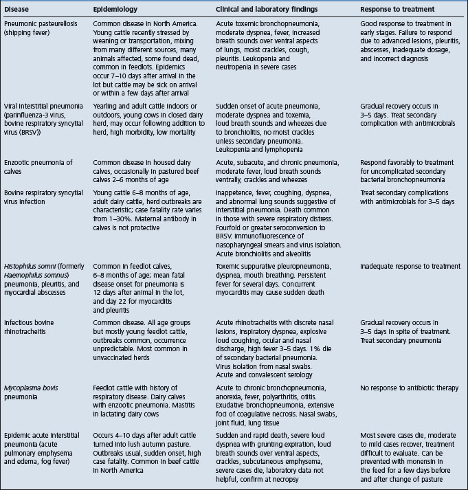
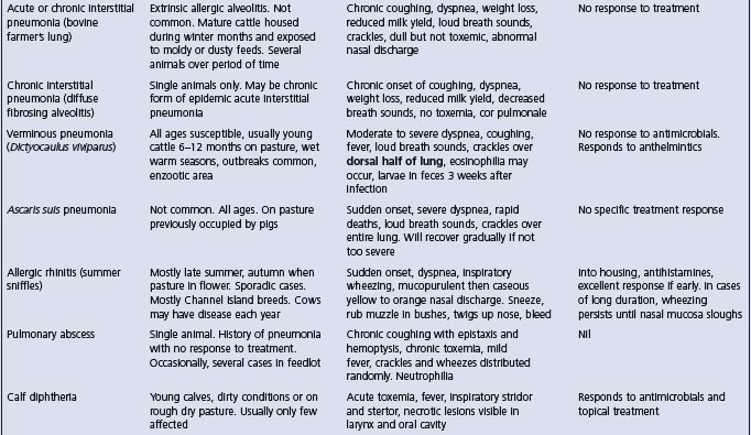
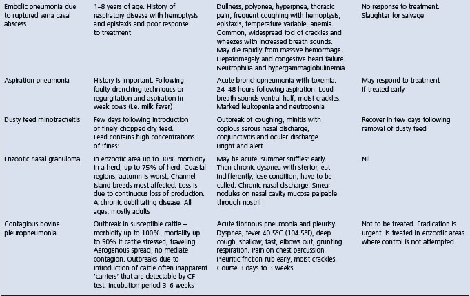
The salient clinical and epidemiological findings of the diseases included in the complex of bovine respiratory disease are summarized in Table 18.5. The common diseases of the respiratory tract of cattle can be broadly divided into those affecting the lower respiratory tract and those affecting the upper respiratory tract. Diseases of the lungs associated with either viruses or bacteria alone or in combination are difficult to distinguish from each other on the basis of clinical findings alone. Thus in a group of cattle with pulmonary disease it may be difficult to distinguish between pneumonic pasteurellosis, Haemophilus pleuropneumonia and the viral interstitial pneumonias with or without secondary bacterial infection. The presence of toxemia, which causes depression and anorexia in bacterial pneumonias, is a useful guide in categorizing the common diseases when making a differential diagnosis list. Cattle affected with uncomplicated viral diseases of the respiratory tract are usually not depressed and anorexic because bacterial toxemia is absent.
The major etiological agents which cause or may be associated with acute UBRD include the following:
The role of the etiological agents in the cause of acute undifferentiated respiratory disease in cattle has been controversial and often uncertain because the major pathogens of bovine respiratory disease are ubiquitous in clinically normal animals. The disease is considered to be the result of the effects of stressors causing immunosuppression, which allows colonization of the respiratory tract by opportunistic pathogens. The spectrum of the immune status of the animals is also a major factor. Animals vaccinated well before natural infection will usually be immune to specific pathogens. Animals that have had the natural infection and have adequate humoral immunity, or cell-mediated immunity in the case of some infections, will be immune to clinical disease.
Many epidemiological studies of bovine respiratory disease have attempted to correlate the level of serum antibodies in feedlot calves on arrival at the feedlot and over the first 30–50 days of the feeding period with morbidity and mortality due to bovine respiratory disease. A low level of antibody to a specific pathogen on arrival followed by significant seroconversion in animals that develop bovine respiratory disease in the first few weeks of the feeding period suggest that the pathogen was an important pathogen in the cause of cases of the disease. Conversely, those animals with a high level of antibody on arrival that do not develop bovine respiratory disease are considered immune. However, some animals with low levels of antibody may remain normal and seroconvert during the early part of the feeding period.
Feedlot cattle commonly seroconvert to the viruses of infectious bovine rhinotracheitis (IBRV), parainfluenza-3 virus (PI-3V), bovine virus diarrhea virus (BVDV), and bovine respiratory syncytial virus (BRSV),3 and to Mycoplasma bovis and Mycoplasma dispar, within the first month after arrival.4 Seroconversion to these pathogens occurs both in animals that develop respiratory disease and those that remain normal within the same group, but the relative importance of each agent and their causative nature is controversial. Seroconversion to M. haemolytica cytotoxin, BRSV and BVDV were predictive of approximately 70% of all respiratory disease cases in Ontario feedlots.3 Calves arriving in Ontario feedlots with high serum antibody levels to Histophilus somni (formerly Haemophilus somnus) had less bovine respiratory disease than calves with lower levels.5
Many respiratory pathogens that could cause disease are present in both individuals and groups of feedlot cattle after they are mixed and during outbreaks of bovine respiratory disease.3 Which ones are most important and causing disease is a major question. It has been suggested that undifferentiated bovine respiratory disease in weaned beef calves entering a feedlot is not a contagious disease and that, while respiratory pathogens may be necessary causes of the disease, they are not sufficient causes.3 This suggests that groups of animals experiencing a high incidence of bovine respiratory disease have been highly stressed and that it may be more important to identify and prevent the environmental and managemental risk factors.
The relationships between bacterial and viral antibody titers and undifferentiated fever and mortality in recently weaned beef calves in western Canada were examined.6 Feedlot calves are commonly exposed to M. haemolytica, H. somni, bovine herpesvirus (BHV)-1 G-IV glycoprotein, BVDV, M. bovis, and Mycoplasma alkalescens in the early feeding period.6 Seroconversion to M. haemolytica antileukotoxin was associated with a decreased risk of undifferentiated fever. Higher arrival BVDV antibody titer was associated with a decreased risk of undifferentiated fever. Higher arrival H. somni antibody titer and increases in H. somni antibody titer after arrival were both associated with a decreased risk of undifferentiated fever. The odds of overall mortality (OR 5.09) and hemophilosis mortality (OR 11.31) in clinical cases were higher than in the controls. In summary, protective immunity to M. haemolytica antileukotoxin H. somni, BHV-1 G-IV glycoprotein, BVDV, and Mycoplasma spp. may be necessary to reduce the occurrence of undifferentiated fever.
The association between exposure to H. somni and M. haemolytica and the risk of acute UBRD was examined using serological evidence of exposure and a vaccine field trial.7 Vaccination with H. somni in combination with M. haemolytica and with M. haemolytica alone reduced the risk of UBRD. There was no association between serological evidence of concurrent exposure to M. haemolytica and UBRD occurrence. Treated animals tend to have lower titers to H. somni compared to untreated animals.
Chronic, antibiotic-resistant pneumonia, sometimes with polyarthritis, occurs in feedlot cattle in western Canada.8,9 M. bovis, BVDV, and H. somni are commonly found in the tissues at necropsy. This coinfection suggests the possibility of synergism between the BVDV and M. bovis in the pneumonia with the arthritis syndrome.
BVDV has been identified as a contributor to respiratory disease in feedlot calves. On arrival in feedlots, 39% of animals were seropositive for BVDV, and those animals treated for UBRD had larger titer increases to the virus than nontreated animals.10 The virus has been demonstrated by experimental infections of the respiratory tract, isolation of the virus and/or identification of BVDV antigen in tissues and demonstration of active infection through seroconversion in groups of cattle with bovine respiratory disease. BVDV-1b strains have been associated with acute pneumonia in commingled calves that were not vaccinated with BVDV vaccines, and in which M. haemolytica and P. multocida were also present in the pneumonic lesions.11 Experimental infection of seronegative and immunocompetent calves with BVDV type resulted in primary respiratory disease.12
Bovine coronavirus (BCV) has been implicated as a cause of UBRD based largely on the isolation of the virus from the nasal cavities of cattle with respiratory disease. However, based on seroepidemiology of BCV titers in feedlot cattle, although higher antibody titers to the virus were associated with a decreased subsequent risk of treatment for UBRD, there was no association between evidence of recent infection (titer increase) and the occurrence of UBRD.13 Other studies have shown that BCV infections are not associated with an increased risk of treatment for UBRD.10
BCV is widespread in the cattle population and can be isolated from nasal swabs from cattle with bovine respiratory disease. The virus can be found in the feces and nasal swabs of recently arrived feedlot cattle and calves with and without clinical signs of bovine respiratory disease.14 Exposure to BCV prior to arrival in the feedlot is common, with 90% of animals being seropositive on arrival.10 BCV can be isolated from feedlot cattle in many different locations and most cattle seroconvert to the virus during the first 28 days after arrival in the feedlot.15 Cattle shedding the virus from the nasal cavity and developing an antibody response to the virus were 1.6 times more likely to require treatment for respiratory disease than cattle that did not shed the virus or develop an immune response. Cattle that shed the virus from the nasal cavity were 2.2 times more likely to have pulmonary lesions at slaughter than cattle that did not shed the virus.16 In natural outbreaks of shipping fever, more than 80% of affected cattle shed BCV at the beginning of the epidemic when the M. haemolytica infection rate was low.17
Intranasal vaccination of backgrounded feedlot cattle with a BCV vaccine reduced the risk of bovine respiratory disease.18 Vaccination had the greatest effect on calves with intranasal BCV, and on those with antibody titers less than 20, on entry.
The role of BCV in epidemics of shipping fever pneumonia in cattle was examined by the collection of nasal swabs and serum samples prior to the onset of the epidemic, during the course of the illness and after death when necropsies were done and samples of lung tissues were examined.19 Respiratory BCV (RBCV) was isolated from the nasal secretions before and after transport, from lung tissues of those cattle that died early in the epidemic but not later. Pasteurella spp. were isolated from all cattle that had severe pneumonia. All cattle were immunologically naive to both infectious agents at the onset of the epidemic but those that died after day 7 had rising antibody titers to RBCV and M. haemolytica. In contrast, the 18 clinically normal and RBCV isolation-negative cattle had high HAI antibody titers to RBCV from the beginning, while their antibody responses to M. haemolytica were delayed. Application of Evan’s criteria for causation were applied to the findings identified RBCV as the primary inciting cause in the epidemics. Because of the multifactorial nature of bovine respiratory disease, the criteria used are as follows.
1. The virus infects the mucosa of respiratory tract passages and lungs of affected cattle
2. The virus can be isolated in cell cultures at high rates from respiratory secretions and lung samples during the pathogenesis of bovine respiratory disease
3. Virus-specific immune responses are observed in cattle that recover from bovine respiratory disease. Rising titers of HAI antibodies against RBCV were detected in all surviving calves that had RBCV infections on days 0, 5 and later. They developed typical primary antibody responses to RBCV infections characterized by rises in IgG1 and IgG2. In contrast, the RBCV isolation-positive cattle with fatal outcomes had no or low titers of HAI antibodies against RBCV in the early stages of the epidemics. These cattle developed only initial IgM responses to RBCV infections before they died
4. Viruses isolated from cattle with bovine respiratory disease are not isolated from clinically normal cattle but they may be detected in the pathogenesis of other respiratory tract diseases
5. Cattle with significant antibody titers against the virus do not develop bovine respiratory disease, which occurs in cattle without such immune protection
6. Elimination of the virus factor prevents or decreases the severity of bovine respiratory disease
7. The whole thing should make biological and epidemiological sense.
The most important part of the clinical and epidemiological examination is to determine the case definition, which includes the following questions.
• Which body system is affected and where in that body system is the lesion?
• Do the animals have pneumonia, rhinitis, laryngitis, tracheitis, bronchitis or combinations of these abnormalities?
The clinician should attempt to make a clinical diagnosis by closely examining several typically affected animals and determine if the lesions are in the lower or upper respiratory tract. The presence of toxemia, depression, fever, anorexia, and agalactia in lactating dairy cattle indicates a primary or secondary bacterial infection. The presence of loud breath sounds (consolidation) and abnormal lung sounds (crackles and wheezes) indicates the presence of pneumonia. Diseases of the upper respiratory tract are characterized by inspiratory dyspnea, stridor, loud coughing, sneezing, wheezing, and lesions of the nasal mucosa. When a single animal is involved, such as a dairy cow, or a group of dairy cows, a close physical examination, including auscultation of the lungs and inspection of the nasal mucosae and the larynx, is usually done.
In commercial feedlots, where large numbers of clinically affected animals are processed daily, and time is an economically important factor, the clinical examination of affected animals may be cursory and limited to taking the rectal temperature of animals that have been identified as sick based on recognition of depression and the absence of clinical findings suggestive of disease of some body system other than the respiratory tract. In such instances, if depressed animals have a rectal temperature above a predetermined level of, for example, 40°C, they are considered to have bovine respiratory disease if no other clinical findings are detectable that are referable to organ systems other than the respiratory system. In such cases, a diagnosis of acute undifferentiated fever of feedlot cattle has been suggested.20 In most cases, the thorax of these animals is not auscultated for evidence of pneumonia. As a result it is probable that the number of animals with respiratory disease is overestimated. Undoubtedly, some sick feedlot animals have a fever of undetermined origin unassociated with respiratory disease or any other inflammatory lesion. In a pen of recently arrived cattle in a feedlot, many animals that appear normal will have rectal temperatures above the critical temperature without any clinical evidence of bacterial infection that requires treatment. The elevated temperatures of these animals will return to within normal ranges within a few days.
The subjective clinical findings of distant examination that have been used by animal attendants in commercial feedlots to identify sick animals that need to be closely examined include: degree of ruminal fill (1, normal; 2, slightly gaunt; 3, moderately gaunt; 4, excessively gaunt), attitude (1, normal; 2, slight lethargy; 3, severe lethargy; 4, nonambulatory), ocular discharge (1, none; 2, slight; 3, moderate; 4, abundant), nasal discharge (1, none; 2, slight; 3, moderate; 4, abundant).
The sounds heard on auscultation of the lungs at three sites along a line extending from the cranioventral to caudodorsal lung fields (1, normal; 2, slightly harsh; 3, moderately harsh; 4, severely harsh).
The feeding and watering behavior of healthy and sick animals in a commercial feedlot has been examined using radiofrequency technology to record individual animal behaviors.21 Eating and drinking behaviors are associated with clinical signs of bovine respiratory disease but there is no obvious predictive association between signs of bovine respiratory disease in recently arrived weaned beef calves and eating and drinking behavior.22 Calves that were sick had greater frequency and duration of drinking 4–5 days after arrival than calves that were not sick. Sick calves had significantly lower frequency and duration of eating and drinking 11–27 days after arrival but had greater frequency of eating 28–57 days after arrival than calves that were not sick. Calves at slaughter that had a higher percentage of lung tissue with lesions had lower frequency and duration of eating 11–27 days after arrival but had greater frequency and duration of eating 28–57 days after arrival. Agreement of calves being sick and having pulmonary lesions at slaughter was adequate. Agreement for calves being removed and having pulmonary lesions at slaughter was low. Experimentally, the electronic acquisition of feeding behavior data for feedlot cattle, when analyzed using cumulative sums (CUSUM) procedures, offers the potential for predicting morbidity before conventional visual methods of appraisal. The feeding behavior during the first 30 days cattle are in a receiving pen may be used to detect animal morbidity approximately 4.1 days earlier than conventional methods typically employed in commercial feedlots.23 Overall accuracy, positive predictive value and sensitivity of the CUSUM prediction method were 87, 91, and 90%, respectively.23
There is a need to improve our clinical diagnostic techniques and to develop new ones that can be applied to making a rapid and accurate diagnosis beside the animal in the field situation. To base a diagnosis of acute respiratory disease on the presence of depression, which has a component of observer subjectivity, and a fever results in the unnecessary treatment of many animals, which is uneconomic and potentially promotes undesirable antimicrobial resistance and residues in milk and meat.
This includes age of animals affected, a single animal or group of animals, and vaccination history. Recently arrived feedlot cattle mixed from many different origins are susceptible to fibrinous pneumonia associated with M. haemolytica.
Are they in the feedlot, on pasture, or housed in a barn with what quality of ventilation?
• How soon after arrival in the feedlot did the animals become affected?
• What stressors may have recently preceded the outbreak?
• What risk factors could have predisposed to this outbreak?
• Have the animals been recently shipped and mixed with animals from another source?
Consideration of the clinical and epidemiological findings can then be correlated, and hypotheses formulated and tested to determine why the disease occurred.
Bovine respiratory disease occurs under many different situations, including all age groups, feedlot animals kept outdoors, housed dairy calves, nursing and recently weaned beef calves, dairy and beef cattle heifers, and adult lactating dairy cows. Epidemics of acute respiratory disease have been described in dairy calves from birth to 6 months of age.24,25 Outbreaks of BRSV can occur in dairy cattle heifers and adult dairy cattle.26
Pneumonic pasteurellosis is most common in recently arrived feedlot calves. In calves 3–5 weeks after arrival in the feedlot, H. somni pleuropneumonia may be more likely. Acute interstitial pneumonia (pasture-induced) is the most likely diagnosis when confronted with an outbreak of acute respiratory disease in mature beef cattle that have been moved from a summer pasture to a lush autumn pasture within the last 4–10 days. Acute pulmonary disease in a few head of feedlot animals several weeks or months after arrival in the lot is probably an acute interstitial pneumonia, which may be associated with 3-methlyindole (3MI) metabolites.27
The risk factors that have been identified in outbreaks of respiratory disease in feedlot cattle include the purchase of cattle from auction markets, whereby cattle arrive at the feedlot over an extended period of time, and mixing of cattle from many different sources.3 An epidemiological study of fatal fibrinous pneumonia in auction-market-derived feedlot calves in western Canada revealed that peak mortality occurred approximately 16 days after arrival at the feedlot.28 The risk of fatal fibrinous pneumonia was consistently greater for calves entering the feedlot in November, shortly after the auction sales had peaked, when the feedlot was reaching capacity.29 Mixing of calves from different farms was considerable, with a median of two calves per farm on truckloads arriving at the feedlot. Increased mixing at the auction markets was associated with increased fatal disease risk. The distance calves were transported by truck from the auction markets to the feedlot was not associated with fatal disease risk.3 When the incidence of fatal fibrinous pneumonia was high, the disease clustered within truckloads or pens. Risk factors positively associated with disease clustering included increased mixing of calves from different farms at the auction markets, month of purchase, number of calves passing through the auction markets, and weather conditions at arrival.28-31
Transportation of feedlot calves increases serum concentration of oxidative stress biomarkers, which are related to episodes of bovine respiratory disease.32 Transportation stress significantly decreases serum total antioxidant capacity and increases malondialdehyde concentrations in steer calves.33 It is proposed that stressors such as marketing (through an auction barn) and transportation to the feedlot precipitate oxidative stress, which reduces the antioxidant defense capacity and increases total body lipid peroxidation, resulting in increased susceptibility to bovine respiratory disease. These biomarkers may be useful to measure the oxidative stress of transported cattle. There is some experimental evidence that acidogenic diets and ketoacidosis may affect lymphocyte function, which may affect vaccine efficacy.34
The literature on how the adequacy of diets of recently arrived feedlot cattle may affect their health and immunity has been reviewed.35 Diets for newly arrived stressed beef cattle must be formulated to compensate for decreased feed intake and known nutrient deficiencies.
The literature on the risk factors for bovine respiratory disease in dairy heifers and the effect of the disease on productivity has been reviewed with relevance to commercial dairy farming in the Netherlands.26 Bovine respiratory disease in dairy heifers increases the risk of mortality directly after the disease episode by up to six times, reduces growth during the first 6 months of life by up to 10 kg, and increases the likelihood of dystocia after first calving. Both herd size and other diseases in dairy heifers are clearly associated with the risk of bovine respiratory disease. Season and colostrum feeding are important. The most important risk factors for mild and severe pneumonia in dairy calves aged birth to 3 months were inadequate air circulation and the purchase of cattle.24
An epidemic of acute respiratory disease associated with BRSV occurred during the winter and spring of 1995 in Norway.36 Data from 431 cattle herds were collected. The risk of acute respiratory disease occurring in cattle was related to herd size and type of production and an expressed interaction between these two variables. The risk of a herd outbreak in a mixed herd of 20 animals was estimated to be 1.7 times greater than in a dairy herd, and 3.3 times greater than in a beef herd of comparable size. On increasing herd size to 50 animals, the risk increased 1.3-fold for a mixed herd, 3.3-fold for a dairy herd and 2.1-fold for a beef herd, compared to a risk for a corresponding type of herd of 20 animals. The disease spread initially from one location to another during the first 6–9 weeks; the rate of transmission between neighboring farms seemed to be higher than for the other districts included in the study. It was hypothesized that one common source of infection was involved in the outbreak and the case herds were clustered in time as well as spatially.37 The average daily milk loss was estimated to be 0.70 kg per cow for 7 days after a herd outbreak compared with the period one week before.38
The clinician is limited in most situations to correlating clinical, epidemiological and necropsy findings in making a diagnosis. Diagnostic laboratories may not be readily available and their resources for microbiological and serological investigations may be much less than is needed for an accurate determination of causes. In the past, investigations of outbreaks of bovine respiratory disease have been incomplete and the interpretation of the findings almost impossible because one or more pieces of information were missing.
A number of important factors contribute to the difficulty of unraveling the etiologies in field outbreaks of respiratory disease. A review of the literature on the morbidity and mortality rates and disease occurrence in North American feedlots found differences in the definition of the terms used that makes the reports difficult to compare.39 In addition, the case definition or the clinical diagnosis is invariably inadequately defined. In feedlots, the morbidity rate will range from 15–45% of cattle within 3 weeks after arrival and the population mortality rate varies from 1–5%. Respiratory diseases account for about 75% of the diseases. Three important items are to be considered:
A systematic method of data collection from the customized records of large feedlots has been developed and validated for use in the National Animal Health Monitoring System. The current collection of data from large feedlots provides an acceptable level of sensitivity and specificity for the program but it is important that the veterinarian makes regular clinical observations to validate the data.
The course of the disease, especially when animals have been treated, alters the gross and microscopic appearance of tissues and the microbiological (bacteriological, virological) and serological findings so that the animal’s status is impossible to determine.
Nasal swabs taken from clinical cases before treatment often yield a pure culture of pasteurellas but M. haemolytica biotype A serotype 1 is the most common isolate obtained from cattle with acute pneumonic pasteurellosis. The same serotype can usually be isolated from in-contact and apparently healthy calves. The antimicrobial sensitivity of the pasteurellas isolated can be done, but interpretation of the results is often difficult because it is not known whether the isolates from nasal swabs represent the organisms causing the lesions. Significant differences may exist between the antimicrobial sensitivities of isolates from nasopharyngeal swabs and those from the lung tissues. Thus it is not yet possible to recommend routine culturing and antimicrobial sensitivity determination of pasteurellas from nasal cavity or nasopharyngeal mucus from cattle with acute bovine respiratory disease. At the individual animal level, nasopharyngeal swabs and bronchoalveolar lavage reveal only moderate agreement; at the group level the isolation rates of various organisms are similar. In healthy calves monitored from the farm to the feedlot there was no relationship between the nasal flora and pulmonary lesions. The results of antimicrobial susceptibilities of bacterial pathogens isolated from the lung tissues of cattle with pneumonia over a period of years may provide some indication of trends in antimicrobial sensitivities but the results are not useful in making decisions about the selection of antimicrobial-affected animals.40
The literature on the principles of antimicrobial susceptibility testing of bacterial pathogens associated with bovine respiratory disease has been reviewed.41 Two different methods are used. The Kirby– Bauer method is the traditional in vitro test of bacterial susceptibility or resistance to antimicrobials, which uses a disk containing a standardized concentration of an antimicrobial. Bacteria grow or fail to grow surrounding the disk, and results are interpreted as resistance or susceptibility of the bacteria to certain antimicrobials. The serial-dilution testing uses a broth or agar medium with selected dilutions of antimicrobials in 1:2 dilution steps. Results are expressed as susceptible, intermediate susceptibility or resistant and also as minimum inhibitory concentrations (MICs), which are considered more reliable. The MIC is defined as ‘the lowest concentration of an antimicrobial that prevents visible growth of a microorganism in agar or broth dilution susceptibility test’.
It is important to adhere to standards set by the National Committee on Clinical Laboratory Standards/Veterinary Antimicrobial Susceptibility Testing Subcommittee (NCCLS/VASTS). Veterinary-specific breakpoints are determined by the NCCLS/VASTS through a consensus process based on reviewing pharmacokinetic, MIC, zone-diameter scattergram and clinical trial data relating to an antimicrobial application. The subcommittee selects MIC breakpoints and zone-interpretative criteria that best fit the definitions of susceptible, intermediate susceptibility and resistant.41
The most veterinary-specific breakpoints for pathogens in bovine respir-atory disease have been determined for five antibiotics: ceftiofur, enrofloxacin, florfenicol, spectinomycin, and tilmicosin.41 The breakpoints for oxytetracycline and chlortetracycline are adapted from human breakpoints developed for tetracycline.41
Nasal swabs may be submitted for isolation or detection of viruses such BVH-1, BRSV, bovine coronavirus.
Serum samples may be submitted for determination of the levels of specific antibody to suspected viral causes of the bovine respiratory disease. Paired acute and convalescent serum samples from both affected and normal animals in the herd are desirable. In a group of animals in a feedlot, or dairy or beef cattle herd, serology for a specific etiological agent may be followed over a period of time to determine seroconversion and its relationship to occurrence or absence of clinical disease.
Plasma fibrinogen concentrations are elevated, paralleling the increase in body temperature, and are a more reliable indication of the presence of the lesion than clinical assessment. Young cattle with clinical signs of acute respiratory disease, a fibrinogen concentration greater than 0.7 g/dL, and a temperature greater than 40°C (104°F) are likely to have pneumonic pasteurellosis. Leukocyte counts are of little value, as a leukocytosis and neutrophilia occur in some animals but in others there may be a neutropenia or no significant change. Acute phase proteins are increased within 24 hours following experimental intratracheal inoculation of M. haemolytica into calves. The availability of a rapid test for acute-phase proteins could assist in the field diagnosis of the disease and its possible differentiation from similar diseases.
Necropsies should be done on all animals that die with the disease. In feedlots this is a means of obtaining daily information on the occurrence of specific diseases and on the efficacy of the animal health management program. The most common pathological finding is fibrinous pneumonia and coagulation necrosis, with varying degrees of the common complications such as pleuritis and pulmonary abscesses. One of the major problems in the diagnosis of feedlot pneumonia has been to assess the age of the lesions. Determining the age of a bacterial pneumonia with some accuracy would help the health management to assess whether or not the pneumonia was already present in the animal on arrival or whether a treatment failure resulted from late detection or from inadequate drug therapy. An attempt to age the lesions of bacterial pneumonia of feedlot cattle indicated that the degree and extent of necrosis and fibrosis offers the best opportunity to age the pneumonia. In acute interstitial pneumonia, the lungs are diffusely red-tan, enlarged, and do not collapse. All lobes are rubbery, wet, and heavy. Emphysema is present mainly in the diaphragmatic lobes and there is white froth in the trachea. In fibrinous pleuritis there are thick sheets of fibrin over the visceral and parietal pleura. The thorax contains fibrin and straw-colored fluid. In caudal vena caval thrombosis, the lungs contain pulmonary arterial aneurysms and abscesses. Blood clots are present in the airways and the liver has an abscess near the vena cava that contains a thrombus.
Tissue samples are submitted for histopathology and bacteriological and virological examination depending on the specific disease suspected. However, the length of time usually required to do the diagnostic work and interpret the results means that the procedure is expensive and to an extent inconclusive because the results are available only when the outbreak is over.
A method has been described of recording pulmonary lesions of beef calves at slaughter and the association of lesions with average daily gain.42 Computer imaging technology can be employed to facilitate the capture of feedlot necropsy data.25 A digital camera is used to capture necropsy findings and the images are electronically transferred to a central reference laboratory for veterinary interpretation and diagnosis.
A large body of information has been generated on the microbiology, and more recently molecular microbiology, of specific pathogens associated with bovine respiratory disease but only a small amount is applicable clinically. Insufficient effort has been directed towards integrating the information and applying it to the effective control of respiratory disease on the farm. Ideally, investigations of outbreaks of bovine respiratory disease should consist of in-depth examinations of a representative sample of the affected group and normal in-contact animals using a multidisciplinary approach involving clinical, epidemiological, and laboratory investigation. These procedures, especially those requiring detailed virological and serological examinations, are expensive and in the light of the economic status of cattle industries not likely to be lightly borne. But it will only be when such a multidisciplinary approach is brought to bear on bovine respiratory disease that we will improve our position with respect to knowing what actually occurs in outbreaks of the disease. Of paramount importance is the identification of risk factors, which, if valid, gives the clinician a powerful clinical tool for the clinical management and control of respiratory disease in cattle. Of even greater importance is the necessity for the clinician to visit the farm and conduct those clinical and epidemiological investigations that are necessary to solve the problem and to monitor the problem and the herd until recovery occurs.
The principles of the clinical management of outbreaks of acute undifferentiated bovine respiratory diseases are:
• The clinician must visit the farm and do the clinical and epidemiological investigations necessary to solve the problem, to assist the owner or the animal attendants with the clinical management of the disease, and to monitor the problem and the herd until recovery occurs. Simply dispensing antimicrobials to the owner without clinical examination of the animals is inadequate and contradicts the intention of the veterinarian–client relationship. The veterinarian is professionally obliged to provide explicit instructions about medication of affected animals and the drug withdrawal requirements, and to keep adequate records of affected animals, treatments given and the results of laboratory examinations. A final report should be prepared by the veterinarian and sent to the owner
• Unless otherwise determined, when toxemia and fever are present it is assumed that a primary bacterial pneumonia is present or, if a viral interstitial pneumonia is suspected, then a secondary bacterial pneumonia may occur. Therefore, antimicrobial therapy is of prime importance
• New cases must be identified as soon as possible. This will require increased surveillance of the group to detect affected animals as soon as clinical abnormalities such as depression, nasal discharge, and dyspnea are noticeable
• New cases must be treated as soon as they are detected. Each treated animal should be suitably identified and a record kept of the initial body temperature and the treatment administered. If the outbreak is due to pneumonic pasteurellosis, failure to respond favorably to antimicrobial therapy or relapse that occurs a few days after an initial apparent recovery is usually due to late treatment. Delaying treatment until 48 hours after an experimental aerosol infection of M. haemolytica can prolong the course of the disease and increase mortality
• Any of the common antimicrobials must be administered parenterally daily for at least 3 days. Oxytetracycline at 10 mg/kg BW intramuscularly, procaine penicillin G at 30 000–45 000 IU/kg BW intramuscularly, or trimethoprim– sulfadoxine at 3 mL/45 kg BW intramuscularly are effective when given early in the course of the disease. Tilmicosin at a dose of 10 mg/kg BW subcutaneously is also effective. Florfenicol at 20 mg/kg BW intramuscularly and repeated 48 h later is also highly effective.20 Florfenicol and tilmicosin are comparable for treatment of undifferentiated bovine respiratory disease in western Canada.43 Florfenicol is superior to tilmicosin for the treatment of undifferentiated fever in feedlot calves that have previously received metaphylactic tilmicosin upon arrival in the feedlot in western Canada.44 Enrofloxacin at 2.5–5.0 mg/kg BW intramuscularly daily for 3–5 days or a single dose of 7.5–12.5 mg/kg BW is effective for the treatment of bovine respiratory disease.45
In lactating dairy cows, antimicrobials with the shortest milk withdrawal times commensurate with effectiveness should be used. Ceftiofur with no withdrawal period is now available for use in lactating dairy cattle. Milk from treated cows must be kept from the bulk milk supply until the stated withdrawal time has elapsed. Other antimicrobials that have been evaluated for the treatment of acute undifferentiated bovine respiratory disease include sulbactam–ampicillin and the fluoroquinolones.
A beneficial response to therapy should be apparent within 12–24 hours. The body temperature should decline significantly and the appearance of the animal and its appetite should be improved.
The response to treatment, or lack of it, is valuable information in making a final decision on cause. Animals that do not respond to treatment and die should be submitted to intensive necropsy examination and culture of affected lungs. One of the emerging problems inherent in such broad policies in treatment is public health concern with the amount of antibiotic residue in meat. Pressure is now being applied to use antimicrobials only when necessary, which necessitates a more accurate diagnosis. A good example of this problem is when cattle are treated with antibiotics for bovine respiratory disease but the diagnosis is then refined in a day or two to interstitial pneumonia and emergency slaughter is then the appropriate course of action. The cattle cannot be slaughtered until the withdrawal period for the specific antibody used has expired, by which time many of the cattle will have died anyway. The regular use of a particular antimicrobial in feedlots may increase the level of resistance to M. haemolytica.
When confronted with an outbreak, one of the major decisions to be made is whether or not to recommend mass medication of the water or feed supplies for several days or to administer an antimicrobial to all in-contact animals in an attempt to treat cases in the preclinical stage. Veterinarians commonly recommend the use of medicated water supplies as an aid in the treatment of outbreaks of acute respiratory disease, and field observations claim beneficial results. However, there is no validated information available to support a recommendation for a medicated water supply for treatment or prophylaxis in the face of an outbreak. Depending on the water supply system it can be difficult to deliver and maintain a constant concentration of a drug in the water supply; palatability of certain drugs can also be a problem. The medication of the water or feed supplies can also create a false sense of security in the animal attendants, who may not be as efficient in the selection of affected animals in the early stages of the disease.
There are no validated reports of the use of medicated feed as an aid to treatment for outbreaks of acute respiratory disease in cattle.
In an outbreak of acute respiratory disease in feedlot cattle when the daily morbidity rate reaches 6–10% the parenteral administration of long-acting oxytetracycline to all in-contact cattle at a dose of 20 mg/kg BW intramuscularly is recommended. Tilmicosin at 10 mg/kg BW subcutaneously is also effective in reducing the morbidity rate when given to beef calves on arrival in the feedlot or 72 hours later.3 The intramuscular injection of two different formulations of oxytetracycline to high-risk feedlot calves on arrival can reduce the morbidity rate due to respiratory disease during the first 2 weeks on feed and for the entire feeding period by 15–19% and the mortality rate from fatal fibrinous pneumonia by 67–84%. Meta-analysis of field trials of antimicrobial mass medication for prophylaxis of bovine respiratory disease in feedlot cattle indicated that prophylactic parenteral mass medication of calves with long-acting oxytetracycline or tilmicosin on arrival at the feedlot would reduce morbidity rates.
The control of outbreaks of acute bovine respiratory disease will depend on mass medication or metaphylaxis, minimizing the effects of the risk factors and enhancing immunity by the judicious use of vaccines.
The parenteral administration of antimicrobials to each animal as a form of mass medication may assist in the reduction of morbidity and mortality rates due to respiratory disease. The use of long-acting oxytetracycline at a dose of 20 mg/kg BW intramuscularly to feedlot cattle on arrival significantly reduced morbidity and mortality rates. The combined use of long-acting oxytetracycline at a dose of 20 mg/kg BW intramuscularly on arrival followed by the oral administration of 25 g of sustained-release sulfadimethoxine on day 3 resulted in a 90% reduction in treatment days per calf purchased. Tilmicosin, given at a dose of 10 mg/kg body weight subcutaneously to calves on arrival in the feedlot or 72 hours later, can significantly reduce the morbidity rate due to respiratory disease and improve the rate of gain.46
A formulation of long-acting oxytetracycline (300 mg/mL) at a dose of 30 mg/kg BW intramuscularly is superior to the standard formulation of long-acting oxytetracycline (200 mg/mL) given at 20 mg/kg BW.47 The 300 mg/mL preparation of oxytetracycline is also more cost-effective than tilmicosin.48
Mass medication of cattle on entry into feedlots in Australia with tilmicosin at 10 mg/kg BW grew 0.08 kg/d faster than cattle medicated with oxytetracycline at 20 mg/kg BW and nonmedicated cattle.49 There was no significant difference in growth rate between oxytetracycline-medicated cattle and those not medicated on entry into the feedlot. Cattle medicated with tilmicosin had fewer treatments for all illnesses compared with cattle not given an antibiotic on entry to the feedlot and compared with cattle mass-medicated with oxytetracycline. Tilmicosin induces apoptosis in pulmonary neutrophils, leading to a reduction in leukotriene B4 synthesis, thereby reducing further amplification of the inflammatory injury of bovine respiratory disease. Preshipment medication with tilmicosin are not more effective than mass medication on arrival.50
The mass medication of feed supplies of newly arrived feedlot cattle has been investigated as a method of reducing the morbidity and mortality due to respiratory disease. The provision of chlortetracycline in the feed at a rate of 1, 2, or 4 g per head daily during the 2-week period after arrival reduced the number of calves that required treatment for respiratory disease.
As a general outline for the control of bovine respiratory disease the following factors are considered as contributing to disease and their effects must be minimized with suitable management and disease prevention techniques.
• Young growing cattle are more susceptible than mature cattle because of a lack of sufficient immunity. The mixing of young cattle of different origins requires increased surveillance to detect evidence of disease. Vaccination of calves at strategic times may be necessary
• Cattle purchased from various sources and mingled in a feedlot are more likely to develop bovine respiratory disease than cattle that have originated from one source. Some cattle will be highly susceptible and others relatively resistant because of differences in nasal flora and immunological, genetic, and nutritional backgrounds. A high level of management and constant surveillance are necessary to recognize, isolate, and treat clinical cases early in order to minimize morbidity and case mortality
• Rapid fluctuations in environmental temperatures and relative humidity, not only during the fall and winter months but also during warm seasons, will commonly precede outbreaks of respiratory diseases. Every practical and economical management technique must be used to provide as much comfort as possible and to avoid overcrowding
• Inadequate ventilation is a major predisposing cause of respiratory disease of cattle raised indoors. This is of major importance in dairy herds during the winter months in temperate climates
• The weaning of beef calves during inclement weather may exacerbate the stress of weaning and commonly results in an outbreak of respiratory disease
• The stress associated with the marketing of cattle is a major factor. The movement of cattle through saleyards – where they may be overcrowded, mixed with cattle of many different origins, temporarily deprived of adequate feed and water, handled roughly while being sorted, weighed, tagged, blood sampled, vaccinated or injected with antibiotics and/or vitamins and then loaded on to uncomfortable vehicles and transported long distances without adequate rest stops – is stressful. The practice of preconditioning cattle before they enter the feedlot must continue to be examined to determine which aspects are most profitable.
Presale vaccination programs are designed to establish an effective immune response to common respiratory tract pathogens well in advance of any natural exposure that may occur while calves travel through the auction market or after they arrive in the feedlot. These programs usually require calves to be castrated, dehorned, and vaccinated against IBRV, PI-3V, BRSV, and BVDV. Some programs also require vaccination against H. somni and M. haemolytica. Presale conditioning programs involve these procedures but also include weaning and nutritional components. Most such conditioning programs require calves to be weaned and adjusted to a roughage and concentrate diet for at least 30 days prior to sale.
Presale vaccination and conditioning programs have increased and decreased in popularity since their introduction in the 1970s. Producers tend to be reluctant to adopt these practices, because there is no assurance they will be rewarded, in terms of price premiums, for their efforts. However, the establishment of special auctions in Ontario that feature large numbers of vaccinated or conditioned calves resulted in an increased interest in these management practices. Producers selling lots of vaccinated or conditioned feeder calves through special auctions received a premium sale price compared with lots of feeder calves sold through conventional auctions.51 Vaccinated and conditioned calves were less likely to receive treatment for bovine respiratory disease during the first 28 days in the feedlot; but there was no difference in mortality.52 Calves that received antimicrobials on arrival at the feedlot had a reduced risk of treatment for bovine respiratory disease compared with calves that did not.
While vaccines are available for the control of acute respiratory disease associated with IBRV, PI-3V, and Pasteurella spp., there are almost no reports available of their efficacy determined under scientifically designed field trials. Based on current immunological technology, efficacious vaccines are considered to be feasible. The vaccines have been evaluated by experimental challenge of vaccinated animals with specific pathogens in a laboratory environment. However, there is little scientific evidence available that the vaccines are protective against acute UBRD as it occurs in the ‘real world’ situation. Preshipment vaccination of beef calves 3 weeks prior to weaning with vaccines containing IBRV, PI-3V, Pasteurella spp., and H. somni did not reduce the incidence of UBRD compared to those unvaccinated.
Pasteurella bacterins and respiratory viral vaccines have been used extensively in an attempt to control bovine respiratory disease. Many veterinarians and feedlot owners maintain that vaccination against respiratory disease is an essential component in their disease prevention programs, both to prevent specific disease of the respiratory tract such as clinical infectious bovine rhinotracheitis and to reduce losses due to respiratory disease in the first few weeks after arrival. However, a review of the literature on the efficacy of the vaccines available for the control of bovine respiratory disease concluded that there are few documented data to support the use of vaccines against respiratory disease under feedlot conditions. Efficacy refers to the ability of the vaccine to reduce overall treatment rate and/or increase weight gains economically.
In North America a large number of bacterial and viral vaccines are available for the control of bovine respiratory disease. There are single antigen or multiple antigen vaccines, modified live virus or inactivated virus vaccines containing one or more of the following antigens: M. haemolytica, H. somni, IBRV, PI-3V, BRSV, and BVDV. There are many multiple antigen vaccines containing combinations of the respiratory viruses, BVDV, H. somni, and M. haemolytica. In western Canada, it is more cost-effective to vaccinate auction-market-derived, fall-placed feedlot calves with a multivalent viral vaccine containing IBR, PI-3, BVD, and BRS viruses than a single univalent viral vaccine containing IBRV only.53
The selection of which vaccine to recommend for the control of UBRD in feedlot cattle is currently not possible based on the efficacy information which is available to the veterinarian. The vaccines are used widely and many anecdotal claims for their effectiveness are made but there is little scientific evidence based on properly designed field trials that the vaccines are effective and economical in reducing the incidence or the consequences of respiratory disease such as suboptimal weight gain. The major problem has been that vaccine manufacturers have not conducted a sufficient number of well-designed field trials to evaluate the efficacy of the vaccine against naturally occurring disease in the field with vaccinated animals and unvaccinated animals as concurrent controls. In most cases the vaccines were approved for sale on the basis of tests for safety in animals, and the potency measured by a serological response to the vaccine or experimental challenge in animals under laboratory conditions.
The information available about the commercial vaccines currently used in Canada for protection against bovine respiratory disease has been reviewed.54 The available vaccines offer protection against only IBRV, BVDV, BRSV, PI-3V, M. haemolytica, and H. somni. Various combination vaccines containing modified live virus, killed virus, bacterins, and/or bacterial culture supernatants/surface extracts are available.
Meaningful field trials to evaluate vaccines for the control of bovine respiratory disease are difficult to achieve. The case definition of what is a ‘case of respiratory disease’ has been very general, such as the presence of anorexia, depression, and a fever. Therefore, when testing a vaccine for the control of pneumonic pasteurellosis, the conclusions reached may be questionable if the cause of the sick animals in either the vaccinated or control group is not known – thus the importance of case definition. In contrast to field trials, the measures used by the manufacturer in the laboratory challenge of the vaccine have been specific. In a field trial, the control group and the vaccinated groups must be comparable. Where more than one vaccine is used to control respiratory disease in vaccinates and controls it is difficult to evaluate one of the vaccines or the components of a multiple antigen vaccine unless large numbers of animals are used. Another problem is the difficulty of having the controls and the vaccinates experience approximately the same risk of being affected with respiratory disease.
Field trials for bovine respiratory disease vaccines are often unsatisfactory because of inadequate planning, unsatisfactory experimental design and lack of monitoring. A check list of key elements to consider when assessing the clinical research published for a particular vaccine has been suggested. The following items should be considered:
• Has the vaccine been laboratory and field tested in randomized controlled field trials? If so, how many trials, and, in each case:
A field trial was conducted to compare the serological responses in weaned beef calves 6–8 months of age vaccinated against IBRV, PI-3V, BRSV, and BVDV. There were significant differences in serological responses among the various commercial vaccines. Antibody titers to IBRV were higher in calves vaccinated with modified-live virus IBRV vaccines than when the inactivated vaccine was used. Following double vaccination with modified-live virus IBRV and PI-3V vaccines, seroconversion rates and antibody titers were higher in calves vaccinated intramuscularly than in those vaccinated by the intranasal route. It is not known if these differences reflect differences in vaccine efficacy under field conditions. The effect of using multiple antigens in the same vaccine on the serological responses is not clear. In some cases, vaccines containing the BRSV antigen resulted in lower titers to BVDV and PI-3V than vaccines that did not contain the BRSV.
The following comments on the use of vaccines as an aid in the control of acute undifferentiated respiratory disease in feedlot cattle are based on the current information available.
Because fibrinous pneumonia associated with M. haemolytica is the most common lesion associated with bovine respiratory disease in feedlot cattle, much of the emphasis has been on the development of effective vaccines for bovine pneumonic pasteurellosis. Based on the immunological and microbiological observations of both naturally occurring and experimentally induced pneumonic pasteurellosis it appears that effective artificial immunization of cattle is possible. High levels of naturally acquired antibody to M. haemolytica have been associated with protection against the disease.
Calves that recover from experimentally induced pneumonic pasteurellosis possess increased resistance to subsequent experimental challenge. Calves that were naturally exposed to M. haemolytica or exposed by vaccination subcutaneously or intradermally to the live organisms developed some resistance to experimental challenge and developed antibodies to all surface antigens and cytotoxin. Resistance to experimental challenge with the organism correlated directly with serum cytotoxin neutralizing titers. This supports the hypothesis that protection against experimental challenge with M. haemolytica may require an immune response to cytotoxin. This is supported by the observation that cattle that died from fibrinous pneumonia due to M. haemolytica had lower cytotoxin neutralizing activity in their sera than cattle from the same group that died from other causes.
Antibodies to leukotoxin and certain bacterial surface components appear to be important for resistance to disease. The basis of a recently introduced pasteurella vaccine is that vaccination of calves with a leukotoxic culture supernatant from pathogenic M. haemolytica provided some protection against experimental challenge with the organism. The vaccine has been evaluated in field trials in feedlots in Canada with variable results. In one trial, the vaccine was used on arrival in fall-placed calves and reduced the pull rate by 4%, the first relapse rate by 11.9% and the second relapse rate by 18.7%. There was an 8.2% decrease in the mean number of treatment days. Overall mortality was decreased by 49% and mortality rate due to fibrinous pneumonia was reduced by 50%. It appeared that vaccinated animals also responded more favorably than nonvaccinated animals. Vaccination of recently shipped nonpreconditioned calves with the vaccine in Ontario resulted in a slight decrease in morbidity, slight improvement in response rates and perhaps an important reduction in relapse rates. When the vaccine was combined with an intramuscular modified-live IBR/PI-3 virus vaccine, the morbidity rate was increased, the response rate was decreased and the mortality rate was increased in some groups. It appears that the use of modified-live virus vaccines in recently arrived calves is contraindicated; this is consistent with earlier observations in the Bruce County Project, where fall-placed calves were vaccinated on arrival with a modified-live virus vaccine.
The Pasteurella vaccine has also been evaluated in a field trial that compared its efficacy in calves vaccinated at the ranch 3 weeks prior to shipping to the feedlot, vaccination of ranch calves on arrival in the feedlot, and auction mart calves assigned to either receive or not receive the vaccine on arrival at the feedlot. The vaccine did not effect a change in morbidity rates or weight gain. Total mortality rates were increased significantly, and mortality rates from respiratory disease tended to be increased in ranch calves that were vaccinated at the ranch. In auction-mart-derived calves, the relapse rates were significantly lower in vaccinated calves. The source of calves was a major factor affecting the incidence and/or effects of bovine respiratory disease. Calves moved directly from the ranches to feedlots, regardless of vaccination status, had lower morbidity and mortality, and better weight gains, than calves purchased from auction marts.
A H. somni bacterin evaluated in a large number of feedlot cattle had no effect on the overall crude mortality rate; however, vaccination appeared to reduce the incidence rate of fatal disease and the mortality rate during the first 2 months in the feedlot, when risk of fatal disease onset was highest. The incidence of fatal disease onset was highest during the first week after arrival, which suggests that inclusion of a H. somni bacterin in a cow– calf preimmunization program might reduce the proportion of disease that occurs during the first week in the feedlot. Crude mortality and incidence of fatal disease onset during the second week were reduced significantly in the vaccinated steers. It was concluded that about 17% of fatal respiratory disease in the unvaccinated steers could have been prevented by vaccination with the H. somni bacterin. Fibrinous pneumonia was the most common pathological diagnosis. Vaccinating calves twice with a killed whole-cell H. somni bacterin reduced the clinical and pathological effects of experimentally induced H. somni pneumonia.
Because prior infection of the respiratory tract with either IBRV or PI-3V may predispose to pneumonic pasteurellosis, the vaccination of beef calves 2–3 weeks before weaning and feedlot cattle 2 weeks before shipment to a feedlot has been recommended as part of a preconditioning program. The results are variable, but vaccination of calves at 3–6 months of age with an intranasal modified-live IBR and PI-3 virus vaccine has provided protection against experimental pneumonic pasteurellosis induced by aerosol challenge with IBRV followed 4 days later by an aerosol of M. haemolytica. It is important to vaccinate the calves at least 2 weeks before they are weaned, stressed, or transported to a feedlot.
A modified-live virus BRSV vaccine given to beef calves prior to weaning, at weaning or immediately after arrival in the feedlot was associated with a significant reduction in the treatment rate in one of three groups immunized prior to weaning and in calves immunized after arrival in the feedlot.25 There was no significant effect of the vaccine on treatment rate in calves immunized at weaning, in calves immunized after arrival in a bull test station, or in yearlings immunized after arrival in the feedlot. It would appear that the vaccine did provide some protection but the small reduction may not justify the cost of the vaccination program.
Some feedlot veterinarians recommend that feedlot cattle be vaccinated on arrival with an M. haemolytica vaccine, the IBRV and PI-3V vaccine, an H. somni vaccine, and the BRSV vaccine. In some cases the BVDV vaccine is also used because some veterinarians feel that the virus is part of the respiratory disease complex. It is expected that control will be achieved if the animals are vaccinated against all the common pathogens that contribute to lesions of bovine respiratory disease. However, there is little, if any, published evidence based on controlled field trials that such blanket recommendations are justifiable.
Barrett DC. Cost-effective antimicrobial drug selection for the management and control of respiratory disease in European cattle. Vet Rec. 2000;146:545-550.
Bowland SL, Shewen PE. Bovine respiratory disease: commercial vaccines currently available in Canada. Can Vet J. 2000;41:33-48.
Coomber BL, Nyarko KA, Noyes TM, Gentry PA. Neutrophil-platelet interactions and their relevance to bovine respiratory disease. Vet J. 2001;161:41-62.
Cusack PMV, McMeniman N, Lean IJ. The medicine and epidemiology of bovine respiratory disease in feedlots. Aust Vet J. 2003;81:480-487.
1 Lonergan GH, et al. J Am Vet Med Assoc. 2001;219:1122.
2 Wittum TE, et al. J Am Vet Med Assoc. 1999;209:814.
3 Cusack PMV, et al. Aust Vet J. 2003;81:480.
4 Martin SW, et al. Can Vet J. 1999;40:560.
5 Martin SW, et al. Can J Vet Res. 1998;62:262.
6 Booker CW, et al. Can Vet J. 1999;40:40.
7 O’Connor A, et al. Can J Vet Res. 2001;65:143.
8 Haines DM, et al. Can Vet J. 2001;42:857.
9 Shahriar FM, et al. Can Vet J. 2002;43:863.
10 O’Connor A, et al. Can J Vet Res. 2001;65:137.
11 Fulton RW, et al. Can J Vet Res. 2002;66:181.
12 Baule C, et al. J Clin Microbiol. 2001;39:146.
13 Martin SW, et al. Can J Vet Res. 1998;62:257.
14 Hasoksuz M, et al. J Vet Diagn Invest. 2002;14:308.
15 Lathrop SL, et al. Am J Vet Res. 2000;61:1057.
16 Lathrop SL, et al. Am J Vet Res. 2000;61:1062.
17 Storz J, et al. J Am Vet Med Assoc. 2000;216:1599.
18 Plummer PJ, et al. J Am Vet Med Assoc. 2004;225:726.
19 Storz J, et al. J Clin Microbiol. 2000;38:3291.
20 Booker CW, et al. Can Vet J. 1997;38:555.
21 Sowell BF, et al. J Anim Sci. 1999;77:1105.
22 Buhman MJ, et al. Am J Vet Res. 2000;61:1163.
23 Quimby WF, et al. Can J Anim Sci. 2001;81:315.
24 Van der Fels-Klerx HJ, et al. Livestock Prod Sci. 2000;66:35.
25 Wildman BK, et al. Can Vet J. 2000;41:124.
26 Van der Fels-Klerx HJ, et al. Netherlands J Agric Sci. 2002;50:27.
27 Ayroud M, et al. Can Vet J. 2000;41:847.
28 Ribble CS, et al. J Am Vet Med Assoc. 1995;207:616.
29 Ribble CS, et al. Can J Vet Res. 1995;59:167.
30 Ribble CS, et al. J Am Vet Med Assoc. 1995;207:612.
31 Ribble CS, et al. Prev Vet Med. 1994;21:251.
32 Van Donkersgoed J. Can Vet J. 1992;33:786.
33 Chirase NK, et al. Am J Vet Res. 2004;65:860.
34 Donovan DC, et al. J Anim Sci. 2003;81:3088.
35 Galyean ML, et al. J Anim Sci. 1999;77:1120.
36 Norstrom M, et al. Prev Vet Med. 2000;44:87.
37 Norstrom M, et al. Prev Vet Med. 2000;47:107.
38 Norstrom M, et al. Prev Vet Med. 2001;51:259.
39 Kelly AP, Jansen ED. Aust Vet J. 1986;27:496.
40 Welsh RD, et al. J Vet Diagn Invest. 2004;16:426.
41 Apley MD. Vet Clin North Am Food Anim Pract. 2003;19:625.
42 Bryant LK, et al. Bovine Pract. 1999;33:163.
43 Hoar BR, et al. Can Vet J. 1998;39:161.
44 Jim GK, et al. Can Vet J. 1999;40:179.
45 Hamm M, et al. Bovine Pract. 1999;33:56.
46 Vogel GJ, et al. J Am Vet Med Assoc. 1998;212:1919.
47 Schunicht OC, et al. Can Vet J. 2002;43:940.
48 Schunicht OC, et al. Can Vet J. 2002;43:355.
49 Cusack PMV, et al. Aust Vet J. 2004;82:154.
50 Duff GC, et al. J Anim Sci. 2000;78:267.
51 Macartney JE, et al. J Am Vet Med Assoc. 2003;223:670.
52 Macartney JE, et al. J Am Vet Med Assoc. 2003;223:677.
Etiology Mannheimia (Pasteurella) haemolytica biotype A serotype 1. Pasteurella multocida biotype A:3
Epidemiology Young growing cattle, especially recently weaned beef calves placed in feedlot. Can occur in nursing calves and mature cows. Stressors include transportation, mixing animals from many different sources, ineffective ventilation of housed animals
Signs Sudden deaths, acute bacterial bronchopneumonia, fever, toxemia, anorexia, abnormal lung sounds; respond to treatment with antimicrobials
Lesions Acute fibrinohemorrhagic pneumonia with pleuritis
Clinical pathology Culture organism from nasal swabs. Hemogram indicates severe infection and increase in fibrinogen
Diagnostic confirmation Culture organism from lung and histopathology of lung
Control Preconditioning programs. Management strategies to reduce stressors. Mass medication with antimicrobials of individual animals on arrival in the feedlot. Vaccines containing antigens of M. haemolytica
Mannheimia (Pasteurella) haemolytica biotype A serotype 1 is the most common cause of the pneumonia.1 Eleven serotypes have been demonstrated within M. haemolytica.2 M. haemolytica serotypes 6, 2, 9, and 11 and untypable serotypes have been found in lesions of pneumonic pasteurellosis.3 M. haemolytica-like bacteria have been isolated from cases of bovine pneumonia, and a careful characterization of such isolates is necessary for a proper identification.4
Biotype T strains have been isolated from cases of pneumonic pasteurellosis and P. multocida is isolated occasionally. P. multocida serotypes A3 and D can cause severe bronchopneumonia in calves.5 The Pasteurella spp. are the final cause of the pneumonia but the mechanisms by which the bacteria enter and colonize the lung and produce the lesions are complex and unclear. Viruses or mycoplasmas may act synergistically to allow the bacteria to be pathogenic. There is often a history of stressors such as:
Pneumonic pasteurellosis is a common disease of young growing cattle in Europe, the UK, and North America. In Canada and the USA the disease occurs most commonly in recently weaned beef calves 6–8 months of age shortly after being placed into feedlots in the fall of the year. Nursing beef calves, yearlings, and mature dairy and beef cows may also be affected, but less frequently. Pneumonic pasteurellosis, also known as shipping fever, is an entity within the bovine respiratory disease complex, characterized clinically by acute bronchopneumonia with toxemia and pathologically by lobar, anteroventrally distributed, exudative pneumonia in which fibrin is usually a prominent part of the exudate and fibrinous pleuritis is common.
The morbidity may reach 35%, the case-fatality rate may range from 5–10%, and the population mortality rate may vary from 0.75–1%. However, these morbidity and mortality data may not be reliable because of wide variations in the methods used to calculate disease incidence and prevalence. A review of the literature of morbidity and mortality rates and disease occurrence in feedlot cattle in North America found deficiencies in the epidemiological data available from feedlots.6 Case definitions of clinical cases are often poorly defined. A summary of 14 comparable studies containing disease incidence rates in calves in the first few weeks following arrival in feedlots found wide variations in how the disease definitions and statistics were used. The incidence of morbidity ranged from 0–69% with most reports between 15% and 45%. The population mortality rate ranged from 0–15% with most reports between 1% and 5%. The peak incidence of disease occurs within the first 3 weeks after arrival of the calves in the feedlot. The most common clinical and pathological diagnosis was respiratory disease, often described as shipping fever.
In order to make valid assessments of morbidity and mortality rates, it is imperative that case definitions are stated clearly, the population at risk is precisely measured and the period of observation is stated or incorporated into the rate.7 The morbidity rates quoted for bovine respiratory disease are actually treatment rates based on treatment of animals that appear depressed, have a fever and have no signs to suggest disease of a body system other than the respiratory tract. Systematic methods of data collection from the customized records of large feedlots are now available8 and have been validated by the National Animal Health Monitoring System.9
An observational study of the epidemiology of fatal fibrinous pneumonia in feedlot calves purchased from auction marts in western Canada and placed in a commercial feedlot between September 1 and December 31 over a 4-year period identified some valid information.1 Peak fibrinous pneumonia occurred approximately 16 days after arrival at the feedlot; the median number of days between arrival and the first treatment of fatal fibrinous pneumonia cases varied from 3–8 days. The fatal fibrinous pneumonia mortality varied 11-fold (0.25–2.73%) between years. The fatal fibrinous pneumonia proportionate mortality varied from 10–57%. Fully 75% of the calves that died of fibrinous pneumonia already were sick within 2 weeks after arrival.1 When the incidence of fatal fibrinous pneumonia was high (greater than 2%), the disease clustered, either within certain truckload groups of calves or within certain pens, or within both.10 Clustering could have been due to contagious factors, noncontagious factors, or both.
Pneumonic pasteurellosis is a major cause of economic loss in the feedlot industry. It is responsible for the largest cause of mortality in feedlots in North America. In addition to the death losses, the costs of treatment (which include the personnel involved in the detection and actual treatment, the drugs used and the vaccines) are considerable. While it has been assumed that there is a loss of production following the illness, this has not been documented. In fact, because of compensatory regrowth in animals that have recovered there may be no correlation between average daily gain, feed conversion and treatment.
The disease occurs most commonly in young growing cattle from 6 months to 2 years of age but all age groups are susceptible. Calves that are nonimmune to M. haemolytica are considered to be more susceptible to the disease than calves that have serum neutralizing antibodies to the organism and its cytotoxin. Calves that have recovered from the experimental disease are resistant to naturally occurring disease. In western Canada, auction market calves that originate from many different farms and are mixed at the market are at high risk. The calves may develop the disease before weaning if subjected to the stress of an early snowstorm in the late fall in Canada. Similar observations in single-suckled calves have been made in the UK.
The disease occurs commonly in outbreaks 7–10 days after cattle have arrived in the feedlot following stressful transportation. This forms a major part of the ‘shipping fever’ complex, which is a major hazard in the practice of rearing beef cattle on range country and then transporting them long distances to other centers for growing and finishing. However, in a western Canadian study, the distance that calves were transported by truck from the auction markets to the feedlot was not associated with an increased risk of fatal fibrinous pneumonia.11 The risk of fatal fibrinous pneumonia was just as high for calves arriving from nearby markets as for calves transported much greater distances.
Although the disease occurs most commonly in young beef cattle soon after their introduction to feedlots it is not uncommon in dairy herds, especially when recent introductions have been made or cattle are returned to their home farms after summer grazing on community pastures or exhibition at fairs. Herd outbreaks of peracute pleuropneumonia due to M. haemolytica have been reported in adult dairy cattle.12 Only adults were affected, many animals were affected, a high proportion of affected cows were in the immediate postcalving period, and all farms had purchased cows and/or heifers within the last 12 months. There was no history of transportation or movement of affected animals. Mature beef cows are also susceptible to pneumonic pasteurellosis if they are subjected to stressors during the summer months or in the fall of the year, usually associated with moving large groups to or from pasture during inclement weather.
The mixing of cattle from different sources is an important risk factor. Mixing of recently weaned beef calves from different sources at auction markets was associated with an increased risk of fatal fibrinous pneumonia in calves moved to feedlots in western Canada, especially in November, shortly after auction sales had peaked and when the feedlot was reaching capacity.13,14
The role of stress as a risk factor in shipping fever pneumonia has been examined experimentally. Experimental transportation and handling to mimic stress, followed by an aerosol of M. haemolytica, did not result in significant lesions of pneumonia but did make the animals susceptible to BHV-1. The effects of transportation and assembling of yearling beef calves can result in an increase in the levels of plasma fibrinogen, which is an indication of some stress. Deprivation of feed and water followed by confinement in unfamiliar surroundings also results in an increase in fibrinogen. The response of the animals was also dependent upon the previous environment and management applied to them before assembly and transportation.
Confinement in drafty or humid and poorly ventilated barns, exposure to inclement weather, transport, fatigue and deprivation from feed and water are commonly followed by outbreaks of the disease in cattle. An increase in virulence of the bacteria is often evident after animal passage; at the commencement of an outbreak only those animals that have been subjected to devitalizing influences are affected but the disease may subsequently spread to other animals in the group. There is little tendency for the disease to become an area problem, sporadic outbreaks occurring with the appearance of conditions favorable to the development of the disease.
The frequency of isolation of Pasteurella spp. from the nasal passages of normal healthy unstressed calves is low but increases as the animals are moved to an auction mart and then to a feedlot.15 Normally, it is difficult to establish long-term colonization of the nasal cavities of healthy, nonstressed calves with M. haemolytica. When calves are inoculated intranasally with IBRV or PI-3V, the nasal passages become much more susceptible to colonization with M. haemolytica even in the presence of antibodies to the organism in the serum and nasal secretion. The prevalence of M. haemolytica biotype A serotype 1 in the nasal cavity and trachea can be low in beef calves from a closed herd that is maintained on range pastures, and serum antibody levels are also low. Over time there may be an increase in the frequency of isolation of the bacteria from healthy calves that were moved to pens, held in low population densities and maintained under low stress conditions. In some cases, serotype 2 predominates while the calves are on the farm, and serotype 1 predominates when the calves are in the feedlot and affected with pneumonia.
There also are relationships between the numbers of bacteria in the nasopharynx and the ambient temperature and humidity. In calves kept at a constant temperature of 16°C (60°F), the bacterial populations in the nasopharynx were at a minimum between 65% and 75% relative humidity and tended to rise at humidities outside that range.
The possibility that infection with several different viruses and mycoplasma may predispose to pneumonic pasteurellosis has been a subject of intense research activity and is presented in more detail under Pathogenesis. Seroepidemiological surveys of cattle in feedlots reveal that IBRV, PI-3C, BVDV, and BRSV were present, active, and associated with respiratory disease.16 The presence of antibody indicates current or recent exposure to the virus but does not indicate resistance. Cattle with low titers to IBRV and/or BRSV were at increased risk of subsequent treatment for bovine respiratory disease. Treated cattle also had greater increases to PI-3V and/or BVDV than control calves. While there is evidence that BVDV can experimentally affect certain immune mechanisms, there is little direct evidence that the virus is a major predisposing factor in the causation of naturally occurring pneumonic pasteurellosis. Seroepidemiological surveys indicate that seroconversion to BVDV is related to increased risk of respiratory disease at both individual and group levels. Serologically there is also evidence of a high prevalence of M. bovis and M. dispar in feedlot calves.17 But the relative importance of these pathogens as a cause and effect relationship is controversial.
Bovine coronavirus has been associated with some natural outbreaks of shipping fever in feedlot cattle. Up to 80% of affected animals shed bovine coronavirus from their nasal cavities when the infection rate with Pasteurella spp. was low.18
The virulence factors of M. haemolytica include fimbriae, polysaccharide capsule, outer membrane proteins, endotoxin (lipopolysaccharide), and leukotoxin, which are described later under pathogenesis. The genes that code for the various virulence factors of M. haemolytica have been cloned for detailed characterization.19 Fibrinogen-binding proteins may be present in the culture supernatants of M. haemolytica serotype 1 and Pasteurella trehalosi serotype 10.20 These bacteria may contribute directly to fibrin formation and the development of fibrinous pneumonia.
M. haemolytica serotypes A1 and A2 can survive for long periods of time in relatively low-nutrient in vivo fluids. Both strains survived for at least 244 days in ovine and 156 days in bovine tracheobronchial washings, respectively.21 This may provide an explanation for the long survival of the organism in the nasopharynx of ruminants.
Tetracycline resistance (tet) genes have been found in isolates of P. multocida, M. haemolytica, Mannheimia glucosida, and Mannheimia varigena from cases of respiratory diseases in cattle and pigs in Germany.22 Tetracycline resistance in P. multocida and M. haemolytica is mediated by at least three different tet genes, most of which are located on the chromosomes. A new tet (H)-carrying plasmid has been identified, and tet (B) has been detected in P. multocida, and tet (G) in M. haemolytica.
Calves that have recovered from the experimental disease are resistant to naturally occurring disease. Numerous M. haemolytica antigens may stimulate the immune response and resistance to disease. These antigens include capsular polysaccharide, leukotoxin, and surface antigens, including iron-regulated proteins,23 a serotype-specific outer membrane protein, and several other antigens that are less well defined. High antibody responses to M. haemolytica surface extract proteins are correlated with resistance to experimental pneumonic pasteurellosis. Resistance to experimental challenge with the organism correlates directly with serum cytotoxin neutralizing titers. Cattle dying from pneumonic pasteurellosis may have lower levels of cytotoxin neutralizing antibody than animals from the same group dying from other causes. Aerosol exposure of calves with M. haemolytica results in the development of toxin neutralizing antibodies in pulmonary lavage samples and an accompanying increase in serum neutralizing titer. Since aerosol exposure of calves to viable M. haemolytica elicits a protective immune response characterized by enhanced clearance of the organism from the lung and by protection against fibrinous pneumonia, it is possible that the presence of pre-existing antibodies to the leukotoxin in the lungs may provide immunity by protecting phagocytic leukocytes from the leukotoxin and by promoting phagocytosis and intracellular killing of the organism.
Passive immunization with antibodies to whole M. haemolytica or leukotoxin-containing supernatants provides protection against experimentally induced pneumonic pasteurellosis similar to the protection provided by active immunization with these antigens.24 In contrast, antibodies to lipopolysaccharide provided little protection against challenge.
Cattle exposed to live organisms produce antibodies to both cell surface antigens and cytotoxin, whereas exposure to the killed vaccine results in the production of antibodies primarily to cell surface antigens.
The experimental lung challenge of calves with formalin-killed P. multocida does not provide subsequent protection to challenge with live P. multocida.25
Transmission of pasteurellas probably occurs by the inhalation of infected droplets coughed up or exhaled by infected animals, which may be clinical cases or recovered carriers in which the infection persists in the upper respiratory tract. M. haemolytica and P. multocida are highly susceptible to environmental influences and it is unlikely that mediate contagion is an important factor in the spread of the disease. When conditions are optimal, particularly when cattle are closely confined in inadequately ventilated barns, or when overcrowded in trucks and trains, or held for long periods in holding pens in feedlots, the disease may spread very quickly and affect a high proportion of the herd within 48 hours. In animals at pasture, the rate of spread may be much slower.
The literature on pathogenesis, including the extensive experimental work done on the role of the virulence factors, has been reviewed.23,26
Considerable research has centered on determining how the pasteurellas, which are part of the normal flora of the upper respiratory tract, colonize first the upper respiratory tract then the lower respiratory tract. Under normal conditions the bovine lung is relatively free of pasteurellas because of an effective lung clearance mechanism. The current hypothesis is that a combination of a viral infection of the respiratory tract and/or devitalizing influences from transportation, temporary starvation, weaning, rapid fluctuations in ambient temperature, the mixing of cattle from different origins and the excessive handling of cattle after arrival in a feedlot can all collectively promote an increase in the total numbers and virulence of pasteurellas in the nasopharynx, which then enter the lung. In clinically normal cattle, M. haemolytica are present in low numbers in the tonsil and nasal passages and those that are isolated are predominantly biotype A serotype 2 which is rarely associated with shipping fever. Exposure of healthy cattle to stressors such as viral infection, change in management practices and environmental changes leads to an explosive growth and selective colonization by M. haemolytica A 1 in the upper respiratory tract.
The experimental intranasal exposure of calves to a leukotoxin-deficient M. haemolytica elicits an increase in the serum antibody titers against the organism and decreased colonization of the nasopharynx by wild-type M. haemolytica.27 This could allow an immune response to develop before transportation and offer protection from nasopharyngeal colonization and pneumonia by wild-type M. haemolytica.
Under normal conditions, alveolar macrophages will effectively clear pasteurellas from the alveoli by phagocytic mechanisms. When the large numbers of organisms enter and colonize the lung they interact with alveolar macrophages. Neutrophils enter the lung within the first few hours after bacterial inoculation.
Bovine alveolar macrophages release superoxide anion when exposed to M. haemolytica and the response is dependent on the presence of opsonizing antibody and the quantity of organisms presented to the phagocyte.28 This may have a major role in the pathogenesis of the acute lung injury associated with pneumonic pasteurellosis. It is an important mechanism by which this phagocyte can initiate microbicidal activity and may provide clues to further study of the defense mechanisms of the lung.
The lung injury caused by the organisms after entry into the lung is dependent on important virulence factors.
Four virulence factors have been associated with M. haemolytica:
The interactions of these virulence factors contribute to the pathogenesis of the disease.23,26 Fimbriae enhance the colonization of the upper respiratory tract. The polysaccharide capsule of the organism inhibits complement-mediated serum killing as well as phagocytosis and intracellular killing of the organism. The capsule also enhances neutrophil-directed migration and adhesion of the organism to alveolar epithelium. The lipopolysaccharide or endotoxin can alter bovine leukocyte functions and is directly toxic to bovine endothelium. It also modifies cardiopulmonary hemodynamics and elevates circulatory prostanoids, serotonin, cAMP, and cGMP. The organism induces morphological alterations in bovine pulmonary endothelial cells, the effects of which can be partially inhibited by indomethacin.29 Tissue factor is involved in intra-alveolar fibrin deposition and coagulopathy associated with pneumonic pasteurellosis in cattle.30
The migration and activation of neutrophils in inflamed tissue are regulated by a complex network of interactions among cytokines, leukocytes, vascular endothelium, cellular adhesion molecules, and soluble chemotactic factors.26 The inflammatory cytokines tumor necrosis factor alpha, interleukin (IL)-1 beta, and IL-8 play a central role in the initiation and orchestration of these interactions.23,26 IL-8 is the dominant cytokine expressed within the lungs during the acute phase of pneumonic pasteurellosis.31
Neutrophil-mediated inflammation in cattle with pneumonic pasteurellosis contributes to the development of severe disease rather than effective clearing of invading M. haemolytica. Leukotoxin is one of the major virulence factors of M. haemolytica responsible for impaired function of neutrophils.
The lipopolysaccharide or endotoxin of M. haemolytica is capable of causing direct injury to bovine pulmonary arterial endothelial cells, which may be a contributing pathogenetic mechanism. Endotoxin interacts with numerous cell types and humoral mediator systems, resulting in widespread injury to the lung. Endotoxin can readily cross the alveolar wall either from the air or blood and interact with cells and humoral mediators. The endotoxin can be found in the neutrophils in the alveolus, interstitial tissue, and capillary lumen; in intravascular, interstitial, and alveolar macrophages; in endothelial cells; and on alveolar epithelial cell surfaces.23 The interaction of endotoxin with cells leads to cell activation and death.
Leukotoxin is produced by all known serotypes and is a heat-labile protein exotoxin, a pore-forming cytolysin that affects ruminant leukocytes and platelets. The bacterium produces the leukotoxin, with maximum production occurring during the log phase of growth, peaking after 6 hours of incubation. Following the inhalation of M. haemolytica into the lung there is an accumulation of neutrophils that, when destroyed by leukotoxin, result in the release of proteolytic enzymes, oxidant products, and basic proteins, which degrade cellular membranes, increasing capillary permeability, which results in fluid accumulation in the interstitium of the alveolar wall, alveolar wall necrosis, and pulmonary edema. Leukotoxin also induces histamine release from bovine mast cells.32 The importance of the neutrophil is supported by experimental evidence that depletion of blood neutrophils in calves made the animals much less susceptible to pulmonary injury following intratracheal inoculation of M. haemolytica.
Exposure to low concentrations of M. haemolytica leukotoxin in vitro induces apoptosis in bovine leukocytes.33 Apoptosis is a process of cell death distinguished from necrosis by various morphological and biochemical criteria. These include chromatin collapse with subsequent chromatin margination in crescent-shaped masses around the periphery of the nucleus, blebbing of the cytoplasmic membrane, and internucleosomal cleavage of DNA into nucleosome-sized fragments. This may represent an important mechanism by which M. haemolytica overwhelms host defenses.
Tilmicosin, a very effective antibiotic for the treatment of pneumonic pasteurellosis, induces apoptosis in neutrophils, which results in reduced levels of the potent proinflammatory mediator leukotriene B4 in lung fields of infected calves treated with tilmicosin. The tilmicosin-induced apoptosis in neutrophils directly enhances phagocytic removal of these cells by macrophages.33,34
Supernatants of the organism can also cause rapid cytolysis of platelets. The genes that code for the synthesis and secretion of the leukotoxin have been cloned. It is a highly immunogenic protein that is produced by all 15 serotypes of M. haemolytica. Antiserum raised to the 105 kDa protein neutralizes its leukotoxic activity. Cattle with high leukotoxin antibody titers have higher survival rates in natural and experimental cases of pneumonic pasteurellosis than animals with low antibody titers. The development of efficacious vaccines will probably depend on using leukotoxin antigens and bacterial surface components to elicit maximum resistance against pneumonic pasteurellosis. Assay tests for determination of cytotoxin neutralizing antibody titers in cattle sera are described. There is positive correlation between ELISA titers to cytotoxin and protection to experimental pneumonic pasteurellosis.
In an attempt to understand the pathogenesis of shipping fever pneumonia the experimental disease has been reproduced using several different methods: the most commonly used is the sequential aerosol infection of calves with either the PI-3 virus or the IBR virus followed by an aerosol of M. haemolytica 3 days or more later.35,36 Exposure of calves to aerosols of PI-3V followed by M. haemolytica at intervals of 3–13 days later results in a purulent bronchopneumonia. The virus interferes with the lung clearance of M. haemolytica when an aerosol of the bacteria is given 7 days following the viral infection. There is little interference after only 3 days and a moderate degree at 11 days.
Pneumonic pasteurellosis similar to the naturally occurring disease can be reproduced experimentally by exposing calves sequentially to aerosols of BHV-1 and M. haemolytica 4 days apart.35,36 The virus infection partly destroys the clearance mechanism of the respiratory tract epithelium and exacerbates the subsequent M. haemolytica infection.35 Both antigens can be detected by immunohistochemical methods in the bronchoalveolar fluid cells.
The viral–bacterial synergism is associated with the release of cytokines, which attract more leukocytes and increase leukocyte expression of CD11a/CD18. In this experimental model, vaccination of the animal against the virus before challenge with the viral–bacterial aerosol sequence is protective. The interaction between the IBR virus and M. haemolytica can persist for up to 30 days after infection with the virus. A sequential aerosol infection of IBRV and P. multocida, or P. multocida alone, can also result in pneumonia. Experimental in vitro studies indicate that IBRV infection does not have a direct effect on the ability of neutrophils to phagocytose M. haemolytica but rather that there is an indirect effect, perhaps through the release of mediators that have an effect on phagocyte function. Large amounts of interferon are produced throughout the course of IBRV infection, which reduces chemotaxis and elevates oxidase activity by bovine neutrophils.
Pneumonic pasteurellosis can also be reproduced by transthoracic intrapulmonic infection of unstressed, conventional calves with only M. haemolytica or P. multocida, endobronchial inoculation intratracheal challenge, or intranasal inoculation of calves with the organism without a preceding viral infection. The intratracheal injection of P. multocida biotype A:3 into 8-week-old calves results in clinical and pathophysiological findings characteristic of bovine pneumonic pasteurellosis and gross pathological and microscopic changes similar to field cases.5 Concentrations of the acute phase proteins haptoglobin, serum amyloid-A and alpha-1 acid glycoprotein increased, suggesting a role for these proteins as markers of the onset of and progress of the disease.
The intratracheal instillation of live M. haemolytica into conscious calves results in acute cardiovascular changes consisting of two systemic hypodynamic and pulmonary vasoconstrictive phases.37 Injection of metrenperone (a 5HT2 receptor antagonist) 2 hours after inoculation abolishes the late increases in pulmonary arterial pressure and pulmonary vascular resistance.28
Experimentally, synergism may occur between M. haemolytica and M. bovis in producing pneumonia in gnotobiotic calves and not in conventional calves.
The role of BVDV in outbreaks of pneumonic pasteurellosis is uncertain. In one study the virus did not impair the pulmonary clearance of M. haemolytica. In a different study the endobronchial inoculation of calves with the virus and M. haemolytica sequentially 5 days apart resulted in a severe fibrinopurulent bronchopneumonia and pleuritis involving up to 75% of the total lung volume. Endobronchial inoculation of the organism only caused a localized noninvasive lesion in the lungs.
In summary, pneumonic pasteurellosis can be reproduced experimentally without a preceding virus infection, and it is likely that the naturally occurring disease can also occur without a preceding viral infection.
The terminal lesion is an acute fibrinous pleuropneumonia. The ventral aspects of the apical and cardiac lobes of the lungs are most commonly affected; in advanced cases a greater portion of the lung becomes affected and other lobes of the lung become involved. Consolidation of affected lobes results in loud breath sounds, and the exudative nature of the lesion causes crackles. The pleuritis causes thoracic pain and pleuritic function rubs. Death is due to hypoxemia and toxemia. Complications include pulmonary abscessation, chronic pleuritis with or without pleural effusion, bronchiectasis, pericarditis and, rarely, congestive heart failure due to cor pulmonale.
The spectrum of clinical findings depends in part on whether the disease is occurring in groups of young cattle in a large commercial feedlot, in a small farm feedlot or in individual animals such as lactating dairy cows, in which illness is more easily recognized based on milk production and feed intake. In the feedlot situation, affected animals must be identified primarily by visual observation followed by closer physical examination.
In the feedlot, the disease usually occurs within 10–14 days after the animals have been stressed or have arrived in the feedlot. It may occur within 1 day after arrival if the animals have been incubating the disease prior to arrival. Animals found dead without any previous warning signs may be the first indication of an outbreak in which many weaned beef calves are obviously affected and some are in the incubation stages of the disease.
When viewed from a distance, affected cattle are depressed and their respirations are rapid and shallow. There may be a weak protective cough, which becomes more pronounced and frequent if they are urged to walk. Those that have been ill for a few days will appear gaunt because of anorexia. A mucopurulent nasal discharge, a crusty nose, and an ocular discharge are common. Although affected cattle are anorexic, they may continue to drink maintenance amounts of water, which may be useful in mass medication of the water supplies.
When the disease has been diagnosed in a group or pen of animals, and new cases are occurring daily, those that are in the earliest stages of the disease are not obviously ill when examined from a distance. If the entire group of animals is put through a chute and examined closely, up to 20%, or even more, of apparently normal animals may have a fever ranging from 40–41°C (104–106°F) and no other obvious clinical abnormalities. Auscultation of the thorax of some of these subclinical cases will reveal rapid shallow respirations and an increase in the loudness of the breath sounds. These animals respond remarkably well to treatment. If not treated at this stage, they may progress to clinical cases within a few days or they may recover uneventfully.
Not all animals that appear depressed have significant lung disease that requires treatment. When the presence of a fever of 40°C, or higher, in animals which are depressed, is used to decide whether or not the animal has pneumonia and requires treatment, some animals are treated unnecessarily. This is a problem in large feedlots that process many ‘sick’ animals daily based on the clinical findings of depression and fever. Using depression and a fever, the sensitivity of detection of sick animals is high but the specificity is low and therefore more animals are treated than is necessary. Improvement of the accuracy of both the diagnosis and the selection of those animals that require treatment will require improvement in the accuracy of the identification of affected animals by visual observation, and the use of rapid and reliable clinical examination techniques of individual animals that can identify animals with evidence of pulmonary disease. Close physical examination techniques, such as auscultation of the lungs, have not been routinely used in feedlots because of the time required to examine individual animals and the perceived inaccuracy of the examination in making a clinical diagnosis.
Outbreaks of the disease in feedlots may last for 2–3 weeks or longer after the first index case, depending on the health status of the cattle when first affected. Outbreaks can be prolonged in feedlots that add groups of newly arrived cattle to an existing pen of cattle every few days in order to fill the pen to optimum capacity. The disease then occurs in each new group of cattle and may spread to previously resident cattle, perpetuating the disease for several weeks.
The origin of the cattle also influences the severity and length of outbreaks. In well-nourished cattle originating from one ranch and maintained as a single group the morbidity may be less than 5% and the mortality nil. The outbreak will last only a few days and the cattle return to normal quickly. In cattle that have originated from a variety of sources and moved through saleyards and then commingled in the feedlot, the disease may persist for several weeks. In these situations, many animals are sick with the disease when they arrive at the feedlot. Some cattle will develop complications, never fully recover and are culled later.
The relationships between clinical and pathological findings of disease in calves experimentally infected with M. haemolytica type A1 indicated that the respiratory rate, rectal temperature and clinical scores are significantly correlated with the extent of consolidation of the lungs.38 The respiratory rate increased from 30 per minute up to 70 per minute as the percentage of lung consolidation increased from 10% to 50%.
The typical case of pneumonic pasteurellosis reveals a fever of 40–41°C (104–106°F), bilateral mucopurulent nasal discharge, gaunt abdomen with rumen atony, coughing, varying degrees of polypnea and dyspnea, and evidence of bronchopneumonia. In the early stages there are loud breath sounds audible over the anterior and ventral parts of the lungs. As the disease progresses these breath sounds become louder and extend over a greater area; crackles become audible, followed by wheezes in a few days, especially in chronic cases. Pleuritic friction rubs may be audible, although their absence does not preclude the presence of extensive adherent pleuritis. In severe cases or those of several days’ duration the dyspnea is marked, commonly with an expiratory grunt, although the respiratory rate may not be elevated.
The course of the disease is only 2–4 days. If treated early, affected cattle recover in 24–48 hours but severe cases and those that have been ill for a few days before being treated may die or become chronically affected in spite of prolonged therapy. Some cattle recover spontaneously without treatment.
A mild diarrhea may be present in some cases but is usually of no consequence. On an affected farm, calves may be affected with pneumonia and young calves may die of septicemia without having shown previous signs of illness.
Nasal swabs taken from clinical cases before treatment often yield a pure culture of pasteurellas, but M. haemolytica biotype A serotype 1 is the most common isolate obtained from cattle with acute pneumonic pasteurellosis. The same serotype can usually be isolated from in-contact and apparently healthy calves. The antimicrobial sensitivity of the pasteurellas isolated can be determined, but interpretation of the results is often difficult because it is not known whether the isolates from nasal swabs represent those causing the lesions. Significant differences may exist between the antimicrobial sensitivities of isolates from nasopharyngeal swabs and those from the lung tissues. Thus it is not yet possible to recommend routine culturing and antimicrobial sensitivity determination of pasteurellas from nasal cavity or nasopharyngeal mucus from cattle with acute shipping fever pneumonia. At the individual animal level, nasopharyngeal swabs and bronchoalveolar lavage reveal only moderate agreement; at the group level the isolation rates of various organisms are similar. In healthy calves monitored from the farm to the feedlot there was no relationship between the nasal flora and pulmonary lesions. There is some evidence that antimicrobial resistance is emerging among field isolates of M. haemolytica, and plasmid-mediated antimicrobial resistance is now known to occur.
Plasma fibrinogen concentrations are elevated, paralleling the increase in body temperature, and are a more reliable indication of the presence of the lesion than clinical assessment. Young cattle with clinical signs of acute respiratory disease, a fibrinogen concentration greater than 0.7 g/dL and a temperature greater than 40°C (104°F) are likely to have pneumonic pasteurellosis. Leukocyte counts are of little value, as a leukocytosis and neutrophilia occur in some animals but in others there may be a neutropenia or no significant change. Acute phase proteins are increased within 24 hours following experimental intratracheal inoculation of M. haemolytica into calves. The availability of a rapid test for acute phase proteins could assist in the field diagnosis of the disease and its possible differentiation from similar diseases.
There is marked pulmonary consolidation, usually involving at least the anteroventral third of the lungs. The stage of pneumonia varies within the affected tissue, commencing with congestion and edema and passing through various stages of airway consolidation with serofibrinous exudation in the interlobular spaces. A catarrhal bronchitis and bronchiolitis, and a fibrinous pleuritis are usually present and may be accompanied by a fibrinous pericarditis. The lung is firm and the cut surface usually reveals an irregular, variegated pattern of red, white, and gray tissue due to hemorrhage, necrosis, and consolidation. Coagulation necrosis of pneumonic lungs is the most characteristic lesion in pneumonic pasteurellosis. In chronic cases there are residual lesions of bronchopneumonia with overlying pleural adhesions. Occasionally, sequestra of necrotic lung tissue are found. P. multocida causes a fibrinopurulent bronchopneumonia without the multifocal coagulation necrosis that is characteristic of the fibrinous lobar pneumonia associated with M. haemolytica.
The sequential gross and microscopic lesions of experimental bovine pneumonic pasteurellosis have been described and may provide guidelines for aging the lesions in naturally occurring cases.39 On days 2–3 after infection the lesion is characterized by soft gray-purple consolidation; on day 6 the affected areas are firm and nodular; on days 9–10 the nodular lesions are more prominent and fibrous tissue encapsulates the lesions and becomes obvious. The initial microscopic changes consist of flooding of the alveoli with edema, fibrin, and hemorrhage. Large numbers of neutrophils and macrophages move into the alveoli by day 2. The classical lesion is visible by day 4 and consists of necrotic tissue surrounded by a dark zone of inflammatory cells. The elongate, ‘oat-cell’ profile of some of these leukocytes is a useful marker in culture-negative cases. In nonfatal cases a walling-off reaction by fibrous tissue isolates the necrotic tissue. Determination of the age of the lesions by gross and/or microscopic examination may assist in correlating the occurrence of the disease with specific health management procedures in the herd.39 In feedlot cattle, determining the age of bacterial pneumonia can help to assess whether or not the pneumonia was present in the animal on arrival or if treatment failure resulted from late detection or from inadequate drug therapy. The degree of necrosis and fibrosis are the main lesions used to age pneumonia.39
In general, M. haemolytica causes a fibrinous pleuropneumonia with extensive thrombosis of interstitial lymph vessels and limited evidence of bronchitis and bronchiolitis. In contrast, bronchopneumonia due to P. multocida is associated with a suppurative bronchitis, minor thrombosis of interstitial lymph vessels and considerably less exudation of fibrin.
The organism is easily cultured from acute, untreated cases but other species of bacteria, including anaerobes, are often found in more chronic cases. More sophisticated tests such as PCR and immunoperoxidase techniques are available for the detection of M. haemolytica but are seldom required in diagnostic cases.
The differential clinical diagnosis of pneumonic pasteurellosis is summarized in Table 18.5.
As a general guideline the common pneumonias of cattle may be divided into bronchial, interstitial, and hematogenous.
• The bronchial pneumonias include pneumonic pasteurellosis and other less common bacterial pneumonias characterized by toxemia and shallow respiration and a good response to early treatment
• The interstitial pneumonias include the viral and parasitic pneumonias, and acute interstitial pneumonias characterized by marked respiratory distress and a slow response or no response to treatment. In viral pneumonias the animals may die acutely in a few days or recover over a period of several days
• The hematogenous pneumonias are associated with vena caval thrombosis and pulmonary aneurysm and are characterized by acute respiratory distress and hemoptysis and no response to treatment.
Pneumonic pasteurellosis of cattle is an acute, toxemic bronchopneumonia with a high fever and a good response to treatment in the early stages. Depression and anorexia are common. The disease is most common in young beef calves that have been recently stressed following weaning or mixed in auction markets and shipped to feedlots. The disease can also occur in mature cattle as a primary or secondary pneumonia.
In viral interstitial pneumonia of calves, young and adult cattle there is characteristic dyspnea, a moderate fever, only a mild toxemia, loud breath sounds over the ventral aspects of the lungs followed by crackles and wheezes in a few days, and recovery may take several days. Pneumonia due to BRSV may be mild with uneventful recovery or severe with dyspnea and subcutaneous emphysema and a high case-fatality rate.
Lungworm pneumonia occurs most commonly in young pastured cattle and is characterized by dyspnea, coughing, only mild toxemia and a moderate or normal temperature; the course may last several days. Usually many cattle are affected. Crackles and wheezes are usually audible over the dorsal aspects of the lungs and the response to treatment is usually favorable if treatment is initiated early when signs are first noticed.
Less common causes of acute pneumonia in calves and young cattle include infection with Klebsiella pneumoniae, Streptococcus spp., and Fusobacterium necrophorum, all of which are characterized by a bronchopneumonia indistinguishable clinically from pneumonic pasteurellosis.
Epidemic acute interstitial pneumonia (fog fever) usually occurs in outbreaks in pastured cattle that have been moved from dry to lush pasture (or just a different species of pasture or on to a recently harvested cereal grain field); the onset is sudden, some cattle may be found dead, while others are in severe respiratory distress with an expiratory grunt.
Infectious bovine rhinotracheitis is characterized by rhinitis, usually with discrete lesions in the nares, tracheitis, loud coughing, high fever and no toxemia unless secondary bacterial pneumonia is present. Recovery usually occurs gradually over 4–7 days.
Contagious bovine pleuropneumonia resembles pneumonic pasteurellosis but occurs in plague form; there is severe, painful, toxemic pleuropneumonia and the case fatality rate is high.
The recommendations for the treatment of bovine pneumonic pasteurellosis are based on clinical experience and the results of clinical field trials.
About 85–90% of affected cattle recover within 24 hours if treated with antimicrobials such as oxytetracycline, tilmicosin, trimethoprim–sulfadoxine, the sulfonamides, and penicillin. Broad-spectrum antimicrobials are used most commonly. One treatment is usually adequate and most economical for most cases but severely affected cattle or those that relapse require treatment daily, or even two to three times daily, depending upon the drug used, for up to 3–5 days.
This will depend on the cost, availability, expected efficacy based on previous experience with the antimicrobial in a particular area, ease of administration, frequency of administration required, concentrations of the antimicrobial that can be achieved in the lung tissues of affected animals, and length of the withdrawal period required before slaughter or withholding of milk in lactating dairy cattle. The choice of antimicrobial also depends on the concentrations that can be achieved in the lung tissues of affected animals. The concentrations of oxytetracyclines are higher in pneumonic lung than in normal lung. Pharmacokinetic studies of oxytetracycline in experimental pneumonic pasteurellosis indicates the need for observance of 12-hour dose intervals.40
The antimicrobials and their dosage schedule in Table 18.6 are recommended as a guideline.
Table 18.6 Antimicrobials for treatment and prevention of bovine pneumonic pasteurellosis
Antimicrobial Individual treatment | Dosage and route of administration |
---|---|
Oxytetracycline | 10 mg/kg BW, IV or IM daily for 3 d; can also use long-acting at 20 mg/kg |
Florfenicol (analog of thiamphenicol) | 20 mg/kg BW, IM; repeat in 48 h |
Trimethoprim–sulfadoxine (40 mg trimethoprim/200 mg sulfadoxine/mL) | 20 mg/kg BW, IM; repeat in 48 h |
3–5 mL/45 kg BW, IV or IM daily for 3 d | |
Penicillin | 20 000–30 000 IU/kg BW IM or SC daily for 3 d |
Sulfamethazine (sustained-release bolus) | 250 mg/kg BW/72 h; severely affected cattle need to be treated parenterally initially with a rapidly acting sulfonamide because of rumen stasis due to toxemia |
Tilmicosin | 10 mg/kg BW SC and repeat 72 h later if necessary |
Enrofloxacin | 2.5 mg/kg BW IM daily 3–5 d; or single dose of 7.5–12.5 mg/kg |
Mass medication (feed and water) | |
Sulfamethazine | 100 mg/kg BW in drinking water daily for 5–7 d |
Oxytetracycline | 3–5 mg/kg BW in feed for 7 d |
Mass medication (individual) | |
Long-acting oxytetracycline | 20 mg/kg BW, IM to all in-contact animals |
Tilmicosin | 10 mg/kg BW SC on arrival and/or 72 h after arrival |
BW, body weight; IM, intramuscularly; IV, intravenously; SC, subcutaneously.
The efficacies of oxytetracycline, penicillin, and trimethoprim–sulfadoxine are not significantly different for the treatment of undifferentiated bovine respiratory disease in cattle within the first 28 days after arrival in the feedlot.41 The response, relapse, and case fatality rates overall were 85.7%, 14.8%, and 1.4%, respectively. In some studies, the use of trimethoprim–sulfadoxine resulted in fewer days of treatment compared to penicillin and oxytetracycline.
Tilmicosin, a semisynthetic macrolide antimicrobial, is highly effective as a single subcutaneous injection at 10 mg/kg BW.42 Tilmicosin is effective in treating experimentally induced pneumonic pasteurellosis as measured by alleviation of clinical signs, reduction in extent of lesions at necropsy and the presence of viable bacteria from the lung.43 Concentrations substantially above MIC for M. haemolytica were present in the lung tissue even at 72 hours following a single subcutaneous injection of 10 mg/kg BW.43 Tilmicosin rapidly accumulates in lung tissue and significantly higher concentrations are found in infected pulmonary tissue than in normal lung. Uniquely, tilmicosin effectively controls M. haemolytica infection, induces neutrophil apoptosis, reduces pulmonary inflammation and does not affect neutrophil infiltration or function.42 Tilmicosin-induced peripheral neutrophil apoptosis occurs regardless of the presence or absence of live M. haemolytica, exhibits at least some degree of drug specificity and promotes phagocytic clearance of dying inflammatory cells.34
Tilmicosin is also effective in reducing the population of M. haemolytica colonizing the nasal cavities of calves with respiratory disease.44 By reducing colonization, prophylactic use of tilmicosin before transportation or at the time of arrival at a feedlot is likely to reduce the incidence of acute respiratory disease in animals during the first several days after arrival.45 Tilmicosin and danofloxacin did not differ in their effect on mean body temperature of beef calves with experimental pneumonic pasteurellosis.46 Some studies have shown that tilmicosin has no effect on neutrophil function or apoptosis.47
Tilmicosin was highly effective as a single-dose treatment of bacterial pneumonia secondary to severe BRSV pneumonia in weaned beef calves.48
Florfenicol, an analog of thiamphenicol, is highly effective for the treatment of acute undifferentiated respiratory disease of feedlot cattle and its use will become widespread.49 Florfenicol and tilmicosin are comparable in the treatment of undifferentiated bovine respiratory disease in western Canada.50
Enrofloxacin at a dose of 2.5–5.0 mg/kg BW subcutaneously daily for 3–5 days or 7.5–12.5 mg/kg BW subcutaneously is effective.51 Difloxacin and enrofloxacin are equally effective for treatment of the experimental disease.52 Danofloxacin is rapidly distributed to the lungs and high tissue concentrations are achieved in the pneumonic lung, including areas of consolidation.53 In calves with experimental pneumonic pasteurellosis, a single dose intravenously is more effective than the same dose administered by continuous infusion.
Ceftiofur preparations are being evaluated. Ceftiofur crystalline-free acid sterile suspension (CCFA-SS), a long-acting ceftiofur at 4.4–6.6 mg ceftiofur equivalents/kg administered subcutaneously in the middle third of the posterior aspect of the ear is effective, safe and practical for the treatment of experimental pneumonic pasteurellosis and the control and treatment of bovine respiratory disease in feedlot cattle.54
A rapidly disintegrating bolus formulation of baquiloprim (an antifolate compound) and sulfadimidine was effective for the treatment of experimental pneumonic pasteurellosis.55 The baquiloprim is absorbed effectively and is synergistic with sulfonamides.
The antimicrobial sensitivity of M. haemolytica varies, depending on the geographical origin of the animals and the previous use of the drug in the herd or the feedlot. Most isolates of M. haemolytica have some degree of multiple antimicrobial resistance, associated with continued use.
A 4-year survey of antimicrobial sensitivity trends for pathogens isolated from cattle with respiratory disease in North America based on MIC determinations using a broth microdilution method, indicated that, overall, resistance to ampicillin, tetracycline, erythromycin, and sulfamethazine was frequently encountered among isolates of M. haemolytica and P. multocida.56 There was widespread resistance to erythromycin, which, because of cross-resistance, may account for the wide variation in sensitivity to tilmicosin. Ceftiofur was very active against the pathogens.
Ampicillin- and tetracycline-resistant Pasteurella isolates from dairy cattle (dairy herds and calf ranches) with pneumonia were spatially clustered within certain geographical areas in California.57 The percentage of M. haemolytica isolates resistant to ampicillin was 21.3%; to P. multocida, 12.3%. The percentage of M. haemolytica isolates resistant to tetracycline was 37%; to P. multocida, 52.5%. This reinforces the need to establish regional estimates of percentages of bacterial isolates which are susceptible to commonly used antimicrobials.
Where large numbers of cattle are involved, early identification is crucial to successful therapy. Affected animals should be removed from their pen, examined, treated, identified with a suitable tag for reference purposes and placed in a hospital pen until recovered. This avoids confusion in deciding which animal requires treatment the next day. The duration of treatment will depend on the severity of the individual case, the response achieved in the first few days, the economic worth of the animal, the extent of complications that may be present, such as pleurisy, pulmonary abscesses, and bronchiectasis, and the results that the veterinarian may expect from prolonged therapy in difficult cases.
Identification of every animal in the feedlot with a unique eartag when it arrives in the lot, and the use of a chute-side computerized records system allows the collection and collation of animal health reports on individual animals, and on pens or groups of animals. Large commercial feedlots now have computerized records systems, which have replaced the manual card system. The case abstract of each animal will include the number of times it has been examined and treated, its body temperature and diagnosis. The abstract is useful when doing a necropsy on animals that have been treated and died. The epidemic curve can be calculated from the number of animals identified each day from each pen. From the cumulative treatment data, the computer can provide information on the diagnostic capability of identifying sick animals based on their body temperature when they were examined the first time, the treatment response, relapses and when they occurred, and mortality rates.
There is much interest in mass medication of the drinking water or feed supply or both. The rationale is that the medication of the feed or water would successfully abort an outbreak by treating those animals incubating the disease, provide convalescent therapy to those that have already been treated individually, and deal with mild cases before they become acutely ill and need individual treatment. However, there are problems. The amount of water that cattle drink is directly proportional to feed consumption. If they are inappetent or anorexic, water consumption will decline to only maintenance requirements and therapeutic levels of drug will not be achieved if the concentration in the water is provided at a level for normal consumption. The other major problem is the provision of a uniform concentration of drug in the water supply, either through automatic water proportioners in the waterline or placing the drug directly into water tanks. Both can be unreliable. There is a need for the development of reliable methods of mass medication of the feed and water supplies of cattle.
The individual treatment of all in-contact animals in an affected group may be useful in controlling an outbreak of the disease. If the rate of new cases each day ranges from 5–10% of the group, each animal in the remainder of the group may be treated with a long-acting preparation of oxytetracycline at the rate of 20 mg/kg BW intramuscularly. Tilmicosin at 10 mg/kg BW subcutaneously is also effective for mass medication of individual animals. This form of group medication will treat cases during the subclinical stages and may prevent new infections. Long-acting oxytetracycline at the same dose rate is also effective for the treatment of clinical cases and reduces the labor and the stress of handling associated with daily treatment, which may be required in severe cases.
Corticosteroids and nonsteroidal anti-inflammatory drugs (NSAIDs) are used by some veterinarians as an ancillary treatment for severe cases. The rationale is their anti-inflammatory effect but no data are available to support the field experience of some veterinarians that they are efficacious and economical.
The causes of failure to respond to therapy include:
• Advanced pneumonia before treatment is initiated
• Presence of viral or interstitial pneumonia or some other pneumonia that is not responsive to antimicrobials
• Inadequate dose of antimicrobials
• Antimicrobial resistance of the bacteria
• Complications such as pulmonary abscess, bronchiectasis, and pleuritis.
Satisfactory economical control of the disease depends on the successful integration of management and perhaps the use of vaccines and antimicrobials prophylactically. It is unrealistic to depend on a vaccine, an antimicrobial or a single management technique to control the disease. Successful control begins with the adoption of effective management techniques when beef calves are still on the range, the judicious use of efficacious vaccines, and care in handling and transportation of cattle.
Because of the common occurrence of the disease at the time of shipment from the range to the feedlot, much attention has been given to reducing the incidence of disease at this time. This led to the development in North America of the concept of preconditioning. The objective of preconditioning was to prepare the weaned calf for the feedlot environment by vaccinating it for all the commonly anticipated diseases before weaning and distributing all stressful procedures such as castration, dehorning, branding, and deworming over a period of time rather than concentrating these at weaning time. Weaning at least 2 weeks before shipment was also considered a desirable practice. This was to result in a weaned calf that could be moved into a feedlot in which the feed bunks and water bowls would not be strange but familiar and the calf would adjust quickly. The most common vaccinations were for IBRV, PI-3V, BVDV, and clostridial disease. In some situations, calves were also vaccinated for H. somni and BRSV, and against pneumonic pasteurellosis.
Preconditioning has not been widely accepted because its economic value has not been proven. Nevertheless, the procedures involved promote health and can be recommended even though their economic value remains to be determined. Some reports claim that preconditioning can benefit both cow–calf producers and the buyers of preconditioned calves. Heavier calves at sale, low cost of average daily gain, and the price differential have the greatest impact on increased net return. Preconditioning programs have motivated cow–calf producers to examine preconditioning as a method to improve the marketability of their calves and to develop a reputation for a high-quality product. Calves perform better in the feedlot if the stresses from dehorning, castration, and vaccination occur at the same time, but before the stress of weaning occurs.
Beef calves should be weaned well in advance of anticipated inclement weather. This is especially true in North America, where the fall months can be cold and windy, and snowstorms can occur. A common successful practice is to begin feeding hay and providing water to calves at least 2 weeks before weaning in the same corral or paddock into which they will subsequently be weaned. Following such a weaning program the calves require only a minimum of adjustment: the only adjustment necessary should be the loss of their dams. Recently weaned calves should be observed at least twice daily for evidence of respiratory disease and treated promptly if necessary. They should not be transported long distances until they appear to be healthy and are eating liberal quantities of hay and drinking water normally. During transportation liberal quantities of bedding are necessary and cattle should not be without feed and water for more than 24–30 hours. For long trips, calves should be rested for 8–12 hours and fed water and hay at intervals of 24 hours. This will minimize the considerable loss of body weight due to shrinkage and the effects of temporary starvation.
The use of creep feed for calves for several weeks prior to weaning has been successful but may not always be economical. A high-energy ration containing cereal grains, a protein supplement and the necessary vitamins and minerals is provided for the calves in a creep arrangement to which the dams do not have access. At weaning time the dams are removed from the calves and the stress on the calves is minimal. This program has been very successful for purebred herds, where it may be economic, but in commercial herds it is only economic when the market value of the calves warrants it.
In the absence of preconditioning programs, conditioning programs have become the usual procedure for preparing beef calves or yearlings for the feedlot. This begins with movement of the animals from their farm source to the feedlot. The ideal situation would be to avoid public saleyards and move the cattle directly from the ranch to the feedlot. This avoids the stress of handling, overcrowding, temporary starvation, exposure to aerosol infection from other cattle, and the unnecessary delays associated with buying and selling cattle. However, large intensified feedlots are unable to buy cattle directly from the herd of origin according to their needs at a particular time and thus inevitably purchase large groups of cattle of different backgrounds. This has necessitated the development of conditioning procedures or processing procedures in which, after arrival, the cattle are individually identified, injected with a mixture of vitamins A, D, and E, treated with a residual insecticide, perhaps given an anthelmintic, injected with a long-acting antimicrobial and vaccinated for clostridial and respiratory diseases. The issue of whether the cattle should be processed immediately after arrival or after a rest period of 2–3 weeks remains unresolved because there are few data to support one time over the other. However, the feedlot industry feels that processing immediately after arrival is most economical.
The feeding and nutritional status of newly arrived cattle is important but there are few scientific data to formulate a sound economic feeding program that will promote rapid recovery from shipping stress. Good results can be achieved when stressed calves are fed a receiving ration consisting of 50–75% concentrate with good-quality hay in a total mixed ration for the 2–3 weeks until the cattle have become adapted to their new environment.
Pasteurella vaccines and respiratory viral vaccines have been used extensively in an attempt to control pneumonic pasteurellosis in cattle. A review of the literature in 1997 on the efficacy of the vaccines available for the control of bovine respiratory disease concluded that there were few documented data to support the use of vaccines against respiratory disease under feedlot conditions.58 Since that time progress has been made in understanding immunity to pneumonic pasteurellosis, and some vaccines with varying degrees of efficacy have been developed. The development of experimental models to reproduce the disease with M. haemolytica has provided a challenge method for evaluating the efficacy of the vaccines.
Various commercial vaccines induce differences in the rapidity and intensity of serum antibody responses to M. haemolytica whole cells and leukotoxin.59 However, well-controlled field trials are necessary to compare efficacy under naturally occurring conditions.
Based on the immunological and microbiological observations of both the naturally occurring and experimental disease it appears that immunization of cattle is possible. Calves that recover from experimentally induced pneumonic pasteurellosis possess increased resistance to subsequent experimental challenge. Cattle that have recovered from the natural disease are resistant to the disease. High levels of naturally acquired antibody to M. haemolytica leukotoxin have been associated with protection against the disease.59
The challenge in the development of an efficacious vaccine against pneumonic pasteurellosis has been to determine the most effective protective antigens of the organism.
Several different Pasteurella vaccines have been developed based on the virulence factors, including exotoxic leukotoxin, lipopolysaccharide with endotoxic activity, capsular polysaccharide, and iron-regulated outer membrane proteins. Each of the vaccines produced may provide some protection against experimental and naturally occurring disease but none provides a high degree of protection.
Several outer membrane proteins of P. multocida type A:3, which occasionally causes a severe bronchopneumonia in cattle, may be important for immunity to the organism.60 A vaccine made of the outer membrane protein fraction of M. haemolytica induces a protective response in calves against intrathoracic challenge exposure with the homologous serovar.61 A tissue-culture-derived M. haemolytica serotype 1 vaccine elicits a serotype-specific inhibition of nasal and tonsillar colonization by the homologous serotype under field conditions.15
Calves naturally exposed to M. haemolytica or vaccinated subcutaneously or intradermally with the whole cell live organisms are resistant to experimental challenge and develop antibodies to all surface antigens and leukotoxin antibody titer. This supports the hypothesis that protection against experimental challenge with M. haemolytica may require an immune response to leukotoxin. Cattle that have died from pneumonic pasteurellosis have lower leukotoxin neutralizing activity in their sera than cattle from the same group that died from other causes. This important observation was followed by vaccination of calves with a leukotoxic culture supernatant from pathogenic M. haemolytica, which provided some protection against experimental challenge with M. haemolytica A1.59 One vaccination of cattle with a M. haemolytica leukotoxin extract vaccine was as effective in enhancing protection against experimental challenge as two vaccinations.62
The use of modified-live M. haemolytica and P. multocida vaccine in dairy calves between 14 and 20 days of age was effective in increasing titers of antibodies against M. haemolytica but did not improve calf health or performance.63 Vaccination of colostrum-deprived calves at 2 and 4 weeks of age with a M. haemolytica A1 culture supernatant vaccine resulted in high titers of IgM antibodies to capsular polysaccharide within 1 week of vaccination.64 All vaccinated calves seroconverted with leukotoxin-neutralizing antibodies but peak antibody levels were low. Following experimental challenge, vaccinated calves had considerable lung injury but survival rate, clinical scores, and amount of lung involvement were better than those of control calves.
Resistance to pneumonic pasteurellosis correlates well with high serum antibody levels to various antigens of M. haemolytica, such as leukotoxin and various capsular antigens, and has led to the use of these components in the development of vaccines.59 High leukotoxin neutralizing antibody titers induced by natural infections have been associated with reduced susceptibility to pneumonic pasteurellosis. Vaccination of calves with a leukotoxic culture supernatant from pathogenic M. haemolytica provides some protection against experimental challenge with the organism.
The efficacy of the leukotoxin extract vaccine has been evaluated in clinical field trials against naturally occurring bovine respiratory disease in weaned beef calves 6–8 months of age entering feedlots in Ontario and Alberta.58 In an initial field trial in Alberta, auction-market-derived calves were given two doses of the vaccine within 1–5 days of arrival. Mortality from all causes was significantly lower in vaccinated calves (4.2% vs 2.1%) and mortality due to fibrinous pneumonia was lower (2.2% vs 1.1%).58 In a trial in Ontario feedlots, recently shipped nonpreconditioned calves were vaccinated within 24 hours after arrival.58 The vaccine resulted in a slight decrease in morbidity, slight improvement in treatment response rates and a reduction in relapse rates. When the vaccine was combined with a modified live virus vaccine containing the IBR and PI-3 viruses, the mortality rate increased. However, the number of calves in each group was insufficient to adequately evaluate the differences. In another trial in Alberta, calves were assigned to one of four groups:
• Vaccinated on the ranch of origin 3 weeks prior to shipment to the feedlot
• Vaccinated only on arrival at the feedlot
• Vaccinated at both locations
• Not vaccinated at either location.58
The vaccine did not result in a change in morbidity or weight gain. Total mortality rates were increased significantly, and mortality rates from respiratory disease tended to be increased in ranch calves vaccinated at the ranch. However, calves moved directly from ranches to feedlots, regardless of vaccination status, had lower morbidity and mortality, and better weight gains, than calves purchased from auction markets. In summary, there were no major benefits from vaccination. One of the problems may be the timing of the vaccination. Ideally, calves should be vaccinated at least 2 weeks prior to arrival at the feedlot; this is consistent with the temporal design of the laboratory studies, in which the first vaccination was given 42–51 days before challenge.
A single vaccination of a M. haemolytica bacterin-toxoid given to calves on arrival in the feedlot reduced overall crude mortality but there were no differences between vaccinates and nonvaccinates in bovine respiratory disease-specific mortality, morbidity, and/or average daily gain.65 Vaccination of seronegative persistently infected BVDV calves with a M. haemolytica bacterin-toxoid does not result in increased M. haemolytica antibodies compared to BVDV negative calves receiving the same vaccine.66
Vaccination of feedlot cattle prior to transport to the feedlot with a M. haemolytica bacterin-toxoid elicited an antibody response but did not have any effect on M. haemolytica colonization of the nasopharynx.67 Florfenicol given on arrival reduced the incidence of respiratory disease, delayed the interval before onset of disease and reduced the incidence of colonization of the nasopharynx by M. haemolytica.
Vaccination of calves after arrival in the feedlot with a genetically attenuated leukotoxin of M. haemolytica combined with bacterial extracts of M. haemolytica and H. somni reduced morbidity due to bovine respiratory disease.68 The same vaccine, along with a modified-live BRSV vaccine, has been used to aid in the control of enzootic pneumonia in beef calves vaccinated at 3–5 weeks of age.69 Vaccination of feedlot calves after arrival with the genetically attenuated leukotoxin of M. haemolytica combined with bacterial extracts of M. haemolytica alone reduced the risk of UBRD.70
Experimental vaccination of calves with live M. haemolytica or P. multocida by aerosol or subcutaneous route effectively reduces the severity of subsequent experimental disease.71 Vaccinated calves develop high serum antibody titers to the somatic antigens of the homologous organism.
Vaccination of pregnant dairy cows at 6 and 3 weeks before parturition with a leukotoxin extract vaccine induced leukotoxin-neutralizing serum antibody titers in the cows, increased titers in colostrum and increased passive leukotoxin colostral antibody titers in the calves.72,73 Vaccination was also associated with increased indirect agglutinating serum antibody titers in the cows. The protective effect of the antibodies against naturally occurring disease in the calves was not determined.
Vaccination of beef cows with a combined genetically attenuated leukotoxin M. haemolytica vaccine and a H. somni vaccine once at 4 weeks prepartum increases passive antibody titers to both organisms in their calves.74 Double vaccination of the calves with pre-existing maternal antibodies at 1 and 2 months of age will increase antibody titers to both organisms until 6 months of age. Vaccination of beef calves with low levels of pre-existing antibody at 3 and 4 months of age will increase antibody titers to H. somni until 6 months of age and to M. haemolytica until 5 months of age.74 Thus prepartum vaccination may be an effective measure for the control of pneumonia in calves under 2 months of age, and vaccination of the calves at 3 and 4 months of age may provide additional protection until the calves are 6 months of age.
Meta-analysis of the published literature on the efficacy of the various vaccines against pneumonic pasteurellosis of cattle indicates that culture supernatants and/or potassium-thiocyanate-extracted outer membrane protein vaccines perform as well as live vaccines.75 Live vaccines are considered to be the best in terms of protective immunity induced against pneumonic pasteurellosis because they replicate at the site of injection and produce the important immunogens that stimulate a protective immune response. However, live vaccines are associated with side-effects such as fever, local abscessation, and lameness.
Commercial vaccines have been evaluated by measuring antibodies in 4–6-week-old calves vaccinated against leukotoxin, capsular polysaccharide, whole cell antigens, and iron-regulated outer membrane proteins.76 A bacterin-toxoid, a leukotoxin culture supernatant, a modified-live M. haemolytica and P. multocida vaccine, and an outer membrane extract of the organism were evaluated. All vaccines induced antibodies to the antigens but there were wide variations between the vaccines: some vaccines demonstrated little if any antibody to leukotoxin or outer membrane proteins. The highest leukotoxin antibody titer did not reach its peak until 14 days after the booster dose of vaccine, which suggests that a second dose of vaccine is necessary for protection.
The efficacy of three commercial vaccines was evaluated against experimental pneumonic pasteurellosis.77 Protective immunity was evaluated by assessment of clinical scores and lung lesions after endobronchial challenge with virulent M. haemolytica. There was significant correlation between lung and serum antibody levels against leukotoxin, capsular polysaccharide and outer membrane proteins. The vaccines did not provide optimal protection but the bacterin-toxoid vaccine was superior to the others. The culture supernatant containing leukotoxin, lipopolysaccharide, and capsular polysaccharide provided the best protection against experimental disease compared to a sodium salicylate extract containing outer membrane proteins, lipopolysaccharide, and capsular polysaccharide, and a combination of the above two. Leukotoxin is an important virulence factor in the disease and its use in vaccines provides significant protection. Muramyl dipeptide analogs may increase the humoral and protective response of calves to capsular polysaccharide.78
Some adverse reactions are associated with live vaccines. Systemic infection due to M. haemolytica occurred 2–18 days following vaccination with an avirulent live culture of M. haemolytica.75 Lesions included injection site inflammation, purulent meningitis, and polyarthritis. Abscess formation at injection sites after vaccination with modified live M. haemolytica vaccines is also possible.75 The purified capsular polysaccharide of M. haemolytica used in combination with other antigens did not provide protection but rather caused a high incidence of anaphylaxis.
Because prior infection of the respiratory tract with either IBRV or PI-3V may predispose to pneumonic pasteurellosis, the vaccination of beef calves 2–3 weeks before weaning and feedlot cattle 2 weeks before shipment to a feedlot has been recommended as part of a preconditioning program. Vaccination of calves at 3–6 months of age with an intranasal modified live IBRV and PI-3V vaccine provides protection against experimental pneumonic pasteurellosis induced by aerosol challenge with IBRV followed 4 days later by an aerosol of M. haemolytica. Using this principle of control it would be necessary to vaccinate the calves at least 2 weeks before they are weaned, stressed, or transported to a feedlot. Vaccination on arrival with modified-live virus vaccines, while commonly done, may be associated with increased mortality. The development of viral vaccines that contain viral glycoproteins and inactivated whole-virion IBRV, which are immunogenic but do not cause disease, may show some promise.
Under ideal conditions, feedlot cattle should be vaccinated twice at a 14-day interval with the M. haemolytica bacterial extract and genetically attenuated leukotoxin vaccine, with the second dose at least 14 days before arrival in the feedlot. However, in commercial feedlots, cattle are usually vaccinated only once after arrival. The second dose is not given because most cases of shipping fever occur within 2 weeks after arrival. Experimentally, one vaccination is as effective as two vaccinations.62 The presence of the organism as a natural commensal in the upper respiratory tract can effectively prime the animal and allow it to respond in an anamnestic mode to only one vaccination.
The breeding herd is vaccinated annually with the M. haemolytica bacterial extract and genetically attenuated leukotoxin vaccine at 4 and 7 weeks prepartum to boost specific colostral antibody levels.
Calves are vaccinated at 3–4 months of age, twice, at a 14-day interval with the M. haemolytica bacterial extract and genetically attenuated leukotoxin vaccine.
Pregnant dairy cattle are vaccinated with the M. haemolytica bacterial extract and genetically attenuated leukotoxin vaccine at 4 and 7 weeks prepartum to boost specific colostral antibody levels. Dairy calves are vaccinated beginning at 4–6 months of age as part of a calfhood vaccination program.
Antimicrobials are used for the control of pneumonic pasteurellosis, particularly in cattle that have just been introduced into a feedlot.
The early onset of pneumonic pasteurellosis in cattle within a few days after arrival in the feedlot requires consideration of mass medication of all animals in a group as part of a health management program. Field trials indicate that administration of antimicrobials to calves at varying times on arrival in the feedlot can reduce subsequent morbidity and mortality from respiratory disease. The administration of antimicrobials to high-risk calves immediately after arrival is particularly effective under commercial feedlot conditions. Meta-analysis of the literature on mass medication for bovine respiratory disease indicates that prophylactic parenteral mass medication of calves with long-acting oxytetracycline or tilmicosin on arrival at the feedlot will reduce bovine respiratory disease morbidity rates.79
The injection of trimethoprim– sulfadoxine or oxytetracyclines on arrival reduced morbidity and mortality from respiratory disease in high-risk calves. The administration of a long-acting preparation of oxytetracycline at a dose rate of 20 mg/kg BW intramuscularly to feedlot cattle on arrival can reduce the morbidity and mortality from respiratory disease. Both the intramuscular and subcutaneous administration of oxytetracycline formulation are comparable to the intramuscular administration of currently available oxytetracycline formulations after arrival in the feedlot. Tilmicosin, at 10 mg/kg BW subcutaneously, given to calves immediately after arrival significantly reduced the treatment rate for bovine respiratory disease during the first 5 days as well as during the first month of the feeding period. The relapse rate and the treatment rate for all diseases during the first month were also reduced. The administration of tilmicosin to calves either upon arrival at the feedlot, or 3 days later, reduced the treatment rate for respiratory disease in the first 30 days. The average daily gain was also improved in the treated group compared to the controls, and the feed conversion efficiency over the first 60 days of the feeding period was superior in the medicated group compared to the nonmedicated animals.
Medication of the feed and water supplies has given variable results because of the difficulties in obtaining adequate levels of antimicrobials in those cattle that most need them. In normal weaned beef calves 6–8 months of age blood levels of sulfamethazine of 5 mg/dL or greater can be achieved in 15–24 hours by providing medicated water at a dose rate of approximately 150 mg/kg BW; at 75 mg/kg BW the same blood levels are achieved by 72–84 hours. A standard recommendation is to provide 150 mg/kg BW for the first 24 hours and reduce the level to 75 mg/kg BW for the duration of the medication period, which may last 5–10 days. A feed additive containing 700 mg/d per head of chlortetracycline and sulfamethazine from the day of arrival at the feedlot to day 56 of the feeding period improved average daily gain and feed conversion and reduced the incidence of bovine respiratory disease for days 0–28 and 0–56, the rate of relapses and mortality for days 0–56 and the incidence of chronic respiratory disease for days 0–28 and 0–56.80 Performance and health improvements attributed to the feed additive were cost-effective.
Medication of the feed and water supplies can result in a false sense of security and the number of advanced cases of disease can actually increase.
Perino LJ, Hunsaker BD. A review of bovine respiratory disease vaccine field efficacy. Bovine Pract. 1997;31:59-66.
Ackermann MR, Brogden KA. Response of the ruminant respiratory tract to Mannheimia (Pasteurella) haemolytica. Microb Infect. 2000;2:1079-1088.
Bowland SL, Shewen PE. Bovine respiratory disease: commercial vaccines currently available in Canada. Can Vet J. 2000;41:33-48.
Coomber BL, Nyarko KA, Noyes TM, Gentry PA. Neutrophil-platelet interactions and their relevance to bovine respiratory disease. Vet J. 2001;161:41-62.
Roth JA, Henderson LM. New technology for improved vaccine safety and efficacy. Vet Clin North Am Food Anim Pract. 2001;17:585-597.
Jeyaseelen S, Sreevatsan S, Maheswaran SK. Role of M. haemolytica leukotoxin in the pathogenesis of bovine pneumonic pasteurellosis. Anim Health Rev. 2002;3:69-82.
1 Ribble CS, et al. Can Vet J. 1995;36:753.
2 Angen O, et al. Vet Microbiol. 1999;65:283.
3 Angen O, et al. Vet Microbiol. 2002;84:103.
4 Al-Ghamdi GM, et al. J Vet Diagn Invest. 2000;12:576.
5 Dowling A, et al. Res Vet Sci. 2002;73:37.
6 Kelly AP, Janzen ED. Aust Vet J. 1986;27:496.
7 Kelly AP, Janzen ED. Aust Vet J. 1989;30:170.
8 Frank GR, et al. J Am Vet Med Assoc. 1988;192:1063.
9 Salman MD, et al. J Am Vet Med Assoc. 1988;192:1069.
10 Ribble CS, et al. Prev Vet Med. 1994;21:251.
11 Ribble CS, et al. J Am Vet Med Assoc. 1995;207:612.
12 Harwood DG, et al. Vet Rec. 1995;136:252.
13 Ribble CS, et al. Can J Vet Res. 1995;59:167.
14 Ribble CS, et al. J Am Vet Med Assoc. 1995;207:616.
15 Frank GH, et al. Am J Vet Res. 1996;57:1317.
16 Cusack PMV, et al. Aust Vet J. 2003;81:480.
17 Nicholas RAJ, Ayling RD. Res Vet Sci. 2003;74:105.
18 Storz J, et al. J Am Vet Med Assoc. 2000;216:1599.
19 Lo RYC. Vet Microbiol. 2000;83:23.
20 McNeil HJ, et al. Vet Immunol Immunopathol. 2002;90:107.
21 Rowe HA, et al. Vet Microbiol. 2001;81:305.
22 Kehrenberg C, et al. J Antimicrob Chemother. 2001;48:631.
23 Jeyaseelan S, et al. Anim Health Res Rev. 2002;3:69.
24 Mosier DA, et al. Am J Vet Res. 1995;56:1317.
25 Dowling A, et al. Vet Immunol Immunopathol. 2004;100:197.
26 Ackermann MR, et al. Microb Infect. 2000;2:1079.
27 Frank GH, et al. Am J Vet Res. 2003;64:580.
28 Desmecht D, et al. Can J Physiol Pharmacol. 1996;74:572.
29 Paulsen DB, et al. Vet Pathol. 1995;32:173.
30 Rashid J, et al. Am J Vet Res. 1997;58:28.
31 Malazdrewich C, et al. Vet Pathol. 2001;38:297.
32 Adusu TE, et al. Can J Vet Res. 1994;58:1.
33 Stevens PK, et al. Infect Immun. 1996;64:2687.
34 Chin AC, et al. Antimicrob Agents Chemother. 2000;44:2465.
35 Narita M, et al. J Comp Pathol. 2000;123:126.
36 Leite F, et al. Vet Immunol Immunopathol. 2002;84:97.
37 Watson GL, et al. Am J Vet Res. 1995;56:1045.
38 Reeve-Johnson L. Vet Rec. 2001;149:549.
39 Daoust PY. Can Vet J. 1989;30:155.
40 Burrows GE, et al. J Vet Pharmacol Ther. 1986;9:213.
41 Bateman K, et al. Can Vet J. 1990;31:689.
42 Chin AC, et al. Am J Vet Res. 1998;59:765.
43 Morck DW, et al. Can J Vet Res. 1997;61:187.
44 Frank GH, et al. Am J Vet Res. 2000;61:525.
45 Frank GH, et al. Am J Vet Res. 2000;61:1479.
46 Fajt VR, et al. Am J Vet Res. 2004;65:610.
47 Fajt VR, et al. J Vet Pharmacol Ther. 2003;26:173.
48 Scott PR, et al. Aust Vet J. 1996;73:62.
49 Booker CW, et al. Can Vet J. 1997;38:555.
50 Hoar BR, et al. Can Vet J. 1998;39:161.
51 Hamm M, et al. Bovine Pract. 1999;33:56.
52 Olchowy TWJ, et al. Am J Vet Res. 2000;61:710.
53 Sarasola P, et al. Antimicrob Agents Chemother. 2002;46:3013.
54 Hibbard B, et al. Vet Ther. 2002;3:22.
55 Dassanayake L, White G. Vet Microbiol. 1994;38:255.
56 Watts JL, et al. J Clin Microbiol. 1994;32:725.
57 Singer RS, et al. J Am Vet Med Assoc. 1998;212:1001.
58 Perino LJ, Hunsaker BD. Bovine Pract. 1997;31:59.
59 Confer AW, et al. Bovine Pract. 2001;35:141.
60 Confer AW, et al. Am J Vet Res. 1996;57:1453.
61 Morton RJ, et al. Am J Vet Res. 1995;56:875.
62 Conlon JAR, et al. Can J Vet Res. 1995;59:179.
63 Aubry P, et al. J Am Vet Med Assoc. 2001;218:1739.
64 Hodgins DC, Shewen PE. Am J Vet Res. 2000;64:3.
65 MacGregor S, et al. Bovine Pract. 2003;37:79.
66 Fulton RW, et al. Vaccine. 2003;21:2980.
67 Frank GH, et al. Am J Vet Res. 2002;63:251.
68 VanDonkersgoed J, et al. Can Vet J. 1993;34:731.
69 VanDonkersgoed J, et al. Can Vet J. 1994;35:239.
70 O’Connor A, et al. Can J Vet Res. 2001;65:143.
71 Panciera RJ, et al. Am J Vet Res. 1984;45:2538.
72 Hodgins DC, Shewen PE. Vet Immunol Immunopathol. 1996;50:67.
73 Hodgins DC, Shewen PE. Am J Vet Res. 1994;58:31.
74 VanDonkersgoed J, et al. Can Vet J. 1995;36:424.
75 Srinand S, et al. Prev Vet Med. 1996;25:7.
76 Srinand S, et al. Vet Microbiol. 1996;49:181.
77 Srinand S, et al. Vet Microbiol. 1996;52:81.
78 Brogden KA, et al. Vaccine. 1995;13:1677.
Mannheimia (Pasteurella) haemolytica is the cause of pasteurellosis in sheep and goats. In the old classification system of M. haemolytica there were two biotypes, A and T, which were further subdivided into serotypes based on antigenic differences in capsular polysaccharide.
• The serotypes within biotype A are now classified as Mannheimia haemolytica with the exception of serotype A11, which is recognized as a separate species, Mannheimia glucosida. M. haemolytica is associated with enzootic pneumonia in sheep and goats and with septicemic disease in young suckling lambs
• M. glucosida comprises a heterogeneous group of organisms that have low virulence and are mainly opportunistic pathogens of sheep
• Biotype T, containing four serotypes, is now classified as Pasteurella trehalosi. P. trehalosi is primarily associated with septicemic disease in weaned sheep
• Population genetics show that bovine and ovine strains of M. haemolytica represent genetically distinct subpopulations that are specifically adapted to, and elicit disease in, either cattle or sheep
• The most common manifestation in sheep is pneumonic pasteurellosis, which occurs in all ages
• M. haemolytica is a secondary invader, and a cause of death, in chronic enzootic pneumonia in sheep associated with Mycoplasma ovipneumoniae
• Other manifestations of M. haemolytica infections in sheep include septicemic pasteurellosis in very young lambs, which often occurs in association with pneumonic pasteurellosis in the same flocks, and mastitis in ewes
• P. multocida is an uncommon respiratory pathogen in sheep in temperate areas but may be of greater importance in tropical areas.
This is a disease of young lambs associated with M. haemolytica biotype A. It occurs in lambs from 2 days to 2 months of age but presents most commonly at 2–3 weeks of age. The young lamb is highly susceptible to biotype A infections, which progress rapidly to a fatal septicemia. The organism is also a primary pathogen in goat kids. Septicemic pasteurellosis in suckling lambs may occur as an isolated disease but more commonly occurs in conjunction with pneumonic pasteurellosis, younger lambs succumbing to the former and ewes and older lambs to the latter. This disease probably does not warrant a separate classification but is kept separate because some outbreaks are manifest only with this septicemia in lambs.
There is a significant difference in the incidence of death from septicemic pasteurellosis in lambs between flocks that are infested with Ixodes ricinus and flocks that are Ixodes-free. It is believed that tick-borne fever can predispose to septicemic pasteurellosis1 and lambs that die are usually 4–8 weeks of age.
A single intramuscular injection of 10 mg/kg tilmicosin or 20 mg/kg oxytetracycline is effective in preventing disease.2
P. multocida is a rare cause of septicemic disease in neonatal lambs but can occur with a high morbidity and high case fatality rate.3 Clinically, it presents with a syndrome resembling watery mouth with marked salivation, abdominal distension and a short clinical course. On postmortem examination there is excess peritoneal and pleural pericardial fluid. Prophylactic long-acting tetracycline at a dose of 100 mg per lamb has prevented further cases.
M. haemolytica is recorded as a major cause of mastitis in ewes in Britain. It has been isolated from the udder of 20% of ewes with subclinical mastitis and from 40% of cases of acute mastitis.4 A variety of typed and untyped strains within biotype A are isolated and serotype A2 is commonly isolated from acute mastitis. The epidemiology of the disease is unclear. Infection may occur from the organism on bedding5 but the source of infection is more probably from the nose and mouth of the suckling lamb, which is supported by the observation that the prevalence of mastitis associated with this organism is less in dairy sheep than in conventional flocks.6 Experimental challenge studies show that strains vary in their pathogenicity for the udder.6
Pasteurella and Mannheimia spp. have been isolated from a number of different species of wildlife but there has been particular concern with outbreaks of acute fatal pneumonic pasteurellosis that have occurred in Rocky Mountain and desert bighorn sheep (Ovis canadensis) following commingling with domestic sheep or feral goats.7 M. haemolytica, P. trehalosi, and P. multocida have been isolated. P. trehalosi serotypes are carried in the tonsils of healthy bighorn sheep8 but bighorn sheep are believed to be particularly susceptible to pathogenic biotype A strains acquired as a result of commingling with domestic sheep on range coupled with the stress of high densities and food shortage.9,10 This association has raised environmentalist concerns for practices of communal grazing of domestic sheep with wildlife species. As with sheep, disease associated with these bacterial species in wildlife has not been prevented by vaccination with current vaccines.11
1 Overas J, et al. Vet Rec. 1993;133:398.
2 Sargison ND, Scott PR. Agri Pract. 1995;16:25.
3 Watson PJ, Davis RL. Vet Rec. 2002;151:420.
4 Jones JET. Proc Sheep Vet Soc. 1985;10:48.
5 Burriel AR. Curr Microbiol. 1997;35:316.
6 Watkins GH, Jones JET. J Comp Pathol. 1992;106:9.
7 Rudolph KM, et al. J Wildl Dis. 2003;39:897.
8 McNeil HJ, et al. Vet Microbiol. 2003;93:145.
9 Monello RJ, et al. Can J Zool. 2001;79:1423.
Etiology Mannheimia haemolytica (Pasteurella haemolytica biotype A)
Epidemiology Organism carried in oropharynx and tonsils of healthy sheep and goats. Carrier prevalence rate increases in late spring/early summer and again in early autumn, which coincides with an increased prevalence of disease. Disease occurs in ewes and young lambs in late spring and early summer and feeder lambs late summer. Commonly there is a history of stress. In pastured sheep outbreaks often associated with changes in climate or management. Outbreaks in housed sheep associated with poor ventilation
Clinical findings Rapid onset and short course. Outbreaks often heralded by sudden death. Fever, cough, respiratory distress, dyspnea. Sheep that recover have persisting ill health
Necropsy findings Fibrinous pleuritis, pericarditis, and bronchopneumonia
Diagnostic confirmation Lesions found on necropsy. Culture, biotyping, and serotyping
Control Antibiotics. Long-acting tetracyclines in the face of an outbreak. Avoidance of stress. Vaccination may be of value
Pneumonic pasteurellosis in sheep and goats is associated with biotype A of Mannheimia (Pasteurella) haemolytica. Serotype A2 is the most prevalent serotype isolated from pneumonic lungs in both sheep and goats. Less common but important serotypes in sheep are A1, A6 to A9, A11, and A12, and in goats A1 and A6, although there is some regional variation.1-6 Some experimental challenge studies suggest that there is a difference in virulence between serotypes, with serotypes A1, A2, A7, and A9 having greater virulence.6 Untypable serotypes can account for approximately 10% of isolates from pneumonic lungs.
Infection with PI-3V or Bordetella parapertussis may predispose to pneumonic pasteurellosis.7
M. haemolytica is a normal inhabitant of the upper respiratory tract of sheep. Colonization of the nasopharynx and the tonsil occurs very shortly following birth, mainly from the ewe but also from the environment, and carriage persists through adult life. M. haemolytica has been isolated from 95% of the tonsils and 64–75% of nasopharyngeal swabs from normal adult sheep.1,8 In healthy flocks there is considerable sheep-to-sheep variation in the biotype and serotype present in the nasopharynx and there are cyclical changes in carrier rate with time and changes in the predominant serotypes that are present. When compared to healthy flocks the prevalence of carriers is higher in flocks that are experiencing disease and there is more decisive prominence of a particular serotype. The carrier prevalence rate also increases in late spring and early summer and again in early fall, which coincides with an increased prevalence of disease.
M. haemolytica is also present on grass and in water in grazing areas and in the bedding of sheep pens; survival in these environments is prolonged in cooler, wet conditions.9
The disease occurs in sheep of all ages and causes losses in sheep in most parts of the world, through both death and depression of weight gain. The seasonal prevalence varies regionally. In the northern hemisphere, outbreaks, as opposed to sporadic disease, are more prevalent in the late spring and early summer. In contrast, in New Zealand and Australia the disease is more prevalent in the late summer and fall, occurring in lambs associated with mustering or transport in hot weather.10
Lambs are most susceptible during the first few months of life and ewes are most susceptible at lambing.4 Outbreaks in lambs can have attack rates up to 40% and a population mortality up to 5%. Both ewes and lambs can be affected in the same outbreak. Outbreaks often start with sudden death in lambs from septicemic pasteurellosis and progress to pneumonic pasteurellosis in the ewes and also in the lambs as they get older. The loss of 400 of 1200 ewes from this disease over a 3-year period is recorded11 and a mean flock population mortality of 2.49% has been calculated from 450 outbreaks in Britain.4
In housed lambs the majority of deaths occur over a 2-week period, 3–6 weeks after entry;12 death losses in feeder and housed lambs are usually of the order of 5%10 but may be as high as 20%.
High mortality has occurred in goats kept in confined quarters after collection from a number of centers.
Pneumonic pasteurellosis can be experimentally reproduced by challenge of specific-pathogen-free lambs with aerosols of M. haemolytica, either given alone or preceded by PI-3V or B. parapertussis challenge5 and by intratracheal inoculation.6
In sheep at pasture, the disease tends to spread slowly and the population mortality rate is lower than in feeder lambs maintained in small areas. Outbreaks in flocks at pasture are frequently associated with changes in climate or management and deleterious changes in environment. In range sheep, confinement for shearing, mating or supplementary feeding may precipitate an outbreak, and severe parasitism or rain and windchill, exposure to bad weather or sudden change in weather may also increase susceptibility,6,13 with deaths starting within 2 weeks of the stress factor.
In housed sheep, risk factors for disease include commingling of animals from different sources, taking sheep to show fairs, drafty or poorly ventilated barns, transport and malnutrition.3,14
Feedlot dust contains significant endotoxin and it has been postulated that inhalation of dust may be a predisposing factor in feedlot sheep. However, an experimental challenge study found no effect.15
PI-3V appears to be an important predisposing factor to pneumonic pasteurellosis in some outbreaks, based on serological evidence of concurrent infection with PI-3V during outbreaks of pasteurellosis in sheep.8,16 Adenovirus may also predispose to naturally occurring and experimentally produced pneumonic pasteurellosis.17,18
The development of pneumonic pasteurellosis in sheep and goats is in general the same as in pneumonic pasteurellosis of cattle. M. haemolytica is a primary pathogen in very young lambs but older lambs are more resistant and predisposing factors are required for the production of disease. Infection with PI-3V impairs the bactericidal activity of ovine neutrophils and the clearance of M. haemolytica by the ovine lung,19 and PI-3V has been incriminated in some outbreaks.16 Experimental infection of specific-pathogen-free lambs with this virus, followed 4 or 7 days later by challenge with M. haemolytica, results in pneumonic pasteurellosis that resembles the naturally occurring disease.
Organisms such as Mycoplasma ovipneumoniae and B. parapertussis colonize the lower respiratory tract in large numbers and attach to cilia, reducing pulmonary clearance of other organisms, which may explain the predisposition of chronic progressive pneumonia to secondary infection with M. haemolytica under stress situations.4,7
M. haemolytica itself has cell-wall-associated components that may help it to become established during an infection and that are putative virulence determinants. It produces a leukotoxin (cytotoxin) that is considered to be an important virulence factor in the pathogenesis of ovine pneumonic pasteurellosis and promotes bacterial proliferation by killing or incapacitating ruminant neutrophils and pulmonary macrophages.20,21 It is a member of the RTX family of Gram-negative bacterial pore-forming cytotoxins. It is specific for ruminant lymphoid cells. Leukotoxin binding occurs via a high-affinity species-specific mechanism and beta-2 integrins are the putative leukotoxin receptor on leukocytes.
Two other components, capsular polysaccharide and lipopolysaccharide, attach to alveolar and bronchial epithelium. Capsular polysaccharide precipitates pulmonary surfactant and M. haemolytica may attach to this surfactant layer in the alveolus by the lectin reaction of its capsular polysaccharide, thus promoting its establishment and colonization in the lung.22
Intraspecific variation in virulence determinants may be responsible for observed differences in host specificity.21 As in cattle, the endotoxic activities of the bacterial lipopolysaccharide are believed to be critical to the development of the pulmonary lesions, and lipopolysaccharide deposited directly into the lung produces lesions similar to those seen in the lung of natural cases.22
In New Zealand, slaughterhouse data consistently show a higher prevalence of lesions of pleurisy and pneumonia in sheep from the warmer areas of the country and this, coupled with the late summer occurrence of clinical disease, suggests heat stress as a predisposing factor, possibly acting through the bypassing of nasal defense mechanisms by open-mouthed breathing.10
Outbreaks often commence with sudden deaths in the absence of premonitory clinical signs. In groups of lambs this occurrence of sudden death without prior illness may continue throughout the outbreak but some older sheep will show signs of respiratory distress, which can be accentuated by driving. As the outbreak progresses, respiratory involvement becomes more evident, signs including dyspnea, slight frothing at the mouth, cough, and nasal discharge. Affected sheep have fever from 40.4–42.0°C (105–107°F) and are depressed and anorectic. In more chronic cases an increase in intensity of air-flow sounds with an increase in pitch is heard over the area of the bronchial hilus, often with fluid sounds. Death may occur as soon as 12 hours after the first signs of illness but the course in most cases is about 3 days. Sheep that recover have evidence of chronic pneumonia and are often culled because of persisting ill-health and poor thrift. In cases produced experimentally, arthritis, pericarditis, and meningitis occur in lambs that survive the acute stages of the disease, but these are not often observed in natural cases.
Most outbreaks are heralded by dead sheep and the diagnosis is established by postmortem examination. There is little information concerning the clinical pathology or the cellular findings in tracheobronchial aspirates in natural case. The organism can survive considerable periods in tracheobronchial washings.23 Nasal swabs for culture are of little value because of the high carriage rate by healthy sheep of the organism in the nasopharynx.
With pneumonic pasteurellosis, petechial and ecchymotic hemorrhages are present throughout the body but the salient findings are in the thoracic cavity. In sheep that have died from peracute pneumonic pasteurellosis there is a greenish gelatinous exudate over the pericardium and large quantities of straw-colored pleural exudate. The lungs are enlarged, edematous, and hemorrhagic. With less acute cases there is consolidation of the lung, usually the apical and cardiac lobes but occasionally in the diaphragmatic lobe. The affected lung is solid and clearly demarcated. Histologically, there is diffuse alveolar necrosis, edema of interlobular septa, and sloughing of bronchial mucosa, and in sheep that survive the peracute phase of the disease there are so-called ‘oat cells’ in zones surrounding the necrotic alveoli. The organism can be cultured in large numbers in lung lesions and exudates.
When deaths occur in lambs without prior clinical illness the primary differentials are:
• Septicemia associated with Histophilus somni (formerly Haemophilus somnus)
• Enterotoxemia associated with Clostridium perfringens type D
Parasitic pneumonia, jaagsiekte, chronic nonprogressive pneumonia, and chronic progressive pneumonia (maedi) are the common chronic clinical pneumonias of sheep, but these should not be confused with pasteurellosis because of their longer course and distinctive features.
Where possible, treatment should be based on sensitivity testing of the isolate associated with the outbreak. Penicillin has commonly been used. Not all strains of biotype A are sensitive to penicillin, but almost all strains are sensitive to oxytetracycline, which may be the drug of choice with the availability of long-acting preparations requiring less handling of the sheep. A commercially available combination of amoxicillin and clavulanic acid has shown good efficacy against the experimental disease.24 Alternate therapies include florfenicol or ceftiofur. Medication of the water supplies with oxytetracycline for 7–10 days may be beneficial.
Environmental and managerial factors that may precipitate outbreaks of the disease should be controlled where possible.
The use of long-acting tetracycline in the face of an outbreak is a common approach to control. At-risk animals are given the drug intramuscularly at a dose rate of 20 mg/kg BW. The treatment may be repeated at 4-day intervals. A controlled field trial of this procedure has shown it to be efficacious.25 The feeding of broad-spectrum antibiotics, especially tetracyclines, to lambs in feedlots is a common method of preventing pneumonia in recently weaned lambs. However, there is no documented evidence of its efficiency.
There has been considerable activity in the development of vaccines for the control of pasteurellosis in sheep and goats; however, the development of an effective vaccine has proved elusive. Early studies showed that sheep that had recovered from infection were resistant to subsequent homologous challenge. Also, immune serum will protect against experimental challenge in specific-pathogen-free lambs.26 Because M. haemolytica A2 is the most common isolate from pneumonic lungs of sheep and goats throughout the world, most research on vaccines for sheep and goats has focused on incorporating either a suitable isolate of M. haemolytica A2 or suitable antigens extracted from the serotype. Different antigen preparations have been used and, whereas most studies have shown that vaccination can give protection against homologous challenge, there is variable protection against challenge with other serotypes of M. haemolytica.27 The approach has thus been to incorporate other serotypes in the vaccine, usually the ones that are common in the region of vaccine manufacture. However, despite the fact that vaccines thus developed can engender protection against experimental challenge with H. haemolytica in sheep and goats, they are not always effective in natural outbreaks and several studies show limited or no protection against natural disease in the field.16,27-30 This may be because the relevant serotype is not in the vaccine formulation. There have been few controlled field studies of vaccination.30 Vaccines containing concentrated components of organisms such as outer membrane proteins show potential for cross-serotype protection with experimental infections but remain to be proved in the field.31
Vaccines containing various serotypes of M. haemolytica stimulate antibody production in pregnant ewes and the lambs receive colostral antibody, which does interfere with the production of antibody by the lambs if they are vaccinated at an early age.
Another approach to control is the vaccination of lambs with PI-3V vaccines in addition to Pasteurella vaccination in an attempt to immunize lambs against challenge exposure with both this virus and H. haemolytica. The experimental vaccination of specific-pathogen-free lambs with an inactivated PI-3V vaccine provides protection against the combined effect of an experimental challenge with PI-3V and H. haemolytica, and field reports suggest that this vaccination is of value.32,33
Gilmour NJL. Pasteurellosis in sheep. Vet Rec. 1978;102:100-102.
Gilmour NJL. Pasteurellosis in sheep. Vet Annu. 1980;20:234-240.
Gilmour NJL, Gilmour JS. Pasteurellosis of sheep. In: Adlam C, Rutter JM, editors. Pasteurella and pasteurellosis. London: Academic Press; 1989:223-262.
Alley MR. Pneumonia in sheep. Vet Annu. 1991;31:52-56.
Martin WB. Respiratory infections in sheep. Compend Immunol Microbiol Infect Dis. 1996;19:171-179.
Watt NJ. Non-parasitic respiratory disease in sheep. Vet Annu. 1996;36:391-398.
1 Sisay T, Zerihun A. Vet Res Communn. 2003;27:3.
2 Prince CV, et al. N Z Vet J. 1985;33:76.
3 Harwood DG. Goat Vet Soc J. 1996;16:12.
4 Alley MR. Vet Annu. 1991;31:51.
5 Odugbo MO, et al. Small Rumin Res. 2003;48:239.
6 Odugbo MO, et al. Vet Res. 2004;35:661.
7 Porter JF, et al. J Comp Pathol. 1995;112:381.
8 Davies DH, et al. Vet Microbiol. 1981;6:173.
9 Burriel AR. Curr Microbiol. 1997;35:316.
10 Alley MR. N Z Vet J. 2002;50:S99.
11 Watkins GH, Jones JET. J Comp Pathol. 1992;106:9.
12 Malone F. Vet Annu. 1990;30:64.
13 Mcllroy SG. Vet Rec. 1989;125:79.
14 Dercksen DP, et al. Vet Q. 1996;18:100.
15 Purdy CW, et al. Curr Microbiol. 2003;46:174.
16 Roger JL. Vet Rec. 1989;125:453.
17 Leamaster BR, et al. J Am Vet Med Assoc. 1987;190:1545.
18 Lehmkuhl HD, et al. Am J Vet Res. 1989;50:671.
19 Davies DH, et al. Vet Microbiol. 1986;11:125.
20 Davies RL, Baillie S. Vet Microbiol. 2003;92:263.
21 Davies RL, et al. Infect Immun. 1997;65:3585.
22 Brogden KA, et al. Am J Vet Res. 1989;50:555.
23 Rowe HA. Vet Microbiol. 2001;81:305.
24 Gilmour NJL, et al. Vet Rec. 1990;126:311.
25 Appleyard WT, Gilmour NJL. Vet Rec. 1990;126:231.
26 Jones GE, et al. Vet Microbiol. 1989;20:57.
27 Cameron CM, Bester FJ. Am J Vet Res. 1986;53:1.
28 Gilmour NJL, et al. Vaccine. 1991;9:137.
29 Purdy CW, et al. Am J Vet Res. 1996;57:1168.
30 Black H, Duganzich D. N Z Vet J. 1995;43:60.
31 Sabri MY, et al. Vet Microbiol. 2000;73:13.
Etiology Pasteurella trehalosi (Pasteurella haemolytica biotype T)
Epidemiology P. trehalosi is carried in oropharynx and tonsils of healthy sheep and goats. Carrier prevalence rate increases in late spring/early summer and again in early autumn, which coincides with an increased prevalence of disease. Septicemic pasteurellosis in weaned lambs commonly follows stress such as transport, marketing, shearing, coupled with a change to better feed
Clinical findings Clinical disease seldom seen, as clinical course is very short. Affected sheep are dull, rapidly become prostrate and toxemic, with frothy bloody nasal discharge in the terminal stages
Necropsy findings Pulmonary congestion and edema. Necrotic ulcers of abomasum. Multifocal hepatitis
Diagnostic confirmation Necropsy lesions. Culture, biotyping, and serotyping
Control Antibiotics. Long-acting tetracyclines in the face of an outbreak. Avoidance of stress. Vaccination may be of value
The disease is associated with septicemic infection with Pasteurella trehalosi, which occurs most commonly following stress. P. trehalosi is a common inhabitant in the upper respiratory tract and tonsils of healthy sheep.1 The organism was previously known as P. haemolytica biotype T and all four serotypes within this biotype have been incriminated in the disease.2,3
This is an important disease in regions of Britain; it occurs in Europe and the USA. While recorded as occurring in Australia or New Zealand it is not a significant disease in these countries.4 The disease occurs predominantly in weaned lambs 5–12 months of age. It occurs following some form of stress such as transport, marketing, or shearing; or coupled with a movement to better feed, which is often to turnips, rape, or alfalfa and crop aftermath grazing. In Britain it is a disease of sheep moved from hill and upland farms to lowland farms for fattening.
In North America it is a disease of feedlot lambs and occurs following a change from roughage to concentrate feeding.
In affected groups a population mortality up to 9% is recorded4 and in some areas septicemic pasteurellosis is the most common cause of death in lambs of this age group.3
The disease has been reproduced by oral inoculation coupled either with rapid change in feed from 100% roughage to 90% concentrate or with drugs eliciting immunosuppression.5
P. trehalosi colonizes the tonsil and in affected sheep necrotic lesions are consistently found in the tonsils and the pharyngeal mucosa and sometimes in the abomasal mucosa.3 It is postulated that bacteria invade from these lesions and enter the bloodstream via venous and lymphatic routes to produce embolic pneumonia with hematogenous spread of the organism to other internal organs.3 The organism proliferates in these areas to produce a fatal bacterial endotoxemia.
Most outbreaks are heralded by dead sheep and the diagnosis is established by the typical lesions at postmortem examination. There is little information concerning the clinical pathology. Nasal swabs for culture are of little value because of the high carriage rate in healthy sheep of the organism in the nasopharynx.
The principal lesions are in the upper alimentary tract, thorax, and liver. Subcutaneous hemorrhages over the neck and thorax are common. The trachea and bronchi contain blood-stained froth, the lungs are congested and edematous, there are subpleural ecchymoses, but there is no pneumonia on gross examination, although an embolic pneumonia is evident histologically. The tonsils and pharyngeal lymph nodes are enlarged and there is ulceration and necrosis of the pharynx and esophagus. There is also hemorrhage, and small necrotic ulcers are present on the tips of the folds of the abomasal mucosa.
Histologically there is acute inflammation and emboli in small arterioles and capillaries. The organism can be isolated in large numbers from the tonsils, lungs, liver, and mucosal lesions of the pharynx and esophagus.
Where possible, treatment should be based on sensitivity testing of the isolate associated with the outbreak. Isolates of P. trehalosi are often resistant to penicillin and oxytetracyclines. Florfenicol or ceftiofur should be considered if the sensitivity is unknown. The whole group should be treated prophylactically by injection or, if there is an appropriate antimicrobial, in the feed or water.
Environmental and managerial factors that may precipitate outbreaks of the disease should be controlled where possible.
Immune serum will protect against experimental challenge in specific-pathogen-free lambs.6 However, despite the fact that vaccines have been developed that engender protection against experimental challenge, they are not always effective in natural outbreaks and several studies show limited or no protection against natural disease in the field.6-8
1 Sisay T, Zerihun A. Vet Res Communn. 2003;27:3.
2 Prince CV, et al. N Z Vet J. 1985;33:76.
3 Harwood DG. Goat Vet Soc J. 1996;16:12.
4 Mackie JT, et al. Aust Vet J. 1995;72:474.
5 Suarez-Guemes F, et al. Am J Vet Res. 1985;46:193.
6 Jones GE, et al. Vet Microbiol. 1989;20:57.
Pasteurella multocida is an important pathogen of pigs. Toxigenic strains, in conjunction with Bordetella bronchiseptica, are recognized as the etiological agents of atrophic rhinitis described under that heading. Pneumonic pasteurellosis and septicemic pasteurellosis are also manifestations of infection with P. multocida in pigs. It has recently been shown that P. multocida capsular type A can cause not only pneumonia in growing pigs but also septicemia and arthritis1 as well as being associated with the presence of skin lesions in sporadic cases of porcine dermatitis and nephropathy syndrome.2,3
P. multocida is commonly isolated from the lungs of pigs with chronic pneumonia, purulent bronchopneumonia, and pleuritis.4,5 Isolates are predominantly capsular serotype A strains with some serotype D strains.6,7 It is possible to serotype P. multocida8 and of the 16 serotypes, serotypes 3 and 5 are the predominant isolates. In most herds there is a single isolate and this is usually A3.9 In a recent study 88% of the lung strains were type A (OMP strains 1:1, 2:1, 3:1, 5:1, and type 6:1).10 These authors suggest that the agent may be a primary pathogen with a relatively high degree of virulence and furthermore there has been a considerable transfer of capsular biosynthesis and tox A genes between the strains representing subpopulations of both type A and type D strains. It has recently been suggested that most P. multocida strains have the tox A gene.11 This is further evidence of a trend reported earlier,12 when it was suggested that there was widespread genetic diversity in the capsular type A strains and that a single clone might be more predominant in a particular pig population. For many years it was thought that toxigenic strains were not found in the lung but in three surveys 25–90% of the pneumonic strains were toxigenic.13-15 The largest and most recent study15 looked at 230 isolates from 250 pigs and found that 200 (88%) were A, 4% were D and 9% could not be identified. The tox A gene was found in 13%, of which 11% belonged to A, 1% to D and 1% could not be typed. Serotype D strains were specifically associated with abscesses in the lung.
P. multocida is a common secondary infection in the lungs of pigs with enzootic pneumonia associated with Mycoplasma hyopneumoniae. The pneumonic lesions from which both organisms are recovered are more severe than those associated with M. hyopneumoniae alone.16 The organism is also a common secondary infection in pneumonia associated with Actinobacillus pleuropneumoniae.
Although found in other species it is generally assumed that there is little interspecies transfer.
It is generally considered that P. multocida is not a primary pathogen of the lower respiratory tract and that its involvement in pneumonia is secondary to infection with other respiratory pathogens. A large-scale survey in Germany17 of 6560 postmortem examinations found that pneumonia was present in 24.4% of cases. In 49.3% of these P. multocida was found and with increasing age there was an increasing rate of recovery of P. multocida. Most of the lung cultures (54.2%) showed multiple infections. Pneumonic pasteurellosis cannot be reproduced by the intranasal or intratracheal challenge of healthy pigs with P. multocida but can be reproduced by challenge to pigs whose pulmonary clearance mechanisms have been compromised by infections with Mycoplasma hyopneumoniae,18 pseudorabies virus,19 or by anesthesia20 and also lungworms. Although atmospheric ammonia may predispose to nasal attachment of P. multocida type D it seems unlikely that this applies to pulmonary infection.21 It has been reproduced when nontoxigenic strains are given repeatedly by intrabronchial injection following A. pleuropneumoniae or M. hyopneumoniae infections.22 Strains vary in their ability to produce secondary pneumonia and pleuritis in these experimental models, suggesting the existence of specific pneumotropic and pleurotropic strains,19 which is supported by epidemiological studies that have found that a single strain predominates in problem herds.23 The organism is carried in the nasal cavity and tonsils of pigs, and carriage rates are higher in herds with a history of chronic respiratory disease.23
Transmission is by aerosol and more probably by direct nose-to-nose contact and thence by inhalation or ingestion. The bacterium has a short-term survival in aerosols, particularly of low humidity (less than 1 h), but survives for longer at high humidity and lower temperature. Heating to 60°C will kill it, but it can survive for up to 14 days in water, 6 days in slurry and up to 7 weeks in nasal washings at room temperature. There is always the feeling that the condition is most common under conditions of poor husbandry, notably overcrowding, and poor hygiene and where environmental stress is high. As a result it is often seen after transport, mixing or moving groups of pigs. The virulence mechanisms are not known but it is known that some may adhere to mucus and some have pili or fimbriae for attachment.24 Serotype A strains are resistant to phagocytosis, which has been attributed to the presence of capsular hyaluronic acid and might allow their colonization of lung lesions.9 Isolates from lung lesions are not invariably toxigenic. A recent study has shown that there is a change in the functional capabilities of the blood cells with oxygen radicle formation and phagocytosing neutrophils elevated after infection.25
There is a possibility of a hyperacute condition in which the only sign is sudden death.
Pneumonic pasteurellosis is a common cause of sporadic cases of acute bronchopneumonia in grower–finisher pigs. Affected pigs have fever of up to 41°C (106°F), are anorectic and disinclined to move (lethargic), and show significant respiratory distress with labored respiration and increased lung sounds, often breathing through the mouth. Cyanosis may occur. Without treatment, death is common after a clinical course of 4–7 days. There is a marked tendency for the disease to become chronic, resulting in reduced weight gain and frequent relapses, and real recovery seldom occurs. Although pneumonic pasteurellosis is secondary to other underlying respiratory disease it can occur as an outbreak, spreading to affect several pigs within a group. The first indication of disease within a group and of an impending outbreak may be the finding of a pig dead with a peracute infection. In an intermediate stage there may be fever, coughing and poor growth rate for about 3–5 weeks before recovery. The disease may also exist in chronic form within pigs in a herd as part of the swine pneumonia complex with little evidence of overt clinical disease but with an effect on growth rate and food conversion efficiency.
At necropsy the lesions are considered to be typical of what is normally called enzootic pneumonia – a chronic bronchopneumonia with abscessation. Pleuritis is common and there may also be pericarditis. In some instances there may be carcass congestion and the trachea may be full of frothy fluid. Experimental infections have caused between 15.5% and 39.4% of lung tissue to be affected with pneumonia.25 Histologically, the airways are filled with degenerate leukocytes but the overall lung pathology is often complicated by other pathogens. Peracute fatalities show an acute necrotizing and fibrinous bronchopneumonia reminiscent of bovine pneumonic pasteurellosis. There is edema, congestion, and hemorrhage with bronchiolar exudation containing bacteria, neutrophils, and macrophages, which are also present in the alveoli. Small bronchi and bronchioles may be completely occluded by the exudates.
Diagnosis is through the clinical signs (fever, dyspnea, cyanosis, sudden death), lesions at gross post-mortem, histopathology, and isolation of P. multocida.
The disease must be differentiated from other causes of respiratory disease in pigs.
Enzootic pneumonia of pigs, unless accompanied by pasteurellosis, is not manifested by a marked systemic or pulmonary involvement.
Dyspnea is a prominent sign in Glasser’s disease but there is obvious arthritis and at necropsy the disease is characterized by arthritis, a general serositis and meningitis.
Pleuropneumonia associated with A. pleuropneumoniae causes a severe pneumonia with rapid death, and differentiation from pasteurellosis is necessary at necropsy.
The septicemic and acute enteric forms of salmonellosis in pigs are often accompanied by pulmonary involvement but these are usually overshadowed by signs of septicemia or enteritis. Chronic pasteurellosis has to be differentiated from lungworm infestations and ascariasis.
The animals are usually severely ill so therefore treatment is firstly by parenteral injection and then by water medication and once they start to eat medication should continue with in-feed antibiotics.
Treatment is with antibiotics, commonly with tetracyclines.26,27 There is also a case for using ceftiofur, penicillin, streptomycin, trimethoprim/sulfonamides, ampicillin, spiramycin, and spectinomycin for 3–5 days. Tilmicosin and telithromycin would also be suitable antibiotics. There is significant variation in the antibiotic sensitivity of isolates28 and the choice of antibiotic should be based on a sensitivity established for the organism for that farm. In a recent survey in the UK, 15% of P. multocida isolates were resistant to tetracyclines and it was also reported that resistance to trimethoprim/sulfonamides, apramycin, and neomycin was found in some isolates.29 A German survey showed that 55% were resistant to sulfonamides.17
Vaccination is ineffective, although autogenous vaccines have been produced that are effective (need to be certain that you have the strain causing the problem). Control depends on management of the risk factors, which are described under enzootic pneumonia of swine, since pasteurellosis is often secondary to that condition. In particular all-in/all-out management with vaccination for enzootic pneumonia is essential. Tiamulin at 40 ppm in the feed has also been used strategically at the time of stress, for example over mixing and moving.
Septicemic disease with death occurring within 12 hours and without signs of pneumonia is occasionally observed in neonatal pigs. Septicemic disease is also recorded in India in association with infection with capsular serotype B.30,31 The disease occurs in all ages of pigs including adults and is manifest with fever, dyspnea, and edema of the throat and lower jaw. A population mortality of 40% in a group of pigs is recorded.30 Acute septicemic disease in grower pigs aged 14–22 weeks and associated with serotype D has been recorded in Australia.18 Recently,32 an outbreak of hemorrhagic septicemia was reported from Australia associated with P. multocida subsp. gallicida in a large pig herd. Affected pigs were found dead with swelling of the pharyngeal region and blue discoloration of the ventral abdomen and ears.
On gross postmortem there was hemorrhage and congestion on serosal surfaces. Histological examination of the viscera showed widespread vascular damage with thrombus formation and intravascular colonies of bacteria.
• Bacteriology – lung, bronchial node (plus liver, spleen, kidney for septicemic form). Culture produces large mucoid colonies 3–5 mm in diameter on blood agar. In the past the recovered organisms were rarely toxigenic. Some isolates did have fimbriae. On a smear Gram-negative coccobacilli may be seen. In early cases aerobic cultures of heart blood and lung lesions will give a pure culture. Anaerobic cultures often yield Bacteroides spp. as well and if Haemophilus cultures are used as well these will also often prove positive. Further identification using electrophoretic typing may be necessary, as in the case of secondary infection in sporadic cases of porcine dermatitis and nephropathy syndrome.2,3 Here a high proportion had a single electrophoretic type (01) isolated from a range of tissues. In the septicemic form1 the organism was readily cultured from the liver, spleen, and lymph nodes
• Histology – formalin-fixed lung (variety of organs for septicemic form) (LM).
1 Ono M, et al. J Comp Pathol. 2003;129:251.
2 Thomson JR, et al. Vet Rec. 2001;149:412.
3 Lainson FA, et al. J Clin Microbiol. 2002;40:588.
4 Jacques M, et al. Infect Immun. 1993;61:4785.
5 Buttenschon J. J Vet Med A. 1991;38:287.
6 Hoie S, et al. Acta Vet Scand. 1991;32:395.
7 Fussing V, et al. Vet Microbiol. 1999;65:61.
8 Pijoan C, et al. J Clin Microbiol. 1983;17:1074.
9 Iwamatsu S, Sawada T. Am J Vet Sci. 1988;50:1200.
10 Davies RL, et al. J Med Microbiol. 2003;52:59.
11 Shin N, et al. Korean J Vet Res. 1999;39:1091.
12 McGollon JD. In: Proceedings of the 15th International Pig Veterinary Society Congress 1998:294.
13 Jaques M, et al. Microbiology. 1994;2:263.
14 Negrote-Abascal E, et al. Curr Microbiol. 1999;38:64.
15 Choi C, et al. Vet Rec. 2001;149:210.
16 Morrison RB, et al. Can J Vet Med. 1985;49:129.
17 Altrock AV, et al. Berl Munch Tierärztl Wochenschr. 1998;111:164.
18 Mackie JT, et al. Aust Vet J. 1992;69:227.
19 Pijoan C, Fuentes M. J Am Vet Med Assoc. 1987;191:823.
20 Hall WF. Can J Vet Res. 1990;54:238.
21 Andreasen M, et al. Acta Vet Scand. 1999;40:197.
22 Chung WB, et al. Can J Vet Res. 1994;58:25.
23 Zhao G, et al. Infect Immun. 1992;60:1401.
24 Isaacson RE, Trigo E. FEMS Microbiol Lett. 1995;132:247.
25 Muller G, et al. Res Vet Sci. 2003;74:23.
26 Andrews JJ, et al. Agri Pract. 1988;9:33.
27 Bousquet E, et al. Vet Rec. 1998;143:26.
28 Cote S, et al. Am J Vet Res. 1991;52:1653.
29 Teale C. Pig J. 2002;49:52.
30 Verma ND. Vet Rec. 1988;123:63.