Enzootic bovine leukosis (bovine lymphosarcoma)

Synopsis

Etiology

Bovine leukemia virus (BLV), the causative agent of enzootic bovine leucosis (EBL), is an exogenous C-type oncovirus in the Retroviridae family

Epidemiology

Occurs worldwide, prevalence of infection varies between countries. Persistent infection is most common, followed by persistent lymphocytosis (PL) in 30% of infected animals, and less than 5% of infected animals develop lymphosarcoma. Clinical disease most common in mature cattle. Infected animal is only source of virus which is transmitted horizontally by infected lymphocytes in blood from parturition, contaminated surgical instruments, rectal palpation, blood-sucking insects. Congenital infection in 4–8% of calves born to infected cows. Genetic makeup of animal determines if becomes infected and develop disease. Economic losses due to loss in milk production traits and premature culling Is not a zoonosis

Signs

No clinical signs during stage of infection and persistent lymphocytosis. Lymphosarcomas characterized by loss of body weight, inappetence, pallor, weakness, and loss of milk production. Enlargement of all lymph nodes. Abomasal ulceration. Congestive heart failure. Paresis and paralyis due to neural involvement. Stertor due to enlargement of retropharyngeal lymph nodes. Eventually weak and recumbent. Sporadic bovine leukosis consists of juvenile lymphosarcoma, thymic lymphosarcoma, and cutaneous forms of tumors which may resemble enzootic bovine leukosis but are BLV-negative serologically

Clinical pathology

Serology for BLV virus using AGID or ELISA

Detect virus by PCR or sheep bioassay

Lesions

Multicentric lymphoid tumors affecting all body systems especially heart, digestive tract, nervous system, reproductive tract

Diagnostic confirmation

Serology and detection of virus by PCR

Differential diagnosis list

Sporadic bovine leukosis

Congestive heart failure due to traumatic pericarditis

Lymphadenitis due to tuberculosis and actinobacillosis

Compression of spinal cord

Fat necrosis

Treatment

None

Control

Test and slaughter seropositive animals in herds and areas with low prevalence of infection. Use bulk tank milk ELISA as screening test. Establish virus free herds and certify by retesting. Control disease in herds and countries with high prevalence of infection by limitation of spread within herds and prevent introduction of infection

ETIOLOGY

Enzootic bovine leukosis and persistent lymphocytosis

The causative agent is bovine leukemia virus (BLV), an exogenous C-type oncovirus in the Retroviridae family. The complete genomic sequence of BLV strain from a cow has been described.1 Infection occurs by iatrogenic transfer of infected lymphocytes from one individual to another and is followed by a permanent antibody response and, less frequently, development of persistent lymphocytosis or lymphosarcoma.2 The virus causes a chronic B-cell proliferative disease in cattle and is an important model for human T-cell leukemia virus type 1 infection because of many shared molecular and biological features. However, EBL is not a zoonosis. It has leukemogenic activity, can be grown in tissue culture and produces specific antibodies in calves2 and sheep.

Sporadic bovine leukosis

Sporadic bovine leukosis affects animals under 3 years of age including:

Juvenile form in calves less than 6 months old, characterized by multiple lymph node enlargement

Thymic form in yearlings less than 2 years old, characterized by a swelling in the neck causing bloat and edema

Cutaneous form in cattle 1–3 years old, characterized by the development of nodes and plaques in the skin

Cutaneous T-cell lymphoma in two Friesian cows in Azores has been described.3

The bovine leukemia virus cannot be cultured from nor antibodies to the virus be detected in animals affected with sporadic bovine leukosis. There is no evidence that these forms of sporadic bovine leukosis are associated with an infectious agent.4

EPIDEMIOLOGY

Prevalence of infection and occurrence of clinical disease

Leukosis in cattle was originally described in Germany in 1871.2 Reports of the disease in cattle became common following World War II and most countries which raise cattle have reported the occurrence of the disease. The infection is now common in cattle in Canada, the United States, and many countries in Europe and South America.2

Serological surveys in cattle in the United States indicate prevalence rates within herds ranging from 0–100%. The disease does not spread rapidly and the number of herds containing positive reactors to the agar gel immunodiffusion (AGID) test is usually small. However, in infected herds the number of seropositive animals may be as high as 80%. Infection with the virus is estimated to be at least 20% in the adult dairy cow population of the United States, 6–11% in Canada, 27% in France, 37% in Venezuela; in the United Kingdom the prevalence of infection is low. In New Zealand, it is estimated that about 6.5% of the dairy herds have infected cattle, with an estimated within herd prevalence of 3.7%.5 The prevalence of infection in beef cattle in Australia is 0.22%. In a national survey in Canada, 40% of the herds contained BLV-infected cows. In Prince Edward Island in Canada, 49.2% of the herds tested had at least one positive reactor, and 5.5% of all the cows tested were positive. In maritime Canadian dairy cattle, the individual cow prevalence was 21% and the herd prevalence 70%.6

The seroprevalence of BLV infection in breeding beed bulls under 2 years of age offered for sale in Kansas was 8.5%.7 This indicates that young bulls purchased for entry into recipient herds could be infected with the virus.

In Argentina the individual seroprevalence is 33%, while the percentage of infected herds with one or more infected animals is 84%.8 The infection occurs in water buffalo in Brazil9 and in draught animals in Cambodia.10

An outbreak of enzootic bovine leukosis in Egypt was associated with the importation of Holstein Friesian heifers and bulls from Minnesota in 1989 to form a closed dairy herd in Upper Egypt.11 In 1996, clinical evidence of enzootic leukosis occurred and ELISA testing revealed a BLV seroprevalence of 37.7% in cattle under 2 years of age and 72.8% in animals over 2 years of age.

The occurrence of clinical lymphosarcoma in countries where the infection occurs is about 1 per 1000 per annum and in infection-free countries, 1 per 50 000 per annum. All of these data are subject to serious error because of the selective nature of the surveys, and until much larger random surveys are conducted, it is not possible to give accurate figures. Even in countries or areas where the infection and the disease are common, there are many herds that remain uninfected. Dairy cattle are much more commonly infected than beef cattle, and have a much higher incidence of lymphosarcoma. In severely affected dairy herds, an annual mortality rate of 2% is unremarkable and it may be as high as 5%.

All breeds of cattle are susceptible to BLV infection. It occurs rarely in animals less than 2 years of age and increases in incidence with increasing age.2 The prevalence of infection is higher in large herds than in smaller herds. The higher prevalence in dairy herds compared to beef herds is probably due to their closer confinement and the higher average age of the herds.

There are a number of forms taken by the disease:

Enzootic bovine leukosis infection alone

Enzootic bovine leukosis with persistent lymphocytosis

Enzootic bovine leukosis with tumors – the common form in adults.

Methods of transmission

Direct contact

Horizontal transmission is the usual method by which the virus is spread under natural conditions. It appears that close physical contact and exchange of contaminated biological materials are required for transmission. The virus is present mostly in lymphocytes and can be found in the blood, milk and tumor masses. Most susceptible cattle become infected by exposure to infected lymphocytes, and not by cell-free virus.2 Either 10 μl (45 240 lymphocytes) or 1 μl (4524 lymphocytes) of whole blood from a BLV-seropositive cow when injected into calves resulted in infection and seroconversion. It is likely that a threshold number of approximately 100 BLV-infected cells is required to establish infection in the recipient.3 Therefore, any means by which BLV-infected lymphocytes can be transmitted from one cow to another is a potential means of transmission. Natural transmission occurs mostly in cattle more than 1.5 years of age, usually during the summer months between in-contact animals and possibly by insect or bat transmission of infected lymphocytes in whole blood. Some observations found an increased risk of infection in dairy cattle during the periparturient period; the crude odds ratio ranged from 4.7–6.0. This suggests that vaginal secretions, exudates and placentas from cows, as well as contaminated calving instruments may serve as sources of infected blood cells. The virus has been found in the nasal secretions of infected cattle for 2–4 years but there is no evidence that transmission to other animals occurred.

Semen, artificial insemination and embryo technology

Most workers have failed to find the virus in semen, and artificial insemination (AI) is not a method of spread, nor is embryo transplantation using zona pellucida intact embryos. Fertilized embryos from donors infected with BVL virus have been transferred without infection of the fetus. It is possible to produce transferable stage in vitro fertilized embryos which are free of in the integrated BLV provirus, from ooctyes which had been exposed to BLV during maturation.12 However, the virus has been found in semen collected by massage of the donor’s urethra and accessory glands per rectum, a procedure which is associated with contamination of semen with blood.2 While transmission by AI has not been demonstrated, it is possible that semen containing infected lymphocytes transmission could serve as a source of the virus. Thus bulls at AI centers will be required to be negative serologically to BVL virus. Properly collected semen from BLV seropositive bulls will not contribute to dissemination of viral infection.13

Transmission experiments suggest that the virus is not present in saliva but it does appear intermittently in urine.2 It is present in nasal and tracheal washings but only in cells, not as a free virus.

Iatrogenic transmission

Transmission can occur via infected blood which contaminates surgical instruments, such as dehorning gouges, ear tattooing pliers and hypodermic needles used on infected and then susceptible animals without disinfection. Transmission can also occur during blood transfusions and vaccines containing blood such as those for babesiosis and anaplasmosis.2 Amounts of blood as small as 0.1 μL are capable of transmitting the infection. Thus the infection can be transmitted via the tuberculin intradermal test. However, while some studies have found that use of common needles for blood sampling infected and non-infected cows at the same time poses a great risk of transmission of the virus to non-infected cows, other studies suggest that the quantities of infective blood passed during injection with common needles is too small to induce infection. The routine practice of brucellosis vaccination, ear-tagging and tattooing in dairy herds did not seem to be associated with the spread of the disease but infection could be reduced from 80% to 4% in heifers between the time of weaning to calving by altering dehorning methods. Transmission via infective milk is possible by the passage of infected lymphocytes through intestinal mucosal epithelium during the first few hours of life. However, infection via this route appears to occur very rarely, if at all, possibly because of the presence of maternal antibodies in the milk.

Rectal palpation

The virus can be transmitted by rectal inoculation of infected blood into cattle and sheep. Using blood-contaminated sleeves from palpating seropositive heifers to palpate seronegative cows resulted in transmission of infection as evidenced by antibody formation.14 This poses the possibility that the virus can be transmitted by rectal examination of cattle, particularly in dairy herds, when a single rectal palpation sleeve is used repeatedly during reproductive tract examinations as part of a health management program when many animals are palpated. Field studies to examine whether using the same sleeve for more than one animal or an individual sleeve for each animal, indicate that rectal transmission is a potential route of spread of BLV, but that it is related to frequency of palpation and age of cattle. Controlled studies of rectal palpation of cows in a dairy herd over a period of 22 months, using a single sleeve per animal or not changing the sleeve between an infected animal and seronegative animals resulted in a 2.8-fold increase in the risk of BLV infection.15 Thus rectal examination without a change of sleeve may be a risk factor in some herds.

Insects

Blood-sucking insects may be involved in transmission of the virus. Evidence implicating arthropod vectors in BLV transmission is indirect, involving experiments in which virus-carrying arthropods or parts of them were transferred to uninfected cattle. In several experiments, infected tabanids, other biting flies, and ticks were placed by hand on cattle and sheep. Minced mouthparts or hematophagus insects previously fed on BLV-infected cattle also were injected into hosts. In some countries there is empirical evidence that the incidence of seroconversion is higher after the tabanid fly season.2 A space–time study found a significant positive geographical correlation between the rate of incidence of BLV infection and the density of the horsefly population. Seasonal variations in the incidence rates also occur; the highest rates are generally observed during summer, and the lowest during winter, spring and early summer. There is also a time link between the rate of seroconversion and the variations in activity of the horsefly population. Experimentally, the virus has been transmitted by horse flies, Tabanus fuscicostatus, from a seropositive cow to recipient calves and goats. Horse flies take relatively large blood meals, have a painful bite, and are often interrupted in feeding and must finish feeding on other animals. This behavior and the large number of flies, and the low volume of blood and small number of lymphocytes required to transmit BLV, make tabanid flies candidates for mechanical vectors of the virus. The stable fly, Stomoxys calcitrans, has an insufficient mouth part volume to carry enough blood lymphocytes to transmit the virus.

Congenital infection

Congenital infection occurs in 4–8% of calves born from BLV-seropositive cows in naturally infected herds. These probably occur as a result of transplacental exposure to the virus during gestation. Calves born from clinically healthy cows naturally infected with BLV are negative for BLV before receiving colostrum, and in utero transmission of BLV may occur but is infrequent.16 Calves born from seropositive cows acquire colostral antibodies if they ingest colostrum and the antibody levels decline during the first 6–7 months of life. In one study the minimum and maximum duration of colostral antibodies were 14 and 147 d, respectively with a half-life of 36). The decay of colostral antibodies and the age at which a calf can be expected to become seronegative is a function of the quantity of BLV antibodies absorbed by the calf and the infection status of the calf.

Prevention of in utero transmission can be done using embryo transfer.

Interspecies transmission

Cattle are the only species infected naturally, although sheep and goats can be infected experimentally. The infection does not spread from cattle to commingled sheep, nor between experimentally infected and non-infected sheep. However, horizontal transmission of a naturally occurring lymphosarcoma in sheep is associated with an antigenically similar virus to the BVL virus. It is assumed that horizontal spread of the BVL virus from cattle to sheep will not occur. The experimental transfer of infection from cattle to sheep is effected so readily that it has become a preferred technique for testing for the presence of a virus.

Source of infection

In cattle, infection with the virus is permanent, spontaneous recovery has not been demonstrated and the virus is maintained in the cattle population. The virus is located in lymphocytes in a covert non-productive state, resulting in an inability of antibodies to arrest the infection, and multiplication of the virus is not necessary for survival or transmission. The virus is also capable of periodic antigenic change and circumventing control by immune mechanisms, thus the infected animal remains a source of infection for long periods, probably for life, regardless of the simultaneous presence of specific antibodies. This virus–host system is the same as that of other retroviruses, especially equine infectious anemia (EIA) and visna–maedi of sheep. In most circumstances, infection occurs when animals are in close physical contact and are more than 12 months old. Infection is established readily by SC and ID injection and by intratracheal infusion, but it does not occur after oral administration.2

Experimental transmission of the infection using tumor material, infected blood or tissue culture virus can be achieved in cattle, sheep, goats and with some doubts to chimpanzees, but the tumors are produced only in the three ruminants. A sheep bioassay can be used to determine the presence of the virus in infected cattle.17

Risk factors

Animal risk factors

The prevalence of infection based on seroprevalence is positively associated with increasing age in both dairy and beef cattle.2 The prevalence of infection in dairy cattle under 17–24 months of age is much lower than in adult cattle and increases sharply after 24 months of age when heifers join the milking herd and are in close contact with older cattle.2 The rate of spread may also be associated with the prevalence of infection; in herds with a prevalence of 13–22% when first tested, the spread was slow; in a herd with a prevalence of 42%, the spread was much more rapid.

Genetic resistance and susceptibility.

Infection with the BLV is not synonymous with clinical disease. Most animals which become infected do not develop neoplastic disease. Once infection has occurred, the subsequent development of only an antibody response, or antibody plus persistent lymphocytosis (PL) or antibody plus lymphosarcoma, with or without PL, is determined by the host’s genetic makeup. Lymphosarcoma, the terminal stage of BLV infection involving the clonal transformation of infected B-cells, occurs in about 1% of BLV-infected cattle and is under genetic control of the host.18

A complex relationship exists among genetic merit, milk production, BoLA genotype, and susceptibility to PL. Cows with high genetic potentials for milk and fat yields are more susceptible to PL than cows with lower genetic potentials, but cows with PL do not produce yields of milk or fat according to their predicted genetic values. Early attempts to quantify the economic impact of subclinical infection emphasized differences in milk production between seropositive and seronegative cattle. However, seropositive cattle may be in different stages of the disease complex. Antibodies to BLV may be present in recently infected cows with no other abnormality, in cows over 3 years of age with PL, and in animals older than 6 years of age with tumors.18 In addition, the genetic potential was not considered. It is now known that genetic merit is correlated with susceptibility to BLV infection and PL and thus inconsistent results are not surprising. When seropositive cows are divided into PL and non-PL categories and the genetic potential for various measures of milk production for each animal are taken into account, the results are much more clear.18

The phenotype frequencies of two BoLA-A alleles are associated with resistance and susceptibility to PL.18 Genetic resistance to PL maps closely to the class II genes of the bovine major histocompatibility complex, BoLA). The frequency of the PL resistance-associated BoLA-A14 allele in any age group was higher in BLV-infected non-PL cattle than in BLV-infected PL cattle in the same age group and increased from 30% in 3-year-old cows to 52% and 59% in 7- and 8-year-old cows which were retained in the herd. In contrast, the frequency of BoLA-A14 in cows with PL decreased in frequency from 7% in 3- to 5-year-old cows to 0% in cows older than 6 years. The relationship between BoLA-A allele frequencies and BLV infection over time, and between BLV infection and milk yield, imply an association of BoLA-A alleles with the full expression of milk and fat production potentials under conditions where BLV infection is prevalent. This suggests that genetic resistance to PL is associated with longevity in the herd, where there is a high prevalence of BLV infection.

Susceptibility to other diseases.

A highly significant correlation was shown between BLV infection and the persistence of Trichophyton verrucosum infection, which suggests that the immune system may be impaired in BLV-infected cows. Observations in Sweden indicate many significant associations between BLV infection status and measures of incidence, reproduction and production, but most were of low magnitude. The risk for other infectious diseases seemed to be greater among BLV-infected herds, while the risk for non-infectious diseases did not differ.

Immune mechanisms

Bovine leukemia virus is a type C retrovirus infecting B-cells and causing enzootic bovine leucosis.19 The disease is divided into three stages: serologically positive, but negative for lymphocytosis; serologically positive and positive for persistent lymphocytosis; and leukemia. Both humoral and cell-mediated immunity are induced in natural BLV infection. The cytokine profiles change as the infection progresses and interleukin-2 contributes to the development of BLV-induced persistent lymphocytosis. Tumor necrosis factor (TNF-α) are involved in the control of B-cell death, the virus-induced B-cell proliferation and the leukemogenesis of B-1 cells.

Following infection, a persistent antibody response occurs primarily to the envelope glycoprotein gp51 and the major core protein p24 of the BLV virions. The time from infection to development of antibodies can be as long as 14 weeks. Experimental infection of calves with the virus results in seroconversion which can be demonstrated with the ELISA in 4–5 weeks after infection.20 Acute lymphocytosis occurs at about the same time after infection.

Environmental and management risk factors
Lack of biosecurity

The introduction of infected animals into a herd has a significant positive effect on the subsequent prevalence of infection and clinical disease.21 The appearance of new outbreaks of leukosis is almost always the consequence of the introduction of BLV-infected animals in farms or areas previously free of the infection. Some outbreaks have followed restocking after brucellosis eradication. Others have occurred following the enlargement of the size of a dairy herd by purchasing animals from a variety of sources. In dairy herds in the Canadian Maritimes, those herds which had routine vaccination practices for other infectious diseases, the seroprevalence for BLV was lower than in herds which did not.22 The infection was introduced into an accredited BLV-free dairy herd following the introduction of 75 pregnant heifers 2.5 years before a clinical case of lymphosarcoma occurred in the herd and recognized at slaughter.23 Some of the heifers orginated from a BLV-infected herd including the animal with lymphosarcoma.

Calf management

The level of calf management in dairy herds is also a major risk factor. Any environmental factor or management practice which allows newborn calves access to infective blood will increase the level of infection in the calves, including:

1. Prolonged close contact between the cow and calf immediately after parturition

2. Feeding of colostrum and milk from infected cows

3. Use of:

Gouge dehorners and ear-tagging equipment
Tattooing equipment
Instruments used for castration or the removal of supernumerary teats
Use of single needles for vaccination
Instruments for control of excessive fly population in calf barns.

Some observations have found positive associations between BLV status of dairy herds and weaning age, housing pre-weaned calves in hutches or separate calf housing, and contact between young-stock and older animals during the winter housing period.24 In Ontario, dairy herds with at least one seropositive cow were more likely to have calves raised in calf hutches in winter, more likely to have calved in separate pens in the winter, and less likely to have cows calved in separate pens during the summer.25 However, the calves housed in hutches were not the same animals sampled for BLV and the results indicate only that those farms using this management procedure had a higher prevalence of BLV. BLV-seropositive cows had a slight, significant increase in calving interval compared to BLV-negative cows.

Pathogen risk factors

The BLV is an exogenous C-type retrovirus closely related to the human T-lymphocyte virus types I and II.2 It is highly cell associated and persists in a subpopulation of peripheral B-lymphocytes which proliferate as a result of the infection. Free virus is rarely or never found in the blood of infected cattle, and therefore not highly contagious. Once an animal is infected, the infection persists for life in the chromosomes of the infected host. The virus can be experimentally transmitted to a variety of animal species such as sheep, goats, pigs, rabbits, rhesus monkeys, chimpanzees, and buffalo.

Economic importance

General comments

Enzootic bovine leukosis causes significant economic losses associated with the costs of control and eradication programs. In Europe, the losses have been significant to the extent that an eradication campaign supported by the European Community has been in place for many years. Denmark has had an established national program for the control of the disease since 195926 and because of its importance, Sweden introduced a control program in 1990 with the aim of complete eradication of BLV from the Swedish cattle population.3

The nature and extent of the economic losses associated with enzootic bovine leukosis (EBL) have been controversial because the evidence has been conflicting. The obvious economic losses include the culling of cattle with lymphosarcoma, shortening of lifespan and loss of production potential, and restrictions on export of cattle and semen to importing countries. In Canada, BLV-seropositive bulls are barred entry into artificial insemination units.

In a spreadsheet analysis of dairy herds in the Maritimes in Canada, total annual costs for an average, infected 50 cow herd were $806.00 for enzootic bovine leukosis, compared to $2472.00 for Johne’s disease. $2412.00 for BVD, and $2304.00 for neosporosis.27 The association between EBL infection and annual value of production on dairy herds in the United States, as part of the National Animal Health Monitoring System’s 1996 dairy herd study, found that compared to herds with no test-positive cows, herds with test-positive cows produced 218 kg less milk per cow.28 The average reduction in average value of production was $59.00 per cow relative to test-negative herds. Most of the economic loss was due to reduced milk production in test-positive herds.

The effects of subclinical BLV infection on milk production, reproductive performance, longevity and culling rate are variable. In some observations, a BLV-seropositive cow had a shorter lifespan than both its seronegative counterpart and the entire milk cow population. Among older dairy cows, BLV-seropositive cows were culled prematurely, compared with uninfected cows.29 The culling rate was higher and milk production was lower in BLV-infected herds compared to BLV-free herds. The effect on reproduction was minor. In other observations, milk production, somatic cell count, age at disposal and culling were not influenced by seropositivity. A comparison of culling rates among dairy cows grouped on the basis of serologic status for BLV did not find any association between culling rate and serological status.30

Economic effects on milk production traits

When the effects of infection were examined according to genetic potential for milk and fat production in dairy cows, the results were surprising. The genetic potential for milk production was significantly greater in seropositive cows with PL and in seropositive hematologically normal cows than in seronegative herdmates. At the individual cow level, infected cows had greater milk production than uninfected cows based on seropositivity to BLV and 305-day mature equivalent fat-corrected milk production.31 Among seropositive cows, those with PL were culled at a younger age and had reduced production in the last lactation relative to other groups. Cows with PL do not produce butterfat according to their potential.

Using data collected over a 6-year period, milk and fat yields in BLV-infected cows with PL declined significantly relative to their BLV-infected non-persistently-infected herdmates.18 The estimated annual loss in milk yield is 366 kg for cows which had PL for 2 years, and 1204 kg for cows with PL for 3 years.18 The economic losses to the dairy industry in the United States have been estimated on the basis of the total number of cows with PL, price of milk, average milk yield, net income per cow, and loss in milk yield in cows with PL. Assuming 70% of cows are infected and 20% of infected cows develop PL, it is estimated that economic loss is about 0.25% of the gross income of milk production18 and the percent annual loss in net profit to the dairy industry due to PL is 3%.

The estimated costs of bovine leukemia infection, including costs of clinical disease and subclinical infection, in a dairy herd representative of the mid-Atlantic region in the US are $412.00 for a case of lymphosarcoma; for a herd with a 50% prevalence of infection, annual incidence of lymphosarcoma was 0.66.32 The mean annual cost of subclinical infection at 50% prevalence of infection was $6406.00. Mean annual cost of a test-and-manage control program was $1765.00. The cost of clinical disease and subclinical infection varied substantially with the prevalence of infection, whereas the cost of control varied with herd size. A basic BLV control program is considered economical in herds in which the prevalence of infection is ≤12.5%.

Economic effects of clinical disease

On an industry basis, the economic losses from lymphosarcoma are not large because only 0.1–5% of seropositive cows and 10–50% of cows with persistent lymphocytosis develop lymphosarcoma. However, for individual farms, a high incidence of the disease can be a major cause of economic loss, particularly in high producing elite dairy herds where pedigreed livestock are sold. In these pedigreed herds, individual animals are kept to a much older age than in the average commercial herd and, because of the increased prevalence of lymphosarcoma in cows over 5 years of age, the death losses are likely to be very severe in exactly the group of cows which is critical to the success of herd. In addition, there is the severe downgrading effect on the salability of stock from a herd known to have a disease in which genetic susceptibility is an important causative factor.

Loss may also result from lymphosarcoma by way of reduced production during the developmental stages of the disease. The course of the disease is usually sufficiently brief to make this a relatively unimportant consideration. Similarly, the immunosuppressive effect of infection with the virus appears not to have influence on the prevalence of other diseases.

Trade restrictions

A major economic effect of the disease lies in import restrictions placed by some countries on infected cattle and on semen either from infected bulls or from non-infected bulls from a positive herd. It is the practice, particularly in countries that do not have the disease, to require proof of freedom from infection with the virus from animals about to be imported into the country. This trend has been increased by the introduction of the infection into the United Kingdom in cattle imported from Canada. This is a matter of major importance when the cattle are purebred and are sold at high prices as breeding animals. Some countries are already demanding a negative blood test for all cattle and meat to be imported, and this could represent a loss of export markets for some countries.

Zoonotic implications

The possibility of transmission of the virus from cattle to humans is a real one; the virus is commonly present in the milk of infected cows and the disease has been transmitted to chimpanzees in this way. However, in spite of exhaustive, but certainly not complete, studies there is no evidence to support the notion that transmission occurs from cattle to humans.2 A case–control study failed to show any relationships between human acute lymphoid leukemia and exposure to dairy cattle and drinking raw milk nor with residence in the general area where dairy cattle are raised.2 The measurement of occurrence of disease in persons living on farms is the critical measurement because short-term pasteurization procedures destroy the infective agent in milk; farm dwellers who take their milk from the supply before the pasteurization points are thus exposed. All the evidence suggests that humans are at minimal risk of acquiring BVL infection and the diseases clinically associated with the virus.2

Using an immunoblot test, a serological survey of 257 humans in California found at least one antibody isotype reactive with BLV in 74% of the sera tested.33 This does not necessarily mean that humans are actually infected with BLV. Only 9% of the subjects indicated any direct contact with cattle or their biological products. The antibodies could be a response to heat-denatured BLV antigens from consumed milk or meat.

Other species

Lymphosarcoma occurs sporadically in all species, but natural infection with the BVL virus has been demonstrated only in sheep and capybaras.

Although there is no evidence of a relationship between bovine viral leukosis and any disease of pigs, there is a record of enzootic leukosis in that species which is inherited.

PATHOGENESIS

Virus and lesion

The virus establishes a persistent infection in a subpopulation of B-lymphocytes by integrating proviral DNA into the host cellular DNA.

The four possible outcomes after exposure of cattle to BVL virus are outlined in Fig. 21.1, including:

1. Failure of the animal to become infected, probably because of genetic resistance

2. Establishment of a permanent infection and the development of detectable levels of antibodies. (These animals are latent carriers of infection)

3. Establishment of a permanent infection; the animal becomes seropositive and also develops a persistent lymphocytosis, a benign lymphoproliferative process. It is not a preclinical stage of lymphosarcoma

4. Infected, seropositive animals that may or may not have been through a stage of persistent lymphocytosis and which develop neoplastic malignant tumors – lymphosarcoma.

image

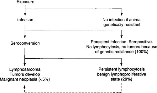
Fig. 21.1 Possible pathways after exposure to BVL virus (percentage figures indicate proportion of seroconverted animals that develop the particular form referred to2).

Whether or not the animal becomes infected or develops any of the other forms of the disease depends on the recipient’s genetic constitution (see Animal risk factors above). The outcome may also be influenced by the animal’s immune status and the size of the infective dose of virus. About 80% of animals with the adult form of the disease have a marked depression of IgM globulins. The immunological responsiveness of leukotic cattle to administered antigens is significantly depressed overall, especially to IgM, resulting from a deficiency in its production in the spleen and lymph nodes.

Lymphomatosis is a neoplasm of the lymphoreticular system. It is never benign and the lesions develop at varying rates in different animals so that the course may be quite short or protracted over several months.

The effects of BLV infection on milk production may not be related solely to overall animal health but may also be mediated directly at a cellular level.34 An in vitro system revealed that the casein production and mRNA synthesis by mammary epithelial cells from a BLV-infected cow were reduced compared with control cell lines without BLV.

Lesions and clinical disease

In adult cattle, almost any organ may be the site of lesions, but the abomasum, heart, and visceral and peripheral lymph nodes are most commonly affected. In calves, the visceral lymph nodes and spleen and liver are the common sites. Depending on the organ which is most involved, several clinical syndromes occur. Involvement of the abomasal wall results in impaired digestion and persistent diarrhea. When the atrial wall is affected, congestive heart failure occurs. In nervous tissue, the primary lesion is in the roots of peripheral nerves and spreads along the nerve to involve meninges and cord. Involvement of the spinal meninges and nerves results in the gradual onset of posterior paralysis. The skin, reproductive tract, and periorbital tissues are commonly affected. In the cutaneous form, intradermal thickenings develop which persist but do not cause discontinuity of the epithelium. They are composed of aggregations of neoplastic lymphocytes. Invasion of periorbital tissues commonly results in exophthalmos. Esophageal obstruction may result from mediastinal lymph node involvement in calves.

The exact nature of the tumor is unclear. The tumors consist of aggregations of neoplastic lymphocytes, but in many cases they may be more accurately described as reticulosarcoma. They are highly malignant and metastasize widely. The hemogram is variable and, although there may be an accompanying lymphocytosis, the presence of large numbers of immature lymphocytes in the blood smear is a more reliable indication of the presence of the disease. Some degree of anemia is common.

CLINICAL FINDINGS

Enzootic (adult) bovine viral leukosis (bovine lymphosarcoma)

This disease is characterized by the occurrence of multiple cases of adult multicentric lymphosarcoma, with tumors developing rapidly in many sites with an accompanying great variation in clinical signs and syndromes. An approximate indication of the frequency with which individual signs appear is set out in Fig. 21.2.35

image

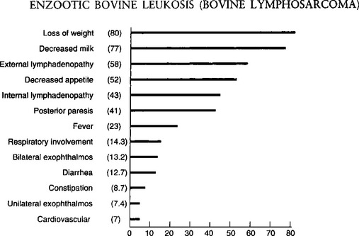
Fig. 21.2 Clinical diagnosis: frequency of predominant signs of bovine leukemia – 1100 field cases.

(By courtesy of Canadian Veterinary Journal34.)

The usual incubation period is 4–5 years with most occurring 4–5 years after the original case was introduced or a blood transfusion from an outside herd was given. This form is rarely seen in animals under 2 years of age and is most common in the 4–8 years age group. Persistent lymphocytosis without clinical signs occurs earlier but rarely before 2 years of age. Many cows remain in the preclinical stage for years, often for their complete productive lifetime without any apparent reduction in performance, but clinical disease appears in a proportion of these cows. The clinical signs and the duration of the illness vary with the number and importance of the sites involved and the speed with which the tumor masses grow.

In 5–10% of clinical cases the course is peracute and the affected animals often die suddenly or unexpectedly without any prior evidence of illness. Involvement of the adrenal glands, rupture of an abomasal ulcer or an affected spleen followed by acute internal hemorrhage are known causes. These animals are often in good bodily condition.

In most cases the course is subacute (up to 7 d) to chronic (several months) and initiated by an unexplainable loss of body condition and appetite, pallor and muscular weakness. The heart rate is not increased unless the myocardium is involved and the temperature is normal unless tumor growth is rapid and extensive, when it rises to 39.5–40°C (103–104°F). Although the following specific forms of the disease are described separately, in any one animal any combination of them may occur. In many cases, clinical illness sufficient to warrant the attention of the veterinarian is not observed until extensive involvement has occurred and the possibility of slaughter of the animal for beef purposes cannot be considered. On the other hand, many cases are examined at a time when diagnostic clinical signs are not yet evident. Once signs of clinical illness and tumor development are detectable the course is rapid and death occurs in 2–3 weeks.

Enlargement of the superficial lymph nodes

Enlargement of the superficial lymph nodes occurs in 75–90% of cases and is often an early clinical finding. This is usually accompanied by small (1 cm in diameter) SC lesions, often in the flanks and on the perineum. The skin lesions are probably enlarged hemolymph nodes and are of no diagnostic significance, often occurring in the absence of other signs of the disease. In many cases with advanced visceral involvement, peripheral lesions may be completely absent. Enlargement of visceral lymph nodes is common, but these are usually subclinical unless they compress other organs such as intestine or nerves. They may be palpable on rectal examination and special attention should be given to the deep inguinal and iliac nodes. In advanced cases, extensive spread to the peritoneum and pelvic viscera occurs and the tumor masses are easily palpable.

Although the enlargement of lymph nodes is often generalized, in many cows only a proportion of their nodes are involved. The enlargements may be confined to the pelvic nodes or to one or more SC nodes. Involvement of the nodes of the head is sometimes observed. The affected nodes are smooth and resilient and in dairy cows are easily seen and their presence may be marked by local edema. Occasionally, the entire body surface is covered with tumor masses 5–11 cm in diameter in the SC tissue.

Digestive tract lesions

Digestive tract lesions are common. Involvement of the abomasal wall results in a variable appetite, persistent diarrhea, not unlike that of Johne’s disease and occasionally, melena due to bleeding of an abomasal ulcer. Tumors of the mediastinal nodes may cause chronic, moderate bloating.

Cardiac lesions

Lesions in the heart usually invade the right atrial wall primarily, causing right-side congestive heart failure. There is hydropericardium with muffling of the heart sounds, hydrothorax with resulting dyspnea, engorgement of the jugular veins and edema of the brisket and sometimes of the intermandibular space. The heart sounds are commonly muffled and other cardiac abnormalities may be obvious. Tachycardia due to insufficiency and arrhthymia due to heart block are common. A systolic murmur is also common along with an abnormal jugular pulse. The liver may be enlarged and palpable caudal to right costal arch, and passive congestion of the liver and visceral edema result in persistent diarrhea.

Nervous system involvement

Neural lymphomatosis is usually manifested by the gradual onset over several weeks of posterior paralysis. Knuckling of the fetlocks of the hind legs while walking is common and one leg may be more affected than the other. This is followed by difficulty in rising, and finally clinical recumbency and inability to stand. At this stage, sensation is retained, but movement is limited or absent. There may be a zone of hyperesthesia at the site of the lesion, which is usually at the last lumbar or first sacral vertebra. Appetite and other functions, apart from the effects of recumbency, are usually normal. Metastases in the cranial meninges produce signs of space-occupying lesions with localizing signs referable to the site of the lesion.

Less common lesions

These include enlargement of the retropharyngeal lymph nodes which may cause stertor and dyspnea. Sometimes clinically detectable lesions occur in the periorbital tissues, causing protrusion of the eyeball (exophthalmos), and in limb muscles, ureter and kidney,36 and genitalia. Involvement of the uterus may be detectable as multiple nodular enlargement on rectal examination.37 Severe bilateral exopthalmos may occur along with generalized lymphadenopathy.38 Periureteral lesions may lead to hydronephrosis with diffuse enlargement of the kidneys while tumors in renal tissue cause nodular enlargements. In either case terminal uremia develops.

BLV particles have been detected by electron microscopy around lymphocytes in the mammary tissue of BLV antibody positive cows affected by subclinical mastitis.39 Whether the virus is a causative agent or an immunosuppressent in bovine mastitis is unknown.

Sporadic bovine leukosis

The calf, thymic and cutaneous forms are designated sporadic bovine leukosis.

Juvenile or calf lymphosarcoma

This occurs in calves at 2 weeks to 6 months of age and is manifested by gradual loss of weight and the sudden enlargement of all lymph nodes, and depression and weakness. Fever, tachycardia and posterior paresis are less constant signs. Death occurs in 2–8 weeks after the onset of signs. Signs of pressure on internal organs, including bloat and congestive heart failure may occur. Diffuse infiltration of major nerves of a limb may also occur.40 Unusually the disease may be fully developed in utero, so that the newborn calf is affected with tumors, or be delayed until 2 years of age. Lymphosarcoma of the pharyngeal region causing retropharyngeal swelling and dyspnea in a 7-month-old beef steer has been recorded.41

Bone and bone marrow necrosis associated with the calf form of sporadic bovine leukosis has been recorded in calves 3 weeks to 8 months of age. Clinical findings included unthriftiness and inactivity, posterior ataxia, superficial lymph node enlargement, lameness and respiratory distress. Involvement of the tibio-tarsal joint, ribs, and spinal canal may also occur resulting in ataxia and paresis.42 Multiple bone infarcts and bone marrow necrosis were present at necropsy. Lymphosarcoma of the mandible of a 2-year-old heifer has also been recorded.

Thymic lymphosarcoma of young cattle

Infiltration of the thymus is a common finding in animals 1–2 years of age and is characterized by massive thymic enlargement and lesions in bone marrow and regional lymph nodes. Jugular vein engorgement and marked brisket edema extending to the submandibular region are common. Moderate bloat due to inability to eructate because of compression of the esophagus may occur.25 The thymic mass is usually not palpable. This form is more common in beef than in dairy cattle. An atypical lymphosarcoma in a mature cow negative for BLV and similar to the thymic form has been reported.43 Metastatic thymic lymphosarcoma in a calf has been recorded36 including a case with metastases causing spinal cord compression. Cases of both the systemic and local forms of sporadic bovine leukosis have been described.25 A large number of the thymic lymphosarcoma occurred in calves in five regions in France over a period of 5 months. Most of the calves had been sired by the same bull, which suggests that the disease had an inherited basis.

Cutaneous lymphoma

This is most common in cattle less than 3 years of age. It is rare and manifested by cutaneous plaques (1–30 cm diameter) appearing on the neck, back, croup and thighs. The plaques become covered with a thick, gray-white scab and the hair is shed; then the center becomes depressed and the nodule commences to shrink. The surface of some placques may become ulcerated and have a serosanguinous exudate. Some of the lesions have a cauliflower-like appearance, appear black, and are ulcerated and foul-smelling. After a period of weeks or months hair grows again and the nodules disappear as does the enlargement of the peripheral lymph nodes. Spontaneous regression of bovine cutaneous leukosis has been recorded. Relapse may occur in 1–2 years with reappearance of cutaneous lesions and signs of involvement of internal organs as in the enzootic form of the disease. In one series of 10 heifers, all animals had lymphadenopathy.44 Some had leukoctyosis and some had lymphocytosis. The body condition may vary from normal to thin and underdeveloped. Some affected animals may have a fever.

Cutaneous T-cell lymphoma in two Friesian cows in the Azores has been reported.4 The lesions consisted of raised pink plaques, with no pruritus or signs of associated pain, which were extensively distributed over both lateral and ventral body regions. Immunocytochemistry found the tumor cells positive for CD3, confirming the T-cell origin of the cells which involved both skin and regional lymph nodes.

Other species

Outbreaks of lymphosarcoma in sheep have been observed with clinical, epidemiological, hematological and necropsy findings similar to those of enzootic bovine leukosis. B-cell leukemia has been described in sheep.45

Infection of other species with BVL virus has not been demonstrated, but epidemic occurrences of lymphosarcoma have been observed in pigs, but only sporadic cases in horses.

In pigs, non-specific emaciation, limb weakness and anorexia are most commonly observed. Sporadic cases in this species are unlikely to be recorded, and although outbreaks have occurred, they and the enzootic form are not commonly encountered. In one herd with the enzootic disease all cases encountered were in pigs less than 6 months of age. There was stunting of growth, development of a pot belly, enlargement of peripheral lymph nodes, and a lymphocytosis, including the presence of immature cells.

In horses, the disease occurs most commonly in animals over 6 years of age. The common clinical manifestations are:

Subcutaneous enlargements which may ulcerate

Enlargement of internal and external lymph nodes

Jugular vein engorgement

Cardiac irregularity

Exophthalmia

Bilateral swelling of the eyelids

Anasarca.

The course varies from acute to chronic, but most affected horses die within a month of first showing signs. Diffuse alimentary lymphoma of the small intestine in adult horses is characterized by:

Malabsorption

Hypoalbuminemia

Increased gammaglobulin levels

Anemia

Chronic alimentary tract dysfunction but without diarrhea.

Malignant lymphoma with ulcerative pharyngitis in horses has been recorded.

CLINICAL PATHOLOGY

A definitive antemortem diagnosis depends on the clinicopathological examination of the animal. Several diagnostic techniques are available and it is important to make the appropriate selection for the particular stage of the disease that is being considered, thus:

Diagnosis of the viral infection is made by serological or virological techniques

Persistent lymphocytosis is identified by hematology

Neoplastic tumors are identified by histological examination of a biopsy specimen.

Because of the increasing economic impact of BLV infection in the cattle industry, the availability of a highly sensitive and specific assay for the identification of BLV-infected animals is of critical importance. Such an assay is needed for the selection of BLV-free cattle for commercial sale, prepurchase testing of breeding animals, and import or export testing, and for control and eradication programs. Ideally, the assay should be practical, inexpensive, and able to be adapted for large-scale use.

Diagnosis of the presence of infection with BVL virus

Serological tests

Virtually all cattle infected with BLV will continuously have antibodies against the major internal (p24) and envelope (gp51) virion proteins in their serum, and serological tests are commonly used for the diagnosis of BLV infection in cattle over 6 months of age.

Radioimmunoprecipitation assay (RIP).

The RIP using gp51 or p24 as antigen, is a highly sensitive and specific method for serologic diagnosis of BLV infection.46 The RIP assay has been used as the criterion-referenced standard to critically evaluate the performance of other diagnostic tests for BLV infection. Detailed comparisons of various BLV assays in a large number of cattle of various origins and ages found that the RIP assay is the most sensitive and specific test. However, its major disadvantage is that it requires a gamma counter and radioistopes, which are expensive.

AGID test.

This is a good screening test to determine the presence of infection in an individual animal or herd.2 The estimated specificity of 99.8% and the sensitivity of 98.5% indicate that the test is a reliable and accurate method to detect BLV infection. False-positive and false-negative results do occur and may be associated with some variability of the immune system or from human error. The AGID test is the official reference test of the Office International des Epizooties and the European Community and is the test recognized by most governments as the official test for purposes of testing imported animals. There is however, a lack of standardization between the BLV-AGID kits used in North America and Europe.47

Radioimmunoassay (RIA).

This is suitable for individual cow testing because of its accuracy. There are several versions of this and the one using the virion gp antigen is preferred.2 It is one of the most sensitive tests and is useful for the detection of BLV antibodies in cattle exposed no longer than 2 weeks, in milk samples, and in serum samples from periparturient dams.

Serum ELISA.

In more recent years, ELISA-based testing has replaced the AGID in eradication programs in several countries. It is more sensitive than other serological tests and can be used on milk.2 The superior sensitivity of the ELISA for pooled serum samples allows detection of antibodies in herds with a prevalence of less than 1%, whereas the AGID test detected only 50% of the herds detected by the ELISA.2 Two commercially available ELISAs and the polymerase chain (PCR) were evaluated and compared with the AGID to detect antibodies to BLV or its nucleic acid.5 The ELISA tests detected about 10% more reactors than the AGID and the elecrophoretic immunoblotting results. Some ELISA positive animals were not detected by the PCR.

Four commercially available BLV-ELISA kits from Europe or the United States were compared to the AGID test officially approved by the Canadian Food Inspection Agency. The ELISA tests were more sensitive than the AGID test kits and the gp51 BLV-ELISA is now recognized as an official test method for the serodiagnosis of bovine leukosis in Canada.48

A highly sensitive and specific blocking ELISA comparable to the radioimmunoprecipitation assay for the detection of BLV antibodies in serum and milk samples has been developed.46

The chronology of seroconversion is important in the serological diagnosis. Calves from infected dams have a 20% chance of being infected in utero and seropositive at birth. If they are serologically negative at birth they seroconvert at their first ingestion of colostrum from seropositive cows and this passively acquired immunity persists for 2–7 months. These calves, and calves from uninfected mothers become positive at varying ages depending on when they come into contact with infection, usually when they are placed into the infected adult herd. This can be as early as 9 months of age, but as a general rule positive reactions are uncommon in cattle which are less than 2 years of age. Seroconversion usually takes place 3–4 months after the negative animals are placed in the infective group, although the interval is longer in the winter than in the summer. Infected animals are seropositive and infected for long periods, usually for life.

Using the ELISA, experimental infection of calves at 3–4 months of age results in seroconversion to the virus at 4–5 weeks after infection.20

In a control and eradication program, early detection of infected calves is difficult because colostral antibodies to BLV cannot be differentiated from antibodies resulting from natural infection. By using measures of colostral antibody concentration, calves infected in utero could be identified by 80 d of age. Calves over 6–8 months of age with a positive immunodiffusion test will likely be infected permanently. Calves which have ingested colostrum from seropositive cows usually have maternal antibodies and polymerase chain reaction tests are necessary to detect the virus and distinguish between infected and virus-free calves.49

Milk ELISA.

This is much more sensitive than the AGID and has been adopted for testing milk from individual cows and pooled milk samples.46,50 A comparison of the ELISA and AGID tests for the detection of BLV antibodies in bovine serum and milk found a high level of agreement.50 The bulk tank milk ELISA is useful for identification of herds which are negative for BLV infection.51 The ELISA identified 80% of herds as positive for BLV and had an apparent sensitivity and specificity of 0.97 and 0.62, respectively.51 But after accounting for the sensitivity and specificity of the AGID test in individual animals, the specificity of the ELISA test for milk was 0.44. With the moderately low specificity, herds identified as positive by the ELISA would require further testing at the individual or herd level to definitively establish their BLV status.

An indirect ELISA to detect antibodies to BLV in bulk-milk samples in Sweden is being used to assist in the eradication of infection from Swedish herds.52 The antibody level in milk is lower than in serum but the sensitivity of the ELISA is as effective as for sera. Testing of bulk milk is a useful and practical method for large-scale epidemiological studies and initial eradication programs. Heifers, bulls, and dry cows are not included when bulk milk is tested and all animals over 1 year of age need to be sampled individually before a herd is declared free of the virus. The sensitivity and specificity of the milk ELISA is estimated to be adequate until the prevalence of BLV-infected individuals in the country is less than 1%.

Detection of virus

Polymerase chain reaction (PCR).

The PCR is a sensitive and specific assay for direct diagnosis of BLV infection in peripheral blood lymphocytes.53 The test is useful for the early detection of BLV infection even before antibodies are present. It is more sensitive than the AGID test or the ELISA in detecting infected cattle where the prevalence of infection is less than 5%.54 The test can identify proviral DNA of BLV in the lymphocytes of calves at birth which are born to infected cows.55 All calves found to be infected at birth were born to BLV-positive cows with persistent lymphocytosis. At birth, the presence of a titer can be due to colostral antibodies or perinatal infection and the PCR test can differentiate uninfected newborn calves with colostral antibodies from BLV-infected calves and detect the presence of the virus in the presence of antibodies.47 The PCR has a practical application in the identification of BLV-infected calves, regardless of colostral antibody, which allows immediate removal of the source from the herd. In a dairy herd with a high prevalence of BLV, a positive PCR assay result provided definitive evidence that a cow was infected with BLV.56 However, sensitivity and specificity were 0.672 and 1.00, respectively. Predictive value of a positive test was 1.00, and predictive value of a negative test was 0.421. Thus PCR assay alone is unreliable for routine detection of BLV in herds with a high prevalence of BLV infection.

The PCR can also be used to ensure that cattle used in the production of a whole blood vaccine for tick-borne disease are free from BLV infection.17 The sheep bioassay, currently in use, requires 4 months of serological testing to insure that donor animals are not infected. Replacement of the sheep bioassay with the PCR could result in considerable saving of time and effort. Use of the PCR requires stringent precautions to prevent false-positive results due to contamination of samples with PCR product.17

A nested PCR identified 98% of BLV seropositive cows from blood and 65% from milk, whereas real-time PCR detected 94% of BLV seropositive cows from blood and 59% from milk.57 BLV was also detected in 10% of seronegative cows most likely because of early detection before seroconversion.

Differentiation between enzootic and sporadic bovine leukosis.

The role of BLV in some cases of sporadic bovine lymphomas needs to be re-examined. The findings of persistently seronegative PCR-positive and seropositive PCR-negative cattle indicates that the BLV cannot be excluded as a causative agent in sporadic bovine leukosis. Enzootic bovine leukosis cannot be distinguished from sporadic bovine leukosis on histopathological examination. The ELISA is recommended as a method of choice to differentiate between EBL and sporadic bovine leukosis (SBL) because it is a rapid, reliable and sensitive test which is inexpensive and easy to perform. In cases where no blood or other fluids are obtained, the PCR test is the most useful method for the direct detection of BLV.

Diagnosis of persistent lymphocytosis (PL)

Approximately 30% of animals infected with the BLV develop PL, which is defined as an increase in the absolute lymphocyte count of three or more standard deviations above the normal mean as determined for that respective breed and age group of animals in leukosis-free herds. The PL is an increase in peripheral B-lymphocytes. It has been suggested that one additional criterion for PL should be that it persists for more than 3 months. When PL was first recognized in herds which experienced clinical lymphosarcoma, it was considered a subclinical expression of the tumor stage of the disease. It became an important diagnostic criterion in control and eradication programs until serological tests became available to more accurately identify infected animals. The majority of cells involved in PL are normal lymphocytes but atypical and abnormal forms have been described and are considered as indicative of preleukemic condition. The total count increases from a normal of 6000 to as high as 15 000/μL. The percentage of lymphocytes in the total white blood cell count increases from the normal of 50–65% is considered a positive result. The presence of 25% or more of the total lymphocyte count as atypical immature cells is also considered a significant aberration. The PL may subside in animals which subsequently develop lymphosarcoma.

The association between the strength of serologic recognition of BLV by the use of ELISA and lymphocyte count in bovine leukosis virus-infected cows has been examined.52 The sample-to-positive ratio, which is the ratio between the test sample and a positive control sample, was compared among lymphocytotic and nonlymphocytotic cows. The sample-to-positive ratio and lymphocyte count were related but cows with high sample-to-positive ratio were not always lymphocytotic. Culling cows on the basis of sample-to-positive ratio will reduce culling of ELISA-positive cows, however, culling on the basis of lymphocyte count will eliminate a greater proportion of the reservoir of infection.

Diagnosis of lymphosarcoma

This can only be done by histopathological examination of a section of tumor material obtained by biopsy or necropsy. A needle aspirate of an enlarged peripheral lymph node may provide a rapid and inexpensive diagnosis. Enlarged lymph nodes or hemolymph nodes are the usual sources, but when the genital tract is involved an exploratory laparotomy is usually performed so that a sample can be obtained. The lymphocyte count may increase to 20 000–30 000 μL, and in some cases may reach values of 50 000–100 000 μL, and even 400 000– 500 000 μL. Conversely, in some cases, the lymphocyte count decreases. Chromosomal changes may be detectable in cells from lymph nodes or in leukocytes from peripheral blood of affected animals. When there is myocardial involvement there may be obvious changes in the electrocardiogram but these are unlikely to be of value in differential diagnosis.

NECROPSY FINDINGS

In cattle, firm white tumor masses may be found in any organ although two rather different patterns of distribution are apparent. In newborn and young animals, the common sites are: kidneys, thymus, liver, spleen and peripheral and internal lymph nodes. This may or may not be a characteristic of the ‘sporadic’ form of the disease. In adults, the heart, abomasum and spinal cord are often involved. In the heart, the tumor masses invade particularly the right atrium, though they may occur generally throughout the myocardium and extend to the pericardium. The frequency of early changes in the subepicardial tissue of the right atrium suggests that this is an area from which tissues should be selected in latent or doubtful cases. The abomasal wall, when involved, shows a gross, uneven thickening with tumor material in the submucosa, particularly in the pyloric region. Similar lesions occur commonly in the intestinal wall. Deep ulcerations in the affected area are not uncommon. Involvement of the nervous system usually includes thickening of the peripheral nerves coming from the last lumbar or first sacral cord segment or more rarely in a cranial cervical site. This may be associated with one or more circumscribed thickenings in the spinal meninges. Affected lymph nodes may be enormously enlarged and be composed of both normal and neoplastic tissue. The latter is firmer and whiter than normal lymphoid tissue and often surrounds foci of bright yellow necrosis. Less common sites include the:

Kidney

Ureters (usually near the renal pelvis)

Uterus (either as nodular masses or diffuse infiltration)

Mediastinal, sternal, mesenteric and other internal lymph nodes

Mandibular ramus.

When performing the necropsy, it is important to remember that lymphosarcoma can appear not only as discrete nodular masses but as a diffuse tissue infiltrate. The latter pattern results in an enlarged pale organ which is easily misinterpreted as a degenerative change rather than as a neoplastic process.

Histologically, the tumor masses are composed of densely packed, monomorphic lymphocytic cells. Attempts to better characterize the nature of these cell populations have been published.58,59 The cleaved variant of the diffuse large cell type with a high mitotic index is characteristic of enzootic lymphoma and this high-grade type of B-cell tumor may be a consequence of the viral etiology of this form of the disease.31 It is possible to confirm viral infection in some cases by a PCR test but such testing is rarely justified. Immunohistochemical staining of formalin-fixed, paraffin-embedded tissue sections of tumors can be used to con-firm that neoplastic lymphocytes are of thymic origin when thoracic masses are examined.60

Samples for confirmation of diagnosis

Histology – formalin-fixed samples of gross lesions, plus enlarged lymph nodes, bone marrow, liver, spleen, thymus, right atrium, abomasum, uterus (LM, IHC)

Virology – neoplastic tissue (PCR).

DIFFERENTIAL DIAGNOSIS

Because of the very wide range of clinical findings, a definitive diagnosis of BVL is often difficult. Enlargement of peripheral lymph nodes without fever or lymphangitis is unusual in other diseases, with the exception of tuberculosis, which can be differentiated by the tuberculin test. In the absence of these enlargements, the digestive form may easily be confused with Johne’s disease. The cardiac form closely resembles traumatic pericarditis and endocarditis, but there is absence of fever and toxemia, and the characteristic neutrophilia of these two diseases is usually absent. Involvement of the spinal nerves of meninges may be confused with spinal cord abscess or with the dumb form of rabies. An examination of cerebrospinal fluid may be of value in determining the presence of an abscess and rabies has a much shorter course and other diagnostic signs. Multiple lymph node enlargements in the abdominal cavity, and nodular lesions in the uterine wall may be confused with fat necrosis, but the nature of the lesion can usually be determined by careful rectal palpation. Stertor caused by enlargement of the retropharyngeal lymph nodes is also commonly caused by tuberculosis and actinobacillosis.

Cases of atypical lymphosarcoma43 and thymic lymphosarcoma41 which are BLV-negative may resemble lymphosarcoma of enzootic bovine leucosis.61

Echocardiography is now being used to detect intracardiac masses which may be compatible with EBL.62

TREATMENT

There is no treatment.

CONTROL

The disease can be eradicated from a herd and even a country, or controlled at a low level. The option chosen depends initially on the prevalence of infection in the herd, the value of the animals in the herd, and whether a governmental indemnity offered for seropositive cows which are culled and sent to slaughter.

History of Compulsory Eradication Program in Denmark

Control and eradication programs have been in effect on a nationwide basis in several western European countries.2 Denmark began an eradication program in 1959 based on the occurrence of clinical lymphosarcoma, and the identification of cattle with PL using the Bendixen hematological key for classifying cattle as normal, suspect or lymphocytic. Leukosis was declared a reportable disease, and all adult cattle from herds in which cases of leukosis originated were subjected to a hematological examination. Affected herds were quarantined, and indemnity was offered to induce owners to have their entire herd slaughtered. This herd-slaughter policy was continued until 1982. When the AGID test became available, the Bendixen key was discontinued and only the AGID test was used in the official program between 1979 and 1982. Routine testing was discontinued in 1982. Surveillance involved testing random sera collected from every sixth adult cow to be slaughtered. According to the official Danish control program, the incidence of tumors in adult cattle at the start of the eradication program was at least 10 times greater than 10 years later. The hematological test was less sensitive than subsequent serological tests but the specificity was fairly high and only a few herds were erroneously classified as leukosis herds.61 When the serological tests were introduced, some herds which were classified as leukosis-free based on the hematological key, were found to be infected.

Voluntary eradication programs using the AGID test have been effective in other member countries of the European Community in the last two decades and have been successful in reducing the prevalence of infection and disease.2 In the Federal Republic of Germany, eradication was achieved in 5 years. These programs have been successful in part because of the low prevalence of infection and the economic losses from culling seropositive cows has not been large.

In Britain, EBL is a reportable disease but is uncommon.41 A national testing program was begun in 1992. All blood samples collected for routine periodic testing for brucellosis have also been tested for BLV and milk samples are collected every 3 months from dairy herds for BLV testing. The prevalence of infection has been low and the source of infection undetermined. Some of the animals had been imported from Canada but in other cases there was no association with importation.

Considering the animal-health aspects and possible consumer reactions against having a widespread retrovirus infection in food-producing animals, and the requirements for exporting cattle and semen, Sweden introduced a national program for the eradication of BLV in 1990.52 An ELISA test was evaluated for detection of antibodies to BLV in individual and bulk milk and serum samples. It is proposed that eradication can be based on using the ELISA on milk samples in combination with other diagnostic tests and the prompt removal of infected animals.

In Canada and the United States, it is considered cost prohibitive to cull and slaughter all seropositive cattle because of the high prevalence of infection. Many seropositive cows are valuable pedigreed animals, and there are no indemnity programs available. Thus all control and eradication programs in these countries are herd based and strictly voluntary. Livestock producers are willing to adopt control programs because of the economic losses associated with export restrictions if their cattle are infected, and the losses due to the occasional clustering of cases of lymphosarcoma.

Enzootic bovine leukosis was eradicated from Finland in 1996.63 The disease was first recognized in 1966 and it required 30 years using the key principle of test and slaughter policy to achieve eradication. The infection status was monitored at meat inspection, and hematologically between 1970 and 1977, serologically between 1978 and 1989. Annual surveys including all dairy herds and samples from beef animals were conducted in 1990–2001. Bulk tank samples represented the dairy herds in the surveys; beef animals were sampled individually at slaughter. The maximum positive herd-level percentage in the survey was 0.03%. The herd level prevalence of infection never exceeded 5%.

Eradication programs

Enzootic bovine leukosis can be eradicated only by:

Test and slaughter of cattle infected with the virus. Programs based on the culling of seropositive cows are effective

The maintenance of a closed herd which permits the entry of only those animals free of infection.

The efficiency of such a program depends on the accuracy of the test used to identify the infected animals, and the repetition of the test at an appropriate interval so that animals that were in the incubation stages of the disease at the time of the first test will have had time to seroconvert. The recommended procedure is:

1. Identify infected animals using the AGID test2

2. Cull and slaughter seropositive animals immediately

3. Retest the herd 30–60 d later

4. Use the PCR assay to test young calves and as a complementary test for clarifying doubtful test results in herds with a low prevalence of infection.54

Testing is repeated until the herd has a negative test. When the herd is negative, testing is repeated every 6 months and the herd declared free when there have been no positive reactors for 2 years. Future introductions into the herd are managed most safely by artificial insemination or fertilized ovum transfer, or importations of animals which have been tested and are seronegative on two tests carried out 30 and 60 days prior to arrival.

In herds where the prevalence is high, a two-herd scheme can be successful. Newborn calves are removed from seropositive cows immediately after birth, fed colostrum from seronegative cows and raised in isolation. All animals over 6 months of age are tested periodically and seropositive animals culled. The parent herd is eventually disposed of as negative replacement animals become available. Only those bulls which are seronegative may be used and they must be tested every 3 months.

Although eradication is biologically feasible, it is unlikely that area eradication programs will be implemented on an extensive scale because losses from the disease are not sufficiently high, and there is a high risk of insect vectors reintroducing it which poses a real threat to maintenance of a BVL-free herd. The cost-effectiveness of an eradication program on a national basis would be a major consideration. For an individual herd, it is feasible provided some steps were taken to increase genetic resistance of the residual stock and to reduce the chances of in-contact infection occurring.

Limitation of spread of infection

In herds with a high prevalence of infection, the test and slaughter method of eradication is not economically viable if the animals have a high economic value because of superior genetic potential. Control of infection in these herds is possible using embryo transfer from infected dams to negative recipients and isolation of newborn calves but these are not practical on a country-wide basis. An alternative method is segregation of BLV-infected and non-infected animals based on the AGID test. This is known as the test and segregation method, which is based on the evidence that the spread of infection between animals is relatively slow and that the virus is spread by movement of infected animals from one herd to another and within a herd. Following the initial testing of the herd, the herd is divided into two groups, BLV-positive and BLV-negative, and kept at least 200 m apart. A third separate location is used for quarantine of replacement animals. Replacement animals must be found negative in two consecutive AGID tests, the first within 30 d before purchase and the second after 60 d of isolation, prior to being moved into the negative group. The AGID tests are conducted every 3 months and the reactors removed to the positive group location until the remaining animals in the herd have attained BLV-negative status by the test. Thereafter, the tests are done every 6 months and continued until at least four consecutive negative tests are obtained for each herd. Variations of this method of test and segregation with subsequent removal of seropositive animals in the routine culling program have been successful. The colostrum and milk fed to calves in the BLV-negative group must be from seronegative cows or be pasteurized to inactivate the virus.

In Canada, cattle owners may enrol in the Canada Health Accredited Herd program to declare freedom from EBL. All reactors must be removed from the herd. If a large number of reactors are detected, two herds on two separate farms can be established: one herd comprised of the reactors and the other of cattle which are seronegative. Calves from the reactor herd can be added to the accredited herd in accordance with strict isolation and testing procedures. To qualify for accredited certification, a herd must have two consecutive negative herd tests, at least 4 and less than 12 months apart. The tests must include all cattle in the herd. The first annual renewal test must occur no more than 12 months following the second qualifying test for certification, and must include all cattle in the herd. Subsequent renewal tests must occur within the same 12-month interval. Only cattle 24 months of age and older must be tested but a herd inventory and audit must be performed on the whole herd. In herds with reactors, the two qualifying tests do not begin until at least 4 months after the removal of the last reactor uncovered during any test. Herd additions can be made during the qualifying test period or after certification has been achieved. Each animal must be accompanied by a health certificate and depending on the enzootic bovine leukosis status of the originating herd, certain testing and isolation procedures could apply. Owners wishing to have their animals attend exhibitions can do so providing they adhere to certain conditions. Properly processed semen and embryos can be introduced without restrictions. Owners are encouraged to follow preventive health management practices to augment the eradication of enzootic bovine leukosis from their herds. These include all areas where blood transfer could occur (needles, dehorning, castrating, extra teat removal, ear tagging, tattooing, hoof trimming, rectal palpations, drenching) and other procedures which transfer leukocytes, and routine insect control.

Prevention of infection in calves and young stock

Several management techniques can be used to prevent infection in calves from birth until they become herd replacements. The feeding of newborn calves with colostrum and milk from seronegative cows has been widely accepted as effective in preventing infection in calves. Postnatal infection in calves can also be minimized by feeding milk replacer, and/or whole milk from non-infected cows. The use of colostrum and milk from non-infected cows permits early serological detection of infected calves. However, feeding colostrum from seropositive cows to newborn calves can provide significant protection from infection during the first 3 months of life. Field studies indicate that colostrum-derived BLV antibodies may prevent as much as 50% of the infection during the first 3 months compared to calves which did not receive colostrum with BLV antibodies. Further reduction in the risk of infection via colostrum can be achieved by pasteurization of the colostrum – 63°C for 30 min.64 The colostrum-derived BLV antibodies will however delay early detection of infection in calves. The replacement of whole milk feeding with high-quality milk replacer may also be considered.

Transmission to newborn calves can also be reduced by avoiding exposure to maternal blood at the time of parturition, housing calves in individual hutches with individual feeders and waterers, and management techniques to avoid iatrogenic transmission.65 When handling a group of calves, the youngest ones should be handled first and the older and sick calves last. Equipment which could act as fomites in transferring blood should be disinfected with chlorhexidine when used between calves. These instruments include:

Nose tongs

Scissors

Forceps

Dehorning instruments

Esophageal tubes

Balling guns

Tattoo equipment

Ear taggers.

Dehorning calves using the electrocautery method before 2 months of age can reduce the prevalence of infection compared to gouge dehorning, which allows the transfer of infected blood between calves. Handling facilities which become contaminated with blood should be cleaned between calves. Fly control should be instituted as necessary. Single needles should be used for vaccination and calves should be tested serologically for BLV infection at about 6 months of age.

A marked reduction in the prevalence of infection within the heifer age groups of a dairy herd with a high prevalence can be achieved by:

1. Use of single needles and individual sleeves for rectal examination

2. Disinfection of tattoo equipment before use

3. Dehorning by use of electrocautery.

Biosecurity

Prevention of entry of infection into herd can be achieved by insuring that all imports into the herd have been tested at least 30 d prior to arrival and are seronegative. Control of insect vectors is highly desirable. Blood transfusions and vaccines containing blood, such as those used for babesiosis and anaplasmosis are particularly potent means of spreading the disease and donors must be carefully screened to insure that they are free of the disease. In the future the selection of cattle with inherent resistance to BLV may be a possibility. Embryo transfer from valuable pedigreed seropositive cows may aid in reducing prenatal infection. Insemination is not a method of transmission so that artificial breeding programs are not disrupted.

Vaccine

The possibility of a vaccine for BLV has been explored.2 However, the prospects are not good thus far. A BLV vaccine would have to be non-infectious, non-oncogenic, and should not interfere with the serological tests commonly used to detect infection.

REVIEW LITERATURE

Johnson R, Kaneene JB. Bovine leukaemia virus and enzootic bovine leucosis. Vet Bull. 1992;62:287-312.

REFERENCES

1 Dube S, et al. Virol. 2000;277:379.

2 Johnson R, Kaneene JB. Vet Bull. 1992;62:287.

3 Peleteiro MC, et al. Vet Dermatol. 2000;11:299.

4 Klintevall K. Bovine leukemia virus: course of infection and means of detection. Dissertation Department of Veterinary Microbiology Faculty of Veterinary Medicine Swedish University of Agricultural Sciences and The National Veterinary Institute Department of Virology Uppsala Sweden, 1995.

5 Reichel MP, et al. New Z Vet J. 1998;46:140.

6 VanLeeuwen JA, et al. Can Vet J. 2001;42:193.

7 Gnad DP, et al. Intern J Appl Res Vet Med. 2004;2:215.

8 Trono KG, et al. Vet Microbiol. 2001;83:235.

9 Molnar E, et al. Vet Rec. 2000;146:705.

10 Meas S, et al. J Vet Med Sci. 2000;62:779.

11 Zaghawa A, et al. J Vet Med B Infect Dis Vet Public Health. 2002;49:123.

12 Bielanski A, et al. Vet Rec. 2000;146:255.

13 Choi KY, et al. J Vet Diagn Invest. 2002;14:403.

14 Wentink GH, et al. Vet Rec. 1993;132:135.

15 Divers TJ, et al. Prev Vet Med. 1995;23:133.

16 Meas S, et al. Vet Microbiol. 2000;84:275.

17 Eaves FW, et al. Vet Microbiol. 1994;39:313.

18 Da Y, et al. Proc Natl Acad Sci. 1993;90:6538.

19 Kabeya H, et al. J Vet Med Sci. 2001;63:703.

20 Klintevall K, et al. Comp Immun Microbiol Infect Dis. 1997;20:119.

21 Casal J, et al. Prev Vet Med. 1990;10:47.

22 Chi J, et al. Prev Vet Med. 2002;55:57.

23 Garazi S, et al. Israel J Vet Med. 2001;56:125.

24 Sargeant JM, et al. Prev Vet Med. 1997;31:211.

25 Hendrick SH. Can Vet J. 2002;43:617.

26 Goetschau A, et al. Am J Epidemiol. 1990;131:356.

27 Chi J, et al. Prev Vet Med. 2002;55:137.

28 Ott SL, et al. Prev Vet Med. 2003;61:249.

29 Pollari FL, et al. Am J Vet Res. 1993;54:1400.

30 Rhodes JK, et al. J Am Vet Med Assoc. 2003;223:229.

31 Pollari FL, et al. Can J Vet Res. 1992;56:289.

32 Rhodes JK. J Am Vet Med Assoc. 2003;223:346.

33 Buehring GC, et al. Aids Res Human Retro. 2003;19:1105.

34 Motton DD, Buehring GC. J Dairy Sci. 2003;86:2826.

35 Reed VI. Can Vet J. 1981;22:95.

36 Sparling AM. Can Vet J. 2000;41:315.

37 Alijarrah AH, et al. J Am Vet Med Assoc. 2004;224:1591.

38 Malatestinic A. Can Vet J. 2003;44:664.

39 Wellenberg GJ, et al. Vet Microbiol. 2002;88:27.

40 Dubreuil P, et al. Can Vet J. 1998;39:431.

41 Ivany JM, et al. Can Vet J. 2000;41:486.

42 Oliver-Espinosa O, et al. Can Vet J. 1994;35:777.

43 Southwood LL, et al. Vet Rec. 1996;138:260.

44 Schweizer G, et al. Vet Rec. 2003;153:525.

45 Valentine BA, McDonough SP. Vet Pathol. 2003;40:117.

46 Gutierrez SE, et al. Am J Vet Res. 2001;62:1571.

47 Simard C, et al. Can J Vet Res. 2000;64:96.

48 Simard C, et al. Can J Vet Res. 2000;64:101.

49 Klintevall K, et al. Vet Microbiol. 1994;42:191.

50 Brenner J, et al. Israel J Vet Med. 1994;49:165.

51 Sargeant JM, et al. Prev Vet Med. 1997;31:223.

52 Nagy DW, et al. J Am Vet Med Assoc. 2002;220:1681.

53 Klintevall K, et al. Vet Microbiol. 1994;42:191.

54 Fechner H, et al. J Vet Med Series B. 1996;43:621.

55 Agresti A, et al. Am J Vet Res. 1993;54:373.

56 Nagy DW, et al. J Am Vet Med Assoc. 2003;222:963.

57 Kuckleburg CL, et al. J Vet Diag Invest. 2003;15:72.

58 Wernau W, et al. Vet Pathol. 1997;34:222.

59 Vernau W, et al. Vet Pathol. 1992;29:183.

60 Alexander AN, et al. J Vet Int Med. 1996;10:275.

61 Gibson LAS. Vet Rec. 1995;136:156.

62 Schmity DG, Seahorn TL. J Am Vet Med Assoc. 1990;205:159.

63 Nuotio L, et al. Prev Vet Med. 2003;59:43.

64 Sprecher DJ, et al. J Am Vet Med Assoc. 1991;199:584.

65 Thurmond MC. Cornell Vet. 1991;81:227.

Bovine immunodeficiency-like virus

ETIOLOGY

Bovine immunodeficiency-like virus (BIV), also known as bovine lentivirus-1, is a lentivirus, within the larger family of Retroviridae. The virus shares structural and genomic similarities with other lentiviruses, such as equine infectious anemia virus, caprine arthritis-encephalitis virus, maedi-visna virus, feline, and the simian and human immunodeficiency viruses. The BIV was first described in cattle in the United States in 1972.1 These viruses replicate primarily in the cells of the host’s immune system following their insertion as provirus into the genome of these target cells, thus establishing a chronic, lifelong infection. The lentiviruses above are usually associated with specific diseases. However, a clear involvement of BIV in the development of a clinical syndrome is not well established.

EPIDEMIOLOGY

Prevalence of infection

Seroepidemiological evidence indicates that BIV infection has a worldwide distribution. Seropositive cattle have been identified in the United States, the Netherlands, New Zealand, Australia,2 Bali,3 Indonesia, Brazil,4 and Canada with estimates ranging from 1–5%.5,6 In Italy, the prevalence is 5.8% in dairy cattle and 2.5% in beef cattle.7 In individual herds the prevalence of infection may be much higher. In the UK, the seroprevalence was found to be 5.9% in dairy cattle and 5.0% in beef cattle.8 The dairy and beef herd prevalences were 60% and 59%, respectively. While the BIV infection in the UK is low, it is widespread. Recent studies, using DNA, derived from semen and buffy coat samples, analyzed by nested PCR, found no evidence of BIV infection in western Canadian cattle.9

A seroprevalence of greater than 50% was present in a dairy herd at a university in the southeastern United States, which is an area with a high prevalence of infection in the cattle population.10 There is some evidence that in some cattle herds with a high incidence of unthrifty animals, the prevalence of seropositive animals may be as high as 95%.11 The prevalence of BIV infection among dairy cattle in Ontario is low and may be associated with an economically important decrease in milk production.6 Dual infection with BIV and BLV have been reported in Mississippi dairy cattle.10

The virus has been found in the seminal leukocytes of 82% of randomly selected semen samples from a bovine stud semen repository12 suggesting the possibility that artificial insemination of dairy cows may have a major role in the transmission of the virus. The BIV may be involved in the pathogenesis of mastitis in cattle due to its immunosuppressive effects but no clear evidence is available.13

Retroviruses are heat labile and readily inactivated at 56°C, and pasteurization of milk for human consumption should provide an adequate safeguard. Feeding milk seeded with the virus and pasteurized before inoculation into calves is effective in inactivating the virus and preventing transmission.14 There is no evidence that the virus is a potential human pathogen.

Methods of transmission

The virus is strongly cell-associated and may be transmitted in infected blood, colostrum, and milk that contains lymphoreticular cells.15 There is some evidence of transplacental infection of the virus in cattle. In dairy cows naturally infected with BIV and seropositive at parturition, 40% gave birth to calves that were BIV seropositive before receiving colostrum;16 seronegative cows do not. Calves born with anti-BIV-specific antibody do not demonstrate increased risk of clinical disease during the neonatal period but the calves born to dams which are seropositive at parturition appear to be at increased risk of occurrence of some clinical signs. The prevalence rate of infection among bulls housed in stud farms was 9.6% using serology, and 12.6% using PCR for the presence of BIV provirus in peripheral blood leukocytes.17

The BIV has no obvious morphological effects on the embryonic development of cattle and it is possible to obtain embryos at the transferable stage free of the virus from cows infected with the virus.18 It is unlikely that BIV is associated with the zona pellucida-intact embryos derived by in vitro fertilization from oocysts obtained from infected animals or with oocysts fertilized with infected semen when embryos are washed as recommended by the International Embryo Transfer Society. Embryos from donors infected with the virus are not likely to transmit the virus to recipients and the resulting offspring.19

PATHOGENESIS

The pathogenetic mechanisms of BIV infection are unclear. Its pathogenicity is controversial. It is uncertain if the virus is a primary pathogen or a primary immunodeficiency virus which predisposes the animal to secondary infections.15 Despite extensive experimental studies the pathogenic significance of the virus is uncertain.

Infection of cattle with BIV is associated with lymphoproliferation, lymphadenopathy, immunosuppression, neuropathy, and progressive emaciation.15

The virus was initially isolated from a cow with persistent lymphocytosis, lymphadenopathy, neuropathy and progressive emaciation. However, overt clinical disease in seropositive cattle is rare and experimentally induced infection in calves has resulted in only mild clinical consequences.20

Early studies of inoculation of calves with the virus resulted in lymphoproliferative disease and lymphocytosis and persistence of the virus.1 Later studies have failed to reproduce significant clinical disease which may in part be due to the long incubation period.11 It is also possible that the lentiviruses have variable virulence due to genetic variation producing viruses with both antigenic and biological heterogenicity in pathogenesis.11 Experimental infection of an 11-month-old calf with the virus was followed by the development of a T-cell lymphosarcoma21 and the bovine leukosis virus was not present.

The virus and its DNA have been detected in the blood and semen of experimentally infected bulls.22 But the virus has not been detected in the semen, blood leukocytes, or semen leukocytes of samples supplied by artificial insemination centers.23

Retroviruses, including the lentiviruses, are characterized by the expression of the unique enzyme, reverse transcriptase, which facilitates the transcription of the RNA of an infectious virus to a complementary DNA copy. The viral DNA has the ability to become incorporated into the host’s cell nucleus as a ‘provirus’. Proviruses are non-infectious, can remain latent for many years and persist in the presence of antibody. Changing the virus from its latent form into an infectious RNA virus can occur and depends on activation of the latently infected cells. The stimuli for activation can include concurrent infection and stress, or both. While other lentiviruses such as equine infectious anemia virus can cause severe clinical disease, the cause and effect relationship between BIV infection in cattle and clinical disease has not yet been documented.

CLINICAL FINDINGS

Naturally occurring BIV infection in Holstein dairy cattle in Louisiana, US, have been described.24 Progressive weight loss was common, and concurrent infections included metritis, subcutaneous abscesses, purulent arthritis, laminitis and infectious pododermatitis, fascioliasis, and mastitis. Reduced vitality, dullness and stupor were also common.

The course of the disease varied from 3 to 40 weeks.

CLINICAL PATHOLOGY

Detection of virus.

A PCR test has been used to detect the BIV in the blood and milk of BIV-seropositive cows.25 The virus can be detected in experimentally infected calves using PCR in peripheral blood mononuclear cells.26

Serological tests.

Using the BIV ELISA, naturally occurring cases in dairy cattle are serologically positive.24,27 An indirect immunofluorescent antibody test has been used to detect seroconversion in experimentally infected bulls by 17 days after infection.22 The sensitivity and specificity of the indirect fluorescent-antibody assay (IFA) and the nested-set PCR have been compared using Bayesian techniques.28 The PCR is the more sensitive assay.

NECROPSY FINDINGS

Moderate to marked enlargement of hemal lymph nodes have been described.24 Lymphoid depletion is common and characterized by an absence of follicular development in nodes draining regions with secondary infections. Encephalitis characterized by meningeal, perivascular and parenchymal infiltration with lymphocytes, plasma cells and macrophages with perivascular edema has been observed.27 Several secondary infections have been observed in cattle with BIV infection but the role of BIV as a prediposing pathogen is uncertain.

REVIEW LITERATURE

Gonda MA. Bovine immunodeficiency virus. AIDS. 1992;6:759-776.

REFERENCES

1 Gonda MA. AIDS. 1992;6:759-776.

2 Burkala EJ, et al. Vet Microbiol. 1999;68:171.

3 Barboni P, et al. Vet Microbiol. 2001;80:313.

4 Meas S, et al. Jpn J Vet Res. 2002;50:9.

5 Jacobs RM, et al. Can J Vet Res. 1995;59:271.

6 McNab WB, et al. Can J Vet Res. 1994;58:36.

7 Cavirani S, et al. Prev Vet Med. 1998;37:147.

8 Scobie L, et al. Vet Rec. 2001;149:459.

9 Gonzalez GC, et al. Can J Vet Res. 2001;65:73.

10 St Cyr Coats. Vet Rec. 1995;136:269.

11 Brownlie J, et al. Vet Rec. 1994;134:289.

12 Nash JW, et al. Am J Vet Res. 1995;56:760.

13 Wellenberg GJ, et al. Vet Microbiol. 2002;88:27.

14 Venables C, et al. Vet Rec. 1997;140:275.

15 Evermann JF, et al. J Am Vet Med Assoc. 2000;217:1318.

16 Scholl DT, et al. Prev Vet Med. 2000;43:239.

17 Jacobs RM, et al. Can J Vet Res. 1998;62:231.

18 Bielanski A, et al. Vet Res Commun. 2001;25:663.

19 Bielanski A, et al. Theriogenol. 2001;55:641.

20 Flaming K, et al. Vet Immunol Immunopathol. 1993;36:91.

21 Rovid AH, et al. Vet Pathol. 1996;33:457.

22 Gradil CM, et al. Vet Microbiol. 1999;70:21.

23 Burger RA, et al. Am J Vet Res. 2000;61:816.

24 Snider III TG, et al. Comp Immunol Microbiol Infect Dis. 2003;26:89.

25 Nash JW, et al. Am J Vet Res. 1995;56:445.

26 Baron T, et al. Arch Virol. 1998;143:181.

27 Snider III TG, et al. Comp Immunol Microbiol Infect Dis. 1996;19:117.

28 Orr KA, et al. Prev Vet Med. 2003;61:79.

Viral diseases characterized by alimentary tract signs

FOOT-AND-MOUTH DISEASE (FMD, APHTHOUS FEVER)

Synopsis

Etiology

Foot-and-mouth disease virus, an aphthovirus

Epidemiology

Affects ruminants and pigs. Highly contagious, usually low mortality but great economic impact worldwide

Pathogenesis

Inhalation/ingestion → oropharyngeal infection → viremia → epidermal cells → signs and lesions enhanced by mechanical trauma

Clinical signs

Fever, profuse salivation, vesicles in mouth and feet, sudden death in young animals

Clinical pathology/diagnostic confirmation

Virus isolation, serology and RT-PCR detection. Typing confirmed in a reference laboratory

Lesions

Vesicular, erosive/ulcerative stomatitis and esophagitis, vesicular/ulcerative dermatitis (feet and teats) and in neonates, interstitial mononuclear and necrotic myocarditis

Differential diagnostic list

Vesicular stomatitis

Vesicular exanthema

Swine vesicular disease

Rinderpest

Bovine viral diarrhea

Treatment

None except symptomatically.

Control

Mass vaccination with killed vaccines in endemic areas, eradication by slaughter when feasible, and strict quarantine during outbreaks

ETIOLOGY

Foot-and-mouth disease is associated with an aphthovirus (family Picornaviridae) which occurs as seven major serotypes: A, O, C, Southern African Territories (SAT) 1, SAT 2, SAT 3 and Asia 1. However, there are a number of immunologically and serologically distinct subtypes with different degrees of virulence, especially within the A and O types. As there is no cross-immunity between serotypes, immunity to one type does not confer protection against the others. This presents difficulties to vaccination programs. Furthermore, there can be great changes in antigenicity between developing serotypes; virulence may also change dramatically. There are also biotypical strains which become adapted to particular animal species and then infect other species only with difficulty. There are strains that are much more virulent for pigs (so-called porcinophilic strains), some for buffalo, and some even for tropical breeds of cattle, which generally react only mildly to endemic strains. Newer techniques for identifying subtypes involve enzyme-linked immunosorbent assay (ELISA), reverse transcriptase-polymerase chain reaction (RT-PCR) and nucleotide sequence analysis.1

EPIDEMIOLOGY

Occurrence

FMD affects all cloven-footed animals and is endemic in Africa, Asia, South America and parts of Europe. The disease can occur in any country but Japan, New Zealand and Australia are disease free. A devastating epidemic occurred in Taipei, China in 1997 and over 4 million pigs died or were slaughtered within a few months.2 The virus was believed to have been introduced from a neighboring country through the smuggling of animal products on fishing boats. Western Europe is essentially free of the disease but cattle imported from Eastern Europe gave rise to an epidemic in Italy during 1993. More recently, there was a massive outbreak in Britain in 2001 which spread to Ireland, France and The Netherlands before it was eventually contained. The outbreak was traced to illegal import to the UK of infected meat products. Spread within the country and to other countries was mostly through the movement of livestock not showing obvious clinical signs. As for North America, the last outbreak in the United States was in 1929, Canada in 1951–1952, and Mexico in 1946–1954. During the outbreaks, movement of cattle and cattle products between the United States and either Canada or Mexico was brought to a standstill. The importance of the Darien Gap in maintaining the disease-free status of North America is well known. This tract of impassable territory between Colombia and Panama prevents any chance of direct contact between cattle populations in North and South America.

Prevalence

There are no reliable figures for the prevalence of FMD in different countries. The disease generally occurs in the form of an outbreak that rapidly spreads from herd to herd before it is controlled. Of the seven standard types, O, A and C are prevalent in all continents where the disease occurs, SAT 1 is found in Africa and Asia, and SAT 2 and SAT 3 are limited to Africa, whereas Asia 1 occurs only in Asia. These limitations are due more to the pattern of the meat trade than to any inherent properties of the serotypes. Overall, outbreaks of types O and A occur more frequently than the others.

Morbidity and case–fatality rate

The morbidity rate in outbreaks of FMD in susceptible animals can rapidly approach 100% but some strains are limited in their infectivity to particular species. However, the case fatality is generally very low, about 2% in adults and 20% in young stock. Nonetheless, severe outbreaks of a more violent form sometimes occur as in the 1997 Taiwan outbreak in pigs where case fatality was 18% and reaching 100% in piglets,2 or in calves of exotic dairy animals in Nigeria.3 During outbreaks in non-endemic countries, most deaths are due to a slaughter policy that usually involves all susceptible animals and herds in contact with, or within a certain radius of, the infected herd.

Methods of transmission

FMD is transmitted by a variety of methods between herds, countries and continents but spread from one animal to another is by inhalation or by ingestion. In endemic areas, the most important method of spread is probably by direct contact between animals moving across state and national boundaries as trade or nomadic cattle. In non-endemic areas such as Europe, the first introduction to a new area is often via pigs which contract infection by ingestion of infected meat scraps. Spread from these pigs to cattle is via movement of people, abattoir waste or animals. Further spread between cattle is more likely to be by airborne means. The virus can persist in aerosol form for long periods in temperate or subtropical climates but not in hot and dry climates. The speed and direction of the wind are important factors in determining the rate of airborne spread. Humidity is also important but rain as such appears not to be. In the most favorable circumstances, it is now estimated that sufficient virus to initiate an infection can be windborne as far as 250 km (156 miles). There are peaks of spread at dawn and dusk. Animals in the United Kingdom are believed to be vulnerable to airborne transmission of the virus from the European mainland. It has been shown that pigs are the most potent excretors of airborne virus and cattle the most susceptible to airborne infections. During the 2001 outbreak in England, there was no indication of airborne spread to the mainland, perhaps because ruminants rather than pigs were mostly affected.

In cattle, the first site of virus infection and subsequent rapid multiplication is the pharynx. Following a few days of viremia, the virus appears in milk and saliva for up to 24 h before vesicles appear in the mouth. All other excretions including urine, feces and semen may be similarly infective before the animal is clinically ill and for a short period after signs have disappeared. However, the period of maximum infectivity is when vesicles are discharging, since vesicular fluid contains the virus in maximum concentration. Although it is generally conceded that affected animals are seldom infective for more than 4 d after the rupture of vesicles, except insofar as the virus may persist on the skin or hair, some animals may remain as carriers and are now believed to be important in the epidemiology of the disease in the field.4 In cattle, carriers may develop during convalescence from the natural disease, or more importantly in vaccinated animals which are exposed to infection. Up to 50% of cattle, sheep and goats may become carriers, but pigs do not.

The nasopharynx is the main site for persistence of the FMD virus and erratic low-level excretion may occur for up to 2 years. The virus may also persist in mammary tissue for 3–7 weeks. Wild fauna may serve as FMD reservoir and in southern, central and eastern Africa, the African buffalo (Syncerus caffer) is a significant reservoir. Humans are often a vehicle for transmission of the virus. It has been recovered from the nasal mucosa of persons working with infected cattle for up to 28 h after contact. Nose-blowing did not eliminate it nor did cotton face masks prevent infection. In a more recent study, the virus could not be detected in nasal secretions 12 h after contact, and contaminated personnel could not transmit the disease to susceptible pigs and sheep after they had showered and changed into clean outer wear.5

The disease is spread from herd to herd either directly by the movement of infected animals, or indirectly by the transportation of virus on inanimate objects, particularly uncooked and unprocessed meat products and other animal products, including milk. The pH and temperature of milk significantly affect survival, which may be as long as 18 h. Flash pasteurization procedures, as distinct from the holding method, do not inactivate the virus in milk – neither does evaporation to milk powder or processing into butter, cheese or casein products.

Introduction of FMD into a herd or country as a result of the use of infected cattle semen for artificial insemination is possible. The virus can also be detected in the semen of infected boars, but this has not been a means of transmitting it. Similarly, it is not transmitted through the transfer of embryos from viremic donor cows.6

Epidemics in free areas occur intermittently and from a number of sources. In England it was estimated that outbreaks arose in the following manner:

Meat products used as pig food – 40%

Completely obscure causes – 28%

Transportation by birds – 16%

Contact with meat and bones other than swill – 9%

Unknown causes (probably swill) – 7%.

The greatest danger appears to be from uncooked meat scraps fed to pigs. A common pattern is the importation of the virus in sheep meat, from sheep which showed no illness, an initial infection in pigs, and then spread to cattle. However, more unusual methods of introduction must not be disregarded. With modern methods of transport, farm workers can carry the virus long distances in their clothing.

Risk factors

Host factors

The disease is most important in cattle and pigs but goats, sheep, buffaloes in India and llama in South America are also affected. Some strains of the virus are limited in their infectivity to particular species. Although cattle, sheep and goats can be carriers, they are not regular sources of infection, and early studies in Kenya showed that goats were infrequent carriers, and sheep not at all.7 Immature animals and those in good condition are relatively more susceptible and hereditary differences in susceptibility have also been observed. Horses are not susceptible to the disease.

A variety of wildlife species such as the deer in England, the water buffalo (Bubalus bubalis) in Brazil and wild ungulates in Africa become infected periodically but are believed to play little or no role as reservoirs of infection for domestic animals. A notable exception is the African buffalo (Syncerus caffer), probably the natural host of the SAT types of the virus and the major source of infection for cattle in southern Africa.8 The disease in buffalo populations is mild but the infection rate is often high and can be persistent. On the other hand, the domesticated Asian buffalo shows typical clinical disease and spread from buffalo to other species. Small rodents and hedgehogs in Europe and capybaras in South America may also act as reservoirs.

Environmental and pathogen factors

The virus is resistant to external influences including common disinfectants and the usual storage practices of the meat trade. It may persist for over 1 year in infected premises, for 10–12 weeks on clothing and feed, and up to a month on hair. It is particularly susceptible to changes in pH away from neutral. Sunlight destroys the virus quickly but it may persist on pasture for long periods at low temperatures. Boiling effectively destroys the virus if it is free of tissue but autoclaving under pressure is the safest procedure when heat disinfection is used. The virus can survive for more than 60 days in bull semen frozen to −79°C (−110°F). In general, the virus is relatively susceptible to heat and insensitive to cold. Most common disinfectants exert practically no effect, but sodium hydroxide or formalin (1–2%) or sodium carbonate (4%) will destroy the virus within a few minutes.

All uncooked meat tissues, including bone, are likely to remain infective for long periods, especially if quick-frozen, and to a lesser extent meat chilled or frozen by a slow process. The survival of the virus is closely associated with the pH of the medium. The development of acidity in rigor mortis inactivates the virus but quick freezing suspends acid formation and the virus is likely to survive. However, on thawing, the suspended acid formation recommences and the virus may be destroyed. Prolonged survival is more likely in viscera, bone marrow and in blood vessels and lymph nodes, where acid production is not so great. Meat pickled in brine, or salted by dry methods may also remain infective. For example, dry-cured Serrano and Iberian hams from experimentally infected pigs were shown to contain viable virus for up to 6 months.9 Fomites, including bedding, mangers, clothing, motor tires, harness, feedstuffs and hides, may also remain a source of infection for long periods. There are claims that the virus can pass unchanged through the alimentary tracts of birds which may thus act as carriers and transport infection for long distances and over natural topographical barriers such as mountain ranges and sea.

Some outbreaks in Europe have been associated with vaccine virus either accidentally escaping from the laboratory or that was incompletely inactivated.10

Immune mechanism

In endemic areas, periodic outbreaks occur which sweep through the animal populations and then subside. A 6-year epidemic cycle has been demonstrated in India.11 This is probably due to the disappearance of immunity which develops during an epidemic and the sudden flaring up from small foci of infection when the population becomes susceptible again. Immunity after natural infection lasts for 1–4 years in cattle and for a shorter time in pigs. When outbreaks follow each other in quick succession, the presence of more than one strain of virus should be suspected. In countries where general vaccination is practiced every year, outbreaks are usually associated with different strains imported in carrier animals or infected meat.

Experimental reproduction

The clinical signs and lesions of FMD can be reproduced by rubbing virus-containing material on the oral mucosa of susceptible cattle or by intradermal inoculation into the dorsum of the tongue. The disease can easily spread from infected to susceptible animals housed in close proximity (cohabitation). With mice and guinea pigs, inoculation of footpads of hind feet is preferred (see Clinical pathology below).

Economic importance

With the possible exception of bovine spongiform encephalopathy (mad cow disease), FMD is the most feared animal disease in the developed world, even though the mortality rate is low. This is because it is the most contagious disease of livestock and has a great potential for causing severe economic loss in high producing animals. Losses occur in many ways although loss of production, the expense of eradication and the interference with movement of livestock and meat between countries are the most important economic effects. There are also significant losses in agriculture and tourism due to restriction on human movement. In Canada, it was estimated in the 1980s that if an outbreak were to occur in 10–15 farms and was eradicated in 6 months, it would still result in farm cash receipts declining by CAN$2 billion in 5 years.12 The 2001 outbreak in the United Kingdom was eradicated within 7 months but resulted in the death of nearly 10 million livestock costing up to 8 billion pounds sterling (about US$12 billion).13 However, in unimproved or low-grade Bos indicus cattle reared under extensive or nomadic system of management, or in pigs in some southeast Asian countries, FMD is often less severe and it impacts less on the subsistent producer. Nevertheless, because of its severity in exotic or improved breeds, and because of its impact in international trade, FMD control and eradication in such countries will still result in a strong benefit–cost ratio in places like Thailand.14

Zoonotic implications

Humans are believed to be slightly susceptible to infection with the virus and vesicles may develop in the mouth or hands. Very few cases have been reported even among people working with infected carcasses and laboratories. However, humans and particularly their clothing can be vehicles for transmission to animals.

Biosecurity concerns

Since FMD is highly contagious, there are biosecurity concerns regarding intentional or accidental introduction of the virus into nonendemic countries. Intentional introduction would be a form of agroterrorism and this would be devastating in any country that is FMD-free, since it would probably take some days before the disease would be recognized and much longer before it could be stamped out. Laboratories working with FMD virus or producing FMD vaccines and reagents must comply with OIE requirements for Containment Group 4 pathogens to ensure that there is no escape of the virus. There are also strict regulations for shipping diagnostic samples to national or international laboratories.

PATHOGENESIS

Virus particles first attach to mucosal epithelial cells, penetrate into the cytoplasm and replicate until the cells disintegrate. This releases more viral particles to infect other cells, including macrophages which drain into the efferent lymphatic system and then the blood. Irrespective of the portal of entry, once infection gains access to the bloodstream, the virus is widely disseminated to many epidermal sites, probably in macrophages, but gross lesions develop only in areas subjected to mechanical trauma or unusual physiological conditions15 such as the epithelium of the mouth and feet, the dorsum of the snout of pigs and the teats. Characteristic lesions develop at these sites after an incubation period of 1–21 d (usually 3–8 d in most species). The initial phase of viremia is often unnoticed and it is only when localization in the mouth and on the feet occurs that the animal is found to be clinically abnormal.

The experimental disease in sheep is characterized by an incubation period of 4–9 d after contact or 1–3 d after virus inoculation. Thereafter, viremia occurs at 17–74 h and hyperthermia from the 17–96 h. Clinical signs are serous nasal discharge, salivation and buccal lesions in 75% and foot lesions in 25% of cases. At the end of viremia, the animal recovers but the virus may persist in the pharyngeal area of convalescent ruminants as previously discussed.

Bacterial complications generally aggravate the lesions, particularly those of the feet and the teats, leading to severe lameness and mastitis, respectively. In young animals, especially neonates, the virus frequently causes necrotizing myocarditis and this lesion may also be seen in adults infected with some strains of the virus, particularly type O.

CLINICAL FINDINGS

In typical field cases in cattle, there is an incubation period of 3–6 d, but it may vary between 1 and 7 d. The onset is heralded by a precipitate fall in milk yield and a high fever (40–41°C; 104–106°F), accompanied by severe dejection and anorexia, followed by the appearance of an acute painful stomatitis. At this stage, the temperature reaction is subsiding. There is abundant salivation, the saliva hanging in long, ropy strings, a characteristic smacking of the lips, and the animal chews carefully. Vesicles and bullae (1–2 cm in diameter) appear on the buccal mucosa, dental pad and tongue. These rupture within 24 h, leaving a raw painful surface which heals in about 1 week. The vesicles are thin walled they rupture easily and contain a thin, straw-colored fluid. Concurrently with oral lesions, vesicles appear on the feet, particularly in the clefts and on the coronet. Rupture of vesicles causes acute discomfort and the animal is grossly lame, often recumbent, with a marked, painful swelling of the coronet.

Secondary bacterial invasion of foot lesions may interfere with healing and lead to severe involvement of the deep structures of the foot. Vesicles may occur on the teats and when the teat orifice is involved, severe mastitis often follows. Pregnant animals may abort or have stillbirths. Very rapid loss of condition and fall in milk yield occur during the acute period and these signs are much more severe than would be anticipated from the extent of the lesions. Eating is resumed in 2–3 d as lesions heal but the period of convalescence may be as long as 6 months. Young animals are more susceptible and may suffer heavy mortality from myocardial damage, even when typical vesicular lesions are absent in mouth and feet.

In most outbreaks, the rate of spread is high and clinical signs are as described earlier but there is a great deal of variation in virulence and this may lead to difficulty in field diagnosis. For example, there is a malignant form of the disease in adults in which acute myocardial failure occurs. There is a typical course initially but a sudden relapse occurs on days 5–6 with dyspnea, a weak and irregular heart action, and death during convulsions. Occasional cases show localization in the alimentary tract with dysentery or diarrhea, indicating the presence of enteritis. Ascending posterior paralysis may also occur. On the other hand, there is a mild form which usually occurs when endemic strains infect only indigenous Bos indicus (Zebu) cattle. This is the form most commonly seen in endemic countries in Africa, Asia and South America.

A sequel to FMD in cattle, due probably to endocrine damage, is a chronic syndrome of dyspnea, anemia, overgrowth of hair and lack of heat tolerance. Affected cattle are described colloquially as ‘hairy panters’. Diabetes mellitus has also been observed as a sequel in cattle.

In sheep, goats and to a lesser extent, pigs, the disease is often mild and go un-noticed, and is important mainly because of the danger of transmission to cattle, but a devastating epidemic involving pigs only occurred in Taiwan in 1997.2 Large vesicles and bullae occur in the snout and feet and these may rupture to expose large, raw surfaces. Adult sheep may develop a syndrome identical to that of cattle so that it becomes a crippling disease with occasional loss of hooves from bacterial complications. Goats are sometimes spared during an outbreak. The more common syndrome in these species is the appearance of a few, small lesions, but with more severe involvement of all four feet. As in cattle, young stock are more susceptible.

CLINICAL PATHOLOGY

Exhaustive laboratory studies are needed for diagnosis, determination of the type of the virus involved and to differentiate the disease from vesicular stomatitis, vesicular exanthema and swine vesicular disease. A handbook of the tests is available on line.1 Fresh vesicular fluid and surrounding epithelial tissue should be collected in glycerol–saline for laboratory tests. This is the sample of choice. If the vesicles are already healing, blood should be collected, along with esophageal-pharyngeal (OP) fluid samples from ruminants or throat swabs from pigs. The OP samples should be collected from up to five animals with the use of a probang cup. The major methods for diagnosis are:

1. Identification of the agent in tissue or fluid.

(a) Virus isolation by inoculation into cell cultures or unweaned mice. The FMD virus is cultivable on tissue culture and in hen eggs, and use is made of this in the preparation of live attenuated or inactivated vaccines. In diagnosis, neutralization of the virus by known antisera is highly efficient
(b) Immunological methods:
Enzyme-linked immunosorbent assay (ELISA): This is the preferred test. The indirect ELISA can detect the FMDV antigen in epithelial cells and in South America, this technique with polyvalent antisera against types O, A and C was more effective for the routine diagnosis of epithelial samples from field cases than was the same technique using monovalent antisera.16 It can simultaneously test for SVD or VS
Complement fixation test (CFT): Direct CFT on epithelial suspension is one of the fastest methods of making a positive diagnosis, within a few hours. But negative samples must be checked in tissue cultures because of the number of false-negatives which occur with the CFT, especially in poorly collected and packaged samples. Type-specific and strain-specific complement-fixing antisera can be prepared and this permits typing of strains in an outbreak. Diagnostic antisera can also be prepared for differentiation from vesicular stomatitis
Nucleic acid recognition methods: These include the reverse transcription polymerase chain reaction (RT-PCR) and the in situ hybridization (ISH). The RT-PCR amplifies fragments of FMD genome in samples and can be used for typing. It is more sensitive than ELISA. A portable real-time RT-PCR that can be performed within 2 h has been described.17 The ISH detects FMD virus RNA in infected tissues including those obtained during necropsy
(c) Virus morphology by electron microscopy.

2. Serological tests for specific antibody response to FMD structural or nonstructural proteins:

Virus neutralization (VN), a prescribed test for international trade. It is serotype specific
Solid-phase competitive enzyme-linked immunosorbent assay (ELISA), another prescribed test
Liquid-phase blocking ELISA
Nonstructural protein (NSP) antibody tests that enable detection of past or current infection, irrespective of vaccination status. They are more useful on a herd basis. The test measures antibody to virus infection-associated antigen (VIAA) by agar gel immunodiffusion (AGIP), or better, antibody to NSPs produced by recombinant techniques.

3. Experimental transmission. The propagation of the virus in unweaned white mice can be used to detect the presence of virus in suspected material, the presence of antibodies in serum and for investigations into the transmission of immunity and the pathogenesis of the disease. In guinea pigs, intradermal injection of fresh vesicular fluid into the plantar pads causes vesicles to appear on the pads in 1–7 d and secondary vesicles in the mouth 1–2 d later. Large animal inoculation may be used for the differentiation of FMD, vesicular stomatitis and vesicular exanthema based on the different species’ susceptibilities to the three viruses (Table 21.3) as well as to test the potency of vaccines. To avoid disseminating the virus, animal inoculation should be done only in specially equipped facilities.

Table 21.3 Differentiation of acute vesicular disease

image

NECROPSY FINDINGS

The lesions of FMD consist of vesicles and erosions in the mouth and on the feet and udder. The erosions often become ulcers especially if secondary bacterial infection has occurred. In some cases, vesicles may extend to the pharynx, esophagus, forestomachs, and intestines as well as trachea and bronchi. The teats and mammary gland are often swollen. In the malignant form and in neonatal animals, epicardial hemorrhages with or without pale areas are also present. Grossly, the ventricular walls appear streaked with patches of yellow tissue interspersed with apparently normal myocardium, giving the typical ‘tiger heart’ appearance. If the animal survives, there is replacement fibrosis and the heart is enlarged and flabby.

Histologically, vesicles start as foci of progressive swelling, necrosis and lysis of infected keratinocytes in the deeper layers of the epidermis and accumulation of fluid in the space. This is followed by necrosis of overlying keratinocytes and rupture of vesicles to form erosions that may extend deep into the dermis to form ulcers, especially on the feet. There is only mild leukocytic infiltration around the erosions and ulcers. Similar changes in mammary gland epithelium lead to acinar necrosis and mild interstitial cellular infiltration. Heart (and occasionally skeletal muscle) lesions in the malignant form are characterized by severe hyaline degeneration, necrosis and occasional calcification of myocardial fibers and marked interstitial infiltration by mononuclear cells. In addition, pancreatic islet and acinar degeneration has been reported in chronically infected cattle.

Tissues to be submitted for histopathology should include oral mucosa and skin containing vesicles or fresh erosions. The heart, mammary gland and pancreas should also be included. Viral antigen can be detected in tissues by immunohistochemistry. Since most animals infected with FMD will not die, and since it is important to make prompt diagnosis from clinical cases, histopathology of necropsy materials is often secondary.

DIFFERENTIAL DIAGNOSIS

The need to identify FMD is of paramount importance in all countries. It is of particular importance in those countries in which the disease is not endemic because of the need to introduce strict control measures quickly. The field veterinarian must be able to recognize suspicious cases, take appropriate samples and submit to a laboratory facility able to confirm the diagnosis promptly. Clinical signs in sheep and goats may be difficult to recognize. In countries where the disease is endemic, there are special difficulties in clinical recognition because of the frequent subdued severity of the oral and feet

lesions even in cattle. Where the other vesicular diseases do not occur, suspicions will be readily aroused, but in North America, the presence of vesicular stomatitis and vesicular exanthema may result in misdiagnosis. Vesicular stomatitis in horses, cattle and swine, vesicular exanthema of swine and swine vesicular disease resemble FMD closely (Table 21.3). Three other vesiculoviruses – Piry, Chandipura and Isfahan – cross-react with vesicular stomatitis virus18 but are much less virulent. The observations that white-skinned pigs fed parsnips or celery and exposed to sunlight will develop vesicles on the snout and feet,19 and that cattle fed on grain treated with caustic soda can develop profuse salivation20 are further confounding factors in the differentiation of the vesicular diseases.

Bluetongue of sheep may also present a problem in differentiation. Details of these are provided separately but a summary is given in Table 21.3. Rapid laboratory differentiation and diagnosis of these diseases may be achieved as described under Clinical Pathology (see above).

Bovine viral diarrhea/mucosal disease, rinderpest, malignant catarrhal fever and lumpy skin disease are easily differentiated by the lesions which develop in the mucosa and sometimes on the feet. The lesions are never vesicular, commencing as superficial erosions and proceeding to the development of ulcers. Pox infections of the mammary gland and foot rot in sheep should also be differentiated from FMD. Ingestion of any caustic material may cause oral vesiculation and salivation.

TREATMENT

Treatment with mild disinfectant and protective dressings to inflamed areas to prevent secondary infection is recommended in endemic countries where a slaughter policy is not in force. A good symptomatic response is reported to the administration of flunixin meglumine.21

CONTROL

Many factors govern the control procedure in a given area. The procedures commonly used are (a) control by eradication and (b) control by vaccination, or a combination of the two. In countries where the disease is endemic, or where there are wildlife reservoirs, eradication is seldom practicable. In areas with only occasional epidemics, slaughter of all infected and in-contact animals is usually carried out. It must be remembered that vaccination is costly and sometimes ineffective and that eradication would be the ideal objective in all countries. For countries in large continents, international cooperation is required for eradication. The European Union phased out mass vaccination in 1991 in order to increase its international competitiveness in trade in livestock and livestock products. Soon after, outbreaks of FMD in Italy were controlled by surveillance and slaughter of thousands of cattle, sheep/goats and pigs in all infected and contact herds.22 A similar procedure was adopted in 2001 in England, Ireland and France and with some modification in The Netherlands, and the outbreaks were successfully controlled within months. Similar results were obtained in Taiwan in 1997.

As in the control of all epidemic infectious diseases, the problems posed for administrators are complex and continually changing. For example, the prospect of making a wrong decision about when to switch from an eradication-by-slaughter program to a containment-by-vaccination program, when an outbreak is raging and public sentiments are running high, is a daunting one. A wrong decision may cost a livestock industry many millions of dollars. To avoid making such errors, it is customary nowadays to develop a mathematical or computer model which simulates the progress of an outbreak in terms of numbers of animals infected, affected and dead, and how these numbers will change under pressure from control procedures, management practices and prevailing weather. An essential aspect of such an analysis is the economic effect of various control programs and their outcomes. The cost–benefit aspects of computer simulation models and the meteorological predictions of the likely spread of the disease are used to determine an appropriate strategy for control. Even then, conclusions from such models may still be controversial as was the case in the 2001 outbreak in England.

Control by eradication

The success of an eradication program depends on the thoroughness with which it is applied. As soon as the diagnosis is established, all cloven-footed animals in the exposed groups should be immediately slaughtered and burned or buried on site. No reclamation of meat should be permitted and milk must be regarded as infected. Inert materials which may be contaminated must not leave infected premises without proper disinfection. This applies particularly to human clothing, motor vehicles and farm machinery. Bedding, feed, feeding utensils, animal products and other articles which cannot be adequately disinfected must be burned. Barns and small yards must be cleaned and disinfected with 1–2% sodium hydroxide or formalin or 4% sodium carbonate solution. Acids and alkalis are the best inactivators of the virus and their activity is greatly enhanced by the presence of a detergent. The effective pH at a disinfection surface may be grossly altered by the presence of organic matter and needs to be adequately maintained. When all possible sources of infection are destroyed, the farm should be left unstocked for 6 months and restocking permitted only when ‘sentinel’ test animals are introduced and remain uninfected. There are strict international requirements for demonstrating freedom from infection.

Recommendations for outdoor sites are difficult to make. Observations in Argentina suggest that contaminated pastures and unsheltered yards are clear of infection if left unstocked for 8–10 d. No animal movement can be permitted and human and motor traffic must be reduced to a minimum. Persons working on the farm should wear waterproof clothing which can be easily disinfected by spraying and subsequently removed as the person leaves the farm. Clothing not suitable for chemical disinfection must be boiled. Because of the rapidity with which the disease may spread, immediate quarantine must be imposed on all farms within a radius of 16–24 km (10–15 miles) of the outbreak.

Although the eradication method of control is favored when the incidence is low, it imposes severe losses on the animal industry in affected areas and is economically impracticable in many countries. However, it must be regarded as the final stage in any control program. The standard strategy is the containment of the disease by ringing the outbreak with a zone of vaccinated animals and setting about reducing the infection rate within the ringed area and eventually eradicating remaining hot-spots by slaughter. Containment of an outbreak is a difficult task with high rewards as shown by various cost–benefit analyses.

The controversy about whether to eradicate or vaccinate is ongoing. For example, the 1967–1968 epidemic in the United Kingdom involving the slaughter of nearly half a million animals at a cost of US$250 million was so damaging financially that it was arranged for vaccination to be available should there be a recurrence of such an epidemic. Nevertheless, the slaughter policy was still adopted in 2001 and a lot more animals killed. Part of the increased concern about a test-and-slaughter policy derives from:

increasing size of herds

risks involved if infection was introduced

environmental concerns regarding carcass disposal if thousands or millions of animals are to be slaughtered within a short time. During the 1997 epidemic in Taiwan, it was reported that a disposal capacity of 200 000 pigs per day was reached despite ring vaccination. In England, disposal capacity was overwhelmed in 2001, even with military intervention, and carcasses were sometimes left for days before burial or burning.

Vaccination

Regular vaccination against FMD is a way of life for most of the world and vaccine production is a major industry. In the endemic countries, eradication does not seem possible within the foreseeable future and countries free of the disease may require regional vaccination during outbreaks. Consequently, it has been estimated that 1.5 billion monovalent doses of the FMD vaccine are administered annually, with South America alone accounting for some 1300 million doses.23

Killed trivalent (containing O, A, and C strains) vaccines are in general use, but because of the increasing occurrence of antigenically dissimilar substrains, the production of vaccines from locally isolated virus is becoming a more common practice. The virus is obtained from infected tongue tissue, a cell culture of bovine tongue epithelium or other cell culture. Baby hamster kidney (BHK) is a favored viral cultural medium and BHK vaccine is now in general use. Its principal virtue is its adaptability to deep suspension culture in contrast with its growth on monolayer culture, enabling large-scale production of virus to be carried out within practicable space limits. Inactivation of the virus to produce a killed vaccine used to be done with formalin but there are disadvantages with its use and more sophisticated agents, especially binary ethylene immine (BEI) are now used. Serviceable immunity after a single vaccination can be relied on for only 6–8 months. Vaccines produced from ‘natural’ virus give longer immunity than those produced from ‘culture’ virus. Vaccines produced in oil-adjuvant offer promise of providing longer immunity, and require only annual revaccination in adult cattle and biannual revaccination for young stock or every 4–6 months in pigs.

A general vaccination program for an area must be planned for that area. Thus in continental Europe, the program until 1991 included an annual vaccination of all adults with an additional campaign every 6 months to vaccinate calves as they reached about 4 months of age. In South America, the specific recommendations are that calves from unvaccinated dams should be vaccinated at 4 months and revaccinated at 8 months of age, but calves from vaccinated cows should be vaccinated twice, the first at 6 months and the second at 10 months of age.24 The important considerations in calves are to avoid vaccination while the calf is still carrying maternal antibodies derived from colostrum and to avoid infection before they can develop active immunity. Calves as young as 1 week old respond as actively to vaccination as adult animals, provided they are free of maternally derived antibody. Immunity is present 7–20 d after vaccination, depending on the antigenicity of the vaccine. It is not usual to include sheep, goats, and pigs in a general vaccination program unless they are also affected during outbreaks. After the outbreak in Taiwan, it was recommended that piglets be vaccinated at 8–12 weeks followed by a boost 4 weeks later, and that sows be vaccinated 3–4 weeks before farrowing or every 4–6 months.25

Because of the short duration of the immunity produced by killed vaccines, attention has been focused on the production of an attenuated living-virus vaccine. The major difficulty encountered so far has been the narrow margin between loss of virulence and loss of immunogenicity. Attenuated vaccines have been produced by passage through white mice, embryonated hen eggs, rabbits and tissue culture. Their use has contributed to the eradication of the disease in cattle in South Africa and it has proved effective in Venezuela, where killed-virus vaccines failed to stem a major outbreak. Provided constant surveillance can be maintained over vaccinated animals, their value in such circumstances cannot be denied. However, their early promise has not been fulfilled, and improved killed vaccines are most generally favored. In spite of the uncertain stability of the lapinized virus, control of the disease in Russia was reported after the use of a rabbit-passaged vaccine. In those countries where vaccination of very large numbers of animals is carried out annually, one of the emerging problems is the quality control of vaccines with respect to innocuity and to immunizing capacity or potency. The techniques to monitor these characteristics are available, but they do add to the costs of the vaccine, and if commercial competition is keen, this aspect of production may be spared. Some outbreaks have been linked to attenuated vaccines.

A great deal has been written about genetically engineered FMD vaccine produced by biotechnological manipulation and their distinct safety advantages over whole-virus vaccines. Initial reports of a polypeptide vaccine (protein VP1) in cattle are encouraging and the peptide can be chemically synthesized and incorporated into the core of hepatitis B virus to produce a vaccine.26 However, much work still needs to be done and these newly developed vaccines cannot yet replace the classical inactivated vaccines.27

General vaccination as a means of control is recommended for countries where the disease is enzootic, or where the threat of introduction is very great, e.g. Israel. If an outbreak occurs, a booster vaccination with the relevant serotype will greatly increase the resistance of the population. However, the strategy of general vaccination has many difficulties. The following disadvantages are suggested:

1. To be effective, the program should consist of vaccination against a number of strains three times yearly. More frequent vaccination may be necessary in the face of outbreaks during optimum conditions for spread. Young animals with maternally derived antibodies do not respond to vaccination.

2. Vaccination of sheep and pigs is also used in control programs. In pigs a bi- or trivalent, inactivated, adjuvant vaccine gives strong immunity for 6 months and some resistance for 12 months. Severe local reactions (abscesses and granulomas) at vaccination sites can be reduced by the inclusion of an oil-adjuvant. However, vaccination of pregnant sows leads to a high rate of abortions and stillbirths. In sheep, monovalent or trivalent vaccines give immunity for 5–6 months but the sheep may act as inapparent carriers.

3. Inapparent infections may occur in animals whose susceptibility has been reduced by vaccination, permitting the existence of ‘carrier’ foci. It has become generally recognized that the number of carrier animals produced by vaccination is very much greater than was previously thought. Apart from the fact that these animals are a potent method of spreading the disease, they also provide an excellent medium for the mutation of existing virus strains, because the hosts are immune. The carrier state in vaccinated and unvaccinated cattle may persist for as long as 6 months and be capable of causing new outbreaks in all species. But the problem must be kept in perspective. The number of carriers produced in this way is directly related to the rate of occurrence of the disease in the population, and if this is kept to a minimum by an assiduous vaccination program and a strict limitation on the movement of infected animals into the population, the rate of occurrence of carriers can be very small. Nevertheless, in FMD-free countries, vaccinated animals are subsequently slaughtered to comply with OIE regulations so as to resume meat export as soon as possible.

4. Importation of vaccinated animals is often prohibited. An additional disadvantage is the production of sensitivity resulting in anaphylaxis in 0.005% of cattle vaccinated repeatedly, especially when the vaccines contain antibiotics or the vaccine contains foreign protein not associated with the antigen, or the virus has been killed with formalin which has also denatured the protein in the vaccine. Edema, urticaria, dermatitis, abortion and fatal anaphylaxis all occur. Cows in early and late pregnancy or otherwise stressed from other diseases, are most susceptible to adverse effects of vaccination.28 Satisfactory purification and standardization of the vaccine can eliminate many of the problems because the hypersensitivity is to the culture medium, and to the agent used to kill the virus, rather than the virus itself.

5. Countries that vaccinate during an outbreak have to re-establish their FMD free status to the satisfaction of their trading partners. This is difficult because currently available vaccines stimulate production of antibodies indistinguishable from those following infection, and because vaccinated animals can be infected and become carriers. The detection of antibodies to nonstructural proteins is helpful in making the distinction at herd level and further research is ongoing to standardize the techniques.29

Alternatives to general vaccination are modified programs including ‘ring’ vaccination to contain outbreaks, ‘frontier’ vaccination to produce a buffer area between infected and free countries and vaccination of selected herds on a voluntary basis when an outbreak is threatened. Such emergency vaccinations can reduce the risk of spreading infection by reducing the rate of virus excretion. It is generally conceded that vaccination of an entire population may be necessary when eradication is incapable of preventing the spread of the disease. For this reason, many countries have strategic reserves of concentrated vaccines, but no such vaccine banks exist in Africa.

Prevention of entry of the disease into free areas is an ever-increasing problem because of modern developments in communications. The following prohibitions are necessary if the disease is to be excluded:

There must be a complete embargo on the importation of animals and animal products from countries where FMD is endemic. The embargo should include hay, straw, and vegetables. Where the disease occurs only as occasional outbreaks, importation of animals can be permitted provided they are subjected to a satisfactory period of quarantine

Particular attention should be given to preventing entry of uncooked meats from ships, airplanes and other forms of transport and in parcels originating in infected areas. In danger areas all swill fed to pigs must be cooked and all food waste satisfactorily disposed of

Personal clothing and other effects of people arriving from infected areas should be suitably disinfected. Persons arriving from endemic countries or countries experiencing outbreaks should keep away from livestock for several days

The risk of introducing the disease through importation of semen or fertilized ova is now thought to be minimal. The virus can survive in frozen bull semen and possibly in some fertilized ova, for example, zona pellucida-free bovine embryos but not in others, for example, zona pellucida-intact bovine embryos.30 However, since even viremic animals do not transmit the disease through their embryos, bovine embryos with intact zona pellucida can be safely imported from enzootic areas regardless of the serological status of the donor.6

Consequently, if exotic or special animals have to be imported from enzootic countries, embryo transfer may be a means of controlling the transmission of FMD.31 Even for Ilama embryos that lack a zona pellucida, the risk of FMD transmission was calculated to be close to zero if favorable epidemiological or ecological conditions exist in the region of origin of the embryos.32

REVIEW LITERATURE

Blackwell JH. Internationalism and survival of foot-and-mouth disease virus in cattle and food products. J Dairy Sci. 1980;63:1019-1030.

Rweyemamu MM, et al. The control of foot and mouth disease by vaccination. Vet Ann. 1982;22:63-80.

Brown F. Review literature Foot-and-mouth disease — one of the remaining great plagues. Proc R Soc Lond Biol. 1986;229:215-226.

Scott GR. Foot-and-mouth disease. In: Sewell MMH, Brocklesby DW, editors. Handbook on animal diseases in the tropics. 4th edn. London: Baillière Tindall; 1990:309-312.

Donaldson AI, Doel TR. Foot-and-mouth disease: the risk for Great Britain after 1992. Vet Rec. 1992;131:114-120.

Thomson GR, Bastos ADS. Foot-and-mouth disease. Coetzer JAW, Tustin RC, editors. Infectious diseases of livestock, 2nd edn., vol 2. Cape Town: Oxford University Press, 2004;1324-1365.

REFERENCES

1 OIE. Manual of diagnostic tests and vaccines for terrestrial animals. http://www.oie.int/eng/normes/mmanual/A_00024.htm, 2003. Chapter 2.1.1. 5th edn.

2 Yang PC, et al. Vet Rec. 1999;145:731.

3 Ikede BO, Onyekwuodiri EO. Am J Anim Prod. 1977;4:227.

4 Salt JS. Br Vet J. 1993;149:207.

5 Amass SF, et al. Vet Rec. 2003;153:137.

6 Mebus CA, Singh EL. Theriogenology. 1991;35:435.

7 House JA, Wilks CR. Proc 8th Ann Mtg US AHA. 1983:276.

8 Dawe PS, et al. Vet Rec. 1992;134:230.

9 Mebus CA, et al. Food Microbiol. 1993;10:133.

10 Barteling SJ, Vreeswijk J. Vaccine. 1991;9:75.

11 Sharma SK, Singh GR. Vet Rec. 1993;133:448.

12 Krystynak RHE, Charlebois PA. Aust Vet J. 1987;28:523.

13 Kao RR. Proc R Soc Lond Series B Biol Sci. 2003;270:2557.

14 Perry BD, et al. Rev Sci Techn (OIE). 1999;18:487.

15 Brown CC, et al. Vet Pathol. 1991;28:216.

16 Alonso A, et al. J Vet Invest. 1992;4:249.

17 Callahan JD, et al. J Am Vet Med Assoc. 2002;220:1636.

18 Murphy FA, et al. Arch Virol Suppl. 1995;10:280.

19 Montgomery JF, et al. NZ Vet J. 1987;35:27.

20 Kilner CG. Vet Rec. 1994;134:222.

21 Benitz AM, et al. Proc 13th World Cong Dis Cattle 1984; p. 71.

22 Maragon S, et al. Vet Rec. 1994;135:53.

23 Della-Porta AJ. Aust Vet J. 1983;60:129.

24 Woodham CB. Foreign Anim Dis Report. 1992;20:11.

25 Liao PC, et al. Taiwan Vet J. 2003;39:46.

26 Brown F. World J Microbiol Biotech. 1991;7:110.

27 Kitching RP. Br Vet J. 1992;148:375.

28 El-Belely MS, et al. Br Vet J. 1994;150:595.

29 Clavijo A, et al. Vet J. 2004;167:9.

30 Singh EL, et al. Theriogenology. 1986;26:587.

31 McVicar JW. Theriogenology. 1986;26:595.

32 Sutmoller P. Rev Sci Tech (OIE). 1999;18:719.

SWINE VESICULAR DISEASE

Synopsis

Etiology

Enterovirus of family Picornaviridae

Epidemiology

Important because resembles foot-and-mouth disease. Occurs in Europe, Hong Kong, Japan, and Malta. Transmitted by direct contact, movement of pigs, feeding uncooked garbage containing pork products

Signs

Fever, lameness, vesicles on coronary bands, recovery in 2–3 weeks

Clinical pathology

Demonstrate antigen in tissues

Lesions

Vesicles

Diagnostic confirmation

Demonstrate virus in tissues

Differential diagnosis

Foot rot of pigs

Differentiate from other vesicular diseases by laboratory examination and virus identification

Treatment

None needed

Control

Control of garbage feeding, movement of infected pigs

The importance of this disease is that the clinical signs of this economically unimportant disease are indistinguishable from those of foot-and-mouth disease which is an economic disaster if it occurs in your country.

ETIOLOGY

The disease is associated with an enterovirus (family Picornaviridae) related to human coxsackie B5 virus, which may have arisen from a variant of this virus that has become adapted to swine. It was once regarded as porcine coxsackie virus.1 Human isolates of coxsackie B5 virus do not cause disease in pigs although swine vesicular disease virus may infect humans and cause an influenza like condition. In animals, the disease is restricted to pigs, although experimental challenge of sheep has produced subclinical infection.

EPIDEMIOLOGY

Occurrence and prevalence of infection

The disease was first recognized as a limited outbreak in Italy in 1966 and was eradicated by slaughter. It then appeared in Hong Kong (1970), England (1972), many countries in Europe, including Poland, Austria, and France (1972–1974), Japan (1973), and Malta (1975). Eradication programs based on a slaughter policy were instituted and in most cases were effective. The disease occurred in 1992 and 1993 in several member states of the European Union, which had never before reported the disease or in which the disease had not occurred since the 1970s.2 Italy had several outbreaks in 1996 and 1997. Serological surveys in 1993, using the virus neutralization (VN) test found no serological evidence of infection in the pigs in the United Kingdom, Denmark, Portugal and Greece.2 Africa, America, and Australasia remain free from infection. There has been some variation in virulence3,4 which is determined at two amino acids in the capsid5 and there may be seven antigenic strains although there is no wide genetic variation. The epidemiological pattern of the disease in the various outbreaks is due presumably to different strains of the virus. The molecular evolution of the virus has been described.6

Methods of transmission

Infection generally occurs through minor abrasions on the feet but may occur through other routes. The incubation period is 2–14 days and the virus may be excreted prior to the onset of clinical signs. During and for a short period following the viremic phase, the virus is excreted in oral and nasal secretions. It is excreted in feces for periods up to 3 weeks, and vesicular fluid and shed vesicular epithelium are potent sources of infection. A chronic infection with shedding of virus for periods up to 3 months has been described.

Large amounts of virus are shed in the immediate vicinity of infected pigs. Transmission occurs by direct contact or contact with infected food or water or infected feces, and the disease spreads rapidly between pigs within the same group.7 Airborne transmission of the virus is not a feature and the spread between groups of pigs is less rapid than that which occurs with FMD. The resistance of the virus and its persistence within the environment allows spread by mechanical methods such as trucks and contaminated boots. Areas which have housed infected pigs may remain infective for a considerable period of time. The potential for contaminated communal livestock trucks and markets to spread infection is considerable due to the occurrence of minor foot abrasions that occur during the movement of pigs. During the period 1972–1981 the major methods of spread were contaminated hauliers, movement of pigs, feeding contaminated waste, contact at markets, movement of equipment or personnel, local spread and recrudescence of previous infection. The biggest group was obscure in origin. In the UK the outbreaks were much fewer in the summer when it was supposed that not much pork was consumed and this resulted in much reduced pig movements.

The disease may be sufficiently mild to escape clinical detection. This plus the occurrence of subclinical infection and the reluctance of farmers to report suspicions of its occurrence facilitates spread by the movement of infected pigs to other farms or through markets. Vertical transmission has not been demonstrated.

The disease may also be spread by the feeding of uncooked garbage but it is believed that more virus is needed to infect pigs via this route. Pigs killed during the incubation period of the disease or with subclinical infection possess a considerable amount of virus in body tissues. There is little reduction in infectivity with cold storage and the virus can persist in pork and pork products indefinitely. Recycling of the virus through garbage feeding with subsequent spread to other piggeries has been a major source of new outbreaks of swine vesicular disease in England. In addition, spread by direct movement of infected pigs to other piggeries or infection following movement through contaminated markets or livestock trucks accounts for the majority of outbreaks investigated.

Risk factors

Pathogen factors

There are minor antigenic differences and variation in virulence between some isolates of swine vesicular disease virus from different countries and two genetically and antigenically distinct variants exist in Europe.8 Swine vesicular disease virus can be grown in tissue culture and has characteristics distinguishing it from the viruses associated with FMD, vesicular stomatitis and vesicular exanthema. The virus is extremely resistant to chemical and physical influences, which has made control of the disease very difficult. It is inactivated only at extremes of pH, (it can survive pH 2–12) and temperatures. It may remain infective in the environment and in manure for periods of at least 6 months. It is resistant to the action of many disinfectants and recommendations for disinfectants include 2% sodium hydroxide, 8% formaldehyde and 0.04% sodium hypochlorite if organic material is not present. It is easily transmitted in infected meat. The virus survives the processing of pork and pork products especially salami, except heating at greater than 68°C (154°F) and can persist in these products indefinitely (salami, 40 days).

Infected carcasses can be held in cold storage for months and then released at neutral pH and 40°C and the virus can still be found after 160 days. It is very stable and therefore difficult to decontaminate the environment, particularly where swine are housed on the soil. The virus can be found in earthworms from above the burial pits.

Economic importance

Although the economic effects of the primary disease are minor, the cost of the slaughter method for eradication is high. Although the morbidity rate with most strains is high, the disease generally runs its course in 2–3 weeks and produces a negligible mortality and only a minor setback to production. The major importance of the disease is its close clinical similarity to other vesicular diseases and the ban on export animals to other countries. The necessity for immediate differentiation of an outbreak from FMD and the problem of having such a similar clinical entity present in the pig population has made eradication of the disease desirable. In most countries this has proved extremely expensive.

PATHOGENESIS

The pathogenesis may be mediated by heparin sulfate which mediates virus attachment to the host cell.9

There is variation in the susceptibility of different sites of the body to invasion by swine vesicular disease virus and in natural outbreaks initial infection is most likely through damaged skin, particularly damaged feet. It has recently been suggested that 90% of the infection may be through the tonsil. A large amount of virus is in the tissues before the clinical signs develop. Once infection is established in a pig, virus excretion is so massive as to result in infection of others in the group through the tonsil and gastrointestinal tract as well as through skin abrasions. Massive amounts of the virus are excreted in the feces. Experimentally, the disease can be reproduced by IV, IM, SC and ID inoculation of virus. Virus spreads at the site of infection and enters the blood stream through the lymphatics. It is followed by viremia which may last 2–3 days. Recent research has suggested that the virus can persist for a longer length of time for up to 63 days but at 119 days post-infection the virus was again found in feces when two groups of pigs were mixed. This suggests that the virus and RNA can persist for a long time and possibly suggests a carrier state10 but the same authors also suggest that persistent infection is rare.11 Most virus is produced during the first week but lesions are infective for a long time. The virus has an especial affinity for epithelium of the coronary band, tongue, lip and snout and for myocardium. Lesions in the brain, especially the brainstem, are seen histologically but nervous signs are not a common clinical finding.

CLINICAL FINDINGS

The incubation period varies from 2–14 days. The disease is usually mild or even inapparent in its manifestation. It may be seen initially just as lame pigs. The morbidity rate varies from 25–65% and up to 100% of pigs within a pen may be affected. A transient fever (40–41°C; 104–105°F) and temporary mild inappetence may be seen. Lameness, arching of the back and other signs of foot discomfort are evident but are less severe than with FMD. Very occasionally they walk on the knees or scream. Both the incidence of lameness and of foot lesions are influenced by management and are less severe on bedding or with soft conditions underfoot. Characteristic vesicles occur at predilection sites frequently associated with trauma. They occur most commonly on the coronary band of the claws, especially at the heel, and of the supernumerary digits. They start as areas of blanching and swelling and progress in 1–2 days to thick-walled vesicles which rupture to give the appearance of an ulcer. Sometimes pigs may have a retracted recovery. In severely affected pigs, the lesions will encircle the coronary band and the horn may be shed as in FMD. Lesions also occur on the tongue, lips and snout and the skin of the legs and belly. They are much less frequent in these areas and frequently do not progress to typical vesicles. An examination of the feet of other apparently normal pigs within the group will often reveal the presence of minor lesions, and the extent of involvement of pigs within the group may be underestimated without careful examination. In some outbreaks, the incidence of clinical lesions has been minimal and even a single vesicle on the pig’s foot should be treated as suspect. Some pigs show no clinical signs but develop significant titers of neutralizing antibody. The course of the disease within a group is generally 2–3 weeks, mortality is very uncommon and there is only a minor setback to production unless complete separation of the horny foot occurs. Nervous signs with ataxia, circling, head pressing and convulsions and paralysis have been observed rarely. Recovered pigs have immunity that protects against re-infection.

CLINICAL PATHOLOGY

Tests for the identification of swine vesicular disease include the demonstration of antigen in tissue and the detection of antibody. Vesicular epithelium provides the best material for direct antigen demonstration and it may be present even in the remnants of 10-day-old lesions. With fluorescent antibody or direct complement fixation, a result may be obtained within 8–12 h. The virus can also be grown on tissue culture and identified. Specific antibody is produced within 4–6 days and may be demonstrable before clinical disease is evident. Antibody may be detected by virus neutralization or the ELISA12 for the diagnosis and surveillance of the disease. Isotype-specific ELISAs were described.13 The direct liquid phase blocking ELISA correlates well with the neutralization test which is used by the European Community authorities.7 An RT-PCR has been developed14 and PCR and PCR-ELISA have been described.15-17 Monoclonal antibody trapping ELISA was used in Canada, Italy, and England to test results against other tests and it was found that virus neutralization should be used as a definitive test.18

NECROPSY FINDINGS

There are no gross or histological findings that differentiate swine vesicular disease from foot-and-mouth disease. Lesions in the skin consist of areas of coagulative necrosis with intraepithelial vesicle formation. Additional necrotic foci are present in the tonsils, renal pelvis, bladder, salivary glands, pancreas and myocardium. There is also non-purulent meningoencephalitis. Intranuclear inclusions are present in the ganglion amphicytes. An ELISA used on vesicular fluid or epithelium can give a result in 4–24 h. It grows well in culture in swine kidney cells and may show effects within 6 h. The intracerebral infection of mice causes paralysis and death.

DIFFERENTIAL DIAGNOSIS

The occurrence of vesicles differentiates this disease from other non-vesicular diseases of pigs. So-called footrot in pigs is associated with lesions on the sole and horn of the claw rather than the epithelial area of the coronary band. The differentiation of swine vesicular disease from other vesicular diseases relies on laboratory examination and virus identification as detailed above.

TREATMENT AND CONTROL

No treatment is described and none is warranted. In most countries where outbreaks have occurred, control has been attempted or achieved by slaughter eradication. Depopulation is followed by thorough cleansing and disinfection and limited repopulation effected after a period of 2–3 months. The disposal of infected carcasses can be important as the disposal site may remain infective.

The detection of infected herds can be a problem. The mild nature of the disease means that it can easily escape detection, especially in darkened pig houses or where conditions underfoot obscure observation of the feet. Mild infections may produce little clinical disease and any vesicular lesions should be treated with suspicion. The reluctance of some farmers to report suspicious lesions can also be important and it is essential to institute educational programs that emphasize the necessity for early detection and diagnosis of outbreaks. Serological surveys to identify present or past infections have proved of value in aiding detection of the disease. Serological single reactors cause a lot of trouble in trade.19

The three most important methods of spread are:

1. Feeding of garbage containing infected pig meat

2. Movement of pigs from infected farms either directly from farm to farm or indirectly through markets

3. Movement of pigs in contaminated transport vehicles.

Control of these methods of spread must include:

Strict enforcement of garbage-cooking regulations

Closing of markets, except perhaps for holding areas for pigs going directly to slaughter

Strict control of movement and sale of pigs

Adequate cleansing and sanitation of infected areas and transport vehicles.

Transmission through feeding of infected meat in garbage appears the most difficult to control and the latent period of this cycle means that outbreaks can recur at a time when eradication was thought to be complete. Disinfection of slurry is also difficult but can achieved by treatment with sodium hydroxide.20

In the United Kingdom, the most crucial item in its control was the introduction of a 21-day movement prevention after the initial movement. Sentinels put in after 8 weeks after the initial disinfection and are observed for about 3 weeks. If they are free after this time they are allowed to restock.

Vaccination has not been used for control in most countries however an inactivated vaccine is reported to provide significant protection in France.

REVIEW LITERATURE

Dekker A, Lin F, Kitching RP. Swine vesicular disease studies on pathogenesis diagnosis and epizootiology; a review. Vet Q. 2000;22:189-192.

Escribano-Romero E, et al. Anim Health Res Rev. 2000;1:119-126.

Kitching P. Swine vesicular disease. In: Morilla A, Yoon KJ, Zimmermann JJ, editors. Trends in emerging viral infections of swine. Ames, Iowa: Iowa State Press; 2002:205-208.

REFERENCES

1 Verdaguer N, et al. J Virol. 2003;77:9780.

2 Mackay DKJ, et al. Vet Rec. 1995;136:248.

3 Niitjar SK, et al. J Gen Virol. 1995;80:277.

4 Kanno T, et al. J Gen Virol. 1999;73:2710.

5 Kanno T, et al. Virus Res. 2001;80:101.

6 Zhang G, et al. J Virol. 1999;80:639.

7 Dekker A, et al. Vet Microbiol. 1995;45:243.

8 Brocchi E, et al. Epidemiol Infect. 1997;118:51.

9 Escribano-Romero E. J Gen Virol. 2004;85:653.

10 Lin F, et al. Epid Inf. 1998;127:135.

11 Lin F, et al. Epid Inf. 2001;129:459.

12 Chenard A, et al. J Virol Meth. 1998;75:105.

13 Dekker A, et al. Epid Inf. 2002;128:277.

14 Nunez JI, et al. J Virol Meth. 1998;72:227.

15 Reid SM, et al. J Virol Meth. 2004;116:169.

16 Lomakina NF, et al. Arch Virol. 2004;149:1155.

17 Callens M, De Clercq K. J Virol Meth. 1999;77:87.

18 Heckert RA. J Vet Diag Invest. 1998;10:295.

19 Lenghaus C, Mann JA. Vet Path. 1976;13:186.

20 Turner C, Williams SM. J Appl Microbiol. 1999;87:148.

VESICULAR STOMATITIS

Synopsis

Etiology

Vesciculovirus in the family Rhabdoviridae

Epidemiology

Disease of cattle and horses and occasionally pigs in the western hemisphere. Clustered outbreaks occur in summer and autumn. Vector, direct and mediate transmission

Clinical findings

Vesicular lesions or healing ulcers on oral mucosa, teats and prepuce

Diagnostic confirmation

Virus isolation or polymerase chain reaction, serology with rising titers

Treatment

None specifically. Supportive

Control

Notifiable disease. Quarantine and movement control

ETIOLOGY

The causative virus is a vesiculovirus (family Rhabdoviridae).1 There are two antigenically distinct serotypes of the virus: vesicular stomatitis New Jersey (VS-NJ) and vesicular stomatitis Indiana (VS-IN). There are three subtypes of the vesicular stomatitis Indiana. Fort Lupton, Alagaos (Brazil) and Cocal (Trinidad). The New Jersey serotype is the most virulent and most common. The virus is much less resistant to environmental influences than the virus of FMD and it is more readily destroyed by boiling and use of disinfectants.

The disease is of major importance because it is indistinguishable from foot and mouth disease, and it can cause disease in humans.

EPIDEMIOLOGY

Occurrence

Geographic occurrence

The disease is limited to the Western Hemisphere and is endemic in Mexico and Panama and south to Venezuela and Peru, with periodic incursions into the United States, Brazil and Argentina to produce epizootic disease. It is also enzootic in Ossabaw Island, off the shore of Georgia in the United States.2 The Island is the only recognized enzootic focus of vesicular stomatitis virus New Jersey (VSV-NJ). The VSV-NJ antibodies have been detected only from feral swine, cattle, horses and donkeys, deer, and raccoons. However, despite high transmission rates, clinical disease is rarely detected.

The first major occurrence of the disease or ‘sore tongue’ in horses, cattle, and swine in the United States was in 1801.1 The disease disabled 4000 horses needed to fight the Civil War in 1862. Major epidemics in US cattle and horses occurred in the Southwestern states from 1889 to 1995. A major outbreak occurred in military horses in the United States during the 1914–1918 war but in recent years, in addition to clinical disease in horses, it has come to assume greater importance in cattle and pig herds.

Vesicular stomatitis is the most important cause of vesicular disease in foot and mouth disease free countries in the Americas causing thousands of outbreaks annually from southern Mexico to northern South America.3 Vesicular stomatitis is endemic from northern South America (Columbia, Venezuela, Ecuador, Peru) to southern Mexico, in which areas outbreaks occur annually. In endemic areas, outbreaks are seasonal, often associated with the transitions between rainy and dry seasons. The NJ serotype accounts for more than 80% of clinical cases and the IN-1 for the remaining. Sporadic cases of the disease occur in Brazil and Argentina where the viruses are related to VSV-IN and classified as VSV-IN2 and VSV-IN3.4

In the United States, there are two different patterns of occurrence; in the southern states (Georgia, Alabama, North Carolina, and South Carolina) a pattern of yearly occurrence of clinical cases in livestock occurred from the 1900s to the 1970s. Since then, viral activity in the region has been focal and limited to isolated wildlife populations. In contrast in the southwestern states (New Mexico, Arizona, Utah, and Colorado) outbreaks have occurred sporadically at approximately 10-year intervals, with last cycle of activity occurring from 1995 to 1998.

The disease occurs seasonally every year in the southwestern United States, southern Mexico, throughout Central America and in northern South America, and emerges from tropical areas to cause sporadic outbreaks in cooler climates during the summer months.1

In the United States, outbreaks occur periodically in the late summer and autumn; a major outbreak occurred in 14 western states in 1982–1983 and another in 1995 involving six states, with sporadic disease in intervening years. The outbreaks occur in the western states, start in the south and progress northerly, and cluster in areas of high livestock density in irrigated and green zone areas.5

In the 1997 outbreak the disease occurred in Arizona, Colorado, New Mexico, and Utah.6 The epidemic curve suggested a propagating epidemic; the number of positive premises peaked during week 39 and then rapidly declined. As in previous outbreaks in the southwestern United States, there was a northerly progression of the disease over time. Nationwide, horses accounted for 88% of examinations done for the disease, and 97% of the premises on which species of infected animal were identified recorded horses positive. Cattle accounted for 10% of examinations carried out, and 3% positive premises on which species were identified had cattle positive.

Host occurrence

Cattle, horses, and donkeys are most susceptible but infection can also occur in pigs, camelids and humans and possibly sheep and goats. Outbreaks of the disease are most common in horses and cattle and to a lesser extent in pigs. Calves are much more resistant to infection than adult cattle. Many species of wildlife are seropositive.

Humans are susceptible – infection causes an influenza-like disease – and the development of high antibody titers in humans often accompanies outbreaks in cattle.

Serological surveys have found that in addition to domestic livestock, many species of wild animals such as bats, deer, monkeys, and humans living in endemic areas of Mexico, Central and South America are exposed to the infection and develop neutralizing antibodies. In the 1995 outbreak in the United States, the overall seroprevalence in livestock in Colorado was less than seroprevalence in epidemic areas, and seroprevalence rates in epidemic areas were greater for horses than cattle.7 The seroprevalence results suggest that some animals had subclinical vesicular stomatitis infection during epidemics and that animals may be exposed to the virus between epidemics. Sentinel premises in Colorado visited quarterly during a 3-year period, when there was not clinical disease, found evidence of seroconversion to both serotypes of virus.8

The morbidity rate varies considerably; 5–10% is usual but in dairy herds it may be as high as 80%. There is usually no mortality in dairy herds but overall case–fatality rates ranging from 0–15% are recorded for beef herds. Morbidity in horse herds is high but there is no mortality. Outbreaks in an area are usually not extensive but the disease closely resembles FMD and has achieved considerable importance for this reason.

Source of infection

Sandflies and blackflies are capable of transovarial transmission and infection of susceptible hosts but the low frequency of transovarial transmission in these insects suggests that there are other natural reservoirs from which insect vectors obtain the virus. The VSV-NJ initially infects the gut of black flies in the natural situation but subsequent spread to the salivary gland may be blocked in older flies decreasing their ability to transmit the virus.9

Antibody to vesicular stomatitis virus has been demonstrated in a large number of wildlife species in Central America but their significance as wildlife reservoirs remains to be determined.1,3 It is possible that outbreaks in the United States originated in Mexico and were transmitted via windborne infection. Feral pigs are believed to be the reservoir and amplifying host on Ossabaw Island.

The saliva and vesicular fluid from clinically affected animals are highly infective but infectivity diminishes rapidly and may be lost within 1 week after the vesicles rupture. However, convalescent cattle have been suspect as perpetuating disease and spreading it with movement to other herds. Vesicular stomatitis virus has been isolated from convalescent cattle 38 d after the disappearance of clinical signs and disease can recur in convalescent cattle. Viral RNA can be detected in the tongue and draining lymph nodes of cattle 5 months after experimental inoculation but there is no evidence for the long-term persistence of replication-competent virus in cattle.

Domestic animals appear to be dead-end hosts in which the virus does not persist and does not return to its natural cycle.4

Method of transmission

The reservoir host is unknown. However, biological transmission by blood-feeding insects, which have been demonstrated repeatedly to be abundant on case-positive premises indicates that the insect-vector hypothesis is plausible.5 The virus can be biologically transmitted by black flies (Simulium vittatum) and mechanically by Culicoides spp. flies (Musca domestica, and M. autumnalis) and eye gnats (Hippelates spp.). Both genera of biting insects are common in the western United States and can inflict thousands of bites per hour on livestock.10 Black flies are the likely vector over long distances. Biological transmission of the New Jersey virus by Simulium vittatum and Simulium notatum can occur.11 Both wild and colonized black flies readily ingest the Indiana serotype and virus is present in a large percentage of susceptible black flies and it is likely that insects, including black flies, are responsible for transmitting the virus to livestock during epidemics. Experimentally, New Jersey serotype infected black flies Simulium vittatum readily transmitted the virus to domestic swine.12 Transmission was confirmed by seroconversion or by the presence of clinical vesicular stomatitis. As in other domestic animal species, where viremia has not been detected naturally or experimentally, viremia did not occur in the pigs infected by infected black flies.

In Ossabaw Island, transovarial transmission has been demonstrated in a phlebotamine sandfly (Lutzomyia shannoni), which may be a biological vector in that region from feral pigs acting as the amplifying host.2 Other suspect vectors for vesicular stomatitis include L. trapidoi and mosquitoes.

Mediate or immediate contagion occurs by contact or ingestion of contaminated materials, especially in large intensive dairies where there is much communal use of water and feed troughs. It also occurs by the ingestion of contaminated pasture. In fed cattle the use of coarse roughage or hard pellets encourages the spread of the infection.

Spread within dairy herds also appears to be aided by milking procedures. The importation of embryos from infected areas is considered a minimal risk for introduction of infection.

Risk factors

Host factors

In Costa Rica, which is an endemic area for vesicular stomatitis in dairy cattle, parity (animals of parity 4 or 5 were 5.3 times more likely to exhibit clinical signs of vesicular stomatitis than animals of parity 3 or lower.13 Animals of parity 6 and higher had an odds ratio of 4.6 times greater than animals of parity 3 and lower. Animals in premountain moist areas were 7.4 times more likely to exhibit clinical signs than those in lower rain forest. Factors associated with seropositivity at birth were farm and breed (Jersey calves had an odds ratio of 14.7 times greater than Holstein calves.

Environmental factors

There is a marked seasonal incidence of the disease, cases decreasing sharply with the onset of cold weather. The disease is enzootic in low-lying coastal countries with tropical climates, heavy rainfall and high insect populations. There is also a greater prevalence in geographically protected areas with heavy rainfall, such as valleys in the mountains and foothills. Areas of low incidence are protected by natural barriers to insect migration. These observations promote the importance of biting insects in the spread of the disease both locally and from infected to clean areas. In enzootic areas there is a much higher risk for dairies in forest land, the presence of sandflies, and a higher risk for clinical disease in older cows and cows in lactation.

The management factors affecting the risk for vesicular stomatitis in horses, cattle and sheep during the 1997 outbreak in Colorado, New Mexico, Utah, and Arizona, were examined.10 Animals with access to a shelter or barn had a reduced risk of developing the disease with an odds ratio (OR) of 0.6. This was more pronounced for horses at an OR of 0.5. When horses had access to pasture, the risk of developing disease was increased with an OR of 2.01. On all premises, where owners reported insect populations were greater than normal, the OR was 2.5. Premises with animals housed <0.5 miles from running water were more than twice as likely to have clinical signs of vesicular stomatitis (OR 2.6). This suggests that rivers are a pathway or a risk factor for vesicular stomatitis which is consistent with outbreaks of the disease following major waterways northward during the summer.

Pathogen risk factors

The two major vesicular stomatitis serotypes are vesicular stomatitis virus-Indiana (VSV-IN) and vesicular stomatitis virus-New Jersey (VSV-NJ). The two serotypes are distinct viruses, with only 50% similarity in the glycoprotein gene sequence. VSV-NJ is more predominant than VSV-IN in North America. Phylogenetic analysis indicates that the 1995 VSV-NJ belongs to a lineage distinct from that of the 1982 to 1985 viruses which caused previous outbreaks in the western United States.14 It is also distinct from strains of the virus from Central America and from the Georgian Hazelburst strain.

In the last 70 years, each sporadic outbreak in the southwestern US has been associated with viral lineages distant from those causing previous outbreaks in the US but closely related to viruses maintained in endemic areas in Mexico. This pattern of viral occurrence contrasts with that observed in endemic areas in Central and South America where viral genetic lineages are maintained in specific ecological areas over long periods of time. Thus the phylogenetic data and the geographical and temporal distribution of outbreaks indicate that vesicular stomatitis does not have a stable endemic cycle in the western United States.

Experimental reproduction

Livestock can be infected with vesicular stomatitis virus by injection or aerosol exposure but not by rubbing virus on intact skin. Intradermal injection causes obvious skin lesions at the inoculation site but intramuscular injection. Experimental inoculation with the virus kills neonatal mice and chick embryos, and most guinea pigs, hamsters, ferrets, and mice, and chicks.1

Experimentally, VS-NJ virus infected black flies when exposed to the abdomen or planum rostrale (snout) of young pigs results in lesions developing post-infection day 1.12 The entire surface of the snout ventral to the nostrils becomes reddened and swollen, with pin point pale raised areas. This proceeds to vesiculation on day 2, and subsequent rupture, erosion, and crusting by day 3. Erosion persists for several days, and by day 7, the vesiculated area is almost healed. Secondary vesicles develop on the upper lips and the tip of the tongue by day 3. Virus can be recovered from tissues surrounding the snout lesions but can-not be isolated from whole blood or plasma.

Pigs can be experimentally infected with the 1995 equine isolate of VS-NJ (Colorado) and the 1997 equine isolate of vesicular stomatitis Indiana (New Mexico).15

Viremia has not been detected in any domestic animal species naturally or experimentally infected with the New Jersey serotype of the virus. Details of the Infection and pathogenesis of vesicular stomatitis virus at the cellular level are available.1

Economic importance

Most cases of vesicular stomatitis recover in a few days. The losses on large dairy farms due to disruption of continuity of milk supplies may cause severe financial loss.1,3 There is also much inconvenience and temporary inability to feed.

There are also losses associated with quarantine such as loss of market opportunities and pasture damage from overgrazing of pastures used for quarantine. Other economic effects result from the cancellation of animal events such as fairs and the cost of loss of international markets.1

In the 1995 epidemic of VSV-NJ in the Western United States, the direct costs for increased labor and veterinary expenses incurred in caring for horses with the disease were estimated at $382.00 per case. In a dairy herd, losses were estimated at $787.00 per animal from increased culling, and in beef ranches the costs were $15 565.00 per ranch.5 State regulations restricting the movement of animals within a zone of 10 miles around premises with confirmed cases for 30 days after the last lesion healed, and declaring a quarantine, all added to economic losses.

Vesicular stomatitis is classified by the Office Internationale des Epizooties as a List A disease, along with such economically important diseases as foot and mouth disease and bovine spongiform encephalopathy. In the United States, all livestock with clinical signs of vesicular disease must be inspected by personnel from the USDA Animal Plant and Health and Inspection Service. Premises confirmed to have vesicular stomatitis positive animals remain quarantined until 30 days after all clinical signs of the disease have disappeared from livestock on the premises. Thus local and national activities involving horses and cattle may be disrupted, and international exports may be prohibited because of meat and livestock embargoes.

Zoonotic implications

Occasional human infections give the disease some public health significance, but the disease is mild, resembling influenza.1

PATHOGENESIS

Local infection of the mucous membrane of the mouth and the skin around the mouth and coronets is followed by the development of vesicles on the lips, muzzle, tongue, and also on the teats and interdigital clefts. The frequent absence of classical vesicles on the oral mucosa of affected animals in field outbreaks has led to careful examination of the pathogenesis of the mucosal lesions. Even in experimentally produced cases, only 30% of lesions develop as vesicles; the remainder dehydrate by seepage during development and terminate by eroding as a dry necrotic lesion.

Immune mechanisms

Following infection, serum neutralizing antibodies develop within a few days and may persist for 8 to 10 years.3 Reinfection can occur in the presence of a high antibody titer. In cattle, horses, and swine, high titers of virus are found at the margins of lesions and in vesicular fluids for a short period after infection. However, viremia is undetectable and there is no known carrier state in cattle, horses, or swine.

CLINICAL FINDINGS

Cattle

In cattle after a short incubation period of 3–15 d, there is a sudden appearance of mild fever and the development of vesicles on the dorsum of the tongue, dental pad, lips and the buccal mucosa. The vesicles rupture quickly and the resultant irritation causes profuse, ropy salivation and anorexia. Confusion often arises in field outbreaks of the disease because of failure to find vesicles. In some outbreaks with thousands of cattle affected, vesicles have been almost completely absent. They are most likely to be found on the cheeks and tongue where soft tissues are abraded by the teeth. At other sites there is an erosive, necrotic lesion. In milking cows there is a marked decrease in milk yield. Lesions on the feet and udder occur only rarely except in milking cows where teat lesions may be extensive and lead to the development of mastitis. Recovery is rapid, affected animals are clinically normal in 3–10 d, and secondary complications are relatively rare.

Horses

In horses, the signs are broadly similar. There is fever, depression, inappetence, drooling of saliva and affected horses may rub their lips on troughs and jaw champ. Vesicles coalesce and rupture with detachment of the epithelium and the formation of shallow ulcers. The period of fever and vesicles is short lived. Not infrequently the lesions seen are limited to the dorsum of the tongue or the lips and are in the coalescing ulcer stage. Other less common sites include the udder of the mare and the prepuce of males. Lesions may occur at the coronary band and lead to lameness and deformity of the hoof wall.

Pigs

In pigs, vesicles develop on or behind the snout or on the feet and lameness is more frequent than in other animals.

CLINICAL PATHOLOGY

Inoculation of Vero cell cultures with epithelial cell material or vesicular fluid and subsequent staining with anti-vesicular stomatitis virus fluorescent antibody conjugate is commonly used for diagnosis. Animal transmission experiments as set out in Table 21.1 may be attempted using fluid or epithelium collected from unruptured vesicles. Typical vesicles develop after inoculation.

Serological tests include virus neutralization, complement fixation, and ELISA tests; the latter has advantages in speed and expense and has comparable sensitivity and specificity.6 Titers, especially those to the serum neutralization test, can persist for years. In the 1997 outbreak in the United States, the definition of the index case was detection of clinical signs of vesicular stomatitis, accompanied by virus isolation or a fourfold increase in the titer (complement fixation test) or serum neutralization test in paired sera collected 7 days apart. A positive result for a competitive ELISA (cELISA), and clinical signs were also used for subsequent case definitions.6

NECROPSY FINDINGS

Necropsy examinations are not usually undertaken for diagnostic purposes but the pathology of the disease has been adequately described.

DIFFERENTIAL DIAGNOSIS

Because of its case-for-case similarity to FMD, prompt and accurate diagnosis of the disease is essential. In most countries the disease is notifiable.

All species

FMD and other vesicular diseases.

Cattle

Bovine virus diarrhea

Bovine malignant catarrh

Pseudocowpox.

Horses

Blister beetle toxicosis

Bullous phemigoid

Phenylbutazone toxicity

Grass seed awns.

TREATMENT

Treatment is seldom undertaken but non-steroidal anti-inflammatories may contribute to the comfort of the animal and the rapidity of recovery.

CONTROL

Hygienic and quarantine precautions to contain the infection within a herd are sufficient control and the disease usually dies out of its own accord. Animal movement off the farm should be prohibited until 30 d after all lesions have healed. There are usually restrictions of movement of animals from infected areas to different jurisdictional areas that are free of clinical disease and vesicular stomatitis is an Office of International Epizootics List-A disease.

Immunity after an attack appears to be of very short duration, probably not more than 6 months, but serological titers persist much longer. An autogenous killed vaccine was approved for use in dairy cattle in infected or at-risk areas during the 1995 outbreak in the United States but vaccine efficacy could not be determined.

A DNA vaccine expressing the glycoprotein gene from VS-NJ virus elicits neutralizing antibody titers in mice, cattle, and horses.16 The level of protection of antibody required for protection is unknown.

A recombinant vesicular stomatitis (Indiana) virus expressing New Jersey and Indiana glycoproteins has been generated and examined as vaccine candidate. When inoculated into pigs it induced neutralizing antibodies and the pigs were protected against homologous high dose challenge.17

REVIEW LITERATURE

Letchworth GJ, Rodriguez LL, Barrera JDC. Vesicular stomatitis. Vet J. 1999;157:239-260.

Schmitt B. Vesicular stomatitis. Vet Clin North Am Food Anim Pract. 2002;18:453-459.

REFERENCES

1 Letchworth GJ, et al. Vet J. 1999;157:239.

2 Stallnecht DE. Ann NY Acad Sci. 2000;96:431.

3 Schmitt B. Vet Clin North Am Food Anim Pract. 2002;18:453.

4 Rodriquez LL. Virus Res. 2002;85:211.

5 Schmidtmann ET, et al. J Med Entomol. 1999;36:1.

6 McCluskey BJ, et al. J Am Vet Med Assoc. 1999;215:1259.

7 Mumford EL, et al. J Am Vet Med Assoc. 1998;213:1265.

8 McCluskey BJ, et al. Ann NY Acad Sci. 2002;969:205.

9 Howerth EW, et al. Ann NY Acad Sci. 2002;969:340.

10 Hurd HS, et al. J Am Vet Med Assoc. 1999;215:1263.

11 Mead DG, et al. Ann NY Acad Sci. 2000;96:437.

12 Mead DG, et al. J Med Entomol. 2004;41:78.

13 Remmers L, et al. Ann NY Acad Sci. 2000;96:417.

14 Llewellyn ZN, et al. Am J Vet Res. 2000;61:1358.

15 Stallnecht DE, et al. Am J Vet Res. 2004;65:1233.

16 Cantlon JD, et al. Vaccine. 2000;18:2368.

17 Martinez I, et al. Vaccine. 2004;22:4035.