Etiology Bovine herpes virus-1 subtypes: BHV-1.1 (respiratory); BHV-1.2a and 1.2b (genital); BHV-1.3 (BHV-5; encephalitic)
Epidemiology Worldwide occurrence in cattle; high prevalence of infection; low incidence of disease; transmitted directly; latent infection characteristic; economic losses due to deaths and abortions, latent infection in breeding animals cause international trade problems and entry into artificial insemination units
Signs Rhinitis with typical nasal lesions, tracheitis, fever, conjunctivitis, coughing, nasal discharge, and recovery in few days; severe systemic disease in newborn calves, abortion outbreaks
Clinical pathology Isolation or detection of virus with tissue culture or PCR; serology with serum-neutralizing titer, ELISA. Bulk tank milk antibodies
Lesions Rhinotracheitis, bronchopneumonia, non-suppurative encephalitis, alimentary tract necrosis in calves with systemic disease, aborted fetuses autolyzed
Differential diagnosis All diseases associated with bovine respiratory tract disease: pneumonic pasteurella, viral interstitial pneumonia, Haemophilus pleuropneumoniae, allergic rhinitis
Treatment Antimicrobials for secondary bacterial infections
Control Vaccination of young breeding herd replacements using modified live virus or inactivated virus vaccines. Subunit and marker vaccines becoming available are superior to conventional vaccines. Some countries eradicating infection by identifying and eliminating seropositive animals
The bovine herpesvirus type-1 (BHV-1), or the infectious bovine rhinotracheitis (IBR) virus is an alphaherpesvirus and the cause of the respiratory disease, abortion, conjunctivitis, and other clinical forms of the disease complex.1 Genetic analyses of various clinical isolates has found at least three distinct BHV-1 subtypes: a respiratory subtype, a genital subtype, and an encephalitic subtype designated as BHV-1.1, BHV-1.2, and BHV-1.3, respectively. BHV-1.3 has been renamed BHV-5.1,2 Antigenic differences between isolates of the virus may account for some of the diverse epidemiological and pathological patterns of behavior of this herpesvirus.
The literature on the evolution of the herpesviruses has been reviewed.3 Four ruminant alphaherpesviruses are related to BHV-1 and have the potential for cross-infection of cattle in Europe: Bovine herpesvirus-5, caprine herpesvirus-1 (CpHV-1), cervine herpesvirus-1 (CvHV-1), and cervine herspesvirus-2 (CvHV-2). Buffalo herpesvirus-1 and elk herpesvirus are also closely related to BHV-1.4 BHV-5 is the cause of fatal meningoencephalitis in calves.5 CpHV-1 causes enteritis and generalized infection in neonatal kids. Most CpHV-1 infections in adults are subclinical, the virus can cause vulvovaginitis, balanoposthitis, or abortion. CvHV-1 can cause ocular disease in red deer and is widespread in free-living and farmed red deer. CvHV-2 has been isolated from reindeer in finland and serological evidence of infection with a virus similar to BHV-1 has been reported in caribou in Canada. Although these viruses differ considerably in their virulence, they are closely related both genetically and antigenically, and can establish latent infections similar to that of BHV-1.4 An immunofluorescence assay using monoclonal antibodies can discriminate between these related herpesviruses.4 Restriction endonuclease and monoclonal antibody analysis has been used to analyze Brazilian isolates of BHV-1 and BHV-5.6
Bovine herpesvirus 4 has been associated with mastitis in cattle.7,8
Disease complexes associated with this virus have been recognized in most cattle-raising countries of Europe, Asia, North America, Africa, and in Australia and New Zealand. The respiratory form of clinical disease is most common in feedlot cattle, and cattle on diary and beef farms without a routine vaccination program.
Seroprevalence surveys have found that 10–50%, or even higher, of cattle are serologically positive to the virus depending on vaccination practices in individual herds, and the frequency of contact between infected and non-infected animals.3 The percentage can also vary between dairy and beef cattle in the same geographical area. The seroprevalence in a country or area also changes over time. A serological survey in Belgium found that infection is endemic in the cattle population.9 In Great Britain in the 1960s, serological surveys indicated that less than 10% of cattle were positive. In the mid- to late-1970s, the incidence of infection increased markedly, and by 1986 35% of the animals and 48% of the herds were positive. In beef herds in northern Australia, up to 96% of bulls and 52% of cows are seropositive, most of which may be venereal because respiratory disease is uncommon in cattle on extensive range conditions. The genital carrier state is important in the maintenance of venereal IBR virus and in the occurrence of sporadic infectious pustular vulvovaginitis (IPV) and infectious pustular balanoposthitis (IPB) in these herds. Outbreaks of IPV and IBP were not uncommon in Britain in the 1960s and 1970s, and then occurred only rarely after the introduction of stricter control measures in artificial insemination centers.10 In 1997, an outbreak of IPV and IBP occurred in a dairy herd with 70–80% of the cows manifesting signs of both the respiratory and genital form of BHV infection.10 Both oculonasal and vaginal isolates were typed as type 2b, which is the same as the Oxford isolate commonly associated with both IPV/IBP and typical low/medium virulence outbreaks of IBR.10
The bovine herpesvirus-1 has not been reported in recent years in a number of countries such as Norway, Sweden, finland, Estonia, Iceland, Denmark, Bulgaria, and Moldova.11 Since 1994, Norway has been free of BHV-1 and certain defined additional guarantees for cattle to be imported are in place to protect the disease status.12 IBR/IBV is a reportable disease in Norway. Some countries in Europe are in the process of eradicating the virus from the cattle population. In beef herds in Yucutan, Mexico, where vaccination is not practised, seropositivity was associated with large and older herds.13
Bovine herpesvirus infections exist in wild ruminants.14 Infections may be endemic in white-tailed deer in certain parts of Canada, and it is suggested a mild form of the disease occurs in these animals. About 29% of both wild and farmed red deer in Britain are seropositive to the closely related herpesvirus of Cervidae 1. Mule deer are susceptible to infection, the disease has occurred naturally in a goat, and antibodies to the virus have been found in pronghorn antelope in western Canada, and in Tanzania in game animals and cattle. According on serological surveys, the virus is widespread in African wildlife, particularly the buffalo, which may be a reservoir of infection among the wildlife population. The virus has been recovered from the wildebeest in Africa, which suggests further that wildlife may serve as reservoirs. Antibodies to the alpha-herpesviruses were found in reindeer (28.5%), roe deer (3.0%) and in red deer (0.5%) in Norway.15 In Saskatchewan, Canada, 52% of Woodland Caribou were seropositive to the bovine herpesvirus-1.16
The uncomplicated form of the respiratory disease in cattle is not highly fatal, most losses being due mainly to secondary bacterial bronchopneumonia. The morbidity and case fatality rates in dairy cattle are about 8% and 3%, respectively, while in feedlot cattle the morbidity rate is usually 20–30% in unvaccinated cattle and may rarely reach 100%. The case fatality rate in feedlot cattle is invariably associated with secondary bacterial tracheitis and bronchopneumonia and may reach 10%, but is usually no more than 1%. Morbidity and mortality are higher in feedlot cattle than in dairy herds because of the frequent introduction of susceptible animals into an enzootic situation. The case fatality rate in the systemic form of the infection in newborn calves is almost 100%.
The main sources of infection are the nasal exudate and coughed-up droplets, genital secretions, semen, and fetal fluids and tissues. Aerosol infection is the method of spread of the respiratory disease. Experimentally, the BHV-1 can be shed from calves into the environment and transported by air over a distance of at least 3.85 m to sentinel calves housed in a separate building.
Venereal transmission is the method of spread of the genital diseases. The virus may survive for up to 1 year in semen frozen at –196°C (–321°F).
Introduction of animals into a group often precedes an outbreak of the disease. However, it can arise simultaneously in a number of dairy farms in an area and spread from these to adjacent farms until the entire area is affected. The same pattern of occurrence simultaneously in a number of foci is seen in feedlots, and from these foci infection spreads to other pens in the lot. An outbreak usually reaches its peak in the 2nd or 3rd week and ends by the 4th–6th week.
To determine whether or not an infection in a herd will spread from a small inoculum, the number of secondary cases resulting from one infectious animal must be estimated. The ratio of secondary cases to originally infected animals is called the reproduction ratio and is commonly denoted by R0. When R0 <1, the infection will not spread and the animal population is effectively protected from the infection. When the infection is established, but R0 is reduced to less than one, the infection will disappear provided that some additional, commonly satisfied assumptions are met. When R0 >1, the infection can spread. Therefore, a strategy for controlling an infectious agent is effective when, and only when, the ratio for that strategy is less than one. The value of R0 for a given host population is determined by a number of different factors including: the transmissibility of the infection; the period over which an infected host is infectious; the population density of hosts; and where appropriate, the density of vectors and the capacity of the vectors to transmit the infection.
All ages and breeds of cattle are susceptible but the disease occurs most commonly in animals over 6 months of age, probably because of their greater exposure. There is no seasonal variation in incidence, except possibly a higher occurrence in feedlot cattle in the fall and winter months when large numbers of susceptible animals are assembled. The disease complexes associated with the virus occur most commonly in animals that lack acquired immunity from previous natural infection or vaccination. An unvaccinated herd of breeding cattle or a group of feedlot animals are highly susceptible to epidemics of respiratory disease and abortion. Newborn calves are highly susceptible to the systemic form of infection if the level of specific antibody to the virus in the colostrum is inadequate or if there is failure of transfer of passive immunity.17
The analysis of the relationship between interferon genotype and severity of clinical disease in cattle experimentally inoculated with BHV-1 revealed that certain alleles of the interferon were significantly associated with the more severe clinical phenotype. A second allele at another locus was associated with the milder disease genotype. Thus, selective breeding programs aimed at altering the frequency of these alleles in cattle populations may potentially improve animal health and lessen the economic impact of BHV-1 infections.18
Several managemental factors have been associated with BHV-1 infection in a herd. Infected herds purchase cattle and participate in cattle shows more often than negative farms. The positive farms have more visitors and are situated closer to other cattle farms.19 The failure to vaccinate regularly and keep reliable records of vaccination dates are commonly associated with inadequate disease control.
In countries with BHV-1 eradication programs, the loss of certification is commonly associated with yearly number of cattle purchased, farm density within a 1 km radius, and cattle density within a 1 km radius.20
The IBR-like viruses are now designated BHV-1.1, and the IPV-like viruses are designated BHV-1.2, with the latter subtype being further divided into two groups given the letter designations a and b.2 Subtype 1.2a isolates cause abortion, 1.2b isolates are not abortifacient.2 Subtype 1.3, or BHV-5, is the encephalitic strain.21,22 Inactivation of the thymidine kinase gene from BHV-1 reduces the abortifacient activity of the virus but does not eliminate the aborifacient activity of BHV-1.2 Currently available vaccines, which are made with 1.1 subtype vaccines, cannot be given to pregnant cattle because they are abortifacient. The currently available MLV BHV-1 vaccines can cause infertility in cattle infected 14 days after breeding.
The BHV-1 genome is not stable during host animal passage, and variations can occur in the restriction endonuclease patterns of the viruses within individual animals during both acute infections, and after viral reactivation or after viral reactivation followed by superinfection with a different subtype of BHV-1 than was used for the primary inoculation.
The virus of IBR is similar to the virus causing IPV in cows and IPB of bulls. Manifestations occurring suggest that strains with differing tissue affinities may exist in the field, and slight differences can be detected by immunological and biochemical means. Only rarely do the respiratory and genital forms of the disease occur together. However, by routine methodology it is difficult, and usually impossible, to distinguish between isolates obtained from the reproductive tract and the respiratory mucosa. Likewise, with the exception of temperature-sensitive mutants, vaccine strains cannot be distinguished from field isolates.
The virulence of several strains of one genotype can vary widely.23 The outcome of BHV-1 infection can vary from subclinical to a systemic infection in neonatal calves that is often highly fatal.24 A subclinical infection of bulls in an artificial insemination unit with BHV-1 can last for several months, even in vaccinated animals.25 Vaccine strains of BHV-1.1 have been associated with outbreaks of meningoencephalitis in feedlot cattle within 7–10 days after routine vaccination intranasally with a vaccine intended for the intramuscular route.26 Newborn calves under 3 days of age are susceptible to the highly fatal systemic form of IBR if vaccinated intramuscularly with a modified live virus BHV-1 and PI-3 vaccine.27
Using DNA restriction endonuclease and polyacrylamide gel electrophoresis (PAGE) it is now possible to compare the antigenic differences between isolates obtained from different clinical syndromes, different species, tissues, and countries. Several different genotypes of the virus have now been identified, which may explain the emergence of a more severe form of the respiratory form of the disease in some countries as well as the different forms of the disease.25 The increasing number of BHV-1 isolates recovered in the United Kingdom, and the sudden increase in the incidence of IBR is probably due to the importation of a BHV-1 isolated within imported North American Holsteins.28 The Colorado and Strichen strains produced the characteristic clinical signs, whereas the Oxford strain produced a mild clinical response with minimal pathological lesions.25 Based on the evidence of epidemiology, molecular genotype analysis, and experimental pathogenicity, the more severe form of IBR that occurred in cattle in Britain in the mid-1970s was associated with BHV-1.1, which had a greater propensity for spread and was probably introduced in imported cattle.25 The herpesviruses isolated from reindeer is a distinct species within the family Herpesviridae.14
Restriction endonuclease analysis of Australian BHV-1 isolates recovered between 1989 and 1993 revealed emergence of a new BHV-1 genotype, BHV-1.2b, which appears associated with severe respiratory disease.29 There is no evidence to date of the BHV-1.1 genotype in Australia.
Outbreaks of IBR have occurred in feedlot cattle vaccinated against the infection.30 The causative viruses were mutants of BHV-1 which did not react with a monoclonal antibody specific for one of the epitopes on glycoprotein D, one of the most important antigens of the virus.
An outbreak of a subclinical form of IBR has been described in a dairy herd of high health status and managed under high standards of biosecurity, and known to be serologically negative for the virus for the previous 15 years.31 Although 70% of the cows had seroconverted to the virus no clinical signs were observed with the exception of an ocular discharge in a few cows, and their performance and productivity were unaffected. The causative virus was isolated after reactivation with corticosteroids and had the DNA profile of a BHV-1 strain normally associated with severe respiratory disease.
The virulence of the virus or its host tissue specificity changes due to unknown factors. It has been suggested that the IPV was transmitted to North America from Europe in infected cattle, but continued to cause lesions in only the genital tract until its introduction into dense populations of cattle in feedlots encouraged rapid passage through many hosts and thus encouraged adaptation to the respiratory tract.
Restriction endonuclease DNA fingerprints of herpesviruses isolated from unrelated epidemics of bovine encephalitis have revealed that they are similar to each other, and totally different from BHV-1 and other ruminant herpesviruses.21 Though antigenically and genetically related to BHV-1, the bovine herpes encephalitis virus is distinctly different.32 A BHV-5 has been isolated from naturally occurring cases of non-suppurative meningoencephalitis in calves21 and can be reproduced experimentally.5
The glycoprotein E (gE) gene is a virulence factor of BHV-1 is important in the development of gE-negative marker vaccines used in eradication programs.23 These marker vaccines, either inactivated or live-attenuated, are deleted in the gene coding for the non-essential glycoprotein E (gE) of BHV-1 in order to allow serological differentiation between vaccinated and infected cattle. A glycoprotein E-deleted BHV-1 strain has been isolated from cattle in the field.33
Immunity to the virus is complex and consists of relationships between local and systemic antibody, and cell-mediated immunity. Following natural infection or vaccination with the modified live virus (MLV) vaccines, both cell-mediated and humoral components of the immune system are activated. The level of humoral immunity has been used as an indicator of previous infection and an indirect measure of resistance to clinical disease. However, the level of serum neutralizing (SN) antibody is not a reliable indicator of resistance to clinical respiratory disease. Animals with low levels of antibody may be immune because of cell-mediated immunity. The level of cell-mediated immunity can be evaluated using the delayed-type hypersensitivity test. Experimentally, the virus-neutralizing (VN) titers are lower in calves inoculated with both the IBR and parainfluenza-3 (PI-3) viruses than in calves infected with a single virus. This suggests that mixed viral infections may result in greater immunosuppression, although infectious virus synthesis may be suppressed by interference.
Following intranasal infection or the use of a MLV IBR virus vaccine intranasally, local secretory antibody and interferon are produced. The interferon appears in 3 days and persists for 10 days. The presence of the interferon does not protect calves against experimental challenge 3 days after vaccination. However, the presence of even low levels of antibody in the serum or nasal secretion, which appears by day 7 following vaccination, provides varying degrees of resistance to clinical disease for 9 months.
Calves acquire colostral antibodies from dams with humoral antibody. The duration of the colostral immunity varies from 1 to 6 months of age dependent on the initial level acquired by the calf. Maternal antibody in the calf may interfere with the successful vaccination of calves before 6 months of age.
BHV-1 infection can cause major economic consequences in a dairy or beef cattle breeding herd, or in a beef feedlot. Losses are incurred due to epidemics of abortion, infertility due to IPV and IPB in bulls, loss of production and deaths from the respiratory form of the disease in all ages of cattle, deaths from the highly fatal systemic form of the disease in newborn calves, and the cost of treatment when secondary bacterial infections of the respiratory tract occur.
The virus causes disease through several different pathways including a primary infection restricted to the respiratory tract, eyes, and the reproductive tract. Systemic spread to many organs by viremia occurs as well as neuronal spread. In addition, the virus can establish latency in neuronal or lymphoid cells. Upon reactivation, the viruses re-establish the lytic cycle of replication.
The BHV-1 virus infects the nasal cavities and upper respiratory tract, resulting in rhinitis, laryngitis, and tracheitis. The pharyngeal tonsil is readily infected by the virus and may be an important lymphoid tissue for early anti-viral responses.34 There is extensive loss of cilia in the trachea leaving the tracheal epithelium covered by microvilli. Intratracheal administration of the virus results in almost complete denudation of tracheal columnar cells, which presumably has an adverse effect on the defense mechanisms of the respiratory tract. Spread from the nasal cavities to the ocular tissues probably occurs by way of the lacrimal ducts and causes conjunctivitis with edema and swelling of the conjunctiva, multifocal plaque formation on the conjunctivae, peripheral corneal edema, and deep vascularization. The virus can also enhance the prevalence and severity of IBR in calves. In neonatal calves, potentially fatal infection, associated with the continued presence of viral antigen and active inflammation, contrasts with repair and clearance of viral antigen in weanling calves.34 Experimentally, the endobronchial inoculation of calves with the BHV-1 causes an interstitial pneumonia.35 The viral antigen can be detected in the desquamated cells and macrophages of bronchoalveolar fluid.
The mechanism by which the brain is infected is presumed to be spread of the virus from the nasal mucosa via the trigeminal peripheral nerve to the trigeminal ganglion, resulting in a non-suppurative encephalitis. However, a viremia has been suspected. Severe encephalitis can be produced experimentally in colostrum-deprived calves with neurovirulent type BHV-1.3.22 Experimental infection with BHV-1.1 produces respiratory disease and a mild encephalitis.22 Intranasal inoculation of young calves and adult cows with BHV-1 can result in non-fatal trigeminal ganglionitis and encephalitis, which may be an important mechanism for latent infection. A rabbit model has been used to study the neuropathogenesis of BHV-5 infection.36
Systemic invasion by the virus is followed by localization of the virus in several different tissues. The virus may be transported by peripheral leukocytes to the placenta and transferred to the fetus to cause abortion. The fetus is highly susceptible to the virus, which causes a peracute infection that is usually fatal. Infection in the last trimester of gestation may result in mummification, abortion, stillbirth, or weak calves with the usual lesions of IBR as well as the lesions of the stomachs and intestines that have been produced by experimental administration of the virulent virus to newborn calves.
The systemic form of the infection in newborn calves is characterized by severe inflammation and necrosis of the respiratory and alimentary tracts, including the pharynx, esophagus, lungs, larynx, lymph nodes, liver, and nephritis and encephalitis. There is severe laryngeal edema and respiratory distress which results in difficulty in swallowing and aspiration pneumonia. A severe, highly fatal syndrome characterized by diffuse erosion and ulceration of the upper alimentary tract, including the oral cavity, has occurred in beef feedlot cattle.
The BHV-1 virus can become latent following a primary infection with a field isolate or vaccination with an attenuated strain.3,37,38 The virus may remain latent indefinitely and recrudescence, reactivation, and shedding of the virus can occur following the use of large doses of corticosteroids which mimic the effects of stress.39 Transportation of cattle with latent infection can reactivate the virus, resulting in re-excretion of the virus and a rise in neutralizing antibodies. Attenuated vaccine strains can remain in a latent stage and vaccination does not provide protection against the establishment of latent infection with a wild strain.40 Vaccination also does not inhibit re-excretion of a wild strain that was in the latent form at the time of vaccination. The vaccine virus and the field isolates can be excreted after live virus vaccination and subsequent field isolate challenge. Colostral antibodies in calves do not prevent initial virus replication, and latency can persist after the decline in colostral immunity and the calves are seronegative.41
The location of latency of the virus in the body varies; the virus remains localized near the site of its first multiplication and during recrudescence will be re-excreted by the tissue primarily infected. The BHV-1 can be isolated from the trigeminal ganglion of clinically normal cattle during the latent period, and trigeminal ganglionitis can be observed during recrudescence.
Latent infection with virulent BHV-1 virus may occur in the trigeminal ganglion of calves previously vaccinated with the MLV vaccine.3,38 The virulent virus may spread along the trigeminal peripheral nerve despite the presence of humoral antibodies in vaccinated calves. Recrudescence of the virus from the trigeminal ganglion and spread along the peripheral nerves by intra-axonal flow to the nasal mucosa can occur in calves treated with corticosteroids and, presumably, occurs following stress. The virus has been isolated from the trigeminal ganglia of 10% of clinically normal cattle at slaughter, 40% of which had SN antibody to the virus.
The practical aspect of latency is that all cattle from endemic herds must be considered as potential sources of BHV-1 virus and capable of spreading infection to previously unexposed animals. Some latent carriers do not possess detectable antibodies. The only method of identification is by treatment with dexamethasone to initiate recrudescence and detection of the virus from nasal secretions, or the PCR examination of the trigeminal ganglion at necropsy.
A combined serological and clinical surveillance of 20 dairy herds over three consecutive years revealed wide variations in the circulation of the virus. In some herds there was no identification of active infection, while in others one or two cycles of infection occurred in calves and yearlings, often without any clinical evidence of disease. Reactivation and shedding of the virus can occur in known carrier bulls at the time of mating, which may explain the higher incidence of titers in bulls than cows in some beef herds. Breeding bulls in an artificial insemination center which were vaccinated with a MLV vaccine were shedding the vaccine virus in the semen, and the virus could be recovered from preputial washings42 2–3 months after the last immunization. However, the frequency of recurrent infections and the amount of virus excreted are reduced after vaccination.
The presence of passively acquired antibodies in calves does not prevent virus replication and establishment of latent infection.43 It is also possible to experimentally produce BHV-1 seronegative passively immunized calves which do not have antibody response after infection but develop a cell-mediated immune response after infection detected by a specific interferon gamma assay. The failure to easily detect such animals presents an epidemiological threat for the control of BHV-1 infections. Marker glycoprotein E-negative vaccines can also establish latency not only in naïve but also in passively immunized neonatal calves after a single intranasal inoculation.44 This indicates that gE-negative vaccines, when used in calves with passive antibodies can result in seronegative vaccine virus carriers.45
The experimental intrapreputial infection of young bulls with BHV-1.2, caused acute balanoposthitis, latent infection, and detection of viral DNA in regional neural (sacral nerve ganglia, pelvic sympathetic plexus) and non-neural tissues (lymph nodes) 50 days after experimental reactivation.46 Following experimental infection in calves the BHV-5 also can result in latent infection of surviving animals.5
Parturition may also be a stimulus for reactivation and shedding of a thermosensitive vaccine strain of the virus in vaccinated animals.22 Reactivation and shedding of the virus has also been observed in cattle that recovered from the respiratory form of the disease and 5 months later were experimentally infected with Dictyocaulus viviparus. The placenta may harbor the virus in a latent stage for up to 90 days without transmitting the virus to the fetus. Recrudescence may be differentiated from primary infection and re-exposure by the intranasal route based on the distribution of antiviral antibody activity among serum IgM, IgG1, and IgG2 isotypes.
The role of the virus in affecting the lung clearance mechanism of cattle in the pathogenesis of pneumonic pasteurellosis has been reviewed and is presented in the section on shipping fever pneumonia in cattle. Experimental aerosol exposure of calves with the BHV-1 virus impairs the function of alveolar macrophages, which allows Mannheimia haemolytica to persist and proliferate in the lung and produce the characteristic lesion. In vitro studies indicate that the BHV-1 virus can interfere with the function of effector cells, such as macrophages, neutrophils, and lymphocytes. Aerosol exposure of calves to BHV-1 can affect the composition of alveolar phospholipids, which can alter the function of lung surfactant and compromise pulmonary defense mechanisms.47 The BHV-1 can cause alteration in the glycoconjugate composition of bovine nasal epithelial surfaces, which may promote Mann. haemolytica proliferation in the early stages of pneumonic pasteurellosis.48 The virus also causes varying degrees of obstructive lung disease, resulting in increased resistance to breathing, retention of carbon dioxide, and increased resting lung volume. Excessive airway constriction and impairment of bronchial relaxation occurs, which may compromise lung defense mechanisms and allow development of secondary bacterial pneumonia. A severe fatal BHV-1 pneumonia can occur.
Experimentally, active BHV-1 infection function affects bovine peripheral blood neutrophils, enhances the binding of Mannheimia haemolytica leukotoxin to bronchoalveolar leukocytes, and increases their killing.47 The virus increases the number of bronchoalveolar leukoctyes, resulting in many more leukotoxin-responsive cells being present in the lung.
The intrauterine inoculation of the BHV-1 into cattle results in an acute necrotizing endometritis in the uterine body and caudal portions of the uterine horns but minimal lesions in the anterior parts of the horns. Experimental inoculation of the virus into heifers on the day after estrus and insemination can result in lesions of the ovaries consisting of focal necrosis and cellular infiltration. Commercially available vaccinal strains of the BHV-1 virus can produce similar lesions. The ovarian lesions have marked effects on luteal function, and plasma progesterone values in the first estrus after inoculation are markedly lower than those in subsequent normal cycles. Whether the BHV-1 virus causes reproductive failure as a result of necrosis of the corpus luteum or embryonic infection remains to be determined. Recently hatched bovine embryos can be infected with any of several strains of BHV-1 and such infection in vitro is embryocidal. Experimentally induced infection during early pregnancy (7–28 days) will cause oophoritis and, in some cases, embryonic mortality. The effects of the virus on the genital tract and on reproductive performance in cattle have been reviewed.1
The BHV-1 and BHV-4 have been associated with mastitis in cattle.7,8 Both viruses, and including the foot-and-mouth disease virus, and the PI-3 virus have been isolated from milk. The BHV-4 has been isolated from cows with clinical mastitis which also developed antibodies against the virus at the time of the mastitis and no bacteria were isolated from the milk samples.7 Bovine umbilical cord endothelial cells were used to culture the virus. Experimental inoculation of the ductus papillaris of the teat has resulted in replication of the virus and subclinical mastitis after BHV-4 infection.8 Simultaneous intramammary and intranasal inoculation of lactating cows with BHV-4 did not induce clinical but subclinical mastitis.49 It is unlikely that BHV-4 is a major mastitis pathogen.
After experimental infection there is an incubation period of 3–7 days, but in infected feedlots the disease occurs 10–20 days after the introduction of susceptible cattle.
There is considerable variation in the severity of clinical signs following natural infection, dependent on the strain of the virus, the age susceptibility, and environmental factors. In North America, where the disease is endemic, the clinical disease is usually mild in dairy cattle and in range beef cattle. A severe form of the disease can occur in feedlots where crowding and commingling from several sources occur. A severe form of upper respiratory tract disease and encephalitis have been reported in neonatal beef calves.50
There is sudden onset of anorexia, loud coughing, fever (up to 42°C, 108°F), severe hyperemia of the nasal mucosa, with numerous clusters of grayish foci of necrosis on the mucous membranes of the nasal septum visible just inside the external nares, a serous discharge from the eyes and nose, increased salivation, and sometimes a slight hyperexcitability. A marked fall in milk yield may be the earliest indication in diary cattle. The respirations are increased in rate and are shallow, but only an increase in the loudness of breath sounds are audible on auscultation of the lungs unless secondary pneumonia is present. A severe primary viral, or secondary bacterial, tracheitis may cause inspiratory dyspnea with abnormal tracheal breath sounds transmitted to the lungs. Respiratory distress is evident on exercise. A short, explosive cough is characteristic of some outbreaks but not in others. Sudden death within 24 hours after first signs appear can result from extensive obstructive bronchiolitis.
In dairy cattle, many animals in a herd become affected within a few days. The disease is usually mild, characterized by inappetence, coughing, profuse bilateral serous nasal discharge, excessive salivation, nasal lesions, moderate fever, moderate drop in milk production, and recovery in a few days. Several animals may have the corneal form of the disease with obvious corneal edema, conjunctivitis, and profuse ocular discharge. The affected animals as a group do not return to full production for 10–14 days. The outbreak of respiratory disease will be followed by abortions in several days up to 90 days after the index case occurred.
In feedlot cattle the illness is often more prolonged, the febrile period is longer, the nasal discharge becomes more profuse and purulent, and the convalescent period is longer. Some deaths may occur in the acute febrile period, but most fatalities are due to a secondary bronchopneumonia and occur after a prolonged illness of up to 4 months in which severe dyspnea, complete anorexia, and final recumbency are obvious signs. Some recovered animals may have a persistent snoring respiration and a grossly thickened, roughened nasal mucosa accompanied by nasal discharge.
Conjunctivitis is a common finding in typical ‘red nose’, but outbreaks of conjunctivitis may occur as the major clinical finding. One or both eyes may be affected, which is easily misdiagnosed as infectious keratoconjunctivitis (pinkeye) associated with Moraxella bovis. However, the IBR lesions are confined to the conjunctiva and there are no lesions of the cornea except diffuse edema. The conjunctiva is reddened and edematous, and there is a profuse, primarily serous, ocular discharge. Calves less than 6 months of age may develop encephalitis, which is marked by incoordination, excitement alternating with depression, and a high mortality rate. Salivation, bellowing, convulsions and blindness are also recorded.1
In newborn calves under 10 days of age, the systemic form of the disease is severe and highly fatal. Sudden anorexia, fever, excessive salivation, and rhinitis, often accompanied by unilateral or bilateral conjunctivitis, are common. The oral mucous membranes are usually hyperemic, erosions of the soft palate covered by tenacious mucus are common, and an acute pharyngitis covered by tenacious mucopurulent exudate is characteristic. The larynx is usually edematous and respiratory distress is common. Bronchopneumonia is common, and loud breath sounds, crackles and wheezes associated with consolidation are present. Outbreaks of the disease commonly occur in highly susceptible herds where the herd immunity has declined, the dams are not vaccinated, and there is minimal, if any, specific colostral immunity. Diarrhea and dehydration, referred to as the alimentary form of BHV-1 infection, occur in some affected calves. The cause of the diarrhea is uncertain but it may be related to the ruminal lesions.
Abortion is a common sequel and occurs some weeks after the clinical illness or parenteral vaccination of non-immune pregnant cows with the MLV vaccine of bovine tissue culture origin. Abortion may occur up to 90 days following vaccination if the virus becomes latent in the placenta and infects the fetus much later than usual. This raises the possibility that vaccination even with safe vaccines may appear to be the cause of abortion if natural infection preceded vaccination. It is most common in cows that are 6–8 months pregnant. Retention of the placenta often follows, but residual infertility is unimportant. However, endometritis, poor conception and short estrus can occur after insemination with infected semen. The infectious bovine rhinotracheitis virus has been isolated from semen 12 months after storage.
Infectious pustular vulvovaginitis is characterized by frequent urination, elevation of the tail, and a mild vaginal discharge. The vulva is swollen, and small papules, then erosions and ulcers, are present on the mucosal surface. Mucosal ulcers may coalesce and sloughing of brown necrotic tissue may occur. Recovery usually occurs in 10–14 days unless there are complications.
Balanoposthitis is characterized by similar lesions of the glans penis and preputial mucosa.
Isolation of the virus from nasal swabs using tissue culture, combined with a four-fold rise in antibody titers, between acute and convalescent phase sera are desirable for a positive diagnosis of the disease.1 When using nasal swabs, cotton and polyester swabs are recommended rather than calcium alginate swabs, which are viricidal within 2 hours. The virus can be detected in nasal swabs by the use of an ELISA, direct and indirect immunofluorescence techniques, immunoperoxidase, and by electron microscopic examination which may reveal herpes-like viral particles. The sensitivity of the direct immunofluorescence techniques is comparable to the cell culture technique. The ELISA is highly sensitive. A combination of the indirect immunofluorescence test and virus isolation from both ocular and nasal swabs of several animals will increase the recovery rate.1
The PCR assay is as sensitive as virus isolation and is a practical alternative for the rapid detection of the virus.1 The results are available in 1 day, compared to virus isolation which requires 7 days. Virus could be detected in nasal swabs for up to 14 days after experimental infection of cattle, and the assay can also detect the virus in bovine fetal serum and semen samples. The PCR assay can be used for detection of virus in semen and is considered equivalent to that of standard virus isolation and dot blot hybridization.51 The PCR assay with Southern blot hybridization is considered to be highly sensitive and can detect the virus in semen before they develop any detectable antibody.52 The PCR assay can also detect five times as many positive semen samples as the virus isolation on egg yolk-extended semen.53
Using restriction endonuclease analysis of viral DNA it is now possible to distinguish field isolates of the virus from vaccine strains, which may be useful in the investigation of vaccine-induced epidemics of the disease.
A PCR assay is used to screen large numbers of milk samples for the presence of BHV-4.54
Several serological tests are available for the detection of antibody and a rise in titer between the acute and convalescent phases of the infection.
The primary immune response to BHV-1 experimental inoculation of cattle is characterized by the formation of IgM and IgG antibodies, primarily IgG1, by postinoculation day 7. Secondary immune responses are characterized primarily by the formation of IgG2 antibody. A secondary immune response resulting from abortion induced by intra-amniotic virus inoculation is characterized by a substantial increase in IgM antibody. A secondary BHV-1 exposure by the intranasal route does not result in secondary IgM antibody formation.
The VN test has been widely used and is the standard by which other techniques have been evaluated.55 The ELISA is a specific, sensitive, and practical test for detection of BHV-1 antibodies and has advantages over the SN test. The IgM– ELISA test is useful for the diagnosis of recent infection with BHV-1 in calves.56 A micro-ELISA test is being used for the control program of BHV-1 infection in Switzerland. The test is simple, rapid, and convenient compared to the SN test, which requires cell culture facilities and is time-consuming.
The detection of latent BHV-1 infection in cattle is important in control programs and in international trade activities. Therefore, tests to detect specific antibodies in serum must be highly sensitive in order to detect low levels of BHV-1-specific antibodies.57 This emphasizes the need for international standardization of tests to detect BHV-1-specific antibodies in cattle. In a comparison of European laboratories to evaluate a panel of test sera, including negative, weak and strong positive samples as well as international reference sera, VN tests and ELISAs demonstrate high specificity.57 The quality of most laboratories was adequate. The VN test and the gB-specific ELISAs were most sensitive for the detection of antibodies in serum; the indirect ELISAs are the tests of choice for assaying milk samples. Most of the ELISAs demonstrate 100% specificity. Discrepancies occur with the low amounts of specific antibody. An indirect ELISA using undiluted test serum demonstrated 100% sensitivity. A commercial anti-BHV-1 blocking ELISA test kit is available to differentiate between cattle immunized with a marker BHV-1 vaccine and naturally infected cattle, but the sensitivity is only 74%.58
An immunofluorescence assay using monoclonal antibodies can discriminate between the four ruminant alphaherpesviruses related to the BHV-1.4
They include the bovine herpesvirus-5, caprine herpesvirus-1 (CpHV-1), cervine herpesvirus-1 (CvHV-1), and cervine herspesvirus-2 (CvHV-2). Buffalo herpesvirus-1 and elk herpesvirus are also closely related to BHV-1.4
Four serological tests have been evaluated to detect serum colostral antibodies to BHV-1 in calves.59 A blocking ELISA demonstrated superior sensitivity in the detection of antibodies in calves up to 9–11 months of age compared to calves up to 7 months of age.59
The serological tests used for the BHV-1 eradication programs in The Netherlands have been compared.60 The combination of the gB-ELISA (for screening), and the Danish test – a blocking and an indirect ELISA (for conformation), provides a sensitivity of >99.0 % and a specificity of >99.9%.
Bulk tank milk testing for BHV-1 antibodies may be useful in eradication and monitoring programs because it offers the possibility of rapid and inexpensive screening.61,62 The correlation between the bulk milk test and the within-herd prevalence of seropositive animals can be as high as 0.86.61 If BHV-1 is detected in the bulk milk, there is a high probability that more than one animal in a herd is infected and that the infection has spread.63 The BHV-1 blocking ELISA is in use on bulk milk samples as part of the Danish surveillance system for BHV-1 infection in dairy herds.64 The test can detect seropositive herds, with prevalence proportions as low as one seropositive cow out of 70 cows.
Specific antibody against BHV-1 may be detectable in fetal fluids and increases the rate of diagnosis of abortion.
In adult cattle, gross lesions are restricted to the muzzle, nasal cavities, pharynx, larynx and trachea, and terminate in the large bronchi. There may be pulmonary emphysema or secondary bronchopneumonia, but for the most part the lungs are normal. In the upper respiratory tract there are variable degrees of inflammation, but the lesions are essentially the same in all anatomical regions. In mild cases there is swelling and congestion of the mucosae. Petechiae may be present and there is a moderate amount of catarrhal exudate. In severe cases the exudate is profuse and fibrinopurulent. When the exudate is removed, the mucosa is intact except for small numbers of necrotic foci in the nasal mucosa but there may be diffuse denudation of epithelium in the upper part of the trachea. Lymph nodes in the throat and neck region are usually swollen and edematous. Histologically, there is acute, catarrhal inflammation of the mucosa. Inclusion bodies are rarely seen in natural cases but do occur transiently in the nuclei of respiratory epithelial cells in experimentally infected animals. Secondary bacterial invasion will cause a more severe necrotizing change, which is usually followed by the development of bronchopneumonia. The virus is usually isolated from affected tissues using cell culture techniques. It can also be demonstrated in paraffin-embedded tissues by utilizing immunohistochemical techniques.
In the systemic form in neonatal calves a severe epithelial necrosis has been observed in the esophagus and rumen, the adherent necrotic epithelium having the pultaceous quality of milk curd. The laryngeal mucosa is congested and edematous, with multiple focal lesions in the mucosa. Bronchopneumonia is common with a thick white exudate coating the tracheal lumen and extending into the bronchi. Histologically, there is necrosis of the pharynx, larynx, associated lymph nodes, esophagus, and liver. Inclusion bodies are evident in many surviving epithelial cells.Aborted fetuses show moderately severe autolysis and focal necrotizing hepatitis. Microscopically, foci of necrosis rimmed by very few leukocytes are visible in the liver and many other organs. Occasionally, intranuclear inclusion bodies can be seen. Viral antigen can be demonstrated in sections of the lung, liver, spleen, kidney, adrenal gland, placenta, and in mummified fetuses using the avidin–biotin complex system. Using this system, the viral antigen can be found in fetal tissues from which virus could not be isolated in cell culture.
The encephalitic form lacks gross lesions but is characterized microscopically by non-suppurative inflammation, neuronal degeneration and gliosis, located particularly in the cerebral cortex and the internal capsule. Inclusion bodies are sometimes present. Both immunoperoxidase and PCR tests are capable of detecting BHV-5 antigen in formalin-fixed brain tissues affected with bovine herpesviral encephalitis.65
• Histology – formalin-fixed samples: abortion/neonate: lung, liver, trachea, kidney, adrenal gland, rumen, esophagus, pharynx; respiratory form: nasal turbinate, trachea, pharynx, lung; encephalitic form: half of midsagittally-sectioned brain (LM, IHC)
• Virology – abortion/neonate: lung, liver, kidney, rumen; respiratory form: lung, trachea, nasal swab; encephalitic form: half of midsagittally-sectioned brain (FAT, ISO, PCR).
Infectious bovine rhinotracheitis is characterized by acute rhinotracheitis, coughing, profuse nasal discharge, nasal septum lesions, bilateral conjunctivitis, anorexia, fever, and gradual recovery in a few days. Secondary bacterial tracheitis and pneumonia can occur. It must be differentiated from the following:
• Pneumonic pasteurellosis is characterized by marked toxemia and depression, coughing, anorexia, gauntness, fever, abnormal lung sounds, good response to antimicrobials
• Bovine virus diarrhea is characterized by depression, anorexia, salivation, oral erosions and ulcers, persistent diarrhea, dehydration and death in a few days
• Malignant head catarrh is characterized by remarkable mental dejection, prominent lesions of nares, severe erosive lesions in oral cavity, interstitial keratitis, enlarged peripheral lymph nodes, high persistent fever, hematuria, terminal encephalitis and death in several days
• Calf diphtheria occurs usually in a single animal and there is depression, fever, unable to suck or eat, inspiratory dyspnea and stridor, fetid oral and laryngeal lesions and the severe toxemia are typical
• Viral pneumonia of calves occurs in a group of calves and characterized mild depression, inappetence, fever, coughing, dyspnea, abnormal lung sounds, no nasal lesions and recovery in a few days
• Allergic rhinitis occurs in cattle on pasture in summer months and is characterized by sneezing and wheezing with inspiratory dyspnea, mouth breathing, normal temperature, profuse thickened nasal discharge caseous and greenish-orange in color
• Systemic form of IBR in newborn calves must be differentiated from acute pneumonia, septicemia, and toxemias.
Broad-spectrum antimicrobials are indicated if secondary bacterial tracheitis and pneumonia are present. Affected cattle should be identified, isolated, and monitored frequently for evidence of secondary bacterial disease accompanied by anorexia and toxemia, and treated accordingly. The tracheitis is particularly difficult to treat; antimicrobials daily for several days are necessary and often slaughter for salvage is the most economical course.
The diseases associated with the virus may occur unpredictably at any time, and even closed herds with no introductions may remain free of the disease for several years and suddenly experience an outbreak. The current strategies for control are natural exposure, biosecurity, vaccination, or eradication of the virus from a herd or even the cattle population of a country.
Cattle that have recovered from a natural infection with the virus are immune to further clinical disease. However, to depend on natural exposure of the herd is risky because not all animals will become infected and become immune. Abortion storms occur in herds that are not vaccinated and depend on natural exposure. Vaccination is therefore recommended in areas where the prevalence of infection is high and eradication is not feasible because of the extensive nature of the cattle population and movement of animals from one area to another.
Biosecurity is any practice or system which prevents the spread of infectious agents from infected animals to susceptible animals or which prevents the introduction of infected animals into a herd, region or country in which the infection has not yet occurred. Biosecurity is an integral part of any successful livestock enterprise, and reduces the risks and consequences of introducing an infectious disease.66 The components of biosecurity, include management and placement programs, farm layout, decontamination, pest control, and immunization. All of these factors directly affect productivity and profitability.
The introduction of new infections into herds can be prevented or minimized by purchasing animals directly from herds known to be free of a particular disease. The adoption of this principle requires awareness of the possibility of purchasing unknown infected animals and testing animals for the infection before entry into the herd. It may also require keeping the introduced animal in quarantine for several weeks after arrival before it is mixed with the other animals.
Veterinarians need to work with their clients to develop a specific disease control and biosecurity protocol for each farm. The benefits of a rigidly enforced biosecurity program need to be stressed. Veterinarians can assist producers in developing methods to handle livestock and to purchase replacement stock by designing protocols which concentrate on general and specific aspects, such as design and construction of isolation rates.
Closed herd. A closed farming system to prevent the introduction of infectious diseases into dairy farms is technically possible and is economical. A closed dairy farming system could prevent the introduction of BHV-1 and can be a good starting point for eradication of infectious diseases from the herd.
In the cattle industry, animals are moved freely from their farms of origin to veterinary clinics, cattle shows and sales, auction markets, 4H club events, and community grazing pastures. Cattle are commonly returned to their farms of origin after being at shows and sales, veterinary clinics and other events where animals from other farms are mixed. Animals may commingle with those from adjacent herds (broken fences or cattle breaching fences from one pasture to another). Breeding bulls may be leased from their farm or origin, used on another farm, and then returned to the farm of origin. The mixing of animals which occurs in all of the above circumstances provides opportunities for the transmission of important infectious agents.
Biosecurity of veterinary practices and veterinary teaching hospitals is now an important aspect of health management of food producing animals.
With currently available diagnostic tests, it is not possible to identify animals which have a latent BHV-1 infection. The next best strategy is to use a well-planned vaccination program.
The rationale for vaccination is based on the following:
• The virus is ubiquitous and the occurrence of the disease unpredictable
• Economic losses from abortion, neonatal disease, and respiratory disease can be high
• Colostral immunity in calves wanes by 4–6 months of age
• The vaccine will prevent abortions due to the virus and provide protection against respiratory disease if given at least 10 days before natural exposure.
Both the aerosol and IM administration of a virulent strain of the virus to colostrum-deprived seronegative cattle (6–12 months of age) results in comparable levels of serum antibody, but no measurable nasal secretory antibody. When these same cattle are challenged later by aerosol exposure they are protected from clinical disease and nasal secretory antibodies will develop.
Based on the effectiveness of active immunization following natural exposure, both MLV and inactivated virus vaccines have been available.
Modified live-virus vaccines. There are two types of MLV vaccines. One is the parenteral Vaccine usually made with bovine fetal kidney tissue culture, and the other is an intranasal vaccine of rabbit tissue culture origin. An intranasal vaccine of bovine tissue culture origin containing a temperature-sensitive mutant is also available.
MLV vaccines offer three advantages over inactivated vaccines:
Protection from infection and disease has been observed within 40–96 hours following intranasal or IM vaccination with MLV vaccines.40 This rapid development may be due to interferon induced locally, but intranasal vaccination also induces secretory IgA antibody and cell-mediated immunity. Vaccinational trials have found the traditional MLV vaccines are safe and effective in preventing clinical disease and are more effective than inactivated vaccines.67
Both the parenteral and intranasal vaccines stimulate the production of humoral antibody. The intranasal vaccine stimulates the production of local interferon and local antibody in the nasal mucosae, is safe for use in pregnant cows, and is highly effective for the prevention of abortion, due to the virus. The parenteral vaccine of bovine tissue culture origin is abortigenic, especially in non-immune cows. The intranasal vaccine provides protection against respiratory disease induced by experimental challenge 72 hours after vaccination. In general, the intranasal vaccine provides effective protection against the respiratory form of the disease but occasionally disease occurs in vaccinated animals. The intranasal vaccines do not cause a significant systemic reaction, and have been used in the face of an outbreak where all in-contact animals are vaccinated in an attempt to reduce the number of new cases.
A major requirement of the intranasal vaccine is that the vaccine virus must multiply on the nasal mucous membranes. If the vaccine is not administered into the nasal cavities carefully, or if the animal is difficult to handle or snorts out the vaccine, vaccination will not occur. The careful administration of a temperature-sensitive vaccine in 2 mL of diluent into one nostril is as effective as a two-nostril vaccination method using a total of 5 mL of diluent. The pre-existence of some local antibody from natural exposure or co-infection with a virulent strain of the virus may also restrict the multiplication of the vaccine virus, especially the temperature-sensitive mutants.
Temperature-sensitive BHV-1 MLV – An intranasal BHV-1 vaccine containing a MLV strain whose growth is restricted to the upper respiratory tract has been developed in Europe. The vaccine strain is chemically treated to produce a temperature-sensitive characteristic, so that it cannot replicate at the body temperature of the animal. Prebreeding vaccination of replacement heifers with the vaccine provides fetal protection.68 The vaccine is efficacious and safe for use in pregnant cattle. Intranasal vaccination stimulates both systemic and local cell-mediated immunity and antibody. Dairy cows develop antibodies within 2 months after the vaccination, and 30 months later 91% of the vaccinated animals that responded still had detectable antibodies.69 A percentage of unvaccinated animals may also seroconvert suggesting, but not proving that vaccine virus has been transmitted to them. Thus vaccine-induced antibodies may persist for years and interfere with control programs that are based on serological monitoring.
Disadvantages of MLV vaccines – The extensive use of MLV vaccines has reduced the incidence of clinical disease but there are some potential disadvantages. MLV vaccines must be stored and handled properly to avoid loss of potency. The parenteral MLV vaccine is potentially abortigenic and cannot be used on non-immune pregnant cattle. The virus in MLV vaccines can also become latent following vaccination. Fatal, generalized BHV-1 infection has been associated with vaccination of beef calves under 3 days of age with MLV containing BHV-1 and PI-3.27 An outbreak of meningoencephalitis occurred in purchased Holstein–Friesian male calves vaccinated intranasally at 1 and 3 weeks of age with a commercial MLV vaccine containing BHV-1, bovine virus diarrhea virus (BVDV), PI-3, bovine adenovirus infection type-7 and bovine respiratory syncytical virus (BRSV).26 Parenteral vaccination was recommended as the proper vaccination protocol. The isolated virus was classified as BHV-1.1.26
Shedding of virus by vaccinated animals – There is some concern that vaccinated calves may shed the vaccine virus, which could then spread to pregnant cattle resulting in abortion. In calves vaccinated with the intranasal vaccines, the virus replicates in the respiratory tract and is shed for 7–14 days. In non-immune calves, replicating virus can be detected 9 hours after vaccination, with peak shedding occurring at 4 days. However, the intranasal vaccination of feeder calves at 7 months of age does not result in transmission of the vaccine virus to non-vaccinated animals co-mingled with the vaccinates. Calves vaccinated with a live temperature-sensitive mutant of BHV-1 vaccine were protected against clinical illness from experimental challenge, but excreted the virus 2 months later following treatment with corticosteroids. This emphasizes the general principle that the use of a MLV vaccine implies a continuing commitment to vaccination which may reduce the incidence of disease but is unlikely to eradicate the infection.
Inactivated vaccines. Inactivated virus vaccines were developed because of some of the disadvantages of MLV vaccines. They do not cause abortion, immunosuppression or latency, although they do not prevent the establishment of latency by field strains. They do not cause shedding and are safe for use in and around pregnant animals. They are also relatively stable in storage.
Inactivated vaccines, however, may not be as efficacious as MLV vaccines because of the potential for destruction of some of the protective antigens during the inactivation process. They require two doses of the vaccine and protection is not observed until 7–10 days following the second dose of the vaccine which is usually given 10–14 days after the primary vaccination.
A major disadvantage of both the MLV and inactivated vaccines is that neither allows for differentiation between vaccinated and naturally infected animals. These factors render conventional vaccines ineffective for a concurrent vaccination and eradication strategy, as well as inappropriate for use in breeding bulls for export market or artificial insemination units which demand BHV-1-free animals. These limitations, along with major advances in molecular biology and protein purification techniques, have encouraged the development of genetically engineered attenuated vaccines as well as nucleic acid-free subunit vaccines.
Subunit vaccines. A subunit vaccine contains only one or more of the antigens of the pathogen necessary to evoke protective immunity, and lacks the components that might cause unwanted side-effects.38 The major surface glycoproteins of the BHV-1 are the antigens responsible for stimulating protective immunity. To produce a subunit vaccine containing only surface glycoproteins, the proteins are isolated from the virus of virus-infected cells, or the peptides can be synthesized. The major glycoproteins of BHV-1 originally designated gI, gIII and gIV are now named gB, gC, and gD,38 and they induce high levels of antibody in cattle that are fully protected from experimental disease. The level of immunity based on serum antibody titers and protection against experimental challenge is much greater with the individual glycoproteins than are those immunized with commercially available inactivated vaccines.
BHV-1 subunit vaccines provide a number of advantages:
• They do not contain live virus and therefore cannot be shed to other animals, cause abortion or establish latent infections
• They prevent infection and disease
• They are not immunosuppressive
• Serological assays, based on one or more antigens not present in the vaccine provide a potential to differentiate vaccinates from naturally infected animals.
Prevention of infection by the use of a BHV-1 subunit vaccine combined with the use of a diagnostic test to identify infected cattle, offers the potential for vaccination of breeding bulls for artificial insemination units and export, as well as for eradication of the virus.
The potential disadvantages of subunit vaccines include:
• Because of the amount of glycoprotein needed, two immunizations may be necessary for protection
• Subunit vaccines will have to be compatible with the commonly available multivalent vaccines
• The efficiency of subunit vaccines is highly dependent upon the use of an effective adjuvant.
An experimental subunit vaccine containing truncated BHV-1 glycoprotein protected beef calves vaccinated at 3 and 7, or at 6 and 7, months of age from experimental aerosol infection with BHV-1, 12 days after the second vaccination. Vaccinated calves had higher levels of SN antibodies and nasal antibodies than control.70 Low levels of maternal antibodies did not interfere with the antibody response of calves to two-dose vaccination at 4 and 5 months of age. The level of protection was similar in calves vaccinated at 3 and 7, or at 6 and 7 months of age, which suggests that calves can be primed with the first vaccination at an early age and respond well to revaccination at weaning. This provides a management strategy of the first vaccination at spring branding, and the second vaccination at weaning. The nasal and serum antibody levels in calves vaccinated at 3 and 4 months of age declined to negligible levels 3 months later which may be a disadvantage.
A subunit BHV-1 vaccine containing only the virus glycoprotein IV along with a recombinant Mann. haemolytica vaccine was compared with a MLV BHV-1 vaccine for the prevention of respiratory disease in feedlot calves.71 The subunit vaccine was considered superior to the MLV BHV-1 vaccine in reducing mortality due to respiratory disease.
Marker vaccines. A marker vaccine is based on deletion mutants of one or more microbial proteins, which allows the distinction between vaccinated and infected animals based on respective antibody responses.72 A marker vaccine must be accompanied by a diagnostic test, which enables distinction of infected from vaccinated animals. These tests detect antibodies against a glycoprotein that is lacking in the vaccine. The desirable characteristics of the companion diagnostic test include:
• Antibodies detectable in 2–3 weeks after infection, both in vaccinated and unvaccinated cattle
• Antibodies must persist for at least 2 years, preferably lifelong
• A low level of virus replication gives rise to detectable antibody formation
• Cattle repeatedly given the matching marker vaccine remain negative in the test
• The test should be suitable to detect antibodies in milk
• A high sensitivity and specificity in comparison with conventional antibody tests.72
Mutants of BHV-1 have been developed by deleting one or more of the non-essential glycoproteins. Marker vaccines offer the advantage of evaluating the effect of vaccination on the circulation of the field virus under naturally occurring conditions. In a randomized clinical field trial the incidence of infection can be determined by measuring the number of cattle that develop antibodies against gE during the experimental period.72 Vaccination of calves with a glycoprotein E-negative vaccine for BHV-1 was effective in reducing both the clinical signs and the excretion of challenge virus as early as 7 days after IM vaccination, or 3 days after intranasal vaccination.73
Experimental results demonstrate the efficacy of the potential marker vaccines. A thymidine kinase-deficient gC deletion mutant protected calves against disease and reduced shedding of virus following challenge.74 A double vaccination with a killed gE-negative vaccine prevented clinical disease after challenge infection, and the duration peak of virus shedding was significantly reduced.75 An intranasal vaccination with a live gE-negative vaccine reduced the challenge virus replication following contact challenge 2 days after vaccination.76 A double vaccination with an experimental subunit gD BHV-1 vaccine was highly efficacious in preventing clinical signs after challenge, but also prevented replication and subsequent excretion of the challenge virus.77 In some cases, the inactivated BHV-1 marker vaccine is more efficacious in reducing virus excretion after reactivation than a live marker vaccine.78
Using a gE-deleted BHV-1 strain, both a killed virus and MLV marker vaccine have been developed.79 These vaccines induce all the relevant immune responses against BHV-1-specific immune reactions, including antibodies against gE. Both vaccines have the capacity to reduce, and even to stop, the spread of BHV-1. A serological test that detects gE-specific antibodies in serum and milk is also available.79 These vaccines have been tested according to the current European requirements for the development of bovine vaccines. The live vaccine is safe in pregnant cattle and is considered safe for all kind of breeding cattle, including bulls. The live virus marker vaccine is also efficacious in the presence of maternal antibody, and vaccination of very young calves, irrespective of their BHV-1 status, can be recommended. An inactivated BHV-1 gE-negative vaccine resulted in only a slight decrease of about 1.4 liters per cow in milk production after a double vaccination.80
Combination or multivalent vaccines. The vaccines available for the control of diseases associated with BHV-1 infection are mostly multivalent antigen vaccines containing other respiratory pathogens such as PI-3, BRSV, and BVDV. Some also contain the antigens for the control of leptospirosis and campylobacteriosis. Vaccines containing only the BHV-1 are not in common use. A Canadian field trial to compare the serological responses in calves to eight commercial vaccines against BHV-1, PI-3, BRSV, and BVDV81 found some differences. Antibody responses to BHV-1 were higher in calves vaccinated with MLV vaccines than in those vaccinated with the inactivated vaccines.81 There were no differences in seroconversion rates and titers to BHV-1 between intranasal and MLV IM vaccines following a single vaccination. However, after double vaccination with MLV BHV-1 vaccines, both seroconversion rates and changes in titers to the virus were higher in calves vaccinated IM than in those vaccinated intranasally. Whether or not these differences in antibody titers reflect differences in vaccine efficacy against naturally occurring disease in the field situation is unknown.
The vaccination of calves with multivalent vaccines containing MLV or MLV and inactivated BHV-1 is associated with virus-specific interferon gamma production and protection from clinical disease due to challenge 5 days after a single vaccination.82
Beef breeding herds. Beef calves should be vaccinated 2–3 weeks before weaning as part of a preweaning preconditioning program. Calves vaccinated with the parenteral MLV BHV-1 vaccine before colostral BHV-1 antibody titers reach low levels do not develop an immediate, active serological response, as indicated by serological titers, but are sensitized to the virus. Revaccination at a later date, when maternal antibodies have decreased to undetectable levels, results in a marked serological response. Heifer and bull replacements are vaccinated at least 2 weeks before breeding. When outbreaks of the respiratory disease occur in unvaccinated beef herds, all cattle in the herd may be vaccinated with the intranasal vaccine. Whether or nor beef herds should be vaccinated annually following the initial vaccination is uncertain. There are field reports of outbreaks of abortion due to the virus in beef cattle that were vaccinated 3 years previously, which suggests that revaccination of breeding females every 2 years may be indicated. Since both natural infection and vaccination results in latent infection it may be that the persistence of the virus, combined with natural exposure, may result in persistence of antibody. The duration of protective immunity following vaccination is uncertain, but usually lasts 1 year. Antibodies last for at least 5.5 years in heifers following experimental infection and complete isolation during that time.
The MLV BHV-1 vaccine given intranasally or parenterally can enhance the prevalence of infectious bovine keratoconjunctivitis in beef calves vaccinated between 4 and 10 months of age, when the risk for the ocular disease is highest. The explanation for the pathogenetic mechanism is uncertain.
Feedlot cattle. Feedlot cattle should be vaccinated at least 10 days before being placed in the lot, especially one in which the disease may be enzootic. If this is not done a high incidence of the respiratory form of the disease may occur in recent arrivals. If vaccination before arrival is not possible, the next best procedure is to vaccinate the cattle on arrival and place them in an isolation starting pen for 7–10 days during which time immunity will develop.
Prevention of pneumonia. A field trial of pre-shipment vaccination of cattle with a combination BHV-1 and PI-3 vaccine administered intranasally 3 weeks before shipment from western Canada to Ontario did not have a significant effect on treatment rates of the animals for pneumonia after their arrival in the feedlot. The vaccination trial was designed to examine the hypothesis that vaccination with a BHV-1 virus vaccine prior to the stress of shipment would decrease the incidence of bovine respiratory disease, most of which is pneumonic pasteurellosis. Experimentally, vaccination of cattle with the BHV-1 virus vaccine prior to inoculation with an aerosol of BHV-1 virus, followed later by an aerosol of Mann. haemolytica, provides protection against the bacterial pneumonia. There was no significant difference between vaccinated and non-vaccinated animals in terms of liveweight gains and incidence rate of subclinical disease. A commercially available MLV BHV-1 intranasal vaccine has been used in veal calves for this purpose but the lack of controls makes it difficult to interpret the results.
Dairy cattle. The necessity of vaccinating dairy cattle will depend on the prevalence of the disease in the area and in the herd, and the movement of cattle in and out of the herd. A closed herd may remain free of BHV-1 infection indefinitely and vaccination may not be indicated. But to avoid unpredictable abortion storms due to the virus in dairy herds, heifer replacements should be vaccinated for the disease 2–3 weeks before breeding. Vaccination of a large dairy herd with a persistent BHV-1 infection has been successful in controlling the respiratory form of the disease. The intranasal vaccine has been used extensively in newborn calves in problem herds, but its efficacy at such an age is unknown. The parenteral vaccination of beef calves under 3 days of age with a MLV BHV-1 and PI-3 vaccine caused high mortality.26 If the systemic form of the disease poses a threat to a potential calf crop, the pregnant cows could be vaccinated with the intranasal vaccine in late pregnancy; this will increase the level of colostral antibody available to the newborn calf and will provide newborn calves with protection against the highly fatal systemic form of the disease.17
Bulls intended for use in artificial insemination centers present a special problem of disease control because the virus in semen can have severe consequences on reproductive performance. Bulls that are seropositive to the virus must be considered as carriers and potential shedders of the virus, and should not be allowed entry to these centers. Not all bulls that are seronegative can necessarily be considered free of the virus, and regular attempts at the isolation of the virus must be made from preputial washing and semen. Bulls that become infected while at the centers should be kept isolated, and culled and replaced with clean bulls. Bulls from herds that routinely vaccinate against BHV-1 should not be vaccinated with conventional vaccines if destined for an artificial insemination center. Cattle destined for export should not be vaccinated in case importing countries prohibit the introduction of seropositive cattle. This will not guarantee that such animals will not become positive from natural infection. The use of marker vaccines has some potential in breeding bulls intended for artificial insemination units and for export.
Eradication of the BHV-1 virus from a single herd or the cattle population in a country can be considered as an alternative to vaccination. One of the prerequisites for the exportation of pedigree cattle is that the animals are serologically negative, which requires that the herd be free of infection with BHV-1. With careful planning and health management, it is possible to establish a seronegative herd. In a preliminary study in one closed beef herd in which there was latent BHV-1 infection, the calves were raised in isolation separate from the cows, following weaning. All of the maternally derived BHV-1 titers in the calves decayed to zero at weaning time and remained seronegative while raised in isolation. Serologically positive animals are removed or culled, and only seronegative animals introduced into the herd.
In 1983, Switzerland began a national program for the eradication of IBR.83 The disease was made notifiable and the use of vaccination was prohibited. The program is based on:
• Annual serological testing of the national herd
• Restrictions on the trade with seropositive animals
• First priority of eradication was given to farms with breeding animals
By 1987, the breeding stock was virtually free of IBR.84 The virus was also eradicated from a beef feedlot farm in Switzerland that raised 750 calves over a period of 12–13 months for market at 500 kg BW. Seropositive animals were identified serologically, kept separate from seronegative animals, and monitored serologically every 3 months. Eradication was complete within 9 months.84
In other countries, control is being attempted by segregation and elimination of seropositive animals and reduction of animal movement to prevent spread. This approach is not feasible in countries with extensive cattle populations and where management practices result in movement of cattle from one region to another.
Eradication using marker vaccines. Some countries are beginning an immunization program with the marker vaccines, which will protect the cattle against disease but still allow differentiation between vaccinated animals and those that have been naturally infected and are potential carriers of the latent virus. These infected animals could be eliminated over a period of time. Successful eradication depends not only on the efficacy of the vaccine but also on the quality of the tests. False-positive test results can lead to unnecessary culling of cattle, an increase of costs, and reduced co-operation of farmers in the eradication program.
In The Netherlands, a compulsory eradication program for BHV-1 began in 1998. The program required that farms either vaccinate all cattle twice yearly or be approved for a certified BHV-1 free status – or SPF (specific pathogen free). To become a certified BHV-1 free herd, cattle have to be sampled individually and all seropositive animals culled as soon as their status is known. The BHV-1 free herd status is monitored by monthly bulk milk samples.85 The spread of BHV-1 between herds can be prevented using a surveillance system of sampling herds annually, both individual milk samples and blood samples.86
Herds with BHV-2 infected (seropositive) animals are required to vaccinate with a glycoprotein E (gE)-negative BHV-1 vaccine.87 The vaccine may be either an inactivated or live vaccine both based on a spontaneous BHV-1 mutant without the complete gE gene.78 These so-called marker vaccines or ‘diva’ (differentiating infected from vaccinated animals) vaccines allow the identification of cattle infected with the wild-type BHV-1 within a vaccinated population using a gE-ELISA or a commercially available gE-blocking ELISA (Idexx) which both specifically detect gE antibodies. The eradication program is based on the presumption that all BHV-1 wild-type strains express gE and induce antibodies which can be measured with a gE-blocking ELISA.
The success of the marker vaccines depends on their capability to reduce transmission of the virus in the field. To prevent major outbreaks on a farm, the transmission ratio R0 between animals must be below 1. If the Ro between animals is below 1 on all farms eradication of BHV-1 would be certain. However, even when Ro between animals is not below 1, eradication can still occur when the Ro between herds is below 1, because the reduced transmission within a farm, makes farms less susceptible and less infectious. For BHV-1 it is possible that vaccination with a live gE-negative vaccine would reduce Ro in a herd below 1.
The efficacy of a live E-negative BHV-1 vaccine to reduce the transmission of BHV-1 in cattle was evaluated in a randomized, double blind, placebo-controlled field trial in 84 dairy herds in the Netherlands.88 The incidence of BHV-1 infections during 17 months was monitored by detecting antibodies against glycoprotein E. The transmission ratio R0 in the placebo-treated herds was estimated at 2.5 and 1.2 in the vaccinated herds. Thus the use of the live gE-negative BHV-1 vaccine reduces the incidence and transmission of infection in the field.88
Before the eradication program began, a large field trial to evaluate the efficacy of the live gE-negative BHV-1 vaccine found that some placebo herds, which were not vaccinated. were seronegative for gE-negative BHV-1, but seropositive for BHV-1 by glycoprotein B (gB) blocking ELISA.87 The source of the gB seropositive occurrence was undetermined. Antibodies against BHV-5 may be differentiated from those of BHV-1 in a BHV-1 glycoprotein E blocking ELISA.89 The gE-negative BHV-1 vaccine strain is not re-excreted after corticosteroid treatments, and not transmitted to susceptible cattle.90
An epidemiological and economic simulation model to evaluate the spread and control of BHV-1 in the Netherlands indicates that compulsory vaccination would be necessary to reach a BHV-1 free status.91 Simulation modeling of BHV-1 control programs at the national level with special attention to sensitivity analysis has been described.92
Between May 1, 1998 and February 22, 1999 it was compulsory for Dutch cattle farmers to take measures to control BHV-1. Cattle on farms not already certified as BHV-1 free had to be vaccinated twice yearly with a gE-negative BHV-1 marker vaccine. In February 1999, the Dutch Animal Service advised all veterinary practices to postpone vaccination against BHV-1 using the marker vaccine.93 Severe disease had occurred in herds vaccinated with the marker vaccine. Using monoclonal antibodies, bovine virus diarrhea virus (BVDV) type 2 was found in the vaccine batch. Batches of vaccine which contained the BVDV type induced clinical signs of BVDV when inoculated into susceptible animals.94 The first clinical signs of illness occurred 6 days after vaccination. Morbidity was up to 70%; on some farms none. During the first week feed intake and milk production decreased. In the second week, some animals become severely ill with fever, nasal discharge, and diarrhea. By the third week the number of affected animals increased rapidly and some died. Necropsy findings included erosions and ulcers of the digestive tract. Contamination of the marker vaccine with the BVDV was a consequence of infection of fetal calf serum used in vaccine manufacture. Vaccination of calves with a BHV-1 marker vaccine containing BVDV type 1 did not induce clinical disease.95
By the end of 1999, 6997 cattle farmers had lodged complaints related to the vaccine.96 During the compulsory vaccination period, 13% of the herd vaccinations resulted in clinical disease and complaints.
Following recognition of the outbreaks associated with the marker Vaccine some dairy farmers identified a ‘chronic wasting’ syndrome in their cows which they attributed to the vaccine. field investigations of these cases could not associate them with the vaccine.97,98 A review of the research into the chronic wasting syndrome of dairy cows found no evidence to associate the syndrome with the vaccine.99 Vaccination of pregnant heifers in their third trimester experimentally with a high dose of the marker vaccine did not have any adverse effects.100 Comparison of performance of dairy herds which were or were not vaccinated with the marker vaccine in 1998 could not discern any differences due the vaccine.98
Loss of certification. The probability of and risk factors for the introduction of BHV-1 into SPF Dutch dairy farms has been examined.85 A total of 95 SPF dairy farms were monitored for 2 years during which time 14 introductions of infectious diseases occurred on 13 of the 95 farms for a total incidence rate per herd-year at risk was 0.09. Outbreaks were usually associated with allowing cattle to return to their farm, more often grazed cattle at other farms, and less often provided protective clothing to the veterinarian. For a successful eradication program, farms should remain BHV-1 free which can be achieved by a more-closed farming system.101 A more-closed farming system is one which rules out the possibility of direct contact with other cattle from other farms. Also, the farmer requests that professional visitors like veterinarians and AI technicians to wear protective farm clothing when handling cattle. Protective farm clothing are coveralls or overcoats and boots which can be worn over ‘off-farm’ clothing and which the farmer provides to the visitors before handling cattle. A sanitary barrier is a covered area outside the barn in which visitors put on protective farm clothing over their ‘off-farm’ clothes. A sanitary barrier has a ‘dirty’ side, where visitors leave, their ‘off-farm’ boots and a ‘clean’ side, where visitors wear protective clothing and can enter the barn. All of these measures would be economical.85
Van Thiry E, et al, editors. Infectious bovine rhinotracheitis and other ruminant herpesvirus infections. Vet Microbiol. 1996;53:1-212.
Turin L, Russo S, Poli G. BHV-1: New molecular approaches to control a common and widespread infection. Mol Med. 1999;5:261-284.
Davison AJ. Evolution of the herpesviruses. Vet Microbiol. 2002;86:69-88.
Thiry E, Lemaire M, Kueser V, Schynts F. Recent developments in infectious bovine rhinotracheitis. Cattle Pract. 2002;10:43-49.
Wellenberg GJ, van der Poel WHM, Van Oirschot JT. Viral infections and bovine mastitis: a review. Vet Microbiol. 2002;88:27-45.
Jones C. Herpes simplex virus type 1 and bovine herpesvirus 1 latency. Clin Microbiol Rev. 2003;16:79-95.
Turin L, Russo S. BHV-1 infection in cattle: an update. Vet Bull. 2003:15R-21R.
1 Thiry E, et al, editors. Infectious bovine rhinotracheitis and other ruminant herpesvirus infections. Vet Microbiol. 1996;53:1-212.
2 Miller JM, et al. Am J Vet Res. 1995;56:870.
3 Davison AJ. Vet Microbiol. 2002;86:69.
4 Keuser V, et al. J Clin Microbiol. 2004;42:1228.
5 Meyer G, et al. Arch Virol. 2001;146:633.
6 D’Arce RCF, et al. Vet Microbiol. 2002;88:315.
7 Wellenberg GJ, et al. Vet Rec. 2000;147:222.
8 Wellenberg GJ, et al. Vet Microbiol. 2002;88:27.
9 Boelart F, et al. Prev Vet Med. 2000;45:285.
10 Pritchard G, et al. Vet Rec. 1997;140:587.
11 Turin L, Russo S. Vet Bull. 2003:15R.
12 Paisley LG, et al. Prev Vet Med. 2001;50:109.
13 Solis-Calderon JJ, et al. Prev Vet Med. 2000;57:190.
14 Rimstad E, et al. Arch Virol. 1992;123:389.
15 Lillehaug A, et al. J Wildlife Dis. 2003;39:779.
16 Jordan LT, et al. J Wildlife Dis. 2003;39:216.
17 Mechor S, et al. Can J Vet Res. 1981;51:542.
18 Ryan AM, et al. Am J Vet Res. 1993;54:73.
19 Thiry E, et al. Cattle Pract. 2002;10:43-49.
20 Vonk Noordegraff A, et al. Prev Vet Med. 2004;62:51.
21 d’Offay JM, et al. Am J Vet Res. 1993;54:534.
22 Belknap EB, et al. Vet Pathol. 1994;31:358.
23 Egyed L, et al. Vet Res Comm. 2000;24:423.
24 van Oirschot JT, et al. Vet Rec. 1995;137:235.
25 Edwards S, et al. Br Vet J. 1991;147:216.
26 Furuoka H, et al. Acta Neuropath. 1995;90:565.
27 Bryan LA, et al. Can Vet J. 1994;35:223.
28 Edwards S, et al. Vet Microbiol. 1990;22:213.
29 Smith GA, et al. Arch Virol. 1995;140:599.
30 van Drunen Little-van den Hurk S, et al. Can J Vet Res. 2000;65:81.
31 Pritchard GC, et al. Vet Rec. 2003;153:113.
32 Miller JM, et al. Am J Vet Res. 1991;52:458.
33 Dipas M, et al. Vet Rec. 2003;153:209.
34 Schuh JCL, et al. J Comp Path. 1992;106:243.
35 Narita M, et al. J Comp Path. 2000;122:185.
36 Meyer G, et al. Vet Microbiol. 1996;51:27.
37 Jones C. Clin Microbiol Rev. 2003;16:79.
38 Turin L, Russo S, Poli G. 1999;5:261.
39 Winkler MTC, et al. Vet Microbiol. 2002;86:139.
40 Castrucci G, et al. Comp Immunol Microbiol Inf Dis. 2002;25:205.
41 Lemaire M, et al. Vet Rec. 1995;137:70.
42 van Oirschot JT, et al. Vet Rec. 1993;132:32.
43 Lemaire M, et al. J Clin Microbiol. 2000;38:1885.
44 Lemaire M, et al. Vaccine. 2001;19:4795.
45 Schynts F, et al. Vet Microbiol. 2001;82:211.
46 Vogel FSF, et al. Vet Microbiol. 2004;98:185.
47 Leite F, et al. Vet Immunol Immunopathol. 2002;84:97.
48 Mosier DA, et al. Vet Pathol. 1995;32:140.
49 Wellenberg GJ, et al. Vet Microbiol. 2002;86:115.
50 Penny CD, et al. Vet Rec. 2002;151:89.
51 Xia JQ, et al. Can J Vet Res. 1995;59:102.
52 Masri SA, et al. Can J Vet Res. 1996;60:100.
53 van Engelenburg FAC, et al. J Clin Microbiol. 1995;33:308.
54 Wellenberg GJ, et al. J Virol Meth. 2001;97:101.
55 Perrin B, et al. Rev Sci Tech Off Int Epiz. 1994;13:947.
56 Ungar-Waron H, Abraham A. Vet Microbiol. 1991;26:53.
57 Kramps JA, et al. Vet Microbiol. 2004;102:169.
58 Perrin B, et al. Vet Rec. 1996;138:520.
59 Cho HJ, et al. Can Vet J. 2002;43:43.
60 de Wit JJ, et al. Vet Microbiol. 1998;61:153.
61 Hartman A, et al. Vet Rec. 1997;140:484.
62 Pritcharc GC, et al. Vet Rec. 2002;150:182.
63 Frankena K, et al. Vet Rec. 1997;140:90.
64 Nylin B, et al. Prev Vet Med. 2000;47:91.
65 Ely RW, et al. J Vet Diag Invest. 1996;8:487.
66 Wells SJ. J Dairy Sci. 2000;83:2380.
67 Castrucci G, et al. Comp Immunol Microbiol Infect Dis. 2002;25:29.
68 Cravens RL, et al. J Am Vet Med Assoc. 1996;208:2031.
69 van der Poel WHM, et al. Vet Rec. 1995;137:347.
70 van Donkersgoed J, et al. Can J Vet Res. 1996;60:55.
71 Harland RJ, et al. Can Vet J. 1992;33:734.
72 van Oirschot JT, et al. Vet Microbiol. 1996;53:43.
73 Makoschey B, Keil GM. Vet Rec. 2000;147:189.
74 Flores EF, et al. J Vet Diag Invest. 1993;5:534.
75 Kaashoek MJ, et al. Vaccine. 1995;13:342.
76 Kaashoek MJ, van Oirschot JT. Vet Microbiol. 1996;53:191.
77 van Drunen Littel-van den Hurk S, et al. Vaccine. 1994;12:1295.
78 Bosch JC, et al. Vaccine. 1997;15:1512.
79 Strube W, et al. Vet Microbiol. 1996;53:181.
80 Bosch JC, et al. Vet Rec. 1997;140:196.
81 van Donkersgoed J, et al. Can Vet J. 1991;32:727.
82 Woolums AR, et al. Vaccine. 2003;21:1158.
83 Ackerman M, et al. Vet Microbiol. 1990;23:365.
84 Ackerman M, et al. Prev Vet Med. 1990;9:121.
85 van Schaik G, et al. Prev Vet Med. 2002;54:279.
86 Graat EAM, et al. Vet Microbiol. 2001;79:193.
87 Mars MH, et al. Vet Rec. 2000;147:328.
88 Mars MH, et al. Vaccine. 2001;19:1924.
89 Wellenberg GJ, et al. Vet Microbiol. 2001;78:79.
90 Mars MH, et al. Vaccine. 2000;18:1975.
91 Vonk Noordegraff AV, et al. Prev Vet Med. 1998;36:219.
92 Vonk Noordegraaf AV, et al. Livestock Prod Sci. 2002;76:153.
93 Barkema HW, et al. Tijdschrift voor Deirgeneeskunde. 2001;126:158.
94 Bruschke CJM, et al. Tijdschrift voor Deirgeneeskunde. 2001;126:189.
95 Antonis AFG, et al. Tijdschrift voor Deirgeneeskunde. 2001;126:208.
96 Van Wuijckhuise L, et al. Tijdschrift voor Deirgeneeskunde. 2001;126:173.
97 De Kruif A. Tijdschrift voor Deirgeneeskunde. 2001;126:166.
98 Bartels CJM, et al. Tijdschrift voor Deirgeneeskunde. 2001;126:191.
99 Meijer GAL, et al. Tijdschrift voor Deirgeneeskunde. 2001;126:223.
100 Bruschke CJM, et al. Tijdschrift voor Deirgeneeskunde. 2001;126:211.
Etiology Unknown, multifactorial
Epidemiology Affects sheep under 12 months of age. Seasonal occurrence, summer and autumn in southern hemisphere. Common disease affecting most flocks but severity varies between farms
Clinical findings Insidious onset. Coughing, nasal discharge and uneven weight gain
Lesions Consolidation of anterioventral lobes of lung. Pleuritis
Diagnostic confirmation Postmortem lesions
Treatment Antimicrobials for severely affected individual sheep
Enzootic pneumonia is defined here as the common, lowly pathogenic disease of sheep, particularly lambs, which is common in all sheep populations. The disease is recognized by different names in different areas of the world. It can be differentiated from the acute fibrinous pneumonia and pleurisy associated with Mannheimia (Pasteurella) haemolytica, which is often called enzootic pneumonia in the British literature, and from the chronic progressive pneumonias, maedi and jaagsiekte.
Although the disease is well-known, its cause is not well defined. This is partly due to its non-fatal character, which leads to incomplete examination of early cases; most of those submitted for examination or necropsy are distorted by the addition of secondary bacterial invaders. It has a multi-factorial etiology. Mycoplasma ovipneumoniae, Bordatella parapertussis, chlamydia, parainfluenza-3 (PI-3) virus, adenovirus, a respiratory syncytial virus, and reovirus have been nominated as causes. Mann. haemolytica is a common secondary infection and may lead to more acute respiratory disease. The disease, which might be most accurately identified as chronic undifferentiated enzootic pneumonia of sheep, is probably a collection of etiologically specific diseases.
M. ovipneumoniae is considered to be important in the disease complex and may be the initiating cause.1-4 It is commonly isolated in large numbers from the lungs of affected sheep, but can also be isolated from the nasal cavity of some normal sheep and less occasionally from normal lung. Experimental challenge with pure cultures of the organism produces minimal lesions, but aerosol or intrabronchial challenge with homogenates of affected lung that contain the organism produces proliferative interstitial and lymphoid pneumonic lesions indistinguishable from the natural disease.3,5 M. ovipneumoniae is a facultative pathogen that requires compromised lung defense mechanisms in order to initiate lesions; infection with this organism subsequently predisposes the lung to secondary infection with organisms such as Past. haemolytica.1,3 There is considerable heterogeneity in M. ovipneumoniae and several different strains may be isolated from a pneumonic lung.5 Differences between strains in pathogenicity are not determined. Other mycoplasma, including M. mycoides subsp. mycoides, M. mycoides subsp. capri, M. putrifasciens, and M. argininii may be associated with chronic enzootic pneumonia in tropical zones.6
B. parapertussis is a common isolate from the nasal cavities and lungs of sheep with chronic enzootic pneumonia and is also believed to have an initiating role in the disease.7 It produces a cytotoxin that damages ciliated epithelium in the trachea and experimental challenge of colostrum-deprived lambs produces mild pulmonary lesions similar to those seen early in the natural disease. B. parapertussis also can predispose pneumonic pasteurellosis.8
PI-3 is a cause of a mild undifferentiated pneumonia in sheep, and surveys around the world have shown that it is a widespread infection.2-4 The disease is clinically mild and marked by the presence of interstitial pneumonia. Antibodies to PI-3 are present in lambs soon after birth, but the half-life is short and lambs are susceptible by the time they are weaned and mixed with other lambs, which is when clinical disease often occurs. In the experimentally produced disease in lambs9 there is a slight seromucosal nasal discharge, coughing, increased sensitivity to tracheal compression, and fever of 40–41°C (104–106°F). At necropsy there is obvious hyperemia of the upper respiratory mucosa, including the trachea, the bronchial lymph nodes are enlarged, and there are small foci of catarrhal inflammation of pulmonary parenchyma of the apical and cardiac lobes.9 However, challenge of lambs at 2 weeks of age with this virus and Mann. haemolytica, while producing disease, did not result in prolonged disease lasting to slaughter, and it was concluded that these agents, without other factors, were not the cause of enzootic pneumonia.10 This conclusion is supported by the results of vaccine trials with PI-3 against enzootic pneumonia.11
BRSV has resulted in pneumonia following experimental challenge of sheep and is evidenced clinically by fever and hyperpnea, and pathologically by multifocal pulmonary consolidation and necrosis of epithelial cells. There is little evidence for BRSV as a cause of significant respiratory disease in sheep.3,4,12
Adenovirus13 and a type-3 reovirus14 have been used experimentally to produce pneumonic lesions, and a vaccine has been produced to protect lambs against the adenovirus infection.15 Similarly, sheep herpesvirus, caprine herpesvirus-1, will produce an interstitial pneumonia in experimentally challenged SPF lambs, but there is no evidence of a causal association with chronic enzootic pneumonia.
Autoantibodies to upper respiratory cilia have been detected in sheep colonized with M. ovipneumoniae and it is suggested that they contribute to the pathogenesis of coughing in this disease.16
Enzootic pneumonia affects animals up to 12 months but may commence as early as 6 weeks of age. The disease can occur in both lambs at pasture and housed lambs. In many affected flocks, 80% of 4–5-month-old lambs have clinical signs and lesions, and the disease is credited with causing a significant depression in growth rate after weaning in lamb flocks with a high prevalence. This has been confirmed in controlled studies on the effect of the experimentally produced disease on weight gain in housed and pasture-fed lambs.2-4
Enzootic pneumonia has a seasonal pattern which differs according to locality and management. In Australia and New Zealand, the period of peak prevalence is in the late summer and autumn. In a longitudinal slaughter study of lambs in New Zealand the prevalence of pneumonic lesions was found to increase from early summer to early autumn with an overall prevalence of pneumonia of 42%. There were significant differences in prevalence between different regions of the country.17 Factors such as co-mingling sheep from different sources and environmental stress can precipitate clinical disease.
In Australia and New Zealand, clinical outbreaks of enzootic pneumonia in lambs aged 5–8 months are often associated with heat stress, frequent yarding after weaning, use of plunge or shower dips, and transport or mustering of sheep in hot dry conditions. Cases commence within 1–3 weeks after transport. In contrast in the United Kingdom and Europe this disease occurs primarily in the late winter and early spring and in the Northern hemisphere the disease is commonly associated with problems in the housing environment. In Ireland, an association has been made between the occurrence of lesions at slaughter and the extent of rain and windchill experienced by the sheep in the 2 months prior to slaughter.18
Death loss from this disease is minor but economic loss is considerable and includes reduced growth rate, prolonged periods on the farm before reaching slaughter weight, the drug and labor costs associated with treatment, slaughterhouse wastage, and downgrading of carcasses with pleural adhesions and an effect on carcass quality. The situation is similar to that with enzootic pneumonia of pigs.
The disease is insidious in onset and persists in a group of lambs for 4–7 months. The disease is mild in its clinical manifestations, and the primary signs are poor and uneven weight gains, an increased nasal discharge, coughing, an increased respiratory rate, and respiratory distress with exercise. Increased intensity and a higher pitch of breath sounds are heard on auscultation over the region of the bronchial hilus, and sounds of fluid in the airways are heard in some cases at rest but can usually be elicited by inducing the lamb to cough. There may be periods of fever.
There is a relationship between the proportion of the lung affected with pneumonia and average daily gain and in one study weight gain was reduced by 50% when greater than 20% of the lung was affected.17 The weight loss is most apparent clinically soon after the disease commences.
Jones GE, et al. A review of experiments on the reproduction of chronic pneumonia in sheep. Vet Bull. 1986;56:251-263.
Alley MR. Pneumonia in sheep. Vet Ann. 1991;31:51-57.
Martin WB. Comp Immun Microbiol Infect Dis. 1996;19:171-179.
Watt NJ. Non-parasitic respiratory disease in sheep. Vet Ann. 1996;36:391-398.
Alley MR, Ionas G, Clarke JK. Chronic non-progressive pneumonia of sheep in New Zealand — a review of the role of Mycoplasma ovipneumoniae. NZ Vet J. 1999;47:155-160.
Ruffin DC. Mycoplasma infections in small ruminants. Vet Clin North Am Food Anim Pract. 2001;17:315-332.
1 Alley MR. Vet Ann. 1991;31:51.
2 McAuliffe L, et al. Vet Rec. 2003;153:687.
3 Martin WB. Comp Immun Microbiol Infect Dis. 1996;193:171.
4 Watt NJ. Vet Ann. 1996;36:39.
5 Alley MR, Clark JK. NZ Vet J. 1980;28:77.
6 Oros J, et al. J Vet Med B. 1997;44:99.
7 Chen W, et al. NZ Vet J. 1988;36:138.
8 Porter JF. J Comp Path. 1995;112:381.
9 Cutlip RC, Lehmkuhl HD. Am J Vet Res. 1982;43:2101.
10 Cutlip RC, et al. Small Rumin Res. 1993;11:171.
11 Davies DH, et al. NZ Vet J. 1983;31:87.
12 Bryson DG. Am J Vet Res. 1988;49:1424.
13 Cutlip RC, Lehmkuhl HD. Vet Path. 1986;23:589.
14 McFarran JB, et al. Res Vet Sci. 1974;17:356.
15 Palfi V, Belak S. Vet Microbiol. 1979;3:191.
16 Niang M, et al. Vet Immunol Immunopathol. 1988;64:191.
Ovine progressive pneumonia and maedi are North American and European terms for slow virus diseases of sheep in which a chronic progressive pneumonia is a major manifestation. The name maedi is derived from the Icelandic term for dyspnea. Maedi-visna virus can also produce visna which is a disease of the nervous system and is discussed elsewhere under that heading. Additional manifestations of infection are arthritis, indurative mastitis, and ill-thrift. These diseases have a close relationship with caprine arthritis encephalitis. La bouhite and Graff–Reinert disease are local names for maedi in France and South Africa, respectively. In the United States it was originally described as Montana progressive pneumonia, and in Holland as zwoergersiekte.
Epidemiology Most sheep infected as lambs. Persistent infection. High prevalence of infection in many countries but low prevalence of clinical disease. Transmission is via infected colostrum and milk but lateral transmission also occurs
Clinical findings Clinical disease of mature sheep, long incubation, long clinical course. Dyspnea and respiratory distress, initially with exercise but eventually also at rest. Some sheep also manifest chronic wasting and/or indurative mastitis
Necropsy findings Lungs uniformly increased in bulk with enlargement of bronchial and mediastinal lymph nodes. Lymphocytic interstitial pneumonia. Discrete or diffuse hardening of mammary glands with lymphoid infiltration
Diagnostic confirmation Clinical signs, pathology and serology. Serology provides confirmation of infection. PCR
Maedi-visna virus and ovine progressive pneumonia virus are ovine lentiviruses; non-oncogenic ovine retroviruses which constitute a species of the lentivirus genus. They induce a persistent infection in sheep that may cause lymphoproliferative changes in the lung, mammary tissues, brain, and joints. There is a high degree of relatedness with the lentivirus associated with caprine arthritis encephalitis, and the ovine and caprine lentiviruses share nucleotide homology and serological properties.1,2
Although they belong to a single virus species, isolates obtained from naturally infected sheep are genetically heterogeneous, antigenic drift is common, and antigenic variation of the surface protein facilitates the persistence of the virus in the host. There is evidence for variation in pathogenic potential between isolates.
There is some evidence that the North American strains of ovine lentivirus may have originated from cross-species transmission of caprine arthritis–encephalitis virus rather than from maedi lentivirus,3 but the similarity in clinical manifestation of maedi and ovine progressive pneumonia permits the discussion of these diseases as a single entity.
The earliest reports of the disease were from South Africa and the United States, but it now occurs in all major sheep-producing countries with the exception of Australia, New Zealand, Iceland, and Finland. The disease was present in Iceland but was eradicated in 1965. Maedi virus was introduced into Iceland in 1933 by the importation of infected sheep, and because of breed susceptibility of the local sheep and management practices it developed to a problem of major national significance. In individual flocks the annual mortality was often 15–30% and in these circumstances sheep farming was not economically feasible. Approximately 105000 sheep are believed to have died of the disease and 650000 sheep had to be slaughtered in order to eradicate it from the country.4
The international movement of sheep has facilitated the spread of the disease and it is believed to have been introduced into Denmark, Norway, Sweden, and Great Britain since the 1970s through the importation of infected sheep. No other country has experienced the severity of disease that occurred in Iceland.
Sheep and goats are the only species known to be susceptible and infection cannot be established by experimental challenge in cattle, deer, pigs, dogs, horses, chickens, mice, and rats.6 Rabbits are susceptible, but infection is limited to the acute stage prior to the production of antibody, and chronic infection does not occur as it does in sheep and goats.5 A serological survey of wildlife in the United States has shown no evidence of infection in bighorn sheep, elk, white tail deer, or antelope.5 All breeds of sheep appear to be susceptible to infection, but there may be differences in breed susceptibility based on breed differences in seroprevalence in flocks with more than one breed of sheep.6,7 Differences in breed susceptibility are not consistent and it is possible that in any one flock they reflect differences in the susceptibility of family lines within the breed.7 Apparent differences in breed susceptibility in surveys could also be a reflection of management differences between flocks and regions.7,8
The prevalence of infection varies between farms, breeds and countries. In the United States, true prevalence is not known, but a seroprevalence of 26% is recorded in samples voluntarily submitted from sheep in 29 States, and 48% of flocks had one or more seropositive sheep. An earlier study found 1–68% reactors in culled ewes from different States. Infection appears more common in western States.9 A random nationwide survey in Canada10 found 19% of sheep over 1 year of age with antibodies; 63% of flocks were infected, and the mean flock prevalence was 12%; similar prevalence rates are recorded for the province of Ontario.11 The disease does not have significant national occurrence in countries such as the United Kingdom where infection has been introduced relatively recently, but there is concern that the prevalence may be increasing.12,13 Prevalence rates vary markedly between countries and between flocks in the same country.4,8,11,14
There is considerable variation in the prevalence of seropositive sheep between flocks. Rates of seropositivity increase with age, and flock seroprevalence is influenced by the average age of the flock. Flock seroprevalence also has been positively associated with the use of foster ewes, allowing lambs older than 1 day to have contact with other lambing ewes, flock size, close contact during confinement for lambing, stocking density on pasture, and the length of time that the flock has been in existence.10,11 Rates of seropositivity are much higher in flocks that also are infected with pulmonary adenomatosis than in those which are not.15
The disease is spread by the respiratory route, by ingestion of infected milk and by in utero infection. The relative importance of these routes appears to vary with the flock and its management, but lateral transmission is important in all.
Lambs may contract the infection at birth, or shortly following, from contact with infected ewes or from ingestion of infected colostrum and milk. Mononuclear cells in the colostrum and milk of infected ewes are infected with virus and virus from these cells may pass through the intestinal wall to infect the lamb.16 Lambs born to seropositive ewes have a significantly greater risk for infection than those from seronegative ewes, and lambs born to ewes that have been infected for a long time are at greater risk for infection. The chance of transmission to lambs from infected ewes increases with the period of contact but can occur within the first 10 hours of life.7,11
Lateral transmission can also occur in older sheep. Lateral transmission appeared an important method of transmission of the disease in Iceland and the Basque area and has been a significant component of the spread of infection of the virus in flocks in the United Kingdom since its initial introduction.12,13,17 In some flocks, the spread of infection can be rapid and the majority of the flock can seroconvert within a few years of the introduction of infected sheep.12,18
Infection can be transmitted in utero but the relative importance of this route in the spread of the disease in flocks is debated.13,19 Virus is also shed in the semen of infected rams that have leukospermia.8
The spread of infection is frequently very rapid in flocks that are concurrently infected with the retrovirus causing pulmonary adenomatosis.12,20 Macrophages are numerous in the lungs of sheep affected with pulmonary adenomatosis, and these cells will be infected with ovine progressive pneumonia virus where there is a dual infection. The copious lung fluid produced by sheep with pulmonary adenomatosis contains maedi–visna virus in dual-infected sheep and is believed to increase the risk for lateral transmission.
The economic importance of the disease rests with losses associated with decreased longevity, mortality with clinical disease, decreased value of cull animals and possible effects of subclinical infection on productivity.
Clinical disease occurs in sheep 2 years old or older, usually in sheep 3 to 4 years of age and is more likely to appear when the infection prevalence exceeds 50%.4 The case fatality rate is 100%. Whereas infection is common in many flocks, the occurrence of clinical disease is commonly rare as most infections are subclinical. High mortality rates occurred in infected flocks in Iceland and have been recorded in Texel flocks in The Netherlands and in some flocks in the USA, but this is the exception and in most infected flocks clinical disease is rare. Despite the high serological prevalence in areas of Canada, ovine progressive pneumonia is not a common diagnosis in the region’s diagnostic laboratories.21
It is possible that the major economic loss associated with infection with these viruses rests with the effects of subclinical infection on productivity of infected flocks. Subclinical infection of breeding ewes in some flocks has been associated with a reduction in conception rate, as well as lowered birth weights in some flocks and/or reduced growth rates in their lambs.14,18,21 The reduction in growth rate of the lamb has a significant assocition with changes in the udder of the ewe and probably results from lowered milk intake.18 This may be reflected in depression of growth rate only in lambs from older parity ewes.14 In other flocks there has been no evidence of effect on the birth weight or growth rate of lambs born of infected ewes.6 Subclinical infection has no effect on mature ewe body weight or greasy fleece weight.6
The virus infects cells of the monocyte– macrophage lineage and attaches to cells by the binding of its envelope glycoprotein to specific receptors on the cell surface. The virus replicates its RNA genome via a DNA intermediate provirus which is integrated into the chromosomal DNA of infected cells. With initial infection there is virus replication; this is followed by an immune response that restricts viral replication but fails to eliminate the virus completely.1 The immune response occurs between 2 and 8 weeks after infection, with antibody to different viral antigens emerging at different times during this period. Serological latency, with some sheep not developing an antibody response until several months after infection, is recorded.8
Replication is restricted and does not proceed beyond the synthesis of provirus in most infected cells. The principal virus replication is in the macrophage, and pulmonary secretions and milk containing infected macrophages are the main source of virus for natural transmission. Diseases such as pulmonary adenomatosis, which increases the number of macrophages in lung secretions, will facilitate transmission of ovine progressive pneumonia virus.
There is persistence and replication of virus in the presence of viral-specific immune responses, with the development of immune-mediated lesions in various organ systems. Persistent production of viral antigen results in lymphocytic hyperplasia. The infected macrophages in the various tissues are surrounded by an inflammatory response creating a focus of mononuclear cell aggregation. The lungs, mammary gland, brain, joints, lymph nodes, and blood vessels are affected by the maedi–visna/ovine progressive pneumonia viruses and, whereas any or all of these organs can be affected in a single sheep, breed and virus differences often lead to a predominance of a single syndrome in a flock.
In the lung there is a gradual development of an interstitial pneumonia without any evidence of healing or shrinkage of tissue, so that the lungs continue to increase in size and weight. The alveolar spaces are gradually filled so that anoxia develops. The pathological lesions develop very slowly during the preclinical and clinical stages of the disease, so that they are very widespread and uncompensatable when clinical signs appear. In the central nervous system there is infiltration of the meninges and white matter with lymphocytes. The demyelination that occurs in visna is believed to result from the direct effect of the virus on oligodendrocytes and astrocytes as well as being the result of an inflammatory response provoked by the presence of viral antigen in these cells.22 Similar infiltrations occur in the udder. Lymphoid follicles are found in the alveolar parenchyma, often with atrophy of the alveolar tissue. Numerous lymphocytic follicles also occur around the lactiferous ducts, some of which may be occluded by lymphocytic aggregates protruding into their lumens.22,23
There is a long incubation period and clinical disease, if it occurs, does not develop before 2 years of age. Most clinical sheep are older than 3 years. The clinical signs develop insidiously, progress slowly, and there is a long clinical course. The earliest signs are usually listlessness and loss of body condition which progresses to emaciation. The presenting syndrome can be one of an increased cull rate of ewes in poor condition. Signs of respiratory involvement are not evident in the initial stages of the disease, but there is exercise intolerance and affected sheep will fall back behind the flock when the flock is moved. Dyspnea with an increase in respiratory rate and flaring of the nostrils, or open mouth breathing, develops later. There is no evidence of excess fluid in the lungs. The respiratory rate is increased to 80–120/min at rest. There may be coughing and some nasal discharge but in most instances this occurs in sheep with secondary bacterial pneumonia. There may be inflammation of the third eyelid. The body temperature is in the high normal range. Clinical illness lasts for 3–10 months and the disease is always fatal. Clinically affected sheep are more prone to diseases such as pregnancy toxemia. In some sheep, clinical respiratory disease is minimal and the major manifestation is wasting and the thin ewe syndrome.
The involvement and induration of the mammary glands is also insidious in onset, and ewes are usually in their third or later lactation by the time the disease fully manifests, although histological change is evident earlier.18,24 In early stages it is more easily detected at the time of drying off. In advanced cases the udder is enlarged and uniformly very firm, but the teats are limp and there is very little milk in the teat cistern. The milk is normal in appearance. Mammary involvement may occur, along with signs of respiratory infection, or affected ewes may show no other clinical abnormality. It is called colloquially hard bag or hard udder. The lambs of ewes with less severe involvement may show growth retardation.
Arthritis is occasionally seen in naturally infected sheep but this manifestation appears restricted to the United States. It occurs in sheep from 1 to 6 years of age. The carpal joints are most commonly involved and show obvious swelling.25 Affected sheep become lame and emaciated.
There is a progressive, moderate hypochromic anemia, with hemoglobin levels falling from 12–14 g/dL down to 7–8 g/dL and some depression of the red cell count. There is a tendency to leukocytosis, and in experimental cases this is observed to be quite marked in the period between exposure and the onset of clinical disease, but the count returns to normal when signs appear. There is also hypergammaglobulinemia. Lymphocytes and neutrophils in bronchoalveolar lavage fluid are increased in number, with an increase in CD+8 cells and decrease in CD+4 cells and an inversion of the CD+4/CD+8 ratio.26
In clinical cases diagnosis is by the presence of the appropriate clinical syndrome, supported by the presence of a positive serological test for the virus. A positive serological test, by itself, has limited value as an aid to diagnosis of disease in the individual sheep, as there is a high prevalence of seropositivity in many flocks, especially in the older animals. A positive test does indicate that the animal is infected but does not indicate that signs or lesions are attributable to infection with the virus. Thus, for example, a positive serological test in a wasting ewe could only be considered as supportive for a diagnosis of this disease as the cause of the chronic wasting.
Flock status with respect to the presence or absence of infection and the determination of the infection status of an individual sheep currently relies on serological testing.
Antigen detection. Antigen can be detected by PCR but this detection method for this disease is not commonly available. PCR is a sensitive method for detection of small amounts of viral nucleic acid27 but its expense is likely to preclude its use in routine diagnosis. It has been used to detect antigen in the third eyelid of infected sheep.28
Serological tests. Flock status with respect to the presence or absence of infection and the determination of the infection status of an individual sheep currently relies on serological testing. Agar gel immunodiffusion (AGID) tests, and ELISA tests are used in most countries. The AGID test is easy to perform and is inexpensive, and for these reasons is probably the most common test in use for routine diagnostic testing. The AGID test with the appropriate antigen is considered to have high specificity, but may lack in sensitivity.8,27 Indirect ELISA and competitive ELISA tests may have better sensitivity, depending on the antigen used.8,27 The slow development of antibody following infection must be considered in the interpretation of a negative test. An analysis of currently available teat systems showed high specificity but sensitivities that were inadequate for diagnosis of infection in individual animals – varying from 64% to 97%.29 A competitive-inhibition ELISA for the detection of antibodies to the surface envelope antigens has a reported high sensitivity and specificity.30
The value of serological testing with current methodologies rests primarily with the establishment of the infection status of the flock. A negative test in an individual sheep could mean that the sheep is free of infection, but can also occur in an infected animal that has not yet responded to infection.
Lesions may be present in the lungs and associated lymph nodes, brain, joints, mammary gland and blood vessels, but gross lesions in most sheep are confined to the lungs and, in some cases, the mammary glands. In advanced cases, the lungs are larger and two to four times as heavy as normal lungs. They collapse much less than normal when the chest is opened, and are gray-blue to gray-yellow in color. There is a diffuse thickening of the entire bulk of both lungs, and the abnormal color and consistency are generalized and unvarying in all lobes. Enlargement of the bronchial and mediastinal lymph nodes is constant. Histopathological changes are characteristic of a chronic interstitial pneumonia, with proliferation of lymphoid tissue and the presence of numerous lymphoid follicles. There is infiltration of lymphocytes and macrophages in the interalveolar septa, which are thickened, and the bulk of the alveolar space is replaced by the thickened alveolar walls. Larger airways are unaffected. There is a complete absence of healing, suggesting that the disease is a progressive one and never reaches a healing stage. A vasculitis is often a significant lesion.
There are frequently associated lesions of arthritis, encephalitis, and mastitis. The mastitic lesion comprises an interstitial accumulation of lymphocytes and the presence of periductal lymphoid nodules with atrophy of alveolar tissue.18 Culture of the virus is difficult, and confirmation of the diagnosis is often limited to the presence of characteristic microscopic lesions, preferably supported by a positive serologic titer to the virus. Nucleic acid probe techniques discussed above are not yet widely available.
In the past, the only control attempted has been eradication of the disease by complete destruction of all sheep in the area and subsequently restocking. However, it is possible to greatly reduce the prevalence by either of two methods.
This requires a management system of separating the lambs from the ewes at birth, giving them no colostrum, or bovine colostrum, and rearing them on milk replacer quite separately from other sheep. This method is effective in establishing an infection-free flock31,32 and is of particular value where there is a requirement to retain genetic lines in the eradication procedure. However, it is very labor-intensive and expensive, and there is no cash flow unless the infected sheep are maintained in production pending the establishment of a mature infection-free flock. This can create a considerable potential for reinfection of the artificially reared flock.
This involves the detection and culling of seropositive animals and is the preferred method where lateral transmission is the dominant mode of transmission in the flock.17 All sheep (and goats) on the farm are serologically tested annually or twice a year, and seropositive animals and their progeny of less than 1 year of age are culled. Ideally they should be slaughtered but this may not be economically feasible, in which case they must be kept separate from the seronegative sheep. The seronegative flock must subsequently be kept isolated from infected sheep, as well as people and equipment in contact with the seropositive animals. Testing is continued semiannually or annually until there are at least two consecutive negative tests. The offspring of older seronegative ewes are kept for replacements.
Future flock introductions with both methods should be from a seronegative flock.
Control procedures that attempt to limit or delay the spread of infection, and consequently the occurrence of clinical disease within an infected flock, have limited success. Shed lambing and close lambing is thought to be very conducive to spread of the disease and its discontinuance is recommended in infected flocks. In flocks that have a high incidence of clinical disease, the determination of the age of onset of clinical disease and the establishment of a culling policy based on this information can reduce the economic impact of the disease.
In countries where the disease is enzootic, there is often a great deal of movement of animals between farms, especially of rams, and in some management systems of replacement ewes. Flock introductions should be from seronegative flocks where possible. In several countries, breed societies, or other bodies, have established certification programs for flocks free of this infection. In the absence of such programs, severe restriction of inter-farm movement and outdoor housing may limit the spread of the disease.
The fact that some sheep in infected flocks retain freedom from infection in the face of continual exposure to infection suggests that there is a genetic resistance to infection. The identification of these determinants may prove to be the future method for the control of this disease. There is currently no effective vaccine.8
Once infection is introduced to a flock it is difficult and expensive to eradicate and all efforts should be directed to prevent its introduction. The specificity and sensitivity of most currently available serological tests are inadequate to determine the infection status of an individual and the results of flock tests of a potential source of replacement sheep should be used coupled with an examination of postmortem records in the potential source flock, if available. Rams and replacement ewes should be acquired from accredited free flocks in countries where these are registered. The sheep should be transported directly from the source farm rather than through a market.
Cheevers WP, McGuire TC. The lentiviruses: maedi-visna caprine arthritis encephalitis and Equine infectious anemia. Adv Virus Res. 1988;34:189.
Cutlip RC, et al. Ovine progressive pneumonia (maedi-visna) in sheep. Vet Microbiol. 1988;17:237-250.
Bulgin M. Ovine progressive pneumonia caprine arthritis encephalitis and related lentiviral diseases of sheep and goats. Vet Clin North Am Farm Anim Pract. 1990;6:691-704.
Watt N, Scott P, Collie D. Maedivisna infection in practice. In Practice. 1994;16:239-247.
Concha-Bermejillo A. Maedi-visna and ovine progressive pneumonia. Vet Clin North Am: Food Anim Pract. 1997;13:13-33.
Knowles DP. Laboratory diagnostic tests for retrovirus infections of small ruminants. Vet Clin North Am Food Anim Pract. 1997;13:1-11.
Tripathy BN. Diseases of the mammary gland of goats and sheep. Vet Bull. 2000;70:1117-1142.
Knowles, DP, Rurangiwa, FR. Equine arthritis/encephalitis maedi-visna. Manual of standards diagnostic tests and vaccines. OIE, 2000.
1 Clements JE, Zinc MC. Clin Microbiol Rev. 1996;9:100.
2 Herrmann LM, et al. Virus Res. 2004;102:215.
3 Karr BM, et al. Virology. 1996;225:1.
4 Petursson G, Hoff-Jorgessen R. Maedi-visna and related diseases. Boston: Kluwer Academic, 1990;185.
5 Cutlip R, et al. Am J Vet Res. 1991;52:189.
6 Snowder GD, et al. J Am Vet Med Assoc. 1990;197:475.
7 Houwers DJ, et al. Res Vet Sci. 1989;46:5.
8 Concha-Bermejillo A. Vet Clin North Am Food Anim Pract. 1997;13:13.
9 Cutlip RC, et al. J Am Vet Med Assoc. 1992;200:802.
10 Simard C, Morley RS. Am J Vet Res. 1991;55:269.
11 Campbell JR, et al. Can Vet J. 1994;35:39.
12 Dawson M, et al. Vet Rec. 1995;137:443.
13 Watt N, et al. In Practice. 1994;16(5):239.
14 Arsenault J, et al. Prev Vet Med. 2003;59:125.
15 Gonzales L, et al. Schweiz Arch Tierheilkd. 1990;132:433.
16 Preziuso S, et al. Vet Microbiol. 2004;104:157.
17 Berriatua E, et al. Prev Vet Med. 2003;60:265.
18 Pekelder JJ, et al. Vet Rec. 1994;134:348.
19 Brodie SJ, et al. J Infect Dis. 1994;169:653.
20 Pritchard GC, Done SH. Vet Rec. 1990;127:197.
21 Dohoo IR, et al. Prev Vet Med. 1987;4:471.
22 Dawson M. J Comp Path. 1988;99:401.
23 Perl S, et al. Israel J Vet Med. 1989;46:34.
24 Tripathy BN. Vet Bull. 2000;70:1117.
25 Bulgin M. Vet Clin North Am Food Anim Pract. 1990;6(3):691.
26 Lujan L, et al. Vet Immunol Immunopathol. 1995;49:89.
27 Knowles DP. Vet Clin North Am Food Anim Pract. 1997;6(3):691.
28 Cappucchio MT, et al. J Comp Path. 2003;129:37.
29 DeMartini JC, et al. Vet Immunol Immunopathol. 1999;71:29.
30 Herrmann LM, et al. Clin Diag Lab Immunol. 2003;19:862.
31 Light MR. J Anim Sci. 1979;49:1157.
32 Cutlip RC, Lehmkuhl HD. J Am Vet Med Assoc. 1986;188:1026.
Etiology Jaagsiete sheep retrovirus
Epidemiology Disease of mature sheep with geographic clustering but low prevalence. Spread probably mainly by respiratory route
Key signs Dyspnea, profuse watery pulmonary discharge, loud fluid sounds on auscultation, long clinical course with progressive emaciation
Diagnostic confirmation Histological changes are diagnostic and histopathological confirmation is the only method currently available
Jaagsiekte is Afrikaans for ‘driving disease’ because of the tendency for affected sheep to show clinical signs when driven. The disease manifests clinically as a chronic progressive pneumonia and is a contagious disease of sheep resulting from the development of a bronchioalveolar adenocarcinoma in the lungs.
The disease is associated with an infectious betaretrovirus, jaagsiekte sheep retrovirus (JSRV) of the family Retroviridae. JSRV has two forms, an exogenous infectious form that alone can produce the disease and an endogenous RSRV-related provirus that is present in all sheep genomes.1 The disease has been transmitted experimentally with partially purified retrovirus from infected lungs, by infection with cloned JSRV,2,3 and supportive evidence for retrovirus as the causative agent includes an inverse dose relationship between reverse transcriptase activity in the infectious inoculum and the incubation period of the experimental disease.2-4
The presence of retrovirus has been demonstrated in the lungs of sheep with jaagsiekte in different countries, there is serological cross-reactivity and strains from different countries have been sequenced.4
A herpesvirus has also been isolated in several countries from the lungs of sheep with jaagsiekte but epidemiological studies show that it is not the causative agent.3
The disease has worldwide distribution and is recorded in most countries that have significant sheep populations, with the exception of Australia and New Zealand.3 Until recently there has been no practical method to detect infected sheep and estimates of the prevalence of jaagsiekte are largely based on clinical or postmortem observations. The prevalence of the disease appears to vary depending upon the breed of sheep and the type of flock management. In most endemically infected flocks annual losses attributable to jaagsiekte are between 2% and 10%, although the tumor is present in a much higher proportion of the flock and infection without lesions is also common.3-6 Annual mortality can be higher in flocks where the infection has recently been introduced and before the disease becomes endemic. PCR analysis of peripheral blood leukocytes of sheep in infected flocks show significantly higher rates of non-clinical infection.
Prevalence varies between countries and there can be areas of high prevalence within countries; in Britain, the Borders and the east coast of Scotland, and East Anglia in England, appear to be foci of infection from which other outbreaks arise.3,6 The prevalence may be higher than generally recognized and, in a biased sample, histological evidence of jaagsiekte was detected in 25% of cases of pneumonia in sheep submitted to a diagnostic laboratory in Scotland over a 6-year period.7 The disease is also a significant cause of mortality in adult sheep in South Africa and Peru, but is a minor disease in the United States and Canada.
The disease occurred in epizootic proportions in Iceland during the same period of time as the maedi-visna epizootic but has been eradicated by a rigorous slaughter policy.
Mature sheep, 2 to 4 years of age are most commonly affected but the disease can occur in younger animals. There are reports of the occurrence of jaagsiekte in goats at very low prevalence rates in India and Greece, and the disease has been experimentally transmitted to goat kids.8,9 The lesions produced were small and circumscribed, and goats have low susceptibility to infection.9
Jaagsiekte has a prolonged clinical course and is uniformly fatal. In some reports there is a greater prevalence of onset of clinical disease in the winter months but in others there is no seasonal variation in clinical onset. Ewes may show a sudden onset of clinical disease in late pregnancy.
The incubation period in natural cases is 1–3 years, but may be as short as 5–12 months after experimental transmission. Clinical disease is rare in sheep younger than 2 years and is most common at 3–4 years of age. Very rarely, cases occur in lambs 3–6 months old and disease can be reproduced in lambs of this age by challenge of very young lambs.3 A genetic or familial susceptibility to the disease is suspected.3,6
Because of the method of spread, the disease is likely to assume more importance in systems of sheep husbandry where there are significant periods of close contact as, for example, occurs with intensified lamb-rearing systems. Close housing during the winter is a potent predisposing cause and probably accounted for the occurrence of the disease in epizootic form in Iceland. However, the disease occurs commonly in range sheep in other countries. Sheep that have a combined infection with jaagsiekte and the maedi-visna lentivirus have an increased ability to transmit maedi-visna infection,10 and flocks with the combined infection can suffer high losses from pneumonic disease.11
Experimental transmission has been effected by pulmonary or IV injection, or by intratracheal inoculation of infected lung material.3 The incubation period of the experimental disease in young lambs is much shorter than that in mature sheep. The disease has also transmitted by inhalation of infected droplets when sheep are kept in close contact, and it is assumed that the natural mode of transmission is by droplet infection from respiratory secretions, which are copious in sheep with clinical disease. A longitudinal study of the natural transmission showed that infection established readily and rapidly in young lambs and also horizontally in adult sheep, but that the majority of infected sheep did not show clinical disease during their commercial life span.5
The virus replicates in the type II pneumocytes in the alveolus. Type II pneumocytes and Clara cells in the terminal brochioles are transformed, and their growth produces intra-alveolar and intrabronchiolar polypoid ingrowths. These cells are surfactant-producing secretory cells and there is also copious production of fluid. The excessive surfactant-like protein produced in the tumor provides a stimulus for the accumulation of macrophages seen in association with this disease. The adenomatous ingrowths of alveolar epitheliums encroach gradually upon alveolar air space so that anoxic anoxia occurs. The lesions produced by experimental inoculation are identical with those of the naturally occurring disease.3
Affected sheep are afebrile and show progressive respiratory distress with loss of weight. Clinical signs are not evident until a significant proportion of the lung is compromised by the tumor. Occasional coughing and some panting after exercise are the earliest signs but coughing is not a prominent sign in this disease unless there is concurrent parasitic pneumonia. Emaciation, dyspnea, lacrimation and a profuse watery discharge from the nose follow. Death occurs 6 weeks to 4 months later. A diagnostic test, colloquially known as the wheelbarrow test, in this disease is to hold the sheep up by the hind legs: in affected animals a quantity of watery mucus (up to about 200 mL) runs from the nostrils. Moist crackles are audible over the affected lung areas and may be heard at a distance, so that a group of affected animals are said to produce a sound like slowly boiling porridge. There is no elevation of body temperature unless there is secondary infection, and the appetite is normal. Advanced cases may have cor pulmonale. Pasteurellosis (Mannheimia haemolytica) is a common complication and often the cause of death.
No immune reaction can be detected in affected animals and there is no serological test. Sheep in advanced stages of the disease may show neutrophilia and lymphocytopenia. The pulmonary fluid contains round or spherical clusters of epithelial cells, which have the hyperplasic adenomatous epithelium typical of pulmonary lesions and increased numbers of macrophages. Earlier reports of a consistent elevation in circulating immunoglobulin concentrations have not been substantiated. JSRV can be detected by exogenous JSVR specific PCR in peripheral blood leukocytes.12,13
Lesions are usually restricted to the thoracic cavity. As in maedi, the lungs are grossly increased in size and in weight (up to three times normal). There are extensive areas of neoplastic tissue, particularly of the anterioventral regions of one or both lungs, with smaller lesions in the diaphragmatic lobes. The affected areas are solid and slightly raised above the adjacent normal lung. This, with the excess frothy fluid in the bronchi, is characteristic. The bronchial and mediastinal lymph nodes are enlarged and hyperplastic, and occasionally contain small metastases. Pneumonic pasteurellosis is a frequent complication, and secondary pulmonary abscesses and pleurisy may develop. Histologically, the alveolus is lined by cuboidal and columnar epithelial cells that form characteristic adenomatous ingrowths of alveolar epithelium into the alveolar spaces.
In Iceland, where the disease assumed epizootic proportions, eradication was effected by complete slaughter of all sheep in the affected areas. In areas where the prevalence is lower, the disease can be satisfactorily controlled, but not eradicated, by slaughter of the clinically affected sheep. There is evidence that the disease is spreading in sheep populations in some countries, and flocks that are free of disease should attempt to obtain replacement sheep from flocks that are free of jaagsiekte. Infected flocks can reduce the prevalence of disease by culling sheep at the onset of clinical signs, and also culling the progeny of affected ewes. PCR can detect infection in the preclinical stages but there has been no trial to establish if eradication from a flock can be achieved with this technology.
Wandera JG. Sheep pulmonary adenomatosis (jaagsiekte). Adv Vet Sci. 1971;15:251-283.
Verwoerd DW, Turbin RC, Payne AL. Jaggsiekte: an infectious pulmonary adenomatosis of sheep. In: Comparative pathobiology of viral diseases. Boca Raton Florida: CRC Press; 1985:53-76.
Sharp JM, Angus KW. Sheep pulmonary adenomatosis: clinical pathological aspects. In: Peturssin G, Hoff-Jorgennsen R, editors. Developments in veterinary virology maedi-visna and related diseases. Boston: Kluwer Academic; 1990:157-176.
Hecht SJ, Sharp JM, De Martini JC. Retroviral aetiopathogenesis of ovine pulmonary carcinoma: a critical appraisal. Br Vet J. 1996;152:395-409.
De Martini JC, York DF. Retrovirus-associated neoplasms of the respiratory system of sheep and goats. Vet Clin North Am Food Anim Pract. 1997;131:55-70.
Fan H, editor. Jaagsiete sheep retrovirus and lung cancer. Curr Topics Microbiol Immunol. 2003;275;1:248.
Salvatori D, De Las Heras M, Sharp M. Ovine pulmonary adenocarcinoma: the story to date. In Practice. 2004;26:387-392.
1 York DF, Querat G. Curr Topics Microbiol Immunol. 2003;275:1.
2 Palmarini M, et al. J Virol. 1999;73:10071.
3 Palmarini M, Fan H. Curr Topics Microbial Immunol. 2003;275:81.
4 Sharp JM, Angus KW. Developments in veterinary virology. In: Peturssin G, Hoff-Jorgennsen R, editors. Maedi-visna and related diseases. Boston: Kluwer Academic; 1990:129-157.
5 Caporale M, et al. Virol. 2005;338:144.
6 Sharp JM, DeMartini JC. Curr Topics Microbiol Immunol. 2003;275:55.
7 Hunter AR, Munro R. Aust Vet J. 1983;139:153.
8 Sharp JM, et al. Vet Rec. 1986;119:245.
9 Turtin RC, et al. Onderstepoort J Vet Res. 1988;55:27.
10 Dawson M, et al. Br Vet J. 1990;146:531.
11 Pritchard GC, Done SH. Vet Rec. 1990;127:197.
Intranasal adenocarcinoma has been recorded as a sporadic disease of sheep and goats for many years but it is now recognized that it is a contagious neoplasm in these species. The disease in sheep and goats is associated with related but different retroviruses, the ovine nasal adenocarcinoma virus (ENT-1) and the caprine adenocarcinoma virus respectively. These retroviruses are highly homologous with the retrovirus that causes jaagsiekte (JSVR) but can be distinguished by unique sequences of the genome.1 Nasal adenocarcinoma is not a component of the disease jaagsiekte, nor are pulmonary tumors present in sheep and goats with nasal adenocarcinoma. Infections with the viruses of enzootic nasal adenocarcinoma and jaagsiekte can occur in the same sheep and this can potentiate the proliferation of jaagsiekte virus in the infected sheep.2
Enzootic nasal adenocarcinoma is recorded in the United States, Canada, Europe, Japan, India, and Africa3 and is believed to occur on all continents except Australia and New Zealand and is not present in the UK.1 The disease occurs sporadically but is often clustered in certain flocks and herds, and is assumed to transmit by the respiratory route. The prevalence in affected flocks varies in different countries. It is generally less than 2% but can be as high as 10–15%.1
There is no seasonal occurrence and no apparent breed or genetic predisposition.
There is no apparent influence of nasal myiasis on the prevalence of nasal adenocarcinoma in infected flocks.4
Clinical disease is recorded occurring as early as 7 months of age but most occurs in mature sheep between 2 and 4 years-of-age. Affected animals are afebrile, have a profuse seromucous or seropurulent nasal discharge, and sneeze and shake their head frequently. There is depilation around the nostrils. The tumor may be unilateral or bilateral.
As the disease progresses, there is dyspnea, stertorous breathing with flaring of the nostrils at rest, and open-mouthed breathing following exercise. Some animals develop facial deformity and protrusion of one or both eyes from tumor growth, and the tumor may protrude from the nostril. There is progressive loss of weight, emaciation, and death after a clinical course of 3–6 months. There is no detectable immune response in affected animals.
At postmortem, the tumor masses are in the ethmoid turbinates, with metastasis to regional lymph nodes in some cases. The tumors may be unilateral or bilateral and are gray or pink in color with a granular surface. The tumors originate in the serous glands of the turbinates and have the histological features of adenocarcinoma.1
The disease has been transmitted experimentally in both goats and sheep with challenge of young kids resulting in disease at 12–16 months of age.5,6
Etiology Eastern encephalitis and Western encephalitis viruses
Epidemiology Disease limited to the Americas. Arthropod, usually mosquito, borne virus. Mammals, including horses, are accidental hosts. Horse is dead-end host for EEE and WEE. Case fatality rate 5–70%. WEE and EEE occur as sporadic cases and as outbreaks
Clinical signs Fever, muscle fasciculation, severe depression, head pressing, incoordination, recumbency, opisthotonos and paddling, and death
Lesions Non-suppurative encephalomyelitis
Diagnostic confirmation Virus isolation and identification. Identification of viral antigen by indirect immunofluorescence. Serological confirmation of exposure, preferably demonstrating an increase in hemagglutination inhibition, virus neutralization, or complement fixation titer
Treatment No specific treatment. Supportive care
Control Vaccination with formalin-inactivated vaccines (EEE, WEE). Insect control
Equine encephalomyelitis is associated with one of the two immunologically distinct arthropod borne alphaviruses (family Togaviridae): Eastern equine encephalomyelitis virus (EEE), Western equine encephalomyelitis virus (WEE).
• There is one EEE virus strain, but two antigenic variants of it: North American and South American1
• WEE likely arose as a recombinant of EEE and Sindbis virus.2 There are strains of WEE from Argentian, Brazil, and South Dakota that differ antigenically2 and there are 4 major lineages of WEE in California whose geographical distributions overlap.1,2
All the viruses are extremely fragile and disappear from infected tissues within a few hours of death. WEE is the least virulent of these viruses in horses and humans.
These encephalitis viruses cause disease in horses, humans, pigs, and various birds including ratites and domestic pheasants.3
Equine Eastern and Western encephalomyelitis viruses are restricted to the Americas. The two viruses have distinct geographical ranges that may overlap: EEE is restricted to South America and North America generally east of the Mississippi river whereas WEE is found west of the Mississippi river and predominantly in the western United States and Canada, although it also occurs in Florida and South America.
Humans, horses, cattle, pigs, dogs, and ratites are accidental hosts of the virus. The EEE and WEE viruses are normally maintained in a host–vector relationship by cycling between mosquitoes, and some other hematophagous insects, and the definitive host. However, there are some important differences in the ecology of the different viruses.
The definitive hosts of endemic WEE are wild birds, which are not clinically affected, and the vectors are the mosquitoes Culex tarsalis (in the western United States) and Culiseta melanura (in the eastern and southern United States).4 Infected mosquitoes bite susceptible birds, usually nestlings or fledglings, that then develop viremia. Mosquitoes are infected by feeding on viremic birds or by vertical transmission.5 Vertical transmission is likely an important over-wintering mechanism in WEE, and possibly EEE.
Epidemics of WEE are uncommon, but sporadic individual cases are not. Epidemics of WEE are associated with factors that increase the number of infected mosquitoes or their feeding on susceptible (unvaccinated) horses. The disease in horses occurs in mid-summer and fall, and is associated with a change in the feeding habits of Culex tarsalis.6 Horses, and humans, are dead-end hosts, as the viremia in these species is not sufficiently severe to allow infection of feeding mosquitoes.
The primary maintenance cycle of EEE virus is transmission between passerine birds by the mosquito Culiseta melanura, an inhabitant of drainage ditches and swamps.4 However, other mosquitoes, including Aedes solicitans and A. vexans, can propagate the virus through infection of large shore birds. The Carolina chickadee and yellow-crowned night-heron are the most common avian hosts in the south-eastern United States.7 Horses are usually dead-end hosts, although viremia may be sufficiently severe in some horses to permit infection of mosquitoes.4 The reservoir of the virus during winter is not known, but may involve the vertical transmission of infection to larvae that survive the winter.
Epidemics of EEE have occurred in the provinces of Ontario and Quebec, in virtually all the states of the United States east of the Mississippi River, and in Arkansas, Minnesota, South Dakota, and Texas, in many of the Caribbean Islands, in Guatemala, Mexico, and Panama, and in Argentina, Brazil, Columbia, Ecuador, Guyana, Peru, Suriname, and Venezuela.1 Eastern equine encephalomyelitis continues to cause significant death losses annually in horses in Florida, primarily in unvaccinated horses.8 It is suggested that the incidence of clinical disease due to EEE in Florida is much higher than reported, and there is a need to increase public awareness about the importance of vaccination, particularly in foals.8 Unexpected epizootics occur in inland States of the United States, and frequently the source of the infection is undetermined, although meteorological factors that allow rapid movement of infected mosquitoes may be important. For instance, in 1972, outbreaks of EEE occurred in Quebec, Canada and in Connecticut, USA that originated with mosquitoes carried on surface winds from Connecticut to Quebec, a distance of 400 km, in 14–16 hours at a speed of 25–30 km/h and a temperature of 15°C.9 There may be a continual cycle of EEE virus in mosquitoes and birds in the southeastern US, from where the virus could be distributed by infected mosquitoes on the wind along the Gulf and Atlantic Coasts and up the Mississippi Valley.10
Recovered horses are resistant to infection for at least 2 years, and vaccination confers immunity of variable duration (see under ‘Control’). Unvaccinated horses are at increased risk of disease – the risk of a vaccinated horse contracting EEE is only 0.14 that of an unvaccinated horse.8,11 The disease is more severe, and mortality is higher, in unvaccinated horses than in vaccinated horses.12 The mortality in young foals from non-immune mares, that are infected with WEE, is always high, often as high as 100%.
Housing and exposure to mosquitoes are important risk factors for EEE, and presumably WEE. During an outbreak in 1831, only horses kept at pasture were affected.13 The use of insect repellants reduces the odds of a horse being infected with EEE to 0.04 that of an unprotected horse.11 Similarly, keeping horses at pasture near woods increases the risk of disease by almost four times, and the presence of swamp land increases the risk by over two times.11 Horses kept in areas with high precipitation have an increased risk of the disease, presumably because of the density of mosquitoes in these areas.14
Morbidity varies widely depending upon seasonal conditions and the prevalence of insect vectors; cases may occur sporadically or in the form of severe outbreaks affecting 20% or more of a group. The prevalence of infections, as judged by serological examination, is much higher than the clinical morbidity. The case fatality rate differs with the strain of the virus; in infection with the WEE virus it is usually 20–30% and with the EEE it is usually between 40 and 80% and may be as high as 90%.
The susceptibility of humans to the causative virus gives the disease great public health importance. Humans can become infected with the EEE virus and the WEE virus.1
Inapparent infection is the mildest form of the disease and may be characterized by only a transient fever. A more severe form of the disease is manifested by tachycardia, depression, anorexia, occasional diarrhea, and fever.
A transitory viremia occurs at the height of the fever. Penetration of the virus into the brain does not occur in all cases and the infection does not produce signs, other than fever, unless involvement of the central nervous system occurs. The lesions produced in nervous tissue are typical of a viral infection and are localized particularly in the gray matter of the cerebral cortex, thalamus and hypothalamus, with minor involvement of the medulla and spinal cord. It is this distribution of lesions that is responsible for the characteristic signs of mental derangement, followed at a later stage by paralysis. The early apparent blindness and failure to eat or drink appear to be cortical in origin. True blindness and pharyngeal paralysis occur only in the late stages.
The diseases associated with EEE and WEE viruses are clinically indistinguishable. The incubation period for EEE is 1–3 days and 2–9 days for WEE. Uncomplicated disease usually lasts about 1 week. In the initial viremic stage there is fever, which may be accompanied by anorexia and depression, but the reaction is usually so mild that it goes unobserved. In the experimental disease, the temperature may reach 41°C (106°F) persisting for only 24–48 hours, with signs of neurologic dysfunction appearing at the peak of the fever. Animals that have signs of neurologic disease for more than 24 hours are often not pyrexic.
Initial signs of neurologic disease include hypersensitivity to sound and touch, and in some cases transient periods of excitement and restlessness, with apparent blindness. Horses can have a period of anorexia and colic before onset of signs of neurologic disease. Affected horses may walk blindly into objects or walk in circles and in severe cases can mimic signs of horses with catastrophic intestinal disease. Involuntary muscle movements occur, especially tremor of shoulder and facial muscles and erection of the penis. A stage of severely depressed mentation follows. Affected horses stand with the head hung low; they appear to be asleep and may have a half-chewed mouthful of feed hanging from the lips. At this stage the horse may eat and drink if food is placed in its mouth. The pupillary light reflex is still present. The animal can be aroused, but soon relapses into a state of somnolence.
A stage of paralysis follows. There is inability to hold up the head, and it is often rested on a solid support. The lower lip is pendulous and the tongue protrude from the mouth. Unnatural postures are adopted, the horse often standing with the weight balanced on the forelegs or with the legs crossed. Head-pressing or leaning back on a halter are often seen. On walking, there is obvious incoordination, particularly in the hindlegs, and circling is common. Defecation and urination are suppressed and the horse is unable to swallow. Complete paralysis is the terminal stage. The horse goes down, is unable to rise and usually dies within 2–4 days from the first signs of illness. A proportion of affected horses do not develop paralysis and survive, but have persistent neurological deficits.
There are no characteristic hematological or biochemical abnormalities. The absence of biochemical indication of liver disease (hyperbilirubinemia, increased activity in serum of liver-specific enzymes such as sorbitol dehydrogenase or gamma glutamyl transferase, absence of hyperammonemia) rules out hepatic encephalopathy.
Diagnostic confirmation is achieved by one or more of several means:6
• Isolation of virus from an affected animal
• Detection of viral antigen or nucleic acid in an animal with appropriate clinical signs
• Seroconversion or an increase in serum titer of sick or recovered animal.
Virus isolation provides definitive proof of infection. However, viremia may have resolved by the time nervous signs have developed, and it may be advantageous to sample febrile animals instead of animals showing more advanced signs of the disease. Virus can be cultured in intracranially inoculated suckling mice, weanling mice, guinea pigs, cell culture, newly hatched chicks, or embryonated eggs.1 Virus isolates can be identified by complement fixation, hemagglutination inhibition, virus neutralization, PCR, IFA, and antigen capture ELISA.1,6 Acute and convalescent sera taken 10–14 days apart for the presence of neutralizing, hemagglutination-inhibiting, or complement-fixing antibodies in the serum of affected or in-contact horses, is of value in detecting the presence of the virus in the group or in the area. A four-fold increase in complement-fixing antibodies is considered positive.
Demonstration of viral nucleic acid in tissue, blood or insects by PCR test may be a useful indicator of the presence of the virus.15 There may be sufficient viral antigen to be detected by ELISA in clinical material, and this may provide a useful test in the early stages of an epidemic.6
The presence of a high hemagglutination-inhibition, complement fixation and neutralizing antibody in a single serum sample obtained from a horse during the acute phase of illness associated with the WEE virus can be used as presumptive evidence of infection with this virus.16 However, antibodies against the WEE virus can persist for years, are produced after vaccination with WEE or WEE/EEE bivalent vaccines, and in foals might be due to colostral immunity. Therefore, a single serum sample cannot be used to make a confirmed diagnosis of WEE using the hemagglutination-inhibition, complement fixation or neutralization tests. Horses infected experimentally or naturally with either the WEE or the EEE virus do not produce detectable hemagglutination-inhibition or neutralizing antibody for 5–10 days after infection.
Circulating antibody appears on or near the day of onset of clinical illness. Infection with the WEE virus results in the production of serum IgM specific to WEE, and the ELISA test is a rapid, sensitive, and specific test for IgM against WEE and EEE viruses.3 Additionally, the ratio of titers of EEE and WEE can be useful in detecting infection by EEE – ratios of >8:1 are highly suggestive of EEE infection.17
The brain meninges may appear congested, but there are generally no gross changes. Histological examination of the brain reveals perivascular accumulations of leukocytes and damage to neurons.18 The gray matter of the forebrain and midbrain are the most severely affected areas. Lesions associated with EEE antigen are also present in myocardium, stomach, intestine, urinary bladder, and spleen.19
Cell culture and transmission experiments utilizing brain tissue as an inoculum are the traditional means of confirming a diagnosis, and require that the brain be removed within an hour of death. Transmission is by intracerebral inoculation of brain tissue into sucking mice or duck embryo tissue culture. Fluorescent antibody tests have been developed to detect EEE virus in brain tissue.20 A PCR-based diagnostic test is available for EEE virus.21 Lesions generally similar to those seen in horses have also been described in a beef cow infected with EEE.22 The disease in piglets is characterized by disseminated perivascular cuffing, gliosis, focal necrosis of the cerebral cortex, and multifocal myocardial necrosis.3
• One half of midsagittally-sectioned brain and liver and spleen should be submitted for fluorescent antibody and PCR testing, virus isolation and bioassay
• One half of midsagittally-sectioned brain, fixed in formalin, should be submitted for light microscopic examination.
Note the zoonotic potential of these organisms when handling the carcass and submitting specimens.
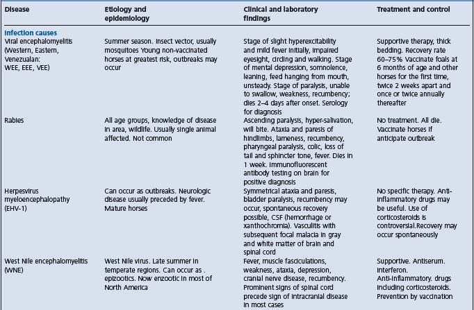
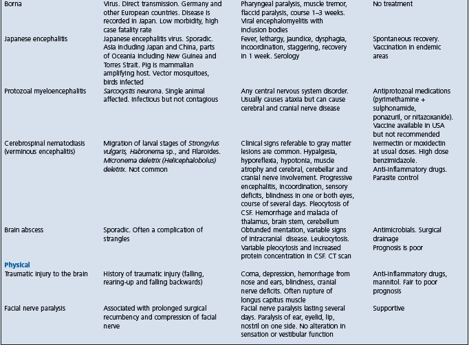
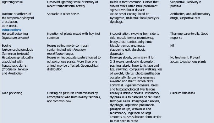
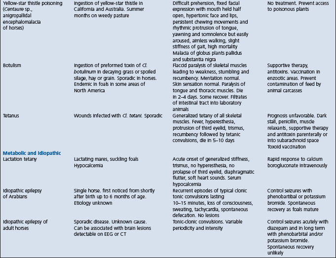
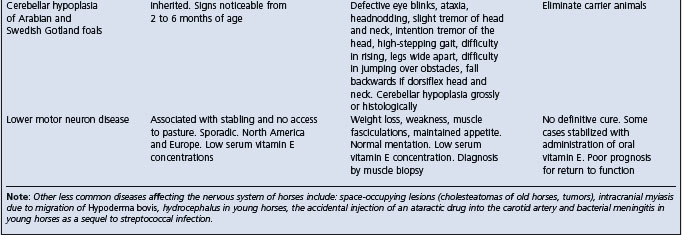
Clinically, the disease has very great similarity to the other viral encephalomyelitides, from which it can often be discriminated by the geographical location of the horse, and to the hepatic encephalopathies and a number of other diseases (see below and in Table 22.1).
Table 22.1 Diseases of horses characterized by signs of intra-cranial or disseminated lesions of the central nervous system
West Nile encephalitis – predominantly a myelitis with later development of signs of neurologic disease whereas EEE and WEE have predominant signs of encephalopathy.
• Borna disease – occurs in Europe
• Japanese encephalitis – occurs in Asia
• Various other viral infections that are geographically restricted
• Hepatic encephalopathy, such as that associated with poisoning by Crotalaria, Senecio, and Amsinckia spp.; acute serum hepatitis or hepatopathy
• Botulism causes weakness evident as muscle fasciculation, recumbency and dysphagia, but does not cause cerebral signs (irritation, behavioral abnormalities)
• Yellow star thistle poisoning (Centauria solstitialis, and poisoning by fumonisins (Fusarium moniliforme) can produce similar clinical signs to that of the encephalitides, with the exception of fever.
There is no definitive or specific treatment. Supportive treatment may be given with the intention to prevent self-inflicted injury, and maintain hydration and nutritional status.
Control of viral encephalomyelitis of horses is based on:16,23,24
• Accurate clinical and laboratory diagnosis of the disease in horses
• Use of sentinel animals to monitor the presence of the virus in the region
• Quarantine of infected horses to stop movement of virus donors
Vaccination of horses is important not only because it minimizes the risk of disease in vaccinated horses but also because of the possibility that for EEE it prevents viremia, subsequent infection of feeding mosquitoes, and propagation of the epidemic.
Formalin-inactivated EEE and WEE virus vaccines are available and are effective although over 50% of horses with EEE had been vaccinated within the previous year.17,25 This apparent poor protection can be explained by many horses not developing a detectable change in antibody titer after vaccination with a bivalent vaccine and rapid decreases in antibody titer from a peak value achieved 2–4 weeks after vaccination.17 Vaccines are available as univalent or bivalent preparations and in combination with other antigens (for instance, tetanus toxoid). Horses should be vaccinated well in advance of the anticipated encephalomyelitis season in a given area. Vaccination against both strains of the virus is advisable in areas where the strain has not been identified or where both strains exist. The currently recommended vaccination schedule consists of two doses of the vaccine initially, 10 days apart, followed by annual revaccination using two doses. Annual revaccination is currently recommended because the duration of effective immunity beyond 1 year is not known. It is probable that the initial two-dose vaccination lasts for up to 3–4 years. The emphasis in a vaccination program should be on the young horses.
Colostral antibody can be detected in the blood of foals from vaccinated dams for up to 6–7 months, after which time it declines rapidly. Foals from vaccinated dams should be vaccinated at 6–8 months of age and revaccinated at 1 year of age. Foals from unvaccinated dams may be vaccinated at 2–3 months of age and again at 1 year of age. Colostral antibodies in the foal will prevent the development of autogenous antibodies, and foals vaccinated when less than 6 months should be revaccinated when they are 1-year-old or, in high-risk areas, foals from vaccinated mares should be vaccinated at 3, 4, and 6 months of age.
Experimental DNA vaccines hold promise for the prevention of WEE.26
Housing of horses indoors at night, especially in flyproofed stables, and the use of insect repellents may restrain the spread of the virus. Use of insect repellents decreases the risk of EEE in horses to 0.04 that of unprotected horses.
Widespread spraying of insecticides to reduce the population of the vector insects has been used in the control of VEE, however, such measures are not practical for preventing sporadic cases of EEE or WEE, and the environmental impact of widespread insecticide use should be considered.
Complete eradication of the virus appears to be impossible because of the enzootic nature of the ecology of the virus. The horse being an accidental host for EEE and WEE virus make elimination of the virus impossible with methods currently available.
Control of the disease in humans in areas where the disease may occur is dependent on insect control, and a monitoring and surveillance early warning system is necessary to decide whether or not to take control measures. In areas where WEE occurs, clinical cases of the disease in unvaccinated horses usually precede the occurrence of the disease in human.27 The establishment of a reporting system whereby practicing veterinarians report all clinical cases of the disease in horses will also assist in predicting potential epidemics of WEE virus infection in the human population. Serological surveys of wildlife may also serve as good indicators of the geographical distribution and seasonality of circulation of these viruses and provide an early warning system prior to the detection of human cases.
Walton TE. Arboviral encephalomyelitis of livestock in the western hemisphere. J Am Vet Med Assoc. 1992;200:1385-1389.
Calisher CH, Walton TE. Japanese Western Eastern and Venezuelan encephalitides. In: Studdert MJ, editor. Virus infections of vertebrates: virus infections of Equines. Amsterdam: Elsevier; 1996:141-155.
1 Walton TE. J Am Vet Med Assoc. 1992;200:1385.
2 Kramer LD, Fallah HM. Am J Trop Med Hyg. 1999;60:708.
3 Elvinger F, et al. J Am Vet Med Assoc. 1994;205:1014.
4 Hayes CG, Wallis RC. Adv Virus Res. 1977;21:37.
5 Fulhorst CF, et al. Science. 1994;263:676.
6 Calisher CH, Walton TE. Studdert MJ, editor. Virus infections of vertebrates: virus infections of Equines 6. Amsterdam: Elsevier. 1996:141.
7 Hassan HK, et al. Am J Trop Med Hyg. 2003;69:641.
8 Wilson JH, et al. Prev Vet Med. 1986;4:261.
9 Sellers RF. Can J Vet Res. 1989;53:76.
10 Ross WA, Kaneene JB. Prev Vet Med. 1995;24:157.
11 Neufeld JL, Nayar GPS. In: Sekla L (ed) Western Equine encephalitis in Manitoba.
12 Winnipeg, Manitoba: Department of Health, 1982; pp. 62–79.
13 Chamberlain RW. Am J Trop Med Hyg. 1987;37:8S.
14 Ross WA, Kaneene JB. J Am Vet Med Assoc. 1996;208:1988.
15 Linssen B, et al. J Clin Micro. 2000;38:1527.
16 Calisher CH, et al. J Am Vet Med Assoc. 1983;183:438.
17 Waldridge BM, et al. Vet Therapeut. 2003;4:242.
18 Erickson GA, Mare CJ. Am J Vet Res. 1975;36:167.
19 Del Piero F, et al. Vet Pathol. 2001;38:451.
20 Weaver SC. Factors in the emergence of arbovirus diseases. Amsterdam: Elsevier, 1997;241.
21 Vodkin MH, et al. Am J Trop Med Hyg. 1993;49:772.
22 McGee ED, et al. Vet Pathol. 1992;29:361.
23 Wong FC, Lillie LE. Am J Publ Health. 1976;67(Suppl 1):15.
24 Omohundro RE. J Am Vet Med Assoc. 1971;161:1516.
25 Bryne RJ. Proc 3rd Intern Conf Equine Infect Dis 1973; 115.