Etiology Sarcocystis species. There are a number of species, with a number of different carnivore species as their final host but generally a specific intermediate host.
Epidemiology High prevalence of infection in most areas. Source of infection is feces of carnivore, primarily farm dogs and cats fed raw meat, or other carnivores if they have access to ruminant carcasses.
Clinical findings Severity of disease dose-dependent. The vast majority of infections are sub-clinical. Abortion, depressed growth rate. Neurological disease and ataxia in sheep. Severe infection with some species results in carcass condemnation.
Clinical pathology Anemia and elevated blood concentrations of enzymes associated with tissue damage in acute disease.
Lesions Non-suppurative encephalitis in sheep with neurological signs. Non-suppurative encephalitis, myocarditis, and hepatitis in aborted fetus. Cysts in carcass in chronic cases.
Diagnostic confirmation Identification of parasite microscopically in biopsy or postmortem material.
Treatment and control No effective treatment, amprolium or salinomycin may aid in control. Proper disposal of carcasses. Raw meat not to be fed to farm dogs and cats. Coyote control.
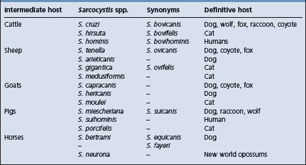
Sarcocystis species are obligate two-host sporozoan parasites in the phylum Apicomplexa. There are a number of species, each with definitive omnivorous or carnivorous species as final hosts. One system of naming the species identifies the intermediate and definitive host in the name, i.e. S. bovifelis, and has been commonly used in the literature. However, currently the organisms are identified by their original names. Table 26.5 shows the currently accepted name of Sarcocystis species of importance in agricultural animals and their definitive hosts.
In all countries where there have been surveys, the prevalence of infection in cattle, sheep, and horses approaches 100% with a lower, but significant infection rate in swine.1 Clinical disease is rare.
Sarcocystis spp. have an obligatory prey– predator live-host life cycle in which the definitive host is a predator or scavenger.1,2 The carnivorous definitive host becomes infected by ingesting tissue of the intermediate host that contains mature sarcocysts. Following ingestion, bradyzoites are released from the sarcocyst in the stomach and intestine, and they transform into micro- and macrogamonts. The male microgamonts mature to release microgametes, which fertilize the macrogamont to form an oocyst. The oocyst subsequently sporulates to release sporocysts which are passed in the feces and contain infective sporozoite.
The prepatent period is variable, approximately 14 days, and there is no illness in the carnivore host in association with this cycle. However, the replicative cycle of the parasite in the intestine results in the presence of large numbers of sporocysts in the feces and the infection is patent for a long period. Intermediate hosts become infected by ingesting sporocysts in the food or water.1,2
Sporocysts are not dependent on weather conditions for maturation, and are quite resistant to environmental influences. Under experimental conditions they can survive freezing, but they are susceptible to drying.2 Consequently they can probably overwinter in the environment. Field studies have shown a lower herd prevalence of sarcocystosis in cattle in arid and semi-arid environments compared to cattle from temperate and tropical areas, which is probably a consequence of the relative aridity as well as the lower density of the definitive and intermediate hosts in arid areas.3,4
Individual species vary in their pathogenicity and in their ability to produce clinical disease in the intermediate host. In cattle, for example, S. cruzi is considerably more pathogenic than S. hominis.1,2
S. tenella is the most pathogenic species for sheep, and S. capricanis for goats and naturally occurring clinical disease in sheep is not observed with S. gigantea or S. medusiformis.5 There is a strong correlation between the number of sporocysts ingested and the severity of disease. The size of the sarcocyst that occurs in the tissues of the intermediate host also varies with the infecting species. Those from cats and occurring in sheep (S. gigantea, S. medusiformis) or cattle (S. hirsuta) are of particular economic importance, as they produce macroscopically visible sarcocysts that can result in meat condemnation. S. cruzi produces microscopic sarcocysts in muscle and will escape gross detection at meat inspection.
There is a positive association between herds infected with Sarcocystis and the presence of working dogs on the farm, the practice of leaving carcasses in the field and the feeding of dogs with raw meat,4 and virtually all of the reported clinical cases of sarcocystosis in cattle in the literature record that the dogs on the farm were fed offal or uncooked beef. Housing of dogs and cattle in the same shed or area can have increased risk for infection and clinical disease,1,6 and cattle pastured close to farm buildings where there are dogs are at greater risk. The presence of foxes on farms is also strongly associated with Sarcocystis infection in those herds that leave carcasses on the field.4
The main risk for cat-associated sarcocystosis is the farm cat that is fed raw meat. Farm cats use hay barns as dens and can contaminate hay and other feedstuffs.7 Feral cats have the potential to distribute sporocysts widely in the grazing environment; however, the presence of feral cats on a farm may not increase the risk for Sarcocystis infection of cattle,4 as scavenged sheep or cattle carcasses are a relatively unimportant part of the diet of feral cats.7
Risk for infection with Sarcocystis is higher with higher stocking densities,4 which may reflect a more intense contamination of pastures by working dogs. Cattle on farms that graze sheep and cattle on the same pastures are less likely to be infected.
In the intermediate host, sporozoites are released from ingested sporocysts in the small intestine where they penetrate the mucosa and enter the endothelial cells of blood vessels. The stages of schizogony and the distribution of merozoites vary according to the species, but in cattle endothelial infection is followed by parasitemia, with merozoites subsequently localizing in striated muscle and nervous tissue where they develop into sarcocysts. Immature sarcocysts can be found in muscle 45–60 days following ingestion of sporocysts and are infective at about 70 days.2
Schizogony in the endothelial cells of the arterioles and capillaries results in widespread hemorrhage and anemia. Fever is associated with the parasitemia, and in the experimental disease coincides with the time of maturation of the first- and second-generation schizonts.1 The vascular lesion appears to be an essential part of the disease’s pathogenesis. It is proposed that the parasite produces growth retardation as a result of changes in plasma concentrations of somatostatin and growth hormone, and changes in cytokine interactions with the endocrine system.10
The severity of the illness and the degree of infection of tissues at postmortem examination in experimentally induced cases increase with the size of the infective dose. The number of asymptomatic infections probably reflects the early ingestion of a few sporocysts that provoke a strong immunity to later challenge. When groups of animals that have not been exposed to infection previously are suddenly brought into contact with heavy contamination, especially from dogs and cats, outbreaks of clinical disease are likely to occur.
Infection and disease can occur at all ages. Clinical disease may be more severe where there is intercurrent nutritional stress, and copper deficiency may be an exacerbating factor. Monensin is suspected of being able to potentiate recent infections to cause a severe myositis.11
Acute illness is recorded with experimental infections but is rarely seen, or recognized, in the field. Illness commences with a rise in temperature and heart rate, followed by anorexia, anemia, weight loss, a fall in milk production, nervousness, muscle twitching, hypersalivation, lameness, abortion, and, in heavy infections, death. The agent is an occasional cause of non-suppurative encephalomyelitis in cattle and manifest with ataxia and recumbency.
Chronic disease in cattle is manifest by poor weight gains, loss of hair of the neck, rump and the switch of the tail (‘rat-tail’), anemia, and abortion.
In sheep, naturally occurring sarcocytosis has been associated with S. tenella and S. arieticanis and presents primarily as a neurological disorder, with muscle weakness, trembling, ataxia of varying severity, followed by hind limb paresis or flaccid paralysis and lateral recumbency. All ages of sheep can be affected, although lambs under 6 months are most susceptible. Attack rates in a susceptible group can be as high as 75% with a high case fatality rate.12-16
Infection may also be manifest with depressed growth, reduced wool growth, and anemia.8,13 Less common manifestations include signs of congestive heart failure associated with endocardial and myocardial infection.17 Infestation of the muscle of the esophagus in sheep is believed a cause of esophageal dysfunction and regurgitation in sheep.18
Natural clinical disease is not recognized. Sarcocystosis produced experimentally in pigs is manifested by cutaneous purpura on the snout, ears, and buttocks, and dyspnea, tremor and weakness or recumbency.19 There is evidence that breed of pig affects the severity of clinical disease with experimental infections and also the subsequent severity of the parasite load.20
Characteristic laboratory findings in the systemic disease include a responsive anemia, a prolonged prothrombin time, and high titers of antibody to Sarcocystis. Blood creatine phosphokinase, lactic dehydrogenase, and aspartate aminotransferase are significantly elevated.19 An indirect hemagglutination test (IHA) and an ELISA test are available for serological surveys. Titers of antibodies are not high at the time of an acute illness, but are at diagnostic levels 1 week to 3 months afterwards.1,14 An ELISA based on antigens from merozoites has high sensitivity and specificity for detection of infection in individual animals, and a 100% sensitivity for detecting herd infection with small sample size.21 Most animals have been exposed to Sarcocystis spp., and serological examination cannot differentiate clinical disease from asymptomatic infection.
Emaciation, lymphadenopathy, laminitis, anemia and ascites are present, but the most obvious feature is the petechial and ecchymotic hemorrhages throughout the body.22 There are also erosions and ulcerations in the oral cavity and esophagus, likely as a result of microvascular damage. Microscopically, schizonts are found in endothelial cells throughout the body, and hemorrhages, lymphocytic infiltration, and edema are seen in heart, brain, liver, lung, kidney, and striated muscle.22 Death is probably a result of the severe necrotizing myocarditis that occurs. There is an association between eosinophilic myositis and sarcosporidiosis, but this relationship is not proven in all cases.1
In sheep presenting with neurological disease there may be no findings at gross postmortem, but a non-suppurative encephalomyelitis on histological examination.12,14,15 Aborted bovine fetuses show non-suppurative encephalitis, myocarditis, and hepatitis.5
A number of options are available to achieve a definitive diagnosis of the species present, including animal transmission studies, immunohistochemistry, electron microscopy and PCR. Such techniques are seldom required for routine diagnostic cases.
• Histology – formalin-fixed heart, skeletal muscle (several sites, including tongue, masseter muscle) (LM).
Diagnosis in clinical cases is difficult because of the non-specific signs observed and the widespread prevalence of infection. Sarcosporidiosis is a consideration in the examination of problems of fever and anemia of undetermined origin in cattle and of ill-thrift in cattle and sheep.
Muscle biopsy may aid in the determination of the presence of infection, but still begs the question of its significance to the clinical disease.
The differential diagnoses for abortion in cattle are covered under brucellosis, and sheep under brucellosis. Causes of encephalitis and ataxia in sheep are listed under those headings.
No approved treatment is available, but amprolium or salinomycin may relieve the signs. Amprolium 100 mg/kg given daily from the time of inoculation reduces the severity of infection in experimentally infected calves and sheep,13,23 and has been used to control an outbreak in sheep.12 Treatment of experimentally infected calves with salinomycin (4 mg/kg BW daily in divided doses for 30 days) reduced the severity of the illness.24 Monensin may have a similar ameliorating effect,9,25 but is also suspected of exacerbating muscle lesions.10 Oxytetracycline, at very high dose rates, and halofuginone may be effective in acute infections.13
Control is difficult as it involves the separation of carnivores and stock, which is not possible on most farms. However, infection in farm dogs and cats could be avoided if all meat fed to them was thoroughly cooked. Freezing will not destroy the infectivity. Coyotes and wild dogs should be controlled and livestock carcasses not left on fields. Prior exposure to small numbers of pathogenic sarcocysts produces a strong immunity, but there is no vaccine available.
Dubey JP, Speer CA, Fayer R. Sarcocystosis of animals and man. Boca Raton, Florida: CRC Press, 1989.
Dubey JP. A review of Neospora caninum and Neospora-like infections in animals. J Protozool Res. 1992;2:40-52.
Anderson ML, Barr B, Conrad PA. Protozoal causes of reproductive failure in domestic animals. Vet Clin North Am Food Anim Pract. 1994;103:439-461.
1 Fayer R, Dubey JP. Sarcocystosis of animals and man. Boca Raton, Florida: CRC Press, 1989.
2 Fayer R, Dubey JP. Comp Cont Educ. 1986;8:F180.
3 Savini G, et al. Epidemiol Infect. 1992;108:107.
4 Savini G, et al. Prev Vet Med. 1994;19:137.
5 Anderson ML, et al. Vet Clin North Am Food Anim Pract. 1994;103:439.
6 Carrigan MJ. Aust Vet J. 1986;63:22.
7 Langham NPE, Charleston WAG. Am J Agric Res. 1990;33:429.
8 Munday BL. Vet Parasitol. 1986;21:21.
9 Dubey JP, et al. J Parasitol. 1989;75(422):980.
10 Fayer R, Elsasser TH. Parasitol Today. 1991;7:250.
11 Jeffrey M, et al. Vet Rec. 1989;124:422.
12 Fitzgerald SD, et al. J Vet Diag Invest. 1993;5:291.
13 Jeffrey M. In Practice. 1993;151:2.
14 Henderson JM, et al. Can Vet J. 1997;38:168.
15 Sargison ND, et al. Vet Rec. 2000;146:225.
16 Caldow GL, et al. Vet Rec. 2000;146:7.
17 Scott PR, Sargison ND. Vet Rec. 2001;149:240.
18 Braun U, et al. Can Vet J. 1990;31:391.
19 Braun U, et al. Can Vet J. 1990;31:392.
20 Reiner G, et al. Vet Parasitol. 2002;106:99.
21 Savini G, et al. Prev Vet Med. 1997;32:35.
22 Johnson AJ, et al. Am J Vet Res. 1975;36:995.
23 Fayer R, Johnson AJ. J Parasitol. 1975;61:932.
Etiology The protozoan parasite Neospora caninum. The dog is identified as the definitive host for N. caninum but the major route of infection in cattle is by vertical transmission.
Epidemiology An infection of cattle worldwide and associated with epidemic and endemic abortion. Point source and congenital infection occurs.
Clinical findings. Abortion in cows, perinatal mortality and encephalomyelitis in congenitally infected calves.
Clinical pathology. IFAT and ELISA serology on maternal serum and fetal fluids.
Necropsy findings. Fetal lesions of multifocal non-suppurative encephalitis, myocarditis, and periportal hepatitis. Infection confirmed by immunohistochemistry.
Diagnostic confirmation. A presumptive diagnosis can be based on the fetal histological lesions and seropositivity of the dam, but the definitive diagnosis requires the demonstration of the protozoa in fetal tissues by immunohistochemical labeling coupled with herd serological examinations.
Control. Feed hygiene and calving hygiene. Cull congenitally infected cattle.
Neospora caninum, a protozoan parasite of the phylum Apicomplexa in the family Sarcocystidae. N. caninum primarily infects dogs and cattle but has a wide host range and infects all the major domestic livestock species, as well as companion animals and certain wildlife species. Dogs are the only known definitive host with cattle as the major intermediate agricultural animal host. Natural infections are infrequently reported in sheep, goats and deer.1 N. caninum is a sporadic cause of encephalomyelitis and myocarditis in several species but its importance is its association with endemic and epidemic abortion in cattle. It is now the most common diagnosis for abortion in cattle in most countries. The organism can be isolated, with difficulty, from infected calves.2
N. caninum was initially associated with abortion in the early 1990s in pastured cattle in Australia and New Zealand, and as a major cause of abortion in large dry-lot dairies in southern California, and dairies in New Mexico, Washington, and Arizona in the United States. Since then, abortion associated with N. caninum has been reported from many countries in cattle under varying management conditions, and has worldwide occurrence.
Abortion may be epizootic or sporadic. In epizootic abortion the number of cows aborting varies. It is usually between 5 and 10%, but up to 45% of cows may abort within a short period. The period of abortion may be a few weeks to a few months. There is no major seasonal occurrence and abortion occurs in both beef and dairy cows.3,4 Sporadic abortions occur predominantly in cows that have been infected congenitally and seropositive cows have greater risk for repeat abortions. Seropositivity in herds can be high, but varies widely. One study in dairy herds reports a within herd seroprevalence ranging from 7 to 70%3 and another in beef herds a within-herd seroprevalence ranging from 6.5 to 67%.5 Seropositive dams have a 3–7 fold higher risk of abortion than seronegative dams.6,7
There are two routes of infection of cattle. The dog is identified as the definitive host for N. caninum and infection of cattle can occur from the ingestion of oocytes in the dog feces contaminating feed or water. However vertical transmission occurs in both cattle and dogs and vertical (congenital) transmission appears the major route for infection in most cattle.1,8
Live-born calves from congenitally infected cows are themselves congenitally infected and the infection is believed to be persistent and lifelong. One study conducted on two dairies found 81% of seropositive cows gave birth to congenitally infected calves. Seroprevalence did not increase with cow age on either dairy, and was stable through the study period. The probability of a calf being congenitally infected was not associated with dam age, dam lactation number, dam history of abortion, calf gender, or length of gestation. Other studies have shown that this route of transmission is highly efficient resulting in infection of 50–95% of the progeny of seropositive dams.9
Congenital infection can result in abortion, or the birth of a normal, infected calf, and an infected cow can give birth to a normal, infected calf at one pregnancy and abort in the subsequent pregnancy.3 The occurrence of infection in some herds can be associated with specific family lines.10
Whereas vertical transmission is the major route of infection that leads to sporadic abortions in cattle associated with N. caninum epidemiological evidence suggests that postnatal (point) infection is often the cause of outbreaks of abortion. Where dog feces are the source of infection many cattle are often exposed and this point source of infection commonly results in outbreaks of abortion. Farm dogs have been shown to have a higher seroprevalence to N. caninum than urban dogs suggesting that the disease cycles between cattle and dogs in rural environments.11
The importance of postnatal infection versus vertical infection in the genesis of abortion may vary between countries associated with differences in management systems.12
Abortion has been produced by experimental challenge of fetuses and pregnant cattle with culture derived tachyzoites of N. caninum.13 Fetal death and resorption or abortion has been reproduced in ewes challenged at 45, 65, and 90 days’ gestation, but not 120 days, and lesions resemble those of ovine toxoplasmosis.14 The disease has also been reproduced experimentally in goats,15 but the importance and prevalence of this infection in naturally occurring abortions in small ruminants remains to be determined. Contaminated placenta, milk, and colostrum can result in infection of calves under one week of age.16
Outbreaks of abortion often appear to be point source infections, but the risk factors, other than probable mass exposure to infected dog feces, are not known. With suspect point source infection the disease in dairy herds frequently occurs as an epizootic, with multiple abortions occurring in a 1 to 2-month period. Severely autolytic fetuses are aborted in the 5th–7th month of pregnancy in most reports, but earlier abortions are recorded in some, and the agent has been associated with outbreaks where the gestational age of fetal loss has ranged from 3 to 8.5 months.3,6,17-20 Heifers may abort earlier in pregnancy than older cows.
Endemic abortion is more likely associated with the presence of congenitally infected cattle in the herd, which are at high risk of aborting, particularly in the initial pregnancy and in the pregnancy during the first lactation.6,20 Cows that have aborted have higher risk for abortion in subsequent pregnancies, but the risk decreases with each subsequent pregnancy. The true frequency of repeat abortions is unknown because cows may be culled for abortion.
It has been postulated that immunosuppression resulting from concurrent infection with agents such as BVD virus may increase the risk for infection with N. caninum and precipitate outbreaks of abortion. Concurrent N. caninum and BVD infections in aborted fetuses have been observed in some studies and one study has found a significant association between abortion and cows with antibody to both N. caninum and BVD.21 Others have found no association22 and whereas concurrent BVD infection might be a risk factor in some outbreaks, it is not in all.
Economic loss is occasioned by the direct cost of abortions and the indirect costs associated with establishing the diagnosis and re-breeding or replacement costs. Seropositivity is also associated with increased risk of stillbirth and increased risk of retained placenta.
Seropositive heifers have been reported to produce less milk than seronegative herdmates.1,3,23 However, this difference in milk production between seropositive and seronegative animals is not apparent in herds that are not experiencing an abortion problem.24 In beef cattle seropositivity has been associated with reduction in average daily weight gain of 0.05 to 0.17 kg/head/day compared with seronegative cohorts and reduced food conversion efficiency rather than decreased food intake appeared the cause.25 However, a subsequent study found no difference in production performance and carcass measures between seropositive and seronegative feedlot cattle.26
Estimates of economic loss associated with epidemic abortion include US$35 million/year in California, AUS$85/year to dairy and AUS$25 for beef cattle in Australia and NZ$17.8 for the dairy industry in New Zealand.9
The organism has a predilection for fetal chorionic epithelium and fetal placental blood vessels producing a fetal vasculitis and inflammation and degeneration of the chorioallantois, and widespread necrosis in the placentome.14 Tachyzoites penetrate host cells and are located in a parasitophorous vacuole. They can be found in macrophages, monocytes, vascular endothelial cells, fibroblasts, hepatocytes, renal tubular cells, and in the brain of infected animals. With neuromuscular disease, cranial and spinal neural cells are infected. Cell death is by the active multiplication of tachyzooites.
Abortion is the only clinical sign observed in infected cows. Fetuses may die in-utero, be reabsorbed, be mummified, stillborn, born alive but diseased, or born clinically normal but chronically infected. Cows that are infected have decreased milk production in the first lactation, producing approximately 1 liter less of milk/cow/day than uninfected cows, are prone to abort, and have a higher risk of being culled from the herd at an early age.
In addition to the occurrence of early abortion, the disease in beef herds is associated with the birth of live-born, premature, low-birth-weight calves. Depending upon the degree of prematurity, these calves can be kept alive with intensive care during the neonatal period.
Most congenitally infected calves are born alive without clinical signs. Congenital infection can occasionally be manifest with ataxia, loss of conscious proprioception, paralysis, and other neurological deficits in the new-born calf,27 but the majority of congenitally infected calves are clinically normal and, surprisingly, epidemiological studies suggest that congenital infection does not necessarily have a detrimental effect on calf health and survival.28
N. caninum infection has been demonstrated in the nervous system of a horse with progressive debilitation, followed by sudden onset of neurological disease with paraplegia.29 It appears a rare cause of neurological disease in horses but should be considered in the differential diagnosis of equine protozoal myeloencephalitis.
Serological examination is with the indirect antibody fluorescent antibody test (IFAT), or by ELISA, and there is good agreement between the two tests. ELISA tests based on recombinant protein have higher sensitivities and specificities than that based on whole-tachyzooite lysates.30 The IFAT is highly sensitive and specific for detection of maternal infection31 and is commonly used. One study compared IFAT titers in maternal sera from 40 cows whose fetuses had been examined for neosporosis by immunohistochemistry. Of the 22 confirmed cases, 21 cows had titers of >1 in 640, whereas only one of the 18 negative cases had a titer of this magnitude.32 The persistence of titers following infection is uncertain, and they may fluctuate during pregnancy. A positive titer in a cow that has aborted indicates exposure but not causality. Recently, IgG avidity patterns have been used to determine the duration of infection.33,34
Diagnosis is also made by the detection of antibody in fetal pleural fluid or sera,31 and IFAT antibody has been found in the sera of from 50 to 65% of immunohistochemistry-confirmed cases of neospora infection, but not in fetuses under 4 months of gestational age.35,36 Antigen can also be detected by PCR.37
Gross findings are of autolysis. The brain may be autolysed, but should still be submitted for examination along with heart, liver, and placenta if available. Histological lesions are of multifocal encephalitis, myocarditis, and periportal hepatitis. Liver lesions may be more prominent in epizootic abortions. Immunohistochemistry using anti-N. caninum serum is used to identify tachyzooites in tissues, and the brain is the organ with the highest detection rate.38 Immunohistochemistry is specific, but insensitive, in diagnosing fetal neosporosis, and maternal serology should be used in conjunction.
There is no treatment that can be used to curtail an ongoing abortion epidemic and possible drug therapies are generally not considered an option because of likely unacceptable milk and meat residues and withdrawal problems.9
Serology using IFAT can confirm infection in individual cows.
Because of the high prevalence of infection, and the occurrence of congenital infection, care must be taken in extrapolating the results of a single positive diagnosis to problems of abortion.3 The high rate of natural congenital infection means that evidence of infection in an aborted fetus is not proof of causation of abortion, and fetal examination should be coupled with serological examination of aborting and non-aborting animals in the herd for statistical differences.
All efforts should be made to exclude the possibility of dog fecal contamination of cattle feed and water and of the grazing environment.39 Placentas, aborted fetuses, and dead calves should be removed and disposed of so that the definitive host and cattle cannot get access to them.
Congenitally infected cows are at high risk for abortion, and abortion rates in infected herds can be substantially reduced by culling these animals.34,40,41 Congenitally infected calves can be identified by serology on precolostral blood samples and culled at a young age. If precolostral blood sampling is not feasible, examination of sera at 6 months of age will determine infected calves, positive titers indicating either congenital infection or postnatal infection.42 Animals purchased into the herd should be seronegative.
It is possible that strategic therapy of pregnant cows with an appropriate antiprotozoal drug could abort the infection. This could be effective in beef cattle, but would probably not be legal, or appropriate, in lactating dairy cattle.
Whereas evidence for increased risk for neospora abortion due to immunosuppression resulting from concurrent infection with BVD virus is equivocal control of BVD infections should be a component of control programs for neosporosis.
A killed tachyzooite vaccine has been approved in the United States for use in pregnant cows and is available commercially. At present there are no controlled studies in the United States on its efficacy in mitigating the effects of bovine neosporosis in dairy cattle. A field trial in dairy herds in Costa Rica measuring abortion as the outcome showed a reduction in the incidence of abortion.43 A study in growing beef cattle showed no significant effects between vaccinated and control animals in cumulative average daily gain or in food conversion efficiency.44 Studies in sheep have shown that vaccination significantly reduces fetal loss in experimentally infected ewes. However, they showed that there was little protection against vertical transmission of N. caninum to the fetuses.45,46
Vaccination of dairy cattle may interfere with a herd test and cull policy.
Dubey JP, Lindsay DS. A review of neospora and neosporosis. Vet Parasitol. 1996;67:1-59.
Dubey JP. Neosporosis in cattle: biology and economic impact. J Am Vet Med Assoc. 1999;214:1160-1163.
Barr BC. Neosporosis. Report of the International Neospora Workshop. Comp Cont Educ Pract Vet. 1997;19(Suppl 4):S120-S126.
Antony A, Williamson NB. Recent advances in understanding the epidemiology of Neospora caninumin cattle. NZ Vet J. 2001;49:42-47.
Buxton D, McAllister MM, Dubey JP. The comparative pathogenesis of neosporosis. Trends Parasitol. 2002;18:546-552.
Dubey PJ. Redescription of Neospora caninum and its differentiation from related coccidian. Int J Parasitol. 2002;32:929-946.
Larson RL, Hardin DK. Review: Neospora caninum-induced abortion in cattle. Bovine Practit. 2003;37:121-126.
Haddad JPA, Dohoo IR, VanLeewen JA. A review of Neospora caninum in dairy and beef cattle — a Canadian perspective. Can Vet J. 2005;46:230-243.
1 Dubey JPJ. Am Vet Med Assoc. 1999;214:1160.
2 Miller CMD, et al. Aust Vet J. 2002;80:620.
3 Thurmond MC, et al. J Vet Diag Invest. 1997;9:44.
4 Crawshaw WM, Brocklehurst S. Vet Rec. 2003;152:201.
5 Sanderson MS, et al. Vet Parasitol. 2000;90:15.
6 Thurmond MC, Hietela SK. Am J Vet Res. 1997;58:1381.
7 Atkinson RA, et al. Aust Vet J. 2000;78:262.
8 Schares G, et al. Vet Parasitol. 1998;80:87.
9 Reichel MP, Ellis JT. Aust Vet J. 2002;50:86.
10 Bjorkman C, et al. J Am Vet Med Assoc. 1996;208:1441.
11 Antony A, Williamson NB. N Z Vet J. 2003;51:232.
12 Antony A, Williamson NB. Aust Vet J. 2001;49:42.
13 Conrad PA, et al. Vet Rec. 1992;130:147.
14 Buxton D, et al. J Comp Path. 1998;118:267.
15 Lindsay DS, et al. Am J Vet Res. 1996;56:1176.
16 Davison HC, et al. Res Vet Sci. 2001;70:163.
17 McAllister MM, et al. J Vet Diag Invest. 1996;8:355.
18 Thornton RN, et al. NZ Vet J. 1991;39:129.
19 Otter A. Vet Rec. 1997;140:239.
20 Hernandez J, et al. J Am Vet Med Assoc. 2002;221:1742.
21 Bjorkman C, et al. Vet J. 2000;159:201.
22 Quinn HE. Aust Vet J. 2004;82:99.
23 Hernandez J, et al. J Am Vet Med Assoc. 2001;219:632.
24 Hobson JC, et al. J Am Vet Med Assoc. 2002;221:1160.
25 Barling KS, et al. J Am Vet Med Assoc. 2001;219:1259.
26 Schmidt PL, O’Connor AM. Proc Am Assoc Bov Practit. 2004;37:164.
27 Gunning RF, et al. Vet Rec. 1994;134:558.
28 Pare J, et al. Can J Vet Res. 1996;60:133.
29 Daft BM, et al. Equine Vet J. 1997;29:240.
30 Louie K, et al. Clin Diag Lab Immunol. 1997;4:692.
31 Otter A, et al. Vet Rec. 1997;141:487.
32 McNamee PT, et al. Vet Rec. 1996;138:419.
33 Bjorkman C, et al. Vet Parasitol. 2005;128:195.
34 Frossling J, et al. Vet Parasitol. 2005;128:209.
35 Wouda W, et al. J Parasitol. 1997;83:508.
36 Barr BC, et al. Vet Rec. 1995;137:611.
37 Ho MSY, et al. J Parasitol. 1997;83:508.
38 Wouda W, et al. J Vet Diag Invest. 1997;9:180.
39 Rinaldi L, et al. Vet Parasitol. 2005;128:219.
40 Hall CA, et al. Vet Parasitol. 2005;128:231.
41 Weston JF, et al. NZ Vet J. 2005;53:142.
42 Thurmond MC, Hietela SK. Bovine Pract. 1995;29:60.
43 Romero JJ, et al. Vet Parasitol. 2004;123:149.
44 Barling KS, et al. J Am Vet Med Assoc. 2003;222:624.
Etiology Cryptosporidium parvum bovine genotype 2 and C. andersoni.
Epidemiology Infection common in ruminant neonates. May cause diarrhea, especially if there is intercurrent infection with other enteropathogens, nutritional, or environmental stress.
Clinical findings Malabsorption-type diarrhea.
Clinical pathology Oocysts in feces demonstrated by immunofluorescent assay.
Diagnostic confirmation Demonstration of lesions and organism.
Treatment Supportive. Halofuginone or paromomycin if approved.
Control Hygiene and management to ensure passive transfer of colostral antibodies and minimization of infection pressure.
Cryptosporidium are protozoon parasites in the Phylum Apicomplexa. Currently, up to 14 species of Cryptosporidium, infecting mammals, fish, and birds, have been proposed but only two of these are of importance to agricultural animals. These include C. parvum which infects many different hosts including cattle, swine, horses, and small ruminants, and the calf genotype of C. muris, now called C. andersoni, which infects cattle.1,2 C. parvum has two distinct genotypes known as human genotype 1 (also known as C. hominis) and bovine genotype 2. Both genotypes are capable of causing disease in humans. Livestock and horses are not commonly infected with genotype 1, although recently infections have been experimentally produced in lambs and piglets3 and mixed infections with genotypes 1 and 2 have been observed in calves.4
Cryptosporidium parvum is a common infection in young ruminants and is found in many species of mammals including humans. There has been controversy about the role of C. parvum as a causative agent of diarrhea because infection can be found in normal healthy animals. However, it is considered a significant cause of varying degrees of naturally occurring diarrhea in neonatal farm animals. Most commonly, the agent acts in concert with other enteropathogens to produce intestinal damage and diarrhea.
Cryptosporidiosis has been recognized worldwide, primarily in neonatal calves, but also in lambs, goat kids, foals, and piglets.5-9 Many studies report prevalence of infection but this does not imply clinical disease.
Infection is common in calves but reported prevalence rates of Cryptosporidium in feces vary widely. Prevalence ranging from 1.1% in a random sample of adult cattle feces in California10 to 79% in symptomatic calves in Maryland, USA have been reported.11 Cryptosporidia have been detected in 70% of 1 to 3-week-old dairy calves based on a single examination of feces.12 A single sampling in a nationwide survey of 7369 calves on 1103 farms in the United States found infection on 59.1% of farms, and in 22.4% of calves.13 Daily sampling of calves in the first 4 weeks of life show higher rates of infection and a period prevalence of infection as high as 100%.14-17 There is also high site prevalence.
A 6 year longitudinal study, on a lowland farm in the United Kingdom, of patterns of fecal excretion of C. parvum in beef cattle, dairy cows, home bred and purchased calves, lambs, rodents in the farm building environment and in the pastures, showed that the parasite was endemic and persistently present in all animal species tested. It found that patterns of infection were variable and that short-term or point prevalence sampling would be unlikely to provide a representative picture of infection rates. Prevalence rates and oocyst numbers in feces were highest in young animals.17
Infection can be detected as early as 5 days of age, with the greatest proportion of calves excreting organisms between days 9 and 14.14-17 This is also the period of greatest intensity of excretion of the organism in the feces of individual calves.
Infection of the calf is followed by the development of resistance to reinfection, and oocyst excretion is less common and intermittent in older and adult cattle,18,19 although high excretion rates are found in adult cattle in some herds.20
Most prevalence studies have found little association between infection and diarrhea, but there are many reports that associate infection in calves with diarrhea occurring between 5 and 15 days of age.
C. parvum is also a common enteric infection in young lambs and goats, and diarrhea can result from a monoinfection, but more commonly is associated with mixed infections. The age pattern of infection and excretion of the organism is similar to that in calves.21 Infection can be associated with severe outbreaks of diarrhea, with high case fatality in lambs from 4 to 10 days of age21-24 and goat kids from 5 to 21 days of age.21
Cryptosporidial infection in pigs occurs over a wider age range than in ruminants and has been observed in pigs from 1 week of age through to market age. In a retrospective study of piglets submitted to a diagnostic laboratory over a 4-year period, cryptosporidia were detected histologically in 5.3% of about 3500 piglets.25 Infection is most common between 6 and 12 weeks of age.26 The majority of infections are asymptomatic, and the organism does not appear to be an important enteric pathogen in this species,26,27 although it may contribute to postweaning malabsorptive diarrhea.
Cryptosporidial infection in foals appears less prevalent and occurs at a later age than in ruminants, with excretion rates peaking at 5–8 weeks of age.28 Infection is not commonly detected in yearlings or adults.28,29 Most studies indicate that cryptosporidiosis is not a common disease in foals, and infections in immunocompetent foals are usually subclinical.26,28 Diarrhea is recorded in foals from 5 days to 6 weeks of age including a report of an outbreak lasting one month where 9 of 30 foals aged between 4 days and 3 weeks of age were affected with a case fatality rate of 33%.30 Persistent clinical infections occur in Arabian foals with inherited combined immunodeficiency.31
Cryptosporidiosis is also recorded in young deer and can be a cause of diarrhea in artificially reared orphans. Infection has also been recorded in red deer calves dying at 24–72 hours of age following a syndrome of severe weakness and depression accompanied by a terminal uremia.32
Experimental infections have shown that a small number of oocysts are required for infection. The replicative cycle in the intestine amplifies a minor infective dose and studies in gnotobiotic lambs indicate a minimum infectious dose as low as one oocyst. The source of infection is feces which contain oocysts that are fully sporulated and infective when excreted. Large numbers are excreted during the patent period in calves resulting in heavy environmental contamination. Transmission may occur directly from calf to calf, indirectly via fomite or human transmission, from contamination in the environment or fecal contamination of the feed or water supply. Infection into the environment of newborn animals, and an increase in contamination of their immediate environment occurs as the result of a periparturient rise in the fecal excretion of oocysts by the dam. This has been recorded in ewes,33 in a beef cattle herd where the rate of infection and the number of oocysts in cattle feces increased significantly at one week post calving34 and in some dairy herds.35,36 Genotype 1 of C. parvum is not host-specific and infection from other species, such as rodents or farm cats, contaminating calf feeds, is also possible.
The factors that make animals susceptible to infection and that predispose infected animals to develop clinical disease are not well understood. Disease in agricultural animals associated with infection with Cryptosporidium is associated with infection with C. parvum and there is little evidence that infection with C. andersoni is associated with disease. Commonly, other enteric infections are present where there is disease attributed to Cryptosporidium. The site of infection with C. parvum is on the enterocyte where it results in cell damage, loss of brush border enzymes and a reduction of villous surface area.
Oocysts are resistant to most disinfectants and can reportedly remain viable for about 18 months in a cool, damp or wet environment, can survive for several months in soil and slurry11,37 but are susceptible to desiccation and temperatures above 60°C.38 The infectivity of the oocysts can be destroyed by ammonia, formalin, freeze-drying and exposure to temperatures below 0°C (32°F) and above 65°C (149°F). Ammonium hydroxide, hydrogen peroxide, chlorine dioxide, 10% formol saline and 5% ammonia are effective in destroying the infectivity of the oocysts. The infectivity of oocysts in calf feces is reduced after 1–4 days of drying.39,40
Concurrent infections with other enteropathogens, especially rotavirus and coronavirus, are common and epidemiological studies suggest that diarrhea is more severe with mixed infections. The rates of single and mixed infections vary with different studies. In general, mixed infections are most common, but cryptosporidial infection can be significant in its own right. For example, in two studies involving diarrheic calves submitted to diagnostic laboratories, cryptosporidia were the only pathogens isolated in 51% and 55% of the cases, while in 25% and 39% of cases the protozoan agent was found in combination with rotavirus and/or coronavirus.41,42 Immunologically compromised animals are more susceptible to clinical disease than immunocompetent animals, but the relationship between disease and failure of passive transfer of colostral immunoglobulins is not clear. The disease can be reproduced in both colostrum-deprived and colostrum-fed calves and, in the field, clinical disease can occur in calves and foals with adequate passive transfer of colostral immunoglobulins. However, the shedding of the organism has been observed to be higher in calves with low absorptive efficiency of IgG from colostrum and low serum IgG concentrations.43
In one series of observations there was a tendency for the prevalence of infection to be higher during the winter months of the year when the calves were confined, which suggests a build-up of contamination,44 but seasonal difference in prevalence is not marked and clinical disease can occur at all seasons.
Case fatality rates in cryptosporidiosis are generally low unless there are other complicating factors. In addition to concurrent infections, these include energy deficits from inadequate intake of colostrum and milk, and chilling from adverse weather conditions.
Age-related resistance, unrelated to prior exposure, has been observed in lambs45 but not calves.46 Infection results in the production of parasite-specific antibody, but both cell-mediated and humoral antibody are important in protection, as well as local antibody in the gut of the neonate.47,48
The disease has been reproduced in colostrum-deprived calves, lambs and kids, in many studies.
Infections in domestic animals and pets may be a reservoir for infection of susceptible humans. In humans, cryptosporidium is considered to be a relatively common non-viral cause of self-limiting diarrhea in immunocompetent persons, particularly in children. In symptomatic immunocompetent patients, cryptosporidiosis most commonly presents with diarrhea that can lead to rapid weight loss and dehydration and require parenteral fluid therapy. The disease is usually self-limiting, symptoms normally lasting between 3 and 12 days. In immunologically compromised persons, clinical disease may be severe. This is particularly serious in human patients with acquired immune deficiency syndrome. The infection is transmitted predominantly from person to person, but direct infection from animals, and indirect water-borne infection from contamination of surface water and drinking water by domestic or wild animal feces can also be important. Animal manures and slurry may contain C. parvum and there is potential for contamination of the food chain as a result of run-off into adjacent surface waters or from direct application of the untreated wastes to crops. Recently, this risk for human infection has been a cause of public concern but this concern has been somewhat mitigated by the recognition that the cattle genotype does not infect humans.
Direct animal contact can result in human infection where there is hand to mouth transmission and infection and disease is recorded in veterinary students49 and is a concern for children at fairs, petting zoos and in educational visits in farm settings.50 Cryptosporidiosis is one of a number of zoonotic infections that have recently emerged in these settings. The apparent increase in prevalence of these infections could be due to the general movement of populations from rural to urban communities and the consequent removal from early exposure to farm animal-derived zoonotic agents. Equally, it could result from better detection and reporting by public health authorities. Regardless, the risk for transmission of zoonotic agents associated with petting zoos, farm animal exhibits, and fairs etc, is real and veterinarians are increasingly asked for advice on this issue. This can be in association with an official capacity as a fair veterinarian or in consultation with farm owners, who desire to bridge the increasing estrangement of urban populations to farm activities by allowing farm tours which frequently involve younger ages and more susceptible humans.
Animal handlers on a calf farm can be at high risk of diarrhea due to cryptosporidiosis transmitted from infected calves, and immunocompromised people should be restricted from access to young animals, and possibly from access to farms.
The life cycle of Cryptosporidium consists of six major developmental events. Following ingestion of the oocyst there is excystation (release of infective sporozoites), merogony (asexual multiplication), gametogony (gamete formation), fertilization, oocyst wall formation, and sporogony (sporozoite formation).1,2,38 Thus the oocysts of Cryptosporidium spp. can sporulate within the host cell, in contrast to the oocysts of Eimeria and Isospora spp., which do not sporulate until they are passed from the host, and they are infective when passed in the feces.3 The infection persists until the immune response of the animal eliminates the parasite. In natural and experimentally produced cases in calves, the cryptosporidia are most numerous in the lower part of the small intestine and occasionally in the cecum and colon. The prepatent periods range from 2 to 7 days in calves, and from 2 to 5 days in lambs. Oocysts are usually passed in the feces of calves for 3–12 days.
The intracellular stages of the organism are within a parasitophorous vacuole, which is confined to the microvillous region of the host cell. The cryptosporidia appear free in the lumen of the intestine and attached to the microvilli of the villous epithelial cells. The parasitophorous envelope of the trophozoites and schizonts are derived from the microvilli, and the intracellular location of the organism is confined to fusion of the organism, with the apical cytoplasm of the epithelial cells and their enclosure by host membranes. Thus the organism is intracellular but extracytoplasmic.
The pathogenesis of the diarrhea is unknown, but the varying degrees of villous atrophy suggest that digestion and absorption may be impaired, resulting in diarrhea. The experimental inoculation of gnotobiotic calves with a monoinfection of Cryptosporidium species treated with peracetic acid to destroy other possible enteropathogens results in lesions of villous atrophy and diarrhea, which indicates that the organism can cause intestinal lesions51 without concurrent infection with other enteropathogens. There is also evidence of hyperplastic crypt epithelium, which along with damaged villous epithelium and atrophic villi indicates that the lesions develop as a result of accelerated destruction or loss rather than decreased production of epithelial cells.51
There are no clinical findings characteristic of diarrhea due to infection with C. parvum in calves. In general, calves are usually 5–15 days old and have a mild to moderate diarrhea which persists for several days regardless of treatment. The age at onset is later, and the duration of diarrhea tends to be a few days longer, than the diarrheas associated with rotavirus, coronavirus, or enterotoxigenic Escherichia coli. Feces are yellow or pale, watery, and contain mucus. The persistent diarrhea results in marked loss of body weight and emaciation in some cases. In most cases, the diarrhea is self-limiting after several days. Varying degrees of apathy, reduced feed intake and dehydration are present. Only rarely does severe dehydration, weakness and collapse occur, in contrast to other causes of acute diarrhea in neonatal calves. Case fatality rates can be high in herds with cryptosporidiosis when the calf feeder withholds milk and feeds only electrolyte solutions during the episode of diarrhea. The persistent nature of the diarrhea leads to a marked energy deficit in these circumstances and the calves die of inanition at 3–4 weeks of life. This syndrome may be particularly common in the winter months where there is additional cold stress affecting energy requirements.
In the experimental disease in calves, depression and anorexia are the earliest and most consistent clinical findings. Feed intake is reduced and, combined with the persistent diarrhea over several days, may cause emaciation. Recovery occurs between 6 and 10 days after the onset of diarrhea.
In the experimental disease in lambs and kids, depression, diarrhea, and reduced feed intake are common and recovery occurs within a few days. More severe clinical manifestations have been observed in the field in lambs subject to environmental cold stress and those that are energy deficient due to an inadequate intake of colostrum.
Diagnosis of cryptosporidiosis is traditionally based on the detection of fecal oocysts. The oocysts can be detected in the feces by examination of fecal smears with certain stains, by fecal flotation, or by immunologically assisted methods. Current diagnostic techniques used in most clinical laboratories include the immunofluorescent assay visualization of fecal oocysts. It has been suggested that, if the diarrhea is associated with cryptosporidia, the feces should contain 105–107 oocysts per mL of feces.52 The oocysts are small (5–6 μm in diameter), relatively non-refractile, and difficult to detect by normal light microscopy. They are readily detected by phase-contrast microscopy. The demonstration of oocysts concentrated from fecal samples is by centrifugal flotation in high specific-gravity salt or sugar solutions.
The modified Ziehl–Neelsen is a simple and rapid procedure well suited for large-scale routine diagnosis of cryptosporidia.14,15 An immunofluorescence technique on fecal smears is available, as are immunoassays.14,15,53
Varying degrees of dehydration, emaciation, and serous atrophy are present in calves that have had persistent diarrhea for several days. There is atrophy of villi in the small intestine. Histologically, large numbers of the parasite are embedded in the microvilli of the absorptive enterocytes. In low-grade infections, only a few parasites are present, with no apparent histological changes in the intestine. The villi are shorter than normal, and there is crypt hyperplasia and infiltration with a mixture of inflammatory cells.51
Several different types of antiprotozoal drugs and antimicrobials have been evaluated with no therapeutic effect.54-56 The exceptions are halofuginone and paromomycin.
Halofuginone is reported to markedly reduce oocyst output and the severity of diarrhea and/or mortality in infected lambs24 and naturally and experimentally infected calves.57-59 Therapy with this drug significantly reduced the severity of diarrhea and dehydration compared with sulfadimidine therapy in calves60 and a randomized double blind trial in calves has shown a significant prophylactic effect when given for 7 days from 1 day of age.61 Preliminary studies suggest that an oral dose of 60–125 μg/kg BW daily for 7 days will protect against clinical disease and will markedly reduce oocyte excretion, but will allow some intestinal infection and thus the development of immunity. The results of these studies suggest that the drug prevents reinfection of the gut by sporozoites and recycling merozoites.
Paromomycin sulfate given orally at a dose of 100 mg/kg BW daily for 11 consecutive days from the second day of age proved successful in preventing natural disease in a controlled clinical field trial in goat kids21 and to reduce but not completely prevent diarrhea in infected lambs.62
Affected calves should be supported with fluids and electrolytes, both orally and parenterally as necessary until spontaneous recovery occurs. Cows’ whole milk should be given in small quantities several times daily to optimize digestion and to minimize loss of body weight. It is important to continue to feed milk to the full level of requirement despite the presence of diarrhea, as a reduction in intake may lead to death from inanition. Several days of intensive care and feeding may be required before recovery is apparent. Parenteral nutrition could be considered for valuable calves.
The disease is difficult to control. The rational approach to prevention is to minimize transmission between the source of the organism and neonatal farm animals, and between the animals. Reducing the number of oocysts ingested may reduce the severity of infection and allow immunity to develop. Calves should be born in a clean environment and adequate amounts of colostrum should be fed at an early age. Calves should be kept separate without calf-to-calf contact for at least the first 2 weeks of life, with strict hygiene at feeding. Disinfectants detailed above should be employed in hygiene.
Diarrheic calves should always be isolated from healthy calves during the course of the diarrhea, and for several days after recovery. Sick calves are commonly treated by the same person who feeds the healthy calves and great care must be taken to avoid mechanical transmission of infection. Calf-rearing houses should be vacated and cleaned out on a regular basis; an all-in all-out management system, with thorough cleaning and several weeks of drying between batches of calves, should be used.
Rats and mice and flies should be controlled where possible and rodents and pets should not have access to calf grain and milk feed storage areas.
Hyperimmune bovine colostrum can reduce the severity of diarrhea and the period of oocyst excretion in experimentally infected calves and lambs.63-66 Protection is not related to circulating levels of specific antibody but requires a high titer of C. parvum antibody in the gut lumen for prolonged periods.
Vaccination with lyophylized C. parvum given orally shortly after birth has given partial protection to experimental calves challenged at 1 week of age.66,67 It was not effective in protecting against natural challenge in a field trial, presumably because natural infection occurred too early to allow development of immunity.68 In the same trial, lactic acid-producing probiotics had no protective effect.
Tzipori S. Cryptosporidiosis in perspective. Adv Parasitol. 1988;27:63-129.
Dubey JP, Speer CA, Fayer R. Cryptosporidiosis of man and animals. Boca Raton, Florida: CRC Press, 1990;1-191.
Casey MJ. Cryptosporidium and bovine cryptosporidiosis: a review. Irish Vet J. 1991;44:2-7.
Odonoghue PJ. Cryptosporidium in man and animals. Int J Parasitol. 1995;25:139-195.
Fayer R, Morgan U, Upton SJ. Epidemiology of Cryptosporidium: transmission detection and identification. Int J Parasitol. 2000;30:1305-1322.
Xiao LH, Fayer R, Ryan U, Upton SJ. Cryptosporidium taxonomy: recent advances and implications for public health. Clin Microbiol Rev. 2004;17:72-97.
1 Fayer R, et al. Internat J Parasitol. 2000;30:305.
2 Xiao LH, et al. Clin Microbiol Rev. 2004;17:72.
3 Ebeid M, et al. Parasitol Res. 2003;90:232.
4 Tanriverdi S, et al. Mol Biochem Parasitol. 2003;130:13.
5 Casey MJ. Irish Vet J. 1991;44:2-7.
6 Dubey JP, et al. Boca Raton, Florida: CRC Press, 1990; pp. 1–191.
7 Fayer R, Ungar BLP. Microbiol Rev. 1986;50:458-483.
8 Odonoghue PJ. Int J Parasitol. 1995;25:139.
9 Tzipori S. Adv Parasitol. 1988;27:63.
10 Hoar BR, et al. Epidemiol Infect. 2001;127:147.
11 Fayer R, et al. Internat J Parasitol. 2000;30:305.
12 Markovics A, et al. Refuah Vet. 1984;41:134.
13 Garber LP, et al. J Am Vet Med Assoc. 1994;205:86.
14 McCluskey BJ, et al. Vet Parasitol. 1995;60:185.
15 Farrington M, et al. Vet Parasitol. 1995;60:7.
16 Korinek J, Chroust K. Acta Vet Brno. 1988;57:39.
17 Sturdee AP, et al. Vet Parasitol. 2003;116:97.
18 Villacorta I, et al. Vet Parasitol. 1991;38:249.
19 Olson ME, et al. Can Vet J. 1997;38:703.
20 Scott CA, et al. Vet Rec. 1994;134:172.
21 Chartier C, et al. Prev Vet Med. 1996;25:357.
22 Angus KW. Cryptosporidiosis in ruminants. Boca Raton, Florida: CRC Press, 1990;83.
23 Castro-Hermida JA, et al. J Parasitol. 2002;88:185.
24 Causape AC, et al. Res Rev Parasitol. 1999;59:41.
25 Sanford SE. J Am Vet Med Assoc. 1987;190:695.
26 Kim CW. Cryptosporidiosis in pigs and horses. Boca Raton, Florida: CRC Press, 1990;105.
27 Lindsay DS, Blagburn BL. Comp Cont Educ. 1991;13:891.
28 Xioa L, Herd RP. Aust Vet J. 1994;26:14.
29 Forde KN. J Equine Vet Sci. 1998;18:38.
30 Grinberg A, et al. Vet Rec. 2003;153:628.
31 Bjorneby JM, et al. Infect Immun. 1991;59:3823.
32 Simpson VR. Vet Rec. 1992;130:116.
33 Xiao L, et al. J Parasitol. 1994;80:55.
34 Ralston BJ, et al. Vet Parasitol. 2003;114:113.
35 Atwill ER, et al. J Parasitol. 2003;89:1234.
36 Faubert GM, Litvinsky Y. J Parasitol. 2000;86:495.
37 Barrick RS, et al. J Dairy Sci. 2003;86:784.
38 Chappell CL, Okhuysen PC. Current Opinion Infect Dis. 2002;15:523.
39 Tzipori S. Microbiol Rev. 1983;47:84.
40 Anderson BC. Am J Vet Res. 1986;47:2272.
41 Moore DA, Zeman DH. J Am Vet Med Assoc. 1991;11:1969.
42 Krogh HV, Henriksen SA. Nord Vet Med. 1985;37:42.
43 Lopez JW, et al. J Dairy Sci. 1988;71:1288.
44 Henriksen SA, Krogh HV. Nord Vet Med. 1985;37:34.
45 Ortega-Mora LM, Wright SE. Infect Immun. 1994;62:5003.
46 Harp JA, et al. Infect Immun. 1990;58:2237.
47 Whitmire WM, Harp JA. Infect Immun. 1991;59:990.
48 Peeters JE, et al. Infect Immun. 1993;61:4440.
49 Preiser G, et al. J Am College Health. 2003;51:213.
50 Stefanogiannis N, et al. N Z Vet J. 2001;114:144.
51 Heine J, et al. J Infect Dis. 1984;150:768.
52 Moon HW, Woodmansee DB. J Am Vet Med Assoc. 1986;189:643.
53 Anusz KS, et al. J Clin Microbiol. 1990;28:2770.
54 Koudela B, Bokova A. Vet Res. 1997;28:405.
55 Moon HW, et al. Vet Rec. 1982;110:181.
56 Tzipori SR, et al. Aust J Exp Biol Med Sci. 1982;60:187.
57 Jarvie BD, et al. J Dairy Sci. 2005;88:1801.
58 Naciri M, et al. Vet Parasitol. 1993;45:199.
59 Jarvie BD, et al. Proc Am Assoc Bov Practit. 2004;37:220.
60 Joachim A, et al. Vet Parasitol. 2003;112:277.
61 Lefay D, et al. Vet Rec. 2001;148:108.
62 Viu M, et al. Vet Parasitol. 2000;90:163.
63 Fayer R, et al. Infect Immun. 1990;58:2962.
64 Naciri M, et al. Int J Vet Parasitol. 1994;25:139.
65 Martin-Gomez S, et al. Vet Parasitol. 2005;129:11.
66 Harp JA, et al. Am J Vet Res. 1996;57:1586.
Etiology Giardia duodenalis. Zoonotic and livestock-specific assemblages.
Epidemiology High prevalence of infection in young farm animals. Fecal–oral cycle of infection from excreting young animals, the dam and fomite contamination in environment. Cross species and water transmission possible.
Clinical findings Most infections asymptomatic. May result in intermittent pasty feces and growth suppression.
Clinical pathology Demonstration of organism in feces by phase microscopy or fluorescent antibody.
Giardia duodenalis (synonyms intestinalis, lamblia) is a flagellate binucleated protozoan that infects a variety of vertebrates including mammals, reptiles, and birds. It is a major cause of diarrhea in humans and has been suspected of causing diarrhea in agricultural animals. Human and animal isolates are similar on the basis of morphology, enzyme activities, and restriction enzyme analysis, although there are differences in DNA banding patterns. Currently the numerous genetic variants from mammals are all placed in the one species and the genotypic designation of G. duodenalis is evolving, with groups, assemblages and genotypes all used. Cattle are susceptible to infection with genotypes in the zoonotic genotype Assemblage A, which infect several different animal species, and the livestock genotype, Assemblage E, which appears restricted to hoofed animals.1
The organism develops in the small intestine where it multiplies by binary fission on the surface of the intestinal mucosa in the trophozooite stage and is excreted in feces as a cyst.
Giardial infection, as opposed to disease, has been reported from most continents and has been identified in all of the common agricultural animals. There is a wide variation in reported prevalence between regions, which probably reflects sampling strategies and detection methodologies.2 Excretion of giardial cysts may be continual or intermittent in young animals. Most prevalence studies have been in calves, and reported point prevalence infection rates in different countries range from 1 to 100%, with the majority of studies showing between 20% and 80% of calves infected and high farm prevalence rates.3-5
A similar large range is evident in more limited studies in lambs, kids, foals, and piglets.5 Longitudinal studies of excretion patterns in grazing beef cattle, feedlot cattle, dairy cattle, calves, and foals show infection rates approaching 100%.1,6-10
Young animals are the primary source of infection, infection being transmitted through the oral–fecal route. High excretion rates and excretion intensities in young animals result in the contamination of the environment and infection via fomites.
The dam is also a source of infection for the young. Relaxation of immunity in terminal pregnancy and a periparturient rise in giardial cyst shedding has been shown in ewes where cyst excretion increased 2 weeks prepartum, peaked at 0–4 weeks postpartum and fell to low levels at 6–8 weeks postpartum.11 A similar periparturient rise is suspected to occur in mares.6 Cross-infection from other species and infection from contaminated water and feed are other sources of infection.
The infectious dose of giardia is thought to be very small.2 Giardia are relatively resistant to environmental influences and at 4°C can survive for 11 weeks in water, 7 weeks in soil and 1 week in cattle feces.12 They do not survive freezing. They are resistant to chlorination and extensive disinfection of the environment of calves does not prevent reinfection.2
Age is a major determinant of infection, and excretion rates are much higher in the young of all livestock species than in adults.13,14 Excretion rates in groups of calves are highest between 3 and 10 weeks of age with the number of cysts in feces highest at 1–6 weeks of age.5 Cyst excretion falls after weaning, but may persist intermittently into adulthood.14 Similar patterns are seen in lambs.11 Infection in foals may initiate later than in ruminants and new infections can occur as late as 22 weeks of age.6 The influence of age on infection in pigs may be confounded by prophylactic medicants routinely used in pig operations.15 No effect of housing, feeding water management or season has been observed in cattle4,13 but hygiene in management practices can affect the age at exposure and the exposure intensity, and can influence infection dynamics. The high and early infection rates in calves and lambs compared with other livestock species are probably a reflection of this. Pigs reared on wire floors are infected later in life than pigs reared on porous concrete floors.16 The prevalence of infection is higher in calves left with their dams to nurse colostrum for 3 days than in calves removed from the dam at birth to individual housing and fed colostrum by nipple bottle.17
Following experimental challenge in calves, there is a prepatent period of 7–8 days and the calves subsequently excrete large number of cysts for periods varying from 60 to 112 days without evidence of clinical disease.18 Infection of 6-week-old specific-pathogen-free (SPF) lambs with Giardia trophozoites has resulted in the occurrence of episodes of diarrhea and soft feces that are temporally associated with the detection of Giardia cysts in feces.19 When compared with controls, infected lambs had reduced rate of gain without reduction in food intakes and took longer to reach market weight.
Evidence for a significant pathogenic and economic importance for the majority of giardial infections in agricultural animals is not convincing.
The majority of giardial infections in cattle are with the livestock-associated assemblage with a small proportion of infections with the zoonotic Assemblage A.3,4,10 Contact with farm livestock is one risk factor for disease in humans.20 The is considerable concern in public health circles that infection of humans could also occur via water bodies receiving agricultural effluent and pasture run off leading to drinking water contamination. There is also concern for fecal dispersion of Giardia in back country watersheds from pack animals.21
Ingested cysts release trophozoites, which multiply and colonize the small intestine. These adhere to the villi of the small intestine by means of a suction disc on the trophozoite’s ventral surface to result in inflammatory cell infiltration, villus atrophy, a reduced villus to crypt ratio and a reduction in brush border disaccharidase enzymes.18,22 Disease, if it occurs in domestic animals, is believed to result from nutrient malabsorption and consequent diarrhea.
There are several reports that detail the demonstration of giardial infection in individual animals with a chronic, malabsorbtive type of diarrhea, and most imply an association with diarrheal disease.5,13 Most of these are in young animals at an age when both undifferentiated diarrhea and Giardia cyst excretion are common, but the evidence for a causal association is not convincing. There are also a number of studies on the incidence of infection in animals that comment that infection is not accompanied by evidence of clinical disease.3-5
A controlled study demonstrating loose feces and reduced rate of gain in experimentally infected lambs gives some credibility to a pathogenic role for this organism.19
In calves and lambs, giardial infection has been associated with a semi-fluid, pasty, intermittent diarrhea containing mucus, lasting 2–3 days but up to 6 weeks in some animals, and growth depression despite a normal appetite.
Giardia cysts can be demonstrated in feces by phase contrast microscopy or immunofluorescent microscopy following flotation. Saturated salt or sugar solutions may disfigure the cyst, and the demonstration of infection is best conducted by sucrose gradient or zinc sulfate solution flotation. Cesium chloride density gradient centrifugation may be more sensitive.2 Immunofluorescence is more sensitive than microscopy for the detection of cysts.2
Findings are in the upper small intestine and are non-specific. Reported changes are an increase in intraepithelial lympho cytes in the jejunum, with moderate to severe diffuse inflammation, villus atrophy, crypt distortion and a reduction in the villus to crypt ratio.18,22 Trophozoites are present in the mucosa and mucosal scrapings of the small intestine.
Giardial infections in agricultural animals have been successfully treated with dimetridazole at a dose of 50 mg/kg BW daily for 5 days17 and is also susceptible to furazolidone, but both drugs are illegal for use in food animals in many countries.
The benzimidazoles, albendazole (20 mg/kg BW daily for 3 days) or fenbendazole (10 mg/kg BW daily for 3 days) are effective in eliminating infection in calves.23,24 The 3-day course is required for effective elimination and some calves become reinfected following treatment.25
Continuous therapy by the incorporation of fenbendazole in a free choice mineral to a concentration of 0.55% was not effective in reducing giardial infection in grazing steers.26
In the absence of an association with significant disease, control procedures for giardiasis have not been developed for livestock. Those described for reduction of exposure in the section on undifferentiated diarrhea are appropriate.
Xiao L. Giardia infection in farm animals. Parasitol Today. 1994;1011:436-438.
Lane S, Lloyd D. Current trends in research into the waterborne parasite Giardia. Critical Rev Microbiol. 2002;28:123-147.
Olsen ME, et al. Update on Cryptosporidium and Giardia infections in cattle. Trends in Parasitol. 2004;20:185-191.
1 Olson ME, et al. Trends Parasitol. 2004;20:185.
2 Lane S, Lloyd D. Crit Rev Microbiol. 2002;28:123.
3 O’Handley RM, et al. Vet Parasitol. 2000;90:193.
4 Hunt CL, et al. Vet Parasitol. 2000;91:7.
5 Xiao L. Parasitol Today. 1994;10:436.
6 Xiao L, Herd RP. Equine Vet J. 1994;26:14.
7 Xiao LH, Herd RP. Vet Parasitol. 1994;55:257.
8 Ralston BJ, et al. Can J Anim Sci. 2003;83:153.
9 Nydam DV, et al. Am J Vet Res. 2001;62:1612.
10 Applebee AJ, et al. Vet Parasitol. 2003;112:289.
11 Xiao LH, et al. J Parasitol. 1994;80:55.
12 Olson ME. J Environ Qual. 1999;28:1991.
13 Wade SE, et al. Vet Parasitol. 2000;93:1.
14 Fayer R, et al. Vet Parasitol. 2000;93:103.
15 Olson ME, et al. Vet Parasitol. 1997;68:375.
16 Xiao LH, et al. Vet Parasitol. 1994;52:331.
17 Quigley JD, et al. J Dairy Sci. 1994;77:3124.
18 Taminelli V, et al. Schweiz Arch Tierhlkd. 1989;131:551.
19 Olson ME, et al. Am J Vet Res. 1995;56:1470.
20 Warburton ARE, et al. CDC Rev. 1994;43:R32.
21 Atwill ER, et al. Equine Vet J. 2000;32:247.
22 Ruest N, et al. Vet Parasitol. 1997;69:177.
23 St Jean G, et al. J Am Vet Med Assoc. 1987;191:831.
24 Xiao L, et al. Vet Parasitol. 1996;61:165.
Etiology Intermediate host-specific tissue cysts of Besnoitia besnoiti, B. caprae, and B. bennetti.
Epidemiology Endemic disease in some tropical and subtropical areas with high morbidity and low mortality. Rare disease elsewhere. Definitive host not known. Possible insect transmission of disease in cattle and goats.
Clinical findings Anasarca, alopecia, hyperpigmentation and scleroderma and infertility.
Inspiratory dyspnea and loss of condition Pin-point nodules (cysts) on the sleral conjunctiva, nasal, pharyngeal, and laryngeal mucosa.
Lesions Parasitic cysts in dermis, subcutaneous, and other fascia.
Diagnostic confirmation Demonstration of bradyzoites in skin biopsy or scleral conjunctival scrapings.
Besnoitiosis is a parasitic disease of cattle, goats, horses, and certain wild animals, and infections in the chronic cystic stage can result in severe production loss.
Besnoitia are coccidian parasites in the family Sarcocystidae. The life cycle involves a definitive host and an intermediate host. There are seven classified species, of which three occur in domestic livestock. These are B. besnoiti in cattle, B. caprae in goats, and B. bennetti in horses, donkeys, and mules. The other four Besnoitia species infect wildlife species. Cats are the definitive host for some Besnoitia infecting wildlife, but the definitive host(s) for the three domestic livestock species are unknown.1-3 Recent studies suggest that B. besnoiti and B. capri are genetically identical, that they also have identical bradyzoite ultrastructure and that they may not be separate species.2,3
Besnoitiosis in livestock occurs as outbreaks in some tropical and subtropical regions and sporadically in other areas. In endemic areas, the disease can affect a large proportion of the herd and cause significant economic loss.1,4-6 Bovine besnoitiosis is recorded in the African continent, southern Europe, South America, Israel, Asia, and the Soviet Union; caprine besnoitiosis in Kenya, Uganda, Iran, and Kazakhstan; equine besnoitiosis in north and east Africa.
Besnoitia are relatively host specific. B. besnoiti infects cattle and in Africa also infects goats and wild ruminants. The Kenyan species of B. caprae does not infect cattle or sheep.6 The natural means of transmission is not known, but is presumed to be by ingestion of oocysts from the definitive host(s). Infection with B. besnoiti and B. caprae can be transmitted experimentally with endozoites and bradyzoites, and mechanically by infections or biting flies.1,4,5 Outbreaks of clinical disease in cattle or goats occur in fly seasons and it is postulated that biting insects may be important vectors. Transmission via semen from infected males is also postulated.6
B. besnoitia is an economically important parasite of cattle in Africa and Israel. Attack rates can be high and although mortality is generally low it can approach 10% in the chronic stages. There is loss of condition and fertility of males in both cattle and goats can be significantly impaired from chronic scrotal skin lesions. Skins have no value for tanning. Equine besnoitiosis appears to have rare occurrence.
Following infection of the intermediate host, the endozoites (tachyzoites) proliferate in macrophages, fibroblasts, and endothelial cells, causing vasculitis and thrombosis, particularly in capil-laries and small veins of the dermis, subcutis, and testes. They then mature to form bradyzoite cysts (cystozoites) within fibroblasts. Replication is accompanied by cellular destruction and the release of inflammatory mediators resulting in anorexia, lethargy, testicular degeneration, generalized edema of the skin, alopecia, and scleroderma.7,8 Besnoitia cysts form in high numbers in the dermis and subcutaneous tissue. Inspiratory dyspnea is associated with infection in the upper respiratory tract.
Typical signs occur in two stages: the acute anasarca stage associated with the proliferation of endozoites and the chronic scleroderma stage associated with cyst formation.
There is fever, an increase in pulse and respiratory rates, and warm, painful swellings appear on the ventral aspects of the body, interfering with movement. There is also generalized edema of the skin. The superficial lymph nodes are swollen, diarrhea may occur, and pregnant cows may abort. Lacrimation and an increased nasal discharge are evident and small, whitish, elevated macules may be observed on the conjunctiva and nasal mucosa. The nasal discharge is serous initially, but becomes mucopurulent later and may contain blood.
As the disease becomes more chronic, the skin becomes grossly thickened, corrugated, and there is alopecia. A severe dermatitis is present over most of the body surface. Affected bulls often become sterile for long periods, especially if the scrotal skin is affected. Cystic stages of the Besnoitia have been found in vascular lesions in the testes of affected animals and may be a major contributor to the sterility. Cysts on the scleral conjunctiva are considered to be of particular diagnostic significance.4
The case fatality rate is about 10% and the convalescence in survivors is protracted over a period of months. In endemic areas, the signs that attract clinical attention are alopecia, and severely thickened and wrinkled skin which is often thrown into folds around the neck, shoulder, and rump region and the carpal and tarsal areas. Small, subcutaneous, seed-like lumps can be palpated.4 In cattle, infections of the teat skin may result in lesions around the mouth in suckled calves.
The acute stage is not commonly seen in goats, and the disease presents like the chronic stage in cattle,5,9 with dyspnea and cutaneous lesions. The cutaneous lesion is a chronic dermatitis of the legs, particularly the carpal and tarsal areas, and the ventral surface of the abdomen. It varies from mild thickening with superficial scaling to marked thickening with hyperpigmentation and a serous discharge. The hair is sparse.
Horses may show exercise intolerance, nasal discharge, and inspiratory dyspnea. Skin lesions, like those in cattle and goats, are present on the ventral abdomen and legs or the whole body surface.8,10 Pin-point white nodules can be seen by endoscopy on the soft palate, pharynx, and larynx.10
There is little information on hematology and blood chemistry. Hypergammaglobulinemia has been reported in one horse.10
Bradyzooite cysts containing a number of banana or spindle-shaped spores can be detected in scrapings or sections of skin, or scleral conjunctival scraping.4 Ear-tip biopsies are commonly used in surveys of goats, and many infected animals show no clinical signs of infection. Serum antibodies to Besnoitia spp. are identifiable by an indirect immunofluorescence technique and by an ELISA, but the tests have only moderate sensitivity.11,12
Necropsy lesions in cattle with the severe form of the disease are characterized by widespread vascular lesions and secondary lesions in skeletal and heart muscle, and lungs.
The parasite is evident in lesions on histological examination. In the chronic form in goats, multiple grayish-white cysts are found in the subcutis of the neck, limbs, thoracic region, and the intermuscular fascia. Cysts are also present in the nasal mucosa, larynx, soft palate, and trachea, and the scrotum and testes of males.1,5
The most efficient and cost-effective method of diagnosis of clinical disease is the demonstration of Besnoitia bradyzoites in skin biopsy smears or scleral conjunctival scrapings.4
There is little information on treatment. Clinical cure of a donkey with a 9-month history of chronic skin disease is reported following prolonged oral administration of trimethoprim–sulfamethoxazole.8 Animals should receive supportive therapy and be treated symptomatically for enteritis or dermatitis.
A vaccine containing Besnoitia besnoiti, grown on tissue culture, and originally isolated from blue wildebeest, has been used to vaccinate cattle. A durable immunity to the clinical form of the disease was produced in 100% of vaccinates, but subclinical infection at a low level did occur.13
1 Bwangamoi O, et al. Zimbabwe Vet J. 1993;24:23.
2 Dubey JP, et al. J Eukaryotic Microbiol. 2000;50:240.
3 Ellis JT, et al. Protist. 2000;151:329.
4 Sannusi A. Vet Parasitol. 1991;39:185.
5 Bwangamoi O, et al. Zimbabwe Vet J. 1996;27:1.
6 Oryan A, Sadeghi MJ. Vet Res Communn. 1997;21:559.
7 Diesing L, et al. Parasitol Res. 1988;75:114.
8 Davis WP, et al. Vet Dermatol. 1997;8:139.
9 Bwangamoi AB, et al. Vet Rec. 1989;125:461.
10 Van Heerden J, et al. J S Afr Vet Assoc. 1993;64:92.
11 Janitschke K, et al. Onderstepoort J Vet Res. 1984;51:239.
Epidemiology Infection from the ingestion of oocytes excreted by cats. Swine can also acquire infection from ingestion of tissue stages of parasites in carrion.
Clinical findings Abortion and stillbirths in ewes is the major veterinary manifestation, minor manifestations in all species are encephalitis, pneumonia, and neonatal mortality. Major importance as a zoonosis from organisms in meat.
Clinical pathology Serological tests, which vary in sensitivity and specificity.
Lesions Granulomatous lesions in organs of all species, with abortions, placentitis, and focal necrotic lesions in brain, liver, and kidney of aborted fetus.
Diagnostic confirmation Demonstration of organism. PCR.
Treatment Not usually indicated. Sulfamethazine and pyrimethamine in abortion outbreak.
Control Reduce exposure to oocysts. In pregnant sheep, prophylactic feeding of monensin or decoquinate, vaccination.
The causative agent Toxoplasma gondii is a systemic coccidian, a universal parasite, a sporozoan, and a member of the suborder Eimeriina. It is a specific parasite of the definitive host (members of the Felidae family), but has a wide range of intermediate hosts. There are three clonal lineages designated type I, II, and III which differ in virulence and epidemiological pattern. Strains isolated from animals are mostly genotype III.1
T. gondii has three infective stages:
1. Tachyzoites – the rapidly multiplying form of the parasite present during the acute stage of infection in the intermediate host
2. Bradyzoites – present in the tissue cysts
3. Oocysts (containing sporozoites) – present only in cat feces.
Oocysts are the infective stage of importance in farm animals, and the only environmental infective stage for herbivores. Oocysts excreted in the feces of cats can survive in soil for many months and are ingested by the intermediate (livestock) host, and the parasite invades tissues to produce tissue cysts. The invasion can include the fetus. An inoculum containing as few as ten oocysts can be infective for goats Tissue cysts in the intermediate host cause damage to the nervous system, myocardium, lung tissue, and placenta. Bradyzoites in animal tissues are a source for toxoplasmosis in humans and pigs.
Toxoplasmosis occurs in domesticated and wild animals and birds in most parts of the world, although surveys indicate differences in area prevalence. The studies in farm animals have been summarized in the literature.2-4
A national survey in swine in the United States found a seroprevalence of 20%.5 The rate of seropositivity was higher in breeding swine than in feeders, and there were geographical differences with the percentage of positive farms varying from 22% to 89% in different States. On a worldwide basis, the seropositive prevalence in swine is 22% with a range of 0–97%.6 Equivalent figures for other species are: sheep, 21%; goats, 25%; horses, 15%.3 The true seroprevalence of toxoplasmosis in cattle is not known due to the inaccuracy of the standard serological tests for cattle.2 The true seroprevalence in cattle is believed to be low, reflective of the relative unimportance of toxoplasmosis in cattle.3
While seroprevalence studies indicate relatively high rates of infection in farm animals, the infection is subclinical and T. gondii has virtually no importance as a cause of clinical disease in farm animals with the exception of that associated with abortion and neonatal disease in sheep. A major importance of toxoplasmosis in farm animals is its zoonotic potential.
The source of infection for sheep, cattle and horses is the oocyst passed in the feces of the cat family. In almost all agricultural areas the feces originate from the domestic cat or feral cats.
Cats become infected, and shed oocysts in their feces, as a result of ingesting tissues of intermediate hosts infected with the parasite. Commonly, these are rodents and small birds, but all animals can be intermediate hosts for T. gondii. Rodents pass the organism from generation to generation through congenital infection and thus can provide a reservoir of infection in an area for a long time with the potential for infection of cats and the triggering of massive oocyst contamination of the environment.7
The prevalence of infection is highest in young cats hunting for the first time. Following infection of the cat, the period of excretion of oocysts is short, approximately 2 weeks, but it is intense and several million oocysts are excreted in the feces. In a given environment the number of cats excreting oocysts in their feces at any point in time is likely to be quite small, but the contamination of the environment over time is significant.
Domestic and barn cats in farm environments tend to nest and to defecate in hay and straw mows, grain stores, or other loose piles of commodity feeds, thus providing the potential for direct infection of livestock feeds with T. gondii. Fields fertilized with manure and bedding from buildings that contain cats can also be a source of infection.8 Feral cats bury feces superficially in the soil, but the action of earthworms and other soil inhabitants can bring infection to the surface to contaminate pastures. Feral cat families can have territories of up to 250 acres and are capable of widely distributing the infective stage of the parasite.7,9 Oocysts can be found in feed, water, and soil in the vicinity of livestock units.10
Oocysts are also the major source of infection for swine, although it is possible for swine to be infected by the ingestion of tachyzoites or bradyzoites present in meat (dead rodents, cannibalized piglets, etc.) or through the ingestion of blood while tail- or ear-biting.11 Toxoplasma infection has been shown in all wildlife mammalian species tested in the environment of swine units.10 Direct sheep-to-sheep transmission by close contact with grossly infected placenta and transmission via the semen of infected rams could occur but are not believed to be of significance. However, a recent study in sheep has shown T. gondii to be present in the placental tissue of a high percentage of successful pregnancies and congenital infection may be an important method of maintenance of infection in sheep flocks in the absence of cats.12
Oocysts are extremely resistant to external influences and can survive in the environment for at least 1 year. They can overwinter in cold climates, but are less viable in arid environments. Fifty grams of infected cat feces can contain as many as 10 million oocysts and infection in farm animals can be established by the ingestion of fewer than 40 oocysts.3,13 Oocysts are destroyed by exposure to temperatures between 90°C (194°F) for 30 seconds and 50°C (122°F) for 2.5 minutes.
In sheep, a high rate of infection has been shown to be related to a high rainfall, which allows longer survival of oocysts on pasture. The prevalence of infection in small ruminants is much lower in hot, dry arid countries than in those with wet climates.14
Sheep raised in cat-free areas have almost no toxoplasmosis, whereas sheep raised in similar environments with cats can have an infection rate as high as 32%.15 In many recorded outbreaks with high prevalence rates in sheep and goats there has been serious exposure to stored feed containing cat feces. Cat access to sows is also a risk factor for disease in swine.16
Other management risk factors include housing. Swine housed outdoors are at significantly greater risk for infection in some areas,17 and prevalence is lower in sows that are kept totally confined.18 Pig meat is a significant source of infection in humans and the trend to ‘animal friendly’ outdoor rearing may increase the risk to humans.
Seroconversion may occur more frequently in sheep during the summer pasturing period than during winter housing.19
Experimental disease can be achieved by challenge with oocysts, tissue cysts and tachyzoites.13 The ewe may show a febrile response during the parasitemic phase 5–12 days following infection. Abortion and fetal mortality occur in sheep that suffer a primary infection during pregnancy. The organism invades the placenta and can be detected in the fetus between 5 and 10 days after the onset of the parasitemia.7 Infection may result in resorption, abortion or the birth of stillborn or congenitally infected live lambs. Infection in early pregnancy (less than 60 days) before the fetus acquires immunological competence usually results in embryonic death and resorption and a barren ewe. Infection in mid-pregnancy generally results in abortions and the birth of stillborn lambs whereas ewes infected in late pregnancy (greater that 110 days) may give birth to live but congenitally infected lambs.
Cattle are relatively resistant to infection. Diarrhea, anorexia, poor weight gain, depression, weakness, fever, and dyspnea follow challenge of calves with pathogenic strains. With strains of low virulence there is a mild fever and lymphadenopathy, and the organisms are detectable only in the lymph nodes and for only a few weeks. Adult cows are also relatively insusceptible and it is apparent that cattle do not readily acquire persistent T. gondii infections, probably because of rapid elimination of parasites from the tissues.2,9,20 Many historical reports of outbreaks of toxoplasmosis in cattle were probably sarcocystosis, as serological tests employed in most studies lacked sufficient specificity for diagnosis of toxoplasmosis.1,2 T. gondii is not important in causing abortion or clinical disease in cattle, but is recorded.21
Large doses of oocysts fed to goats cause a febrile, anorectic, fatal illness, and pregnant does abort.9 The pathogenesis of the abortion is as for sheep. The reaction in buffalo calves is described as peracute, with pulmonary consolidation, necrotic foci in all organs, and fluid accumulations in body cavities.
Infection is relatively easily established in pigs, but is generally not associated with clinical disease or only with a short period of fever and growth suppression. There is a greater susceptibility to infection in young pigs, with piglets below the age of 12 weeks much more susceptible than older animals. With a mild strain and pigs of 8–10 days of age, the reaction is minor, but day-old pigs may have a high mortality rate.22 Infections induced by tissue cysts are generally less severe than those induced by the ingestion of oocysts and consist of inflammatory and degenerative changes in numerous organs as with other animal species. Congenital toxoplasmosis is not easily reproduced experimentally despite its association with syndromes of neonatal mortality in the field.23
Abortion and neonatal mortality in sheep and goats are the major clinical manifestations of infection with T. gondii and result when primary infection occurs during pregnancy. Ovine abortion and neonatal mortality due to T. gondii are important problems in New Zealand, Australia, Canada, United States, and the United Kingdom; in most countries they are second in importance only to chlamydial abortion. Perinatal mortality rates (including abortions and neonatal deaths) in affected flocks may be as high as 50%, and in non-clinical flocks may still result in low rates of loss. In the United Kingdom, toxoplasmosis is the primary cause of loss in 10–20% of flocks with an abortion problem, and has an annual incidence of 2% in the breeding ewe population.7 Abortion, with associated mummification of fetuses and perinatal deaths, due to toxoplasmosis also occurs in goats.2,21
Humans are intermediate hosts for T. gondii, and approximately one-half the population of the United States is infected.3 Infection can result from the ingestion of oocysts from cat feces that contaminate waterways and food, that contaminate the hair of domestic dogs and cats, or that are inadvertently ingested because of poor hygienic practices. However, the major risk for human infection rests with ingestion of bradyzoites and tachyzoites in meat or tissues that are eaten or handled. The risk is with raw or undercooked meats. Beef is a minor source of infection, with pig and to a lesser degree sheep meat having greater risk.3
Tachyzoites are secreted in the milk of goats challenged with oocysts and raw goats’ milk has a public health risk for toxoplasmosis, although the risk is minimal.3,24
There is usually no clinical disease in humans infected with T. gondii, or the disease is mild and self-limiting.3 Significant disease can occur in humans suffering from acquired immune deficiency (AIDS) or malignancy, in those treated with cytotoxic or immunosuppressive drugs, and in the very young and the very old. There is also the risk in pregnant women for abortion or congenital infection of the fetus with resultant hydrocephalus, intracranial calcification, and retinochoroiditis. Maternal infection in the first and second trimester may result in severe congenital toxoplasmosis and death of the fetus in-utero and abortion. Later infection may result in the birth of apparently normal children that have a risk for developing chorioretinitis later in life.
Toxoplasmosis poses an occupational risk for veterinarians, farmers, and slaughterhouse workers who handle infected material. The risk is particularly high with contact with lambing ewes in infected flocks; veterinarians and farm workers, especially if pregnant or immunocompromised, should take precautions to avoid infection when handling infected material.
T. gondii is an intracellular parasite that attacks most organs, with predilection for the reticuloendothelial and central nervous systems. Sporozoites from oocysts, or bradyzoites from tissue cysts, invade and penetrate cells by an active process25 and multiply in the intestinal epithelium. After invasion of a cell, the parasite multiplies and eventually fills and destroys the cells. Liberated toxoplasma reach other organs via the bloodstream after release from their development site. The stage of parasitemia commences approximately 5 days after initial infection and declines with the development of immunity 2–3 weeks after infection, at which stage the organism localizes in tissue cysts.
The clinical character of the disease varies with the organs attacked, which itself varies depending on whether the disease is congenital or acquired. The principal manifestations are encephalitis when infection is congenital, and febrile exanthema with pneumonitis and enterocolitis when very heavy infections occur postnatally. However, the vast majority of infections occur without any clinical signs, and tissue cysts can be found in many animals and appear to cause no harm. When the immunity of the animal falls because of stress, disease or immunosuppressive therapy, tissue cysts rupture and large numbers of inflammatory cells invade surrounding tissue. The characteristic granulomatous lesions are thought to be the result of a hypersensitivity reaction.
Abortion and fetal mortality occur in sheep that suffer a primary infection during pregnancy. In the ewe, the infection is limited by the developing immune response; however, this does not limit the infection in the placenta. The fetus and the ability of the fetus and its associated placenta to respond with a protective response depends upon the age of the fetus at the time of infection.
Immunocompetence to T. gondii is not present before 60 days of gestation7 and infection in early or mid-pregnancy results in fetal death, with resorption or mummification. Some lambs infected in mid-pregnancy may survive to near term and be stillborn, or may survive to parturition but are weak and die shortly following birth. Parasite multiplication in the placenta results in multiple foci of necrosis, and placental abnormality may contribute to abortions and to the birth of weak lambs. Also, congenital brain infection may result in locomotory and sucking dysfunction. Only those sheep that become infected during pregnancy abort. With infection in late pregnancy, the fetus can mount an immune response and is usually born live, infected, and immune. Infection of pregnant and non-pregnant sheep provokes sufficient immunity to prevent abortion in future pregnancies.
The clinical syndrome and the course of toxoplasmosis vary a great deal between species and between age groups. The only clinical syndrome recognized with any regularity in the field is abortion and neonatal mortality in sheep. The other, less common, syndromes are as follows.
In cattle, the disease usually runs an acute course – fever, dyspnea, and nervous signs, including ataxia and hyperexcitability, in the early stages, followed by extreme lethargy. Stillborn or weak calves that die soon after birth may also be observed. Toxoplasmosis plays no significant role in bovine abortion. Congenitally affected calves show fever, dyspnea, coughing, sneezing, nasal discharge, clonic convulsions, grinding of the teeth, and tremor of the head and neck. Death occurs after a course of 2–6 days.
Pigs are highly susceptible, and in outbreaks pigs of all ages can be affected. In adult pigs there is debility, weakness, incoordination, cough, tremor, and diarrhea, but no fever. Young pigs are often acutely ill with a high fever of 40–42°C (104–107°F), they develop diarrhea, and die after a course of several weeks. Pigs of 2–4 weeks of age have additional signs, including wasting, dyspnea, coughing, nervous signs, especially ataxia. Pregnant sows commonly abort, piglets are premature or stillborn, or survive and develop the above syndrome at 1–3 weeks of age. Toxoplasmosis may be the cause of a resident problem of abortions and stillbirths in a pig herd.
In sheep, although a syndrome of fever, dyspnea, generalized tremor, abortions, and stillbirths can occur, the clinical manifestation of the systemic disease in the ewe is rare. The principal manifestations of toxoplasmosis in sheep are fetal resorption, abortion, the birth of mummified or stillborn lambs, neonatal death, and the birth of full-term lambs that show locomotor and sucking disorders.
Abortion commonly occurs during the last 4 weeks of pregnancy and the rate may be as high as 50%. Full-term lambs from infected ewes may be born dead, or alive but weak, with death occurring within 3–4 days of birth. Lambs affected after birth show fever and dyspnea, but a fatal outcome is uncommon. Fetal resorption can occur in ewes infected in early pregnancy.
Serological tests available for the detection of humoral antibodies to T. gondii include the Sabin–Feldman dye test, the indirect hemagglutination assay, the indirect fluorescent antibody test (IFAT), the modified agglutination test (MAT), the latex agglutination test (LAT), the enzyme-linked immunosorbent assay (ELISA), and the immunosorbent agglutination assay test (IAAT). Serological tests are commonly used to determine the presence of toxoplasmosis, but the sensitivity and specificity vary with the test and with the same test between species. Tests in cattle pose a particular problem.1,2,26-28 In cattle and swine, agglutination tests that use whole T. gondii tachyzooites are suitable, but the latex agglutination tests that use soluble antigens, and the Sabin–Feldman dye test lack sensitivity and specificity.26,27
Serological testing to establish toxoplasmosis as the cause of abortion is of limited value. A negative titer will rule out toxoplasmosis but, since antibody persists for years, a positive titer will only indicate that the animal has been exposed to infection at some stage of her life. Seroprevalence rates are normally high in sheep and swine. Rising titers in paired samples are more informative, but may be of limited value in the diagnosis of abortion in sheep where infection and antibody response may precede the abortion storm. In sheep, it is more informative to test pleural or peritoneal fluid of aborted fetuses for the presence of antibody. An IgG avidity ELISA using a dominant membrane protein (P30) of T. gondii is claimed to be able to differentiate between acute and chronic infections in sheep.29 Polymerase chain reaction assay (PCR) can be used to detect T. gondii in infected fetal tissues.30
MAT, IFAT and a commercially available ELISA test have been shown to have equivalent sensitivity and specificity in swine and can be used for serological and epidemiological studies in this species.31,32 Meat juice taken from heart or tongue from pig carcasses after slaughter can be tested for antibody and a PCR for detection of the infection in meat is described.33
Multiple, proliferative, and necrotic granulomata are characteristic of toxoplasmosis, and in cattle the lesions may undergo calcification. The lesions occur most commonly in the nervous system, myocardium, and lungs. When there is visceral involvement, pneumonitis, hydrothorax, ascites, lymphadenitis, intestinal ulceration, and necrotic foci in the liver, spleen, and kidneys may be observed.
In sheep, there may be involvement of the uterine wall, the placenta, and the fetus. The lesions in the fetal lambs are usually limited to focal necrotic lesions in brain, liver, kidney, and lungs; pathological lesions are much more common and severe in the placenta.12,34 The characteristic lesions are confined to the cotyledons and consist of foci of inflammation and necrosis, which may produce macroscopically visible white foci. On histological examination, granulomatous, necrotic lesions can be found in the viscera and in the brain. Toxoplasma can be found in the cells of most organs, particularly the lungs and brain. The organism is not easily demonstrated in aborted sheep fetuses or in their placentas.
In swine, the prominent lesions are necrotic placentitis, non-suppurative encephalomyelitis, and myocardial degeneration. In contrast to sheep, grossly visible areas of necrosis are not present in the placenta, but numerous organisms may be visible on microscopic examination of the placenta.23 Experimentally, there is also myocardial degeneration, necrosis, and mineralization. It is probable that many cases previously diagnosed as bovine toxoplasmosis were actually cases of neosporosis or sarcosporidiosis.
Immunohistochemical staining can be used to identify the parasite in formalin-fixed material. Serological testing of fetal thoracic fluid can be useful in those fetuses that are immunocompetent at the time of abortion. A PCR can be used for the detection of antigen in ovine tissue and can be used on autolysed tissue.24
On rare occasions, bioassay must be performed to confirm the identity of the parasite and is the most sensitive method of detecting infection. Aseptically collected brain, lung, and diaphragm is administered orally, or by intracerebral or intraperitoneal injection, to mice, or orally to cats. A positive diagnosis depends upon the presence of toxoplasma cysts in the brains of the mice 8 weeks after the injection or the secretion of oocysts by the cat. Cats are a more sensitive assay because of the volume of tissue that can be tested.
• Parasitology – fresh or chilled brain, lung, placenta (BIOASSAY) (rarely required)
• Serology – fetal thoracic fluid (IHA)
• Histology – placental cotyledons, lung, liver, brain, spinal cord, kidney, heart (LM, IHC).
Toxoplasmosis is rarely considered in a primary diagnostic list other than with problems of abortion and associated neonatal mortality. The differential diagnosis of abortion in cattle is dealt with under brucellosis, in sheep under brucellosis, and in pigs under leptospirosis. The causes of encephalitis in animals are listed under that heading, and of pneumonitis under pneumonia.
Treatment with a combination of sulfamethazine and pyrimethamine has proved effective in mitigating the effects of experimentally induced toxoplasmosis in pregnant ewes; it should be considered for therapy in the face of an outbreak of toxoplasma abortion. Treatment was administered over 3 days for three periods with an interval of 5 days between the start of each treatment period.35 These drugs are effective against the proliferating parasites in the acute stage of the disease, but will usually not eradicate infection and have limited activity on the organisms in tissue cysts.
There are two concerns in the control of toxoplasmosis in agricultural animals. The first is the concern to reduce the economic effects of infection in agricultural animals, and the second is to reduce the risk for human disease associated with consumption of infected meat.
The elimination of cats in the farm environment will preclude feed contamination and contamination of pasture areas. While it is possible to ban domestic cats from the farm, this will not, in general, totally eliminate the risk for toxoplasmosis due to the range of activities of cats from adjacent areas, the presence of feral cats and the occurrence of wind-borne spread of oocysts.9 Nevertheless, risk for infection will be reduced by eliminating cats from the farm environment or restricting them to neutered animals. Where possible, feeds should be stored in cat-proof areas. In swine units, control of rodents and of access of pigs to any carrion is an important control measure. On all farms, the carcasses of infected or suspect animals should be totally destroyed, or at least be made inaccessible to carnivores.
With sows housed both indoors and outside, serological testing to determine seroprevalence and seroconversion associated with the two housing areas may determine if housing management needs change. Serological testing can also be used to determine housing/pasture areas of risk for sheep, risk for abortion in sheep, and whether anti-toxoplasmal drugs or vaccination should be used for protection.
There is an effective and long-lasting immunity following primary toxoplasma infection and ewes that have aborted should be kept in the flock. Exposure of ewes to natural infection in a contaminated environment prior to breeding would be an effective method of preventing reproductive disease, but is difficult to achieve with certainty.
Feeding monensin at a dose of 15 mg/head per day during the first 100 days of pregnancy has been shown to reduce lamb loss following experimental infection, as has decoquinate fed at 2 mg/kg daily.12,28 Decoquinate is more palatable and has less risk of toxicity,28 and medication offers an option for control in ewes that are seronegative for T. gondii antibodies and likely to be exposed in pregnancy to feed, water or an environment contaminated with toxoplasma oocysts. Both drugs are best fed to ewes before they encounter infection and are not effective as therapeutic agents.
Tachyzoites from an incomplete strain, S48, of T. gondii are used in a vaccine for sheep which is available commercially in some countries. S48 tachyzoites readily infect seronegative sheep, but do not initiate chronic infection or tissue cysts and the parasite cannot be demonstrated in muscle or brain 6 weeks after vaccination.36,37 Ewes should be vaccinated at least 3 weeks before mating, and a single injection will protect the life of the sheep. In flocks where toxoplasmosis is a cause of lamb loss, initial vaccination of the whole flock followed by vaccination of replacement ewes is a better economic option than that of just vaccinating replacement ewes.38 Vaccination does not entirely protect the pregnant ewe against parasitemia or the infection of the fetus following challenge with virulent T. gondii oocysts, but there is a significant reduction in the birth rates of non-viable lambs. It has been postulated that vaccination results in reduced numbers of tachyzoites invading the gravid uterus or fetus, with a consequent reduced potential for inducing significant pathology in the placenta and the fetus.36 The immunity appears to be cell-mediated.37 Experiments with an adjuvenated vaccine in pigs show protection from clinical challenge and a reduction in recoverable toxoplasma from tissues of vaccinated challenged pigs.39
Oocysts from cat feces are a major cause of infection in man, but the ingestion of cysts in meat from sheep, swine and, to a lesser extent, cattle is also a major cause of human infection. The implementation of control procedures on the farm will reduce that risk and the major influence will be by the reduction or elimination of cats on the farm.37 The infectivity of meat can be destroyed by irradiation and proper cooking. Discussions of other strategies for control of food-borne toxoplasmosis are available.3,40
Dubey JP, Beattie CP. Toxoplasmosis in animals and man. Boca Raton, Florida: CRC Press, 1988;220.
Reports of the scientific program; section on toxoplasmosis. J Am Vet Med Assoc. 1990;196:177-384.
Smith JL. Food-borne toxoplasmosis. J Food Safety. 1991;12:17-58.
Buxton D, Innes EIA. A commercial vaccine for ovine toxoplasmosis. Parasitology. 1995;110:S11-S16.
Hill D, Dubey JP. Toxoplasma gondii. Transmission diagnosis and prevention. Clin Microbiol Infect. 2002;8:634-640.
1 Ajzenberg D, et al. Int J Parasitol. 2004;34:1185.
2 Dubey JP. Vet Parasitol. 1986;22:177.
3 Smith JL. J Food Safety. 1991;12:17.
4 Dubey JP. J Am Vet Med Assoc. 1990;196:257.
5 Patton S, et al. J Eukaryotic Microbiol. 1996;43:121.
6 Dubey JP. Vet Parasitol. 1986;19:181.
7 Buxton D. J Roy Soc Med. 1990;83:509.
8 Faull WB, et al. Vet Rec. 1986;119:491.
9 Langham NPE, Charlerton WAG. Am J Agric Res. 1990;33:429.
10 Dubey JP, et al. J Parasitol. 1995;81:723.
11 Dubey JP, et al. J Am Vet Med Assoc. 1986;189:55.
12 Duncanson P, et al. Int J Parasitol. 2001;31:1699.
13 Buxton D. Vet Ann. 1993;33:45.
14 Deconinck P, et al. Rev Med Vet. 1996;157:377.
15 Munday BL. Res Vet Sci. 1972;13:100.
16 Weigal RM, et al. J Parasitol. 1995;81:736.
17 Dubey HR, et al. J Parasitol. 2002;88:1234.
18 Smith KE, et al. Vet Parasitol. 1992;42:199.
19 Lunden A, et al. Acta Vet Scand. 1994;35:273.
20 Munday BL, Dubey JP. Aust Vet J. 1988;65:150.
21 Canada N, et al. J Parasitol. 2002;88:1247.
22 Beverley JKA, et al. Res Vet Sci. 1978;24:139.
23 Dubey JP, et al. Vet Pathol. 1990;27:411.
24 Steuber S, et al. Dtsch Tierarztl Wschr. 1995;1022:91.
25 Dowsel T, Solda D. Current Opin Microbiol. 2004;7:388.
26 Dubey JP, et al. Am J Vet Res. 1995;56:1030.
27 Dubey JP, et al. Am J Vet Res. 1993;54:270.
28 Buxton D, et al. Vet Rec. 1996;138:434.
29 Sager H, et al. Parasitol Res. 2003;91:171.
30 Hurtado A, et al. Vet Parasitol. 2001;102:17.
31 Gamble HR, et al. Vet Parasitol. 2005;128:177.
32 Minho AP, et al. Pesquista Vet Brazil. 2004;24:199.
33 Jauregui LH, et al. J Clin Microbiol. 2001;39:2065.
34 Buxton D, Findlayson J. J Comp Pathol. 1986;96:319.
35 Buxton D, et al. Vet Rec. 1993;132:409.
36 Buxton D, et al. Vet Rec. 1991;129:89.
37 Buxton D, Innes EA. Parasitology. 1995;110:S11.
38 Rolfe SJ, et al. Proc 20th Proc Soc Vet Epidemiol Prev Med. 2002;20:31. Cambridge.