ZINC POISONIng

Synopsis

Etiology Soluble zinc salts in drinking water.

Epidemiology Rare occurrence due to contamination of water from galvanized vessels, or industrial pollution.

Clinical signs

Pigs: lameness due to degenerative arthritis.

Cattle: constipation, reduced milk yield.

Clinical pathology Elevated serum and tissue levels of zinc.

Necropsy lesions

Pigs: degenerative arthritis.

Cattle: degenerative lesions in all organs especially pancreas.

Diagnostic confirmation Elevated serum and tissue levels of zinc.

Treatment

Primary: remove zinc source.

Supportive: symptomatic treatment.

Control Rinse galvanized pipes and utensils after each carriage of milk or milk products. Supplement diet with additional calcium.

ETIOLOGY

Toxic doses are not well defined but drinking water containing 6–8 mg/kg of zinc is associated with constipation in cattle and 200 g of zinc as lactate fed over a period of 2 months as a 0.1% solution is associated with arthritis in pigs. The maximum amount tolerated by pigs is 0.1% zinc (as zinc carbonate) in the diet. Experimental zinc poisoning in sheep and cattle is associated with reduced weight gains and feed efficiency when zinc is fed at the rate of 1 g/kg BW. At 1.5–1.7 g/kg BW there is reduced feed consumption in both species and depraved appetite in cattle.

EPIDEMIOLOGY

Occurrence

Zinc poisoning in livestock is a rare occurrence and poorly documented.

Source of toxin

Zinc released from galvanized surfaces when:

subjected to electrolysis when galvanized and copper pipes are joined
galvanized bins flake zinc when used for storage of pig swill
an outbreak of poisoning has occurred in pigs fed buttermilk from a dairy factory. The buttermilk was piped to the pig pens each day through a long galvanized iron pipe. The buttermilk lay in pools in the pipe after each batch was run through; souring occurred and the lactic acid produced was associated with the formation of zinc lactate which was passed to the pigs in the next batch of buttermilk. The concentration of zinc in the milk (0.066%) was slightly higher than the minimum toxic strength (0.05%)

The addition of zinc to pig rations as a preventive against parakeratosis is unlikely to be associated with poisoning because of the unpalatability of rations containing excessive amounts

Zinc chromate used as a paste in joining electrical cables

Fumes from a nearby galvanizing factory

Zinc, often associated with cadmium, is a common pollutant from industrial plants handling a variety of ores; nearby pasture may contain more than 500 mg/kg of zinc

Zinc-based paints, with a 50–55% zinc content when cattle lick freshly painted ironwork

Zinc added to calf-grower rations as a non-specific dietary supplement

Accidental inclusion of zinc oxide in a prepared dairy cow ration

Zinc dust as an industrial hazard; dose rates up to 45 mg/kg BW have no effect on cattle; 50 mg/kg is associated with anemia; daily dose rates of 110 mg/kg BW are associated with deaths

Careless use of zinc sulfate as a prophylactic and treatment for:

poisoning by fungi, especially Pithomyces chartarum
ovine footrot
lupinosis. It is apparent that daily doses of 50–100 mg zinc/kg BW in these circumstances can be associated with severe abomasal lesions, pancreatic damage and death in sheep, provided the material is administered with a drenching gun. The same dose administered by ruminal intubation is non-toxic, because the zinc triggers a closure of the reticular groove resulting in its immediate deposition in the abomasum

Accidental oral dosing with large doses of zinc oxide can also be associated with hypocalcemia and a syndrome comparable to milk fever.

Dietary levels of zinc associated with poisoning in different species have been summarized.1 Pigs develop abnormal articular cartilage at 500 ppm dietary zinc, while 2000 ppm zinc in the ration is associated with copper deficiency, anorexia, and subcutaneous hematomata. For horses, appproximately 3600 ppm in the diet, or 90 mg/kg body weight reduces growth rate. Sheep and cattle generally are adversely affected by 900 ppm zinc in their diet.

Importance

Zinc is one of the least important agricultural industrial poisons but individual farms may suffer serious losses.

PATHOGENESIS

Zinc obtained orally is absorbed primarily from the proximal small intestine and approximately one-third of absorbed zinc is protein bound in the plasma.2 Phytic acid content of plant proteins interferes with absorption of zinc in monogastric diets. Other nutrients or elements that reduce zinc absorption include calcium, cadmium, and copper.3 Once absorbed, zinc accumulates rapidly in liver and pancreas with slower accumulation in muscle and bone. Excretion is primarily in feces contributed from bile and from secretion via intestinal mucosa and bile.4

The pathogenesis of zinc poisoning has not been determined, but it is likely that the arthritic lesions observed will be due to faulty calcium absorption. This lesion in equines has also been suggested related to interactions of zinc and copper with interference in collagen metabolism.5 The development of anemia in some animals is poorly understood, but may be a result of interations of zinc, copper, and calcium.6

CLINICAL FINDINGS

Acute poisoning

Cattle.

Large doses are associated with light green-colored diarrhea and drastic reduction in milk yield. Severe cases show additional signs including somnolence and paresis.

Pigs.

Large doses are associated with decreased food intake, arthritis, hemorrhages in the axillae, gastritis, and enteritis. Death may occur within 21 days.

Chronic poisoning

Dairy cattle show chronic constipation and a fall in milk yield.

Pigs fed buttermilk containing zinc show anorexia, lethargy, unthriftiness, rough coat, subcutaneous hematomas, stiffness, lameness, progressive weakness with enlargement of the joints, particularly the shoulder joint, and finally recumbency.

Horses.

Chronic poisoning is associated with a non-specific, degenerative arthritis especially at the distal end of the tibia. The lesion is accompanied by an effusion into the joint capsule and the obvious enlargement of the hock joint. There may also be a generalized osteoporosis, lameness, and illthrift.

Experimental dosing with large quantities of soluble zinc salts is associated with diarrhea, dysentery, or subcutaneous edema, jaundice, posterior weakness and death. Zinc fed experimentally to foals is associated with pharyngeal and laryngeal paralysis, stiffness and lameness resulting from swelling of the epiphyses of long bones.

CLINICAL PATHOLOGY

After experimental feeding high levels of zinc are detectable in tissues, especially liver, pancreas and kidney, and serum and liver levels of copper are reduced. Serum zinc levels in affected cattle may be as high as 500 μg/mL, in contrast with the usual levels of about 140 μg/mL in normal cattle. Estimated as zinc protoporphyrin, the levels in poisoned donkeys and mules reach 900–1900 μg/mL.7 Fecal levels of zinc are likely to be elevated from an average of 220 mg/kg in normal animals to 8740 mg/kg in affected ones.

NECROPSY FINDINGS

Severe, acute poisoning in sheep is associated with an abomasitis and duodenitis, in which the mucosa may appear green in color. In survivors a severe, fibrosing pancreatitis may develop. Acute poisoning in cattle has been accompanied by generalized pulmonary emphysema, a pale flabby myocardium, renal hemorrhages, and severe hepatic degeneration. Chronic poisoning in this species may result in lesions in many organs but the most consistent damage is in the pancreas. Atrophy of exocrine pancreatic acini with extensive interstitial fibrosis have also been described in piglets receiving a total parenteral nutrition diet.8

In chronic zinc poisoning in pigs there is a non-specific, degenerative arthritis affecting particularly the head of the humerus, the articular cartilage being separated from the underlying osteoporotic bone. In foals, similar joint lesions and nephrosclerosis may be seen.

The hepatic zinc content in normal animals is high (30–150 mg/kg wet matter in calves) and may reach levels of 400–600 mg/kg wet matter after continued ingestion of zinc chromate paste without being accompanied by signs of zinc poisoning. In acute poisoning by zinc oxide in cattle, levels of 2000 mg/kg dry matter in liver and 300–700 mg/kg dry matter in kidney may be achieved; tissue copper levels in these animals may be reduced to 10–20 mg/kg. Tissue levels in calves dying of experimental zinc poisoning are much lower: 200–400 mg/kg.8

Samples for confirmation of diagnosis

Toxicology – 50 g liver, kidney; 500 g suspect feed or ingesta (ASSAY (Zn))

Histology – formalin-fixed pancreas (LM).

Tissue assay

The zinc content of liver in normal animals is high (30–150 mg/kg wet matter in calves) and may reach levels of 400–600 mg/kg wet matter after continued ingestion of zinc chromate paste without being accompanied by signs of zinc poisoning. In acute poisoning by zinc oxide in cattle levels of 2000 mg/kg dry matter in liver and 300–700 mg/kg dry matter in kidney may be achieved; tissue copper levels in these animals may be reduced to 10–20 mg/kg. Tissue levels in calves dying of experimental zinc poisoning are much lower – 200–400 mg/kg.

DIFFERENTIAL DIAGNOSIS

Diagnostic confirmation of zinc poisoning depends on identification of elevated levels of zinc in fluids or tissues.

Differential diagnosis list

Diseases with similar clinical profiles include, in pigs:

Rickets limited in occurrence to young pigs

Erysipelas

TREATMENT

Primary: Other than removal of the source of the zinc, none are recommended. Supportive treatment is limited to symptomatic treatment.

CONTROL

Galvanized utensils and piping should be rinsed after each use in carrying milk. The addition of extra amounts of calcium to the diet of pigs is capable of preventing the toxic effects of zinc if the calcium supplementation is heavy and the zinc intake is not too high.

REFERENCES

1 Dziwenka MM, Coppock R. Clinical Veterinary Toxicology. St. Louis: Mosby, 2004;221-226.

2 Ogden L, et al. Vet Hum Toxicol. 1988;30:577.

3 Miller WJ. J. Dairy Sci. 1970;53:1123.

4 Poulsen HD. Livestock Prod Sci. 1995;43:235.

5 Glade MJ. Equine Vet Sci. 1986:175.

6 Pritchard GC, et al. Vet Rec. 1985;23:545.

7 Ostrowski SR, et al. Vet Human Toxicol. 1990;32:53.

8 Gabrielson KL, et al. Vet Pathol. 1997;33:692.

SULFUR POISONING

ETIOLOGY

Poisoning occurs due to ingestion of toxic amounts of the element or inhalation of sulfur dioxide gas. Sulfur and sulfates in the feed and drinking water play a significant role in the etiology of polioencephalomalacia. The feeding of 85–450 g per head to cattle has been fatal, as has 45 g of sulfur in feed pellets to ewes, and the minimum lethal dose of a sulfur-protein concentrate for sheep is estimated to be 10 g/kg BW. Continuous feeding of sulfur at the rate of 7 g per day can be fatal to adult sheep. Sulfur given to adult horses at a dose level of 0.2–0.4 kg per horse has been associated with poisoning.

EPIDEMIOLOGY

Accidental or uninformed use of sulfur (flowers of sulfur) occurs in the conventional uses of the element:

Fed to livestock as a tonic and to control external parasites

Used in feedlots to restrict the consumption of feed by lambs and thus reduce the incidence of enterotoxemia.

Sodium metabisulfite and sulfur dioxide gas are used in the preparation of ensilage but at the levels used are unlikely to have toxic effects in animals eating the ensilage. Hydrogen sulfide gas is often present in gases emanating from oil and natural gas wells, in cesspools, and in wells. However, animals are not likely to be exposed to concentrations of the gas which are sufficiently high to be associated with illness, although a slatted floor system of manure disposal, if functioning imperfectly, might present problems.

PATHOGENESIS

In small doses the substance is relatively non-toxic, but excessive doses can be associated with fatal gastroenteritis and dehydration. Conversion of the sulfur to hydrogen sulfide in the rumen, and the absorption of the gas can result in the development of polioencephalomalacia.1 It is possible that sulfur is most toxic when fed in a ration containing a high level of protein. Sulfur can react with metalloproteins or proteins containing disulfides leading to production of H2S which is inhaled and inhibits cytochrome oxidase and is directly cytotoxic.2 Elevated hydrogen sulfide in blood also is associated with depression of respiratory and cardiovascular control in the CNS.

CLINICAL FINDINGS

The syndrome in sulfur poisoning is characterized by dullness, abdominal pain, muscle twitching, black diarrhea, and a strong odor of hydrogen sulfide on the breath. Dehydration is severe and the animals soon become recumbent and dyspneic, develop convulsions and die in a coma. Pigs exposed to an environment containing 35 mg/kg of sulfur dioxide for long periods show increased salivation accompanied by clinical and histological evidence of irritation of the conjunctiva and respiratory mucosa.

NECROPSY FINDINGS

The lungs are congested and edematous, the liver is pale, the kidneys congested and black in color, there is severe gastroenteritis with peritoneal effusion and petechial hemorrhages occur extensively in all organs and in musculature. Polioencephalomalacia, unresponsive to thiamin, may occur in a high proportion of cases.1

REFERENCES

1 Bulgin MS, et al. J Am Vet Med Assoc. 1996;208:1063.

2 Gunn MF, et al. Can Vet J. 1987;28:188.

POISONING BY ORGANIC IRON COMPOUNDS

Fatal poisoning in piglets and in horses and calves is associated with the injection of organic iron preparations as prophylaxis against juvenile anemia.

Piglets

Within 1 or 2 hours of injection sudden deaths occur, sometimes accompanied by vomiting and diarrhea. By necropsy examination there is severe myodegeneration of skeletal but not cardiac muscle. The progeny of vitamin E deficient sows are most susceptible, and the most toxic compounds are those which contain a high proportion of their iron in ionic, and therefore readily absorbable, form. In the state of deficiency of vitamin E the muscle cell membranes are damaged and extensive biochemical changes result, including a great increase in extracellular potassium levels causing cardiac arrest and sudden death.

Age resistance.

Pigs at 2 days of age are much more susceptible to the toxic effects of these iron compounds than are 8-day-old pigs. A suggested reason for this age resistance is the older pigs’ better renal functional ability to excrete iron. Another possible reason is the greater mobilization of calcium by older pigs in response to iron administration. This mobilization, or calciphylaxis, can be great enough to result in deposition of calcium in damaged tissues or to cause death. This effect appears to be precipitated by simultaneous or immediately preceding (within 24 hours) injection of vitamin D3 but the injection is not essential to it.

Much of the administered iron is taken up by the reticuloendothelial (monocyte-macrophage) system, and this blockage of the system by the iron preparation may remove the buffering action of the system against absorbed inorganic and bacterial toxins.

There is an additional possible damaging effect of iron injection in young pigs, the development of asymmetric hindquarters. In this condition there is asymmetry but the muscles are normal in composition and appear to have asymmetric blood supplies.

Horses

Deaths have occurred in horses within a few minutes of intramuscular injection of iron compounds. Others have shown severe shock but recovered. Death when it occurs appears to be due to acute heart failure. Newborn foals also die soon after the oral administration of a ‘digestive inoculant’ which contains ferrous fumarate (16 mg/kg) or the iron compound alone. Naturally occurring cases1 and experimental cases point to acute hepatitis as the critical lesion, and the iron compound as the cause. Commencing 2 days after dosing the signs include depression, ataxia, recumbency, jaundice, and nystagmus. The mortality rate is 66% and the lesions are hepatitis and jaundice.

Cattle

Acute hepatitis and sudden deaths have occurred in 6–9-month-old bulls about 24 hours after injection of an organic iron preparation.2

REFERENCES

1 Mullaney TP, Brown CM. Equine Vet J. 1988;20:119.

2 Holter JA, et al. J Vet Diag Invest. 1990;2:229.

IODINE POISONING

ETIOLOGY

Poisoning with inorganic iodine is not as common and is associated with illness in animals, because the toxic dose is so great. Doses of 10 mg/kg BW daily are usually required to produce fatal illness in calves. There is a special occurrence of goiter in foals when the foal and the dam are fed excessive amounts of iodine, when kelp is fed as a dietary supplement. Intakes of 35–40 mg iodine/day to a mare can be associated with the development of goiter in her foal. Toxicity has also occurred at much lower levels of intake (e.g. 160 mg/day per cow) and appears to be a practical risk when cows or calves are fed organic iodides such as ethylene diamine dihydroiodide constantly as a prophylactic against footrot.

CLINICAL FINDINGS

Inorganic iodine.

In cattle and sheep signs include heavy dusting of the hair coat with large-sized dandruff scales, hair loss, dryness of the coat, lacrimation, hyperthermia, nasal discharge, hypersalivation, coughing due to bronchopneumonia, and anorexia. Exophthalmos occurs in some cases and severely affected animals may die of bronchopneumonia. In horses alopecia and heavy dandruff are characteristic.

Organic iodine.

Toxic effects include a serious increased susceptibility to calf pneumonia and to abortion in pregnant cows.

CLINICAL PATHOLOGY/NECROPSY

Squamous metaplasia of tracheal and parotid duct epithelium is visible histologically, and serum vitamin A levels are reduced. Serum iodine levels are elevated above the normal level of 5–10 μg/100 mL up to 20–130 μg/mL.

REVIEW LITERATURE

Stowe CM. Iodine, iodides and iodism. J Am Vet Med Assoc.. 1981;179:334-335.

CADMIUM POISONING

There is much interest in cadmium as an environmental pollutant and the likelihood of its entering the human food chain via animals used as food. The chances of cadmium accumulating in lean meat are not very great because the levels of ingestion required to produce significant levels are so high that they would be associated with observable clinical illness. However, kidney and liver do accumulate cadmium much more readily than other tissues. Naturally occurring cases of poisoning by cadmium salts are rare in animals, most cases result from accidental administrations of farm chemicals, e.g. a cadmium-containing fungicide.

Sewage and sewage sludge may have higher than desirable levels of cadmium but have not been shown to be associated with poisoning when used as pasture top-dressing or as feed. Conversely, cattle are seen to be effective screens or filters between the high content of cadmium in the diet and the human consumer of the meat.

Ruminants

Chronic poisoning in cattle is associated with inappetence, weakness, loss of weight, poor hoof keratinization, dry brittle horns, matting of the hair, keratosis, and peeling of the skin. At necropsy there is hyperkeratosis of forestomach epithelium and degenerative changes in most organs. In sheep, levels of 60 μg/g of feed for 137 days are needed to be associated with illness. Experimental poisoning of sheep is associated with anemia, nephropathy, and bone demineralization at a dose rate of 2.5 mg/kg body weight per day. Abortion, congenital defects, and stillbirths are also listed as toxic outcomes. The cutaneous lesions can be partly offset by the administration of zinc.

Pigs

In young pigs, levels in the feed of 50 mg/kg for 6 weeks reduce growth rate and are associated with an iron-responsive anemia.

CHROMIUM POISONING

Use of protein concentrates prepared from tannery waste as an animal feed is not recommended because of the material’s high chromium content. Trivalent chromium salts given orally to pigs at the rate of 0.5–1.5 and at 3 mg/kg BW is associated with transient diarrhea. With the higher dosage there is also tremor, dyspnea, and anorexia.

VANADIUM POISONING

Experimental and natural poisoning of adult cattle and calves are recorded. Signs include diarrhea, sometimes dysentery, oliguria, difficulty in standing, and incoordination. Field cases are only likely to be encountered when industrial contamination of pasture occurs. Careful ploughing-in of the pasture, especially when the vanadium is contained in a fertilizer such as basic slag, reduces the toxic risk.1

REFERENCE

1 Frank A, et al. Science of the Total Environment. 1996;18:73.

BROMIDE POISONING

Accidental access to sodium bromide can result in a syndrome of somnolence, lateral recumbency, drooping of the ears, eyelids, and tail and dribbling of urine.

COBALT POISONING

Overdosing with cobalt compounds is associated with weight loss, rough hair coat, listlessness, anorexia, and muscular incoordination. Toxic effects appear in calves at dose rates of about 40–55 mg of elemental cobalt per 50 kg BW per day. Sheep are much less susceptible, ingesting 15 mg/kg BW of cobalt without apparent effect. Pigs tolerate up to 200 mg cobalt/kg of diet but intakes of 400 and 600 mg/kg are associated with growth depression, anorexia, stiffness of the legs, incoordination, and muscle tremors. Supplementation of the diet with methionine, or with additional iron, manganese, and zinc alleviate the toxic effects.

BORON POISONING

Because boron is now accepted as an essential plant food, it is being added to agricultural fertilizers and adding another toxic chemical to the list of farm hazards for animals. To improve the availability of the element a solubilized form of it is used, increasing its toxicity and its palatability. Signs include depression, weakness, tremor, ataxia, and short seizures of gait spasticity, dorsiflexion of the head, flutter of the periorbicular muscles, followed by stumbling backwards and sternal recumbency, then lateral recumbency and a quiet death. The case fatality rate is 100%. There are no gross lesions on necropsy examination.

Experimental dosing with the fertilizer in goats is associated with the above syndrome plus head-shaking, ear-flicking, star-gazing (staring), phantom dodging, oral champing, restless weight shifting from foot to foot, sawhorse stance, mild diarrhea, and frequent urination. The goats do not eat or drink but paw and nuzzle the food and water as though they are hungry but unable to prehend.1

REFERENCE

1 Sisk DB, et al. Vet Human Toxicol. 1990;32:205.

Diseases associated with farm chemicals

Poisoning of animals by agricultural chemicals has become a major area of study in farm-animal medicine. It is a problem area because of the multiplicity of the products used, their extraordinary potency, and the difficulty of determining their generic composition from their trade names. It is possible to arrive at a diagnosis by observing clinical signs and necropsy lesions if one of the more common compounds has been used, but in many other instances it is not possible to do so. It is necessary in the case of any suspected poisoning to inquire in great detail into the history of exposure of the affected animals to any noxious materials. Having identified possible exposure it is then necessary to identify exactly the compound used and then consult a suitable information source, usually the manufacturer, about toxicity problems.

Poisoning of animals has been the prime concern of veterinarians when dealing with agricultural chemicals, but there is now an additional involvement, the need to certify animals and their products as being free of residues of agricultural chemicals. Part of this responsibility is to adequately warn owners of the dangers when dispensing drugs which are regarded as contaminants in human food. The additional involvement for the veterinarian is to identify the source of violative residues when public health authorities advise that rules concerning food purity have been disregarded.

The subject has now become so large that it forms a complete new literature. It is not possible to review all the known toxic compounds in a few pages, and only the more common substances are dealt with here.

REVIEW LITERATURE

Burrows GE, editor. Clinical toxicology. Vet Clin North Am Food Anim. Pract. 1989;5;2:237-447.

POISONING BY ANTHELMINTICS

Carbon tetrachloride poisoning

Synopsis

Etiology Overdosing or standard dose to susceptible animals.

Epidemiology Outbreaks occur when cold or nutritionally stressed, or when lactating sheep or animals with liver damage are drenched with a recommended dose.

Clinical signs

Inhalation dose: collapse, coma, death within a few minutes.

Ingestion dose: acute hepatic insufficiency (anorexia, depression, jaundice) 3 days after dosing.

Clinical pathology Elevated SGOT levels.

Necropsy lesions Acute hepatitis nephrosis.

Diagnostic confirmation History of dosing plus hepatic lesion.

Treatment Inhalation dose: artificial respiration. Ingestion dose: supportive treatment for hepatic insufficiency.

ETIOLOGY

Carbon tetrachloride is sometimes accidentally administered in excessive quantities but deaths more commonly occur when sheep are given standard doses or cattle are dosed by mouth, instead of by injection. Standard doses of 2 mL per sheep to kill adult Fasciola hepatica or 1 mL/10 kg BW to obtain efficiency against immature forms, have been widely used but in some circumstances these doses can be highly toxic. Doses as low as 0.5 mL/10 kg BW can be associated with liver damage in calves and clinical effects are apparent at 1 mL/10 kg BW in goats.

EPIDEMIOLOGY

Risk factors

Factors which increase the likelihood that carbon tetrachloride will be toxic in a particular dosing incident are:

Lush pasture

Concurrent administration of a hepatoxic anthelmintic, or dieldrin or phenobarbital

Damage to the liver by plant or chemical poisons

Ingestion of oxalate-rich plants

Cold stress in shorn sheep

Lactating ewes are more susceptible than dry sheep

Accidental administration of the dose directly into the respiratory tract or inhalation of vapor due to faulty placement of the dose.

PATHOGENESIS

Inhalation of carbon tetrachloride is associated with an immediate and acute depression of the central nervous system and peripheral and circulatory collapse. Diffuse pulmonary edema occurs and sheep that survive show hepatic and renal damage.

Ingestion of toxic doses may result in death within 24 hours due to anesthetic depression and severe pulmonary edema, or may occur 3–7 days later resulting from renal and hepatic insufficiency. Deaths are associated with almost complete liver and kidney failure.

CLINICAL FINDINGS

In gross overdosing or inhalation there is an immediate onset of staggering, falling, progressive narcosis, collapse, convulsions, and death due to respiratory failure. Animals that survive this stage or, as in the most common form of carbon tetrachloride poisoning in which animals absorb insufficient dose to produce narcosis, additional signs may be manifested in 3–4 days. These comprise anorexia, depression, muscle weakness, diarrhea, and jaundice. After a further 2–3 days affected sheep go down and mild-to-moderate clonic convulsions may occur, but death is always preceded by a period of coma. Survivors are emaciated and weak, and may develop photosensitization or shed their wool. They are very susceptible to environmental stresses, particularly inclement weather, and isolated deaths may occur for several months.

CLINICAL PATHOLOGY

In the first 3 days after dosing, liver dysfunction is suggested by a pronounced elevation of AST levels and renal dysfunction by an elevation of blood urea levels. After 4 days from dosing the AST levels return to normal but elevated blood urea levels remain. The BSP test is highly positive and gamma-glutamyltransferase levels are increased.

NECROPSY FINDINGS

Animals dying after inhalation of the drug show marked pulmonary, hepatic, and renal damage. Those dying of massive oral overdosing may show abomasitis and inflammation of the duodenum. In addition acute hepatic swelling, pallor, and mottling accompanied by centrilobular necrosis and fatty degeneration, and renal lesions of extensive tubular necrosis and degeneration, are observed in animals which die after the ingestion of small doses.

DIFFERENTIAL DIAGNOSIS

The history of deaths during dosing or commencing in sheep 3–4 days after drenching with carbon tetrachloride usually suggests the diagnosis. Diagnostic confirmation depends on demonstration of lesions of acute hepatitis.

Differential diagnosis list

Diseases manifested by acute hepatitis include:

Facial eczema which requires the presence of Pithomyces chartarum

Lupinosis

Poisoning by Senecio, Crotalaria, or Heliotropium spp.

TREATMENT

Primary.

In inhalation poisoning, artificial respiration and respiratory center stimulants are indicated. For the hepatitis there is no specific treatment.

Supportive treatment.

should include the parenteral administration of calcium solutions and the provision of readily digestible carbohydrate. In valuable and seriously affected animals, the latter are probably best provided by the repeated parenteral injection of glucose and protein hydrolysate solutions.

CONTROL

Alternative, less toxic flukicides are almost universally used now. Carbon tetrachloride should not be used if sheep are stressed by cold or lack of feed, or if they have been grazing hepatotoxic plants. It should never be given simultaneously with anthelmintics that damage the liver. Sheep should be drenched into the pharynx when standing naturally so that the dose can be swallowed immediately.

Phenothiazine poisoning

Exposure to phenothiazine and potential poisoning has occurred in the past from its extensive use as an anthelmintic. Today, one of the remaining common uses may be for control of small strongyles in horses. Keratitis, the noteworthy sign of poisoning, occurs most commonly in calves, rarely in pigs and goats, and usually after a heavy single dose of phenothiazine, but it can occur in a program of daily intake in a dietary premix. Phenothiazine is absorbed from the rumen as the sulfoxide, conjugated in the liver and excreted in the urine as leucophenothiazine and leucothionol. As urine is voided, further oxidation turns the metabolic products to a red-brown dye, phenothiazine and thionol which may be confused as hematuria or hemoglobinuria.1 Cattle are unable to detoxify all the sulfoxide and some escapes into the circulation and can enter the aqueous humor of the eye, causing photosensitization. Other photodynamic agents which cannot enter the eye may also be produced, and they, with the sulfoxide, are associated with photosensitization of light-colored parts of the body. Hyperlacrimation, with severe blepharospasm and photophobia commences 12–36 hours after treatment and is followed by the development of a white opacity on the lateral or dorsal aspects of the cornea, depending on which is exposed to sunlight. Most animals recover within a few days, particularly if kept inside or in a shaded paddock. If the animals continue to be exposed a severe conjunctivitis with keratitis may result.

Tetrachlorethylene poisoning

Tetrachlorethylene rarely produces incoordination which may be evident for 1 or 2 hours after dosing in cattle or sheep. Treatment is not usually necessary.

Hexachloroethane poisoning

Hexachloroethane is preferred to carbon tetrachloride for the treatment of fascioliasis in cattle but it is not completely without danger. Deaths are rare (1 in 20 000 cattle treated), and in sheep (1 in 40 000) but non-fatal illness is not uncommon. Susceptible groups may show narcosis, muscle tremor, and recumbency after administration of the standard dose (cattle: 15 g per 6 months of age up to a maximum of 60 g; sheep: 0.4 g/kg BW); such animals should be given half this dose on two occasions at 48-hour intervals.

CLINICAL SIGNS

In severe cases these take the form of ataxia, dullness, anorexia, dyspnea, ruminal tympany, and sometimes abdominal pain, diarrhea, and dysentery. The groups include:

Emaciated animals

Occasional idiosyncratic animals

When the liver damage is associated with fluke, infestation is severe

Animals on high protein diets, or grazing rape or kale

Parturient or heavily lactating cows.

Necropsy lesions include acute abomasitis and enteritis, edema of the abomasal mucosa, and hepatic centrilobular necrosis.

Treatment with calcium borogluconate as in milk fever elicits a good response.

Hexachlorophene

At high dose rates (25–50 mg/kg BW) hexachlorophene is associated with atrophy of seminiferous epithelium of the testis of young adult rams. Repeated dosing is associated with periportal fatty changes in liver.

Rafoxanide and closantel

Rafoxanide and closantel, both highly regarded anthelmintics, and clioxanide, a discredited drug, are all halogenated salicylanilides, and have approximately the same low level of toxicity. They are capable of causing temporary or permanent blindness if overdosed.2 In the latter case there is degeneration of optic nerve tracts and other optic pathways in the brain.

Nicotine poisoning

Nicotine poisoning seldom occurs in animals except in lambs and calves where nicotine sulfate is still incorporated in some vermifuges. Doses of 0.2–0.3 g nicotine sulfate have been toxic for lambs weighing 14–20 kg. Animals in poor condition are more susceptible than well-nourished animals. Animals are affected within a few minutes of dosing and show dyspnea with rapid shallow respirations, muscle tremor and weakness, recumbency, and clonic convulsions. Animals that survive the acute episode may show abdominal pain, salivation, and diarrhea. At necropsy there may be abomasitis and inflammation of the duodenum.

Treatment should include artificial respiration and the administration of respiratory center stimulants. Oral dosing with tannic acid preparations will precipitate the alkaloid and retard further absorption.

Piperazine

Piperazine compounds are relatively non-toxic but poisoning can occur in horses on normal or excessive doses. Signs follow a delay of 12–24 hours and include incoordination, pupillary dilatation, hyperesthesia, tremor, somnolence, and either swaying while at rest or lateral recumbency. Recovery follows in 48–72 hours without treatment.

Thiabendazole (2-(4′-thiazolyl)-benzimidazole)

At an oral dose rate of 800 mg/kg BW in sheep transient signs of salivation, anorexia, and depression appear. There are similar signs at larger dose rates and death is likely at a dose rate of 1200 mg/kg BW. Toxic nephrosis is the cause of death and is reflected in the clinical and pathological findings of hypokalemia, hypoproteinemia, and uremia.

Levamisole

All commercial preparations of levamisole consist of the levo isomer. Its mechanism of action is similar to nicotine by causing prolonged depolarization and neuromuscular junction blockade. In pigs, concurrent treatment with levamisole and pyrantel tartrate resulted in enhanced toxicity of the levamisole.3 Following treatment at standard doses, some cattle and, more rarely, sheep show signs of lip-licking, increased salivation, head-shaking, skin tremors, and excitability. The excitability is more marked in calves; when released they tend to raise their tails and run around the paddock. Coughing may commence within 15–20 minutes, but this is due to the death and expulsion of lung worms and stops in 24 hours. With higher doses the signs are more pronounced, defecation is frequent and hyperesthesia in the form of a continuous twitching of the skin may be seen. Double doses in goats produce mild depression and ptosis, while higher doses produce, in addition, head-shaking, twitching of facial muscles, grinding of teeth, salivation, tail-twitching, increased micturition, and straining. Accidental injection of pigs caused vomiting, salivation, ataxia, recumbency, and a high mortality within a few minutes of injection.

Parbendazole, cambendazole, and albendazole

Parbendazole and cambendazole are teratogens and are specifically contraindicated in pregnant animals especially during the first third of pregnancy and at dose rates higher than normal. The safety margin is small and their use at any dose level is not recommended in these females. Defects produced include rotational and flexing deformities of the limbs, overflexion of the carpal joints, abnormalities of posture and gait, vertebral fusion and asymmetric cranial ossification, cerebral hypoplasia and hydrocephalus. Albendazole at 4 times the standard dose also produces some abnormalities if given early in pregnancy.

Fenbendazole

A dose of fenbendazole and the flukicide, bromsalans, to cattle either simultaneously or within a few days of each other may be accompanied by deaths. As fenbendazole and the other tertiary benzimidazoles, oxfendazole, and albendazole, are extremely valuable in removing dormant Ostertagia ostertagi larvae, it is suggested that fascol (bromsalans) should not be used where this is an important problem or that 2 weeks should elapse between treatments.

Ivermectin

The intravenous injection into horses of a cattle formulation of ivermectin, contrary to the recommended usage, may cause immediate collapse with coma and periodic nystagmus. Treatment intravenously with flumethasone and flunixin meglumine is effective. Intramuscular injection of the ivermectin is associated with ventral midline edema, due possibly to a reaction to dead microfilariae, edema of limbs and eyelids, fever, dyspnea, disorientation, colic, and sudden death. Transient swelling at the injection site is common.

Occasional outbreaks of severe neurological disorders occur in only Murray Grey cattle after the administration of normal dose rates. Signs include incoordination, knuckling of the fetlocks, a swaying gait, muscle fasciculations, ear droop, blindness, and drooling of saliva, all apparent within 24 hours of dosing. The tongue is paralyzed and protrudes in some. Exercise enhances the syndrome and the patients collapse. Some are dead within 24 hours, some survive for 3 weeks, but all die. There are no gross necropsy lesions but the concentration of ivermectin in the brain is 10 times normal. A genetic penetrability of the meninges is suspected.

Organophosphatic anthelmintics

These are dealt with in the next section on insecticides. Industrial organophosphates and organophosphatic defoliants are dealt with in the section on miscellaneous farm chemicals.

Sumicidin

Sumicidin (fenvalerate) is a synthetic pyrethroid anthelmintic capable of causing non-fatal restlessness, yawning, frothing at the mouth, dyspnea, ear and tail erection, pupillary dilatation, ruminal tympany, regurgitation of ruminal contents, staggering, tremor, clonic convulsions and recumbency after a single oral dose. Single oral doses of >450 mg/kg are lethal. Repeated daily dosing (113 or 225 mg/kg BW) also causes death after 5–15 days.4

REFERENCES

1 Beasley VR. Veterinary Toxicology, IVIS Web Publication. http://www.ivis.org/advances/Beasley/Cpt11a/chapter_frm.asp?LA=1, 2004.

2 Obwolo MJ, et al. Aust Vet J. 1989;66:229.

3 Cook WO, et al. Vet Hum Toxicol. 1985;27:388.

4 Muhamed OSA, Adam SEI. J Comp Pathol. 1990;102:1.

INSECTICIDES

CHLORINATED HYDROCARBONS (ORGANOCHLORIDES)

Synopsis

Etiology Poisoning by any of the group of insecticides including aldrin, hexachloride, chlordane, DDT, dieldrin, endrin, heptachlor, isodrin, lindane, methoxychlor, toxaphene.

Epidemiology Accidental or misinformed overdosing. Usage on animals now superceded by other less toxic compounds. Stored or leftover products may accidentally be accessed by animals. Importance now due to residues in animal products used in human food chain.

Clinical signs Excitement, tremor, intermittent convulsions, hyperthermia, death.

Clinical pathology Assay of compounds in animal tissues.

Necropsy lesions No consistent significant lesions; some animals show pale musculature.

Diagnostic confirmation

Chemical assay of liver or brain for acute poisoning; fat or other animal tissue for chronic poisoning.

Treatment Primary: nil. Supportive: by sedation, control of hyperthermia, removal of residual chemical; activated charcoal for oral detoxification.

Control Use alternative insecticides. Avoid mixed farming enterprises which include use of these insecticides for insect control in crops.

ETIOLOGY

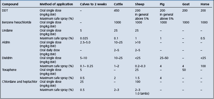
This group of poisons includes DDT, benzene hexachloride (and its pure gamma isomer – lindane), aldrin, dieldrin, chlordane, toxaphene, methoxychlor, DDD, isodrin, endrin, and heptachlor. General toxicity data for the more common compounds are given in Table 32.4. Methoxychlor is less toxic than DDT, and isodrin and endrin are more toxic than aldrin and dieldrin. Camphor (2-bornanone) is chemically similar to toxaphene and is associated with a similar syndrome when fed accidentally.

Table 32.4 Toxic oral doses and maximum concentrations of insecticides1

image

EPIDEMIOLOGY

Occurrence

Poisoning with these compounds has been recorded in all animal species. The chlorinated hydrocarbons have come under so much criticism as environmental contaminants that they are rarely used directly on animals nowadays, so that outbreaks of clinical illness associated with them are much less common than they were.

Source of toxin

Ingestion, inhalation, aspiration, and percutaneous absorption are all possible portals of entry so that contamination of feed and application of sprays and dips can all be associated with poisoning. Organochlorides are closely regulated and banned in some countries but still widely used in agriculture, principally on growing plants to control insect pests, and on stored seed grain to control fungi. If the plants or grain, even milled and by products, e.g. bran, are fed to animals they can be associated with problems of tissue residues; if they are fed in sufficient quantities they can be associated with clinical illness. Many outbreaks are associated with the application to animals of products intended for crops, e.g. endosulfan, and labelled specifically ‘Not For Animal Use’. These insecticides may also contaminate soil and persist there for many years. Rooting animals such as pigs are particularly susceptible to this source of poisoning. These compounds are also sometimes fed accidentally and in large amounts in lieu of feed additives, and are associated with acute poisoning. In feedlotted or shedded animals cases may continue for periods as long as a year because of repeated contamination from the environment. Insect baits, e.g. grasshopper baits containing toxaphene and chlordane, used on pasture and for leaf-eating insects on market gardens can be associated with poisoning in livestock, which may eat large quantities of them. These insecticides, especially heptachlor, are incorporated in the soil before the crop of potatoes or maize is sown to control soil pests. Subsequent grazing of the field will cause contamination of the livestock for several years.

Method of application

Dipping of animals is the most hazardous method of application because entry may occur through all portals. Spraying is safer, percutaneous absorption and inhalation being the only portals of entry. The small particle size of the compound and concentration of animals in confined spaces while spraying, increase the possibility of poisoning. Oily preparations are not used for animal treatment but are used inadvertently and are readily absorbed through the skin.

Formulation used

Concentrations of insecticide in formulations used for spraying barns are much higher than those used for animals. Amongst spray preparations simple solutions are most dangerous followed by emulsions and least of all suspensions of wettable powder. Dusting is safest and is preferred to other methods. Preparations for use on plants are often unstable emulsions, which come out of suspension quickly when they reach the plant. If these preparations are used in animal dips the first few animals through the dip can be heavily contaminated and suffer acute, lethal toxic effects. Although the treatment of pastures to control their insect pests is usually safe to animals grazing, the treated pasture or hay made from it can cause contamination of animal products. This contamination can be avoided by incorporating the insecticide into superphosphate granules (‘prills’) instead of applying it as sprays or dusts. The use of chlorinated hydrocarbons to protect stored seeds provides a hazard to animals if they are fed on the treated seed.

Risk factors

The compounds vary in their ability to pass the skin barrier. Benzene hexachloride, aldrin, dieldrin, and chlordane are readily absorbed. Species susceptibility to skin absorption also varies widely. Very young animals of any species are more susceptible than adults, and lactating and emaciated animals also show increased susceptibility.

When these compounds were first used in dips, skin and foot infections occurred frequently because of contamination of the dip in the absence of a bactericidal agent. Cases of otitis media have occurred for the same reason, but more rarely.

Importance

All of these compounds are capable of causing death due to acute poisoning but these are increasingly rare. Because the compounds are soluble in fat and accumulate in body stores of it they are formidable threats to the meat industry. They are also excreted in significant amounts in milk and enter the human food chain at this point. They are concentrated still further in cream and butter. They also represent a threat to sucklings, but the degree of contamination in fetuses and suckling animals is much less than in their dams.

The principal importance of organochlorine poisoning is the contamination of animal tissues at levels which are not acceptable by modern health standards. These contaminations become the subject of veterinary investigations, and are susceptible to standard techniques of epidemiological examination.

PATHOGENESIS

The mode of action of organochlorides is to induce repetitive discharge of motor and sensory neurons by interference with axonal transmission of nerve impulses. After absorption, cyclodiene insecticides are activated by the mixed function oxidase (MFO) system and any prior chemical or environmental exposures that increase the MFO system may exacerbate the onset of poisoning. The diphenyl aliphatic (DDT) organochlorines affect sodium channels, prolonging sodium influx and inhibiting potassium efflux at the nerve membrane. The cyclodiene organochlorines competitively inhibit the binding of gamma amino butyric acid (GABA) at receptor sites, resulting in loss of GABA inhibition and resultant stimulation of the neuron. In all organochlorine poisonings recovery may occur, but with smaller animals paralysis follows and finally collapse and death ensue.

Most of the substances accumulate in the fat depots, where they are a potential source of danger in that sudden mobilization of the fat may result in liberation of the compound into the bloodstream and the appearance of signs of toxicity.

CLINICAL FINDINGS

The speed of onset of illness after exposure varies from a few minutes to a few hours, depending on the portal of entry and the compound and its formulation, but it is never very long.

The toxic effects produced by the members of this group include complete anorexia, increased excitability and irritability followed by ataxia, muscle tremor, weakness and paralysis and terminal convulsions in severe cases. Salivation and teeth grinding occur in large animals and vomiting in pigs. Variations on this clinical syndrome which is common to all organochlorine intoxications include:

DDT and methoxychlor chronic poisoning may be associated with moderate liver damage

Benzene hexachloride, lindane, chlordane, toxaphene, dieldrin, endrin, aldrin, heptachlor are associated with an exaggerated syndrome including teeth grinding, champing of jaws, dyspnea, tetany, snapping of the eyelids, auricular spasms, opisthotonus, frequent micturition, frenzied movements, walking backwards, climbing walls, violent somersaults, and aimless jumping. Fever of 5–7% above normal may occur, possibly a result of seizure activity. Seizures may persist for 2 or 3 days if the animal does not die.

CLINICAL PATHOLOGY

Blood, hair, and ingesta can be assayed chemically for specific toxins. The removal of a biopsy from the fat pad near the cow’s tail offers a satisfactory means of providing samples for tissue analysis. Organochlorine residues in acutely poisoned animals may reach 4–7 ppm in brain or liver.

NECROPSY FINDINGS

At necropsy there are no specific major lesions in the nervous system but toxic hepatitis and tubular nephritis appear in some cases. For assay specimens of hair, if the portal is percutaneous, and of the ingesta, if oral intake is probable, are appropriate. Tissue levels need to be high to be good indicators of recent intoxication. If possible the specimens should be deep frozen and the suspected compound should be nominated as assay procedures are long and involved.

DIFFERENTIAL DIAGNOSIS

The predominantly nervous syndrome attracts attention to encephalitis, encephalomalacia and toxic and metabolic encephalomyelopathies in all species. Diagnostic confirmation depends on a positive assay on animal tissues.

Differential diagnosis list:

Lead poisoning, confirmed by a positive assay for lead on tissue

Rabies confirmed by histopathological examination

Pseudorabies of cattle in early stages with pruritis and the accompanying frenzy

Polioencephalomalacia confirmed by histopathological examination

Thromboembolic meningoencephalitis in cattle confirmed by the isolation of Histophilus somni

Salt poisoning in pigs identifiable by the characteristic lesions of eosinophilic meningoencephalitis.

TREATMENT

There is no specific primary treatment.

Supportive treatment includes sedation with pentobarbital sodium; repeated doses until signs disappear are preferred, with intravenous injections of glucose and calcium and the administration of a non-oily purgative. Activated charcoal (2 g/kg) given early by stomach tube will bind pesticide in rumen and reduce further absorption. Residual chemical should be removed from the coat, and this may be facilitated by judicious washing with soap and copious water rinse.

Toxic residues.

Treatment to reduce the contamination of tissues is unsuccessful, and in most cases the time required for the contamination to subside naturally is long, of the order of 3–6 months, but varying between specific compounds. For example, cows fed DDT prepartum have required an average of 189 days from parturition for the level in the milk fat to decline to 125 ppm. Contamination in other species and with other chlorinated hydrocarbon compounds also tends to be persistent. After the source of contamination is removed drenching of cows with up to 2 kg of activated charcoal followed by daily incorporation in their feed for 2 weeks, or small amounts of mineral oil by mouth at short intervals, have been recommended for this purpose. Neither of these procedures is really practical in the average farm operation. The common procedure for reducing the level of contamination in animals is to put them in a feedlot without any contact with pasture and feed them on energy intensive rations. Sheep decontaminate much more quickly than cattle and animals on a high plane of nutrition eliminate the toxins more quickly.

CONTROL

Mixed enterprise farms, especially vegetable cropping and cattle farming, are the main sources for contamination incidents. Avoidance of the use of the compounds is recommended.

REVIEW LITERATURE

Raisbeck MF, et al. Organochlorine insecticide problems in livestock. Vet Clin North Am Food Anim Pract. 1989;5(2):391-410.

Ensley SM. Organochlorine Insecticides. In: Plumlee KH, editor. Clinical Veterinary Toxicology. St. Louis: Mosby; 2004:186-188.

ORGANOPHOSPHORUS COMPOUNDS AND CARBAMATES (ORGANOPHOSPHATES)

Synopsis

Etiology Poisoning by accidental exposure or overdosing with any one of the very large number of insecticides in these two groups of organic compounds.

Epidemiology Outbreaks occur due to overdosing, use of oil-based preparations formulated for use on non-animal surfaces, dehydrated animals, drift of spray from orchards, field crops to pasture.

Clinical signs

Acute disease in cattle: salivation, diarrhea, tremor, pupillary constriction, dyspnea, moist rales, ataxia, weakness.

Horses: abdominal pain, diarrhea, dyspnea, ataxia; bilateral laryngeal paralysis.

Delayed neurotoxicity occurs only with some compounds, characterized by incoordination, posterior paralysis, with few or no cholinergic signs. Congenital defects in piglets.

Clinical pathology Marked depression of blood cholinesterase levels.

Necropsy lesions Acute disease: no diagnostic lesions. Delayed neurotoxicity: degenerative lesions in peripheral nerves and spinal cord.

Diagnostic confirmation Depressed cholinesterase levels in blood; organophosphate or carbamate in feed or environment.

Treatment Acute disease: primary treatment is atropine in large doses to effect or atropine plus oxime, remove residual toxin from hair coat and prevent absorption from GI tract with activated charcoal and cathartics.

Control Avoid use in stressed, especially dehydrated, animals. Special constraints with Chlorpyrifos.

ETIOLOGY

Organophosphorus compounds and carbamates act in essentially the same way therapeutically and toxicologically, but bonding of the compound to the esterase enzyme is irreversible in the organophosphorus compounds and spontaneously degradable with the carbamates, rendering the carbamates potentially less dangerous. A large number of compounds are included in the group and those used for the direct treatment of animals have been selected for their low toxicity. A vast amount of information is available on the relative toxicities of the many compounds but it is not possible to provide details here and the information does not lend itself to summarization.

EPIDEMIOLOGY

Occurrence

All animal species are affected. OP compound and carbamate poisoning is a common poisoning encountered by veterinarians in animal agriculture but the losses are still very small.

Source of toxin

Grazing in recently sprayed areas, particularly orchards where the most toxic compounds are frequently used

Spray used on cereal crops and in orchards carried by wind onto pasture fields

Hay or cubes made from plants sprayed with organophosphate compounds

Inadvertent access to granular insecticides intended for crops

Use of old insecticide containers as feeding utensils

Contamination of water supplies

Too high a concentration of the insecticide in a spray

Application to animals of products containing oily bases and designed specifically for spraying on walls or plants.

Risk factors

Host factors

These include susceptible groups such as:

Young animals (but with some compounds adults are more so), stressed, water-deprived, and chilled animals. The increased susceptibility due to restriction of water intake is noted especially after oral treatment to control warble-fly infestations

Pregnant females in that congenital defects occur in their offspring

Brahman and Brahman-cross cattle appear to be more susceptible to some compounds than other cattle

Dorset Down sheep may be especially susceptible

Chlorpyrifos is more toxic for male animals with high blood levels of testosterone and is not recommended for use in bulls over 8 months of age.

Toxin factors

These include:

Formulation used, especially the solvent or vehicle used and droplet size

Method of application, e.g. the toxicity of pour-ons is delayed by 24 hours compared to sprays

Toxicity of some compounds appears to increase with storage.

Importance

The introduction of these compounds into animal therapeutics as treatments for nematode, botfly, sheep nasal botfly, and warble-fly infestations and as insecticidal sprays on plants and soil has increased their importance as possible causes of poisoning, and as causes of pollution of milk, meat, and eggs. They also have a role in the poisoning of the native birdlife. They are now one of the most important causes of poisoning of agricultural animals.

PATHOGENESIS

Organophosphorus compounds are highly toxic and are readily absorbed by ingestion, inhalation and by percutaneous and perconjunctival absorption. Once absorbed, sulfur containing organophosphates (phosphorothioates and phosphorodithioates) are metabolized by MFOs and sulfur is exchanged for oxygen thus increasing toxicity. There are two forms of toxicity, cholinesterase inactivation and an organophosphorus-induced, delayed neurotoxicity.

Cholinesterase inactivation

The inactivation of cholinesterase by these organophosphorus compounds is associated with an increase in acetylcholine in tissues and increased activity of the parasympathetic nervous system and of the postganglionic cholinergic nerves of the sympathetic nervous system. The toxic effects thus reproduce the muscarinic and nicotinic responses of acetylcholine administration. Differences between the toxicities of compounds depend on the stability of this bonding between esterase and compound, and the toxicity of the substance formed by the bonding.

The muscarinic effects of acetylcholine are the visceral responses of the respiratory system and include marked respiratory distress due to a decrease in dynamic lung compliance and arterial oxygen tension and an increase in total pulmonary resistance; there is bronchial constriction and increased mucous secretion by bronchiolar glands. In the alimentary tract there is increased peristalsis and salivation. Effects in other systems include hypotension and bradycardia, pupillary constriction, sweating and abortion.

The nicotinic effects are the skeletal muscle responses of twitching, tremor and tetany, convulsions, opisthotonos, weakness and flaccid paralysis. There is a difference in the relative muscarinic and nicotinic responses between species, the visceral effects being more marked in ruminants and the muscular effects more evident in pigs in which posterior paralysis is the common manifestation.

Organophosphorus-induced delayed neurotoxicity

This form of toxicity is manifested by distal axonopathy commencing 1 or 2 weeks after the poisoning incident. There is a dieback of neurons causing regional flaccid paralysis, especially in long neurons. The pathogenesis of this lesion is the toxic end-product produced by the interaction between some OP compounds and the esterase, a phosphorylated neuropathy toxic esterase. Typical examples of this effect are:

Congenital defects in young carried by poisoned pregnant females

Bilateral laryngeal hemiplegia in horses

Possibly the paralytic ileus is associated with by chlorpyrifos

The most severe effects in this category are associated with industrial organophosphorus compounds and are discussed under that heading.

Haloxon has this neurotoxic effect in that it is associated with only a slight depression in cholinesterase levels, but a neurotoxic response in the form of hindlimb ataxia has been reported in a proportion of treated sheep and pigs. The susceptibility of sheep is determined by their ability to metabolize this class of organophosphorus compound and this is genetically controlled.

CLINICAL FINDINGS

Acute poisoning

Illness may occur within minutes of inhalation or ingestion of solutions of the more toxic compounds and deaths may commence 2–5 minutes later. After cutaneous application of dichlorvos to calves clinical signs appear within 30 minutes, peak at about 90 minutes and disappear in 12–18 hours. With less toxic compounds in solid form signs may not appear for some hours and deaths may be delayed for 12–24 hours.

Cattle, sheep and goats

In acute cholinesterase inactivation cases in ruminants the premonitory signs, and the only signs in mild cases, are salivation, lacrimation, restlessness, nasal discharge, cough, dyspnea, diarrhea, frequent urination, and muscle stiffness with staggering. Grunting dyspnea is the most obvious, often audible from some distance because of the number affected. Additional signs include protrusion of the tongue, constriction of the pupils with resulting impairment of vision, muscle tremor commencing in the head and neck and spreading over the body, bloat, collapse, and death with or without convulsions or severe respiratory distress. In sheep and goats1 the signs are similar and include also abdominal pain. Signs disappear at 12–18 hours.

In delayed neurotoxicity cases signs do not appear for at least 8 and up to 90 days after the poisoning. Signs include posterior incoordination and paralysis. Chlorpyrifos is a specific example of this kind of poisoning. It should neither be applied to adult dairy cattle nor to any mature bulls. The signs include anorexia, depression, recumbency, a distended abdomen, ruminal stasis and diarrhea, and fluid splashing sounds on percussion of the right flank. Severe dehydration develops and may cause death.

Pigs

Cholinesterase inactivation syndrome

Visceral effects except vomiting are less pronounced than in ruminants and salivation, muscle tremor, nystagmus, and recumbency are characteristic. In some instances, the syndrome is an indefinite one with muscle weakness and drowsiness the only apparent signs. Respiratory distress and diarrhea do not occur.

Delayed neurotoxicity syndromes

Outbreaks of posterior paralysis occur 3 weeks after dosing with an organophosphorous anthelmintic; clinical signs vary in severity from knuckling in the hindlimbs to complete flaccid paralysis. The hindlimbs may be dragged behind while the pigs walk on the front legs. Affected pigs are bright and alert and eat well.

Piglets with congenital defects of the nervous system manifested clinically by ataxia and tremor are produced by sows dosed with OP compounds during pregnancy. Teratogenicity may be a characteristic of only some organophosphorus compounds, e.g. trichlorfon is teratogenic, dichlorvos is not.

Horses

Cholinesterase inactivation syndrome

Signs include abdominal pain and grossly increased intestinal sounds, a very fluid diarrhea, muscle tremor, ataxia, circling, weakness, and dyspnea. Increased salivation occurs rarely.

Delayed neurotoxicity syndrome.

Bilateral laryngeal paralysis develops in foals after dosing with an organophosphatic anthelmintic. It is described under laryngeal hemiplegia.

Miscellaneous OP poisoning

Syndromes include:

A significant drop in conception rate when the administration is at the beginning of estrus

The fluid diarrhea which is a transient sign in moderate intoxication in foals may be expanded to a severe gastroenteritis with heavier dose rates

Most organophosphorus compounds are associated with only temporary interference with cholinesterase and are not associated with any permanent effects in recovered animals but with some compounds, especially coumaphos and ronnel, the recovery period may be quite long, up to 3 months in the case of ronnel, because of slow excretion of the compound and the combined compound-esterase complex

Absorption of an organophosphorus compound may also be associated with significant changes in the patient’s cholinesterase status without causing clinical signs

Potentiation of the action of succinylcholine chloride for up to 1 month after the administration of the organophosphorus compound in horses. The administration of the relaxant to a sensitized horse can be followed by persistent apnea and death. This, and a number of other interactions with drugs which may themselves have toxic effects, mean that the manufacturer’s instructions with organophosphorus compounds must be followed explicitly.

CLINICAL PATHOLOGY

The estimation of cholinesterase in body tissues and fluids is the most satisfactory method of diagnosing this poisoning, but it is essential that proper methods and standards of normality be used. Convincing figures are of the order of 50–100% reduction from normal controls. The degree and the duration of the depression of blood cholinesterase levels varies with the dose rate and the toxicity of the compound used. Blood cholinesterase levels are depressed for much longer than the clinical signs, e.g. after dichlorvos poisoning the depression of cholinesterase level in the blood does not reach bottom until 12 hours after application and the return to normal levels takes 7–14 days.2 Similarly, cholinesterase levels in cattle poisoned with terbufos, an agricultural insecticide, do not commence to rise toward normal until 30 days and are not normal for 150 days after the poisoning incident. Unlike organophosphate insecticides, carbamate insecticide cholinesterase inhibitors may spontaneously reverse binding and cholinesterase depression may not be detectable in recently poisoned animals.

Suspected food material can be assayed for its content of organophosphorus compounds but assay of animal tissues or fluids is virtually valueless and may be misleading.

NECROPSY FINDINGS

There are no gross or histological lesions at necropsy in acute cholinesterase inactivation cases, but tissue specimens could be collected for toxicological analysis. Material sent for laboratory analysis for cholinesterase should be refrigerated but not deep frozen.

Distinctive degenerative lesions in peripheral nerves and spinal cord can be seen in delayed neurotoxicity cases, and hypoplasia is visible in the cerebrum, cerebellum, and spinal cord in congenitally affected piglets.

DIFFERENTIAL DIAGNOSIS

Outbreaks of a syndrome of dyspnea, salivation and muscle stiffness and constriction of the pupils after exposure plus a history of exposure and depressed blood levels of cholinesterase suggest intoxication with these organophosphorus compounds but diagnostic confirmation requires positive assay results on suspected toxic materials. In cattle the morbidity and case fatality rates approximate 100% but in pigs the recovery rate is good and all pigs may recover if the intake has been low and access is stopped. With the other poisons listed below death is much more common in pigs and residual defects, including blindness and paralysis occur in a proportion of the survivors.

Differential diagnosis list:

Diseases with a similar clinical profile include:

In cattle:

Sporadic cases of anaphylaxis

Groups of cattle affected by acute bovine pulmonary emphysema and edema (fog fever) may show a sudden onset of dyspnea but pulmonary edema is obvious on auscultation, and salivation and muscle stiffness are absent.

Early stages of nicotine poisoning, with transient muscle tremors and salivation.

In pigs:

Arsenic poisoning

Rotenone poisoning

Salt poisoning

Mercury poisoning

Avitaminosis-A.

TREATMENT

Primary treatment is urgent and critical, especially in cattle because of the usually high case fatality rate. Atropine in large (about double the normal) doses is the rational and approved treatment.3 Recommended doses are 0.25 mg/kg BW in cattle and 1 mg/kg BW in sheep. In very sick animals about one-third of this dose should be given very slowly intravenously in a dilute (2%) solution and the remainder by intramuscular injection. Injections may have to be repeated at 4–5-hourly intervals as signs return, and continued over a period of 24–48 hours. Multiple continued dosing with atropine may not be effective and could result in atropine overdose. Cows that have received very large doses of the poison may not respond to treatment. Atropine does not reverse the nicotinic effects of the OP compound, i.e. the tremor, spasms, and convulsions, but it does block the muscarinic effects. Atropine appears to have low efficacy in sheep. This is not a serious drawback as sheep are much less susceptible than cattle to larger doses of atropine. It should be remembered that reactions in cattle following organophosphorus treatment for warble flies may be due either to the drug or to the damaged grub, and treatment with atropine would be contraindicated in the latter.

Oximes have some efficiency in the treatment of OP compounds, but not all carbamates some of which may have their toxicity enhanced; this is because of their ability to reactivate acetylcholinesterase (ACHE) by dephosphorylation. Also their usefulness as antidotes declines rapidly with the passage of time after the poisoning occurs, and they are of doubtful usefulness after 24 hours. For these reasons oximes are not recommended unless their specific relationship to the particular carbamate is known.4 The oxime trimedoxime bromide is superior to 2-pyridine aldoxime methiodide (2-PAM) and diacetylmonoxime (DAM). Recommended dose rates for 2-pyridine aldoxime methiodide are 50–100 mg/kg BW given intravenously and for trimedoxime bromide 10–20 mg/kg BW. These dose rates can also be used for subcutaneous and intraperitoneal injection. Administration by any route is as a 10% solution in normal saline. In horses 2-pyridine aldoxime methiodide at doses of 20 mg/kg BW has given good results. Treatment may need to be repeated for up to 10 days to counteract slower acting compounds such as coumaphos.

Combined oxime and atropine, e.g. 2,3-butanedione monoxime and atropine, is recommended as superior to either drug alone.5 The value of the treatment is greatest if it is carried out early, before 24 hours after poisoning, and is of little value after 72 hours.6

Removal of residual toxin.

Animals that have been dipped or sprayed should be washed with water to which soap, soda or a detergent is added to remove residual organophosphorus material. When oral intake has occurred activated charcoal will adsorb residual toxin in the gut.

CONTROL

Most outbreaks occur after accidental access to compounds and this cannot always be avoided. Animals to be treated orally with organophosphorous insecticides should be permitted ample fresh drinking water beforehand. Chlorpyrifos is restricted to use in beef cattle and then not in calves less than 12 weeks old nor in bulls over 8 months of age.

REVIEW LITERATURE

Abdelsalam EB. Factors affecting the toxicity of organophosphorus compounds in animals. Vet Bull. 1987;57:441-448.

Barrett DS, et al. A review of organo phosphorus ester-induced delayed neurotoxicity. Vet Human Toxicol. 1985;27:22-37.

Meerdink GL. Organophosphorus and carbamate insecticide poisoning in large animals. Vet Clin North Am Food Anim. Pract. 1989;5(2):375-389.

Meerdink GL. Anticholinesterase Insecticides. In: Plumlee KH, editor. Clinical Veterinary Toxicology. St. Louis: Mosby; 2004:178-180.

REFERENCES

1 Mohamed O, et al. Br Vet J. 1990;146:358.

2 Awal MA. Vet Human Toxicol. 1992;34:229.

3 Bakima M, et al. Res Vet Sci. 1989;13:127.

4 Dawson RM. Vet Rec. 1994:134-687.

5 Raina R, Malik JK. Vet Res Communn. 1991;15:319.

6 Mosha RD, Gyrd-Hansen N. Vet Human Toxicol. 1990;32:6.

ROTENONE

Rotenone has commonly been used in the past to control bovine Hypoderma larvae (cattle grubs). It has a reputation for low mammalian toxicity but relatively high toxicity to aquatic life. The mammalian oral LD50 is 100–300 mg/kg while the LD50 for fish is less than 100 ug/L of water. Oral absorption in mammals is limited but enhanced by fat in the diet. Rotenone is relatively non-toxic but toxic effects appear in pigs fed a ration containing 2.5% rotenone. Pyrethrins are believed to enhance toxicity of rotenone. Ingesta at necropsy may contain as much as 2000 ppm of rotenone. Signs include salivation, muscle tremor, vomiting, ascending paralysis, incoordination, quadriplegia, respiratory depression, coma, and death. Accidental oral exposure may be treated with activated charcoal and an osmotic cathartic for decontamination followed by control of seizures are needed. Phenothiazine tranquilizers are contraindicated in rotenone toxicosis.

REVIEW LITERATURE

Osweiler GD. Toxicology. Philadelphia: Williams & Wilkins, 1996;243-245.

AMITRAZ

This acaricide is widely used in most species but is prohibited in horses; when it is applied to them accidentally it is associated with a syndrome characterized by somnolence, incoordination, depression, reduction of intestinal sounds; impaction of the large intestine may occur within 24–48 hours. The susceptibility of the compound for equids is probably due to its much greater persistence in the body.1 Salivation, depression, anorexia, ataxia, tremors, and coma are signs attributed to amitraz in other species.2 Concentration of the dipping fluid, environmental temperature and the condition of the skin may influence absorption of the compound and the susceptibility of the animal. Residual amitraz should be removed from affected animals by hosing with cold water and the animal should be treated with large volumes of lubri-cant by stomach tube at intervals of 12–24 hours. Oral fluids containing electrolytes should be given by stomach tube to counter dehydration and intravenous fluids may be necessary.

REFERENCES

1 Pass MA, Mogg TD. J Vet Pharmacol Therap. 1995;18:210.

2 Gwaltney-Brant S. Insecticides and Molluscacides. In: Plumlee KH, editor. Clinical Veterinary Toxicology. St. Louis: Mosby; 2004:177-178.

HERBICIDES

Herbicides vary widely in their composition and also in their toxicity.

Arsenicals is associated with arsenic poisoning

Sodium chlorate is also toxic by causing methemoglobinemia

One hazard of the relatively safe organic compounds set out below is their contamination by highly toxic ones as a result of faults in the manufacturing process, e.g. the dioxins which have been found to be significant contaminants of the 2,4,5-T chemical. Today, restrictions on registration and changes in the manufacturing process have drastically reduced contamination by dioxins.

Some herbicides, e.g. glyphosate, make pasture that is sprayed with them more palatable, thus creating their own toxicity hazard. The phenoxy acetic acid herbicides can increase palatability of some plants after spraying as well as induce elevated nitrate concentration in plants for several days after spraying.

Dinitrophenol compounds

Dinitrophenols.

Animals can be poisoned accidentally by inhalation, ingestion or percutaneous absorption of these compounds which have the effect of increasing the basal metabolic rate. Poisoning is manifested by an acute onset of restlessness, sweating, deep rapid respiration, fever, and collapse. In ruminants, but not in non-ruminants, the metabolites of these compounds are associated with intravascular hemolysis, methemoglobinemia and hypoproteinemia. Death may occur 24–48 hours later.

Dinitrophenol (DNP) and Dinitro-orthocresol (DNOC) are the commonest members of this group. In all species, doses of 25–50 mg/kg BW are usually toxic but much smaller doses produce toxicity when environmental temperatures are high. There is no accumulation of the drug within the body. Dinoseb, now rarely used, is a highly toxic DNP.

Hormone weed killers

2,4-D, silvex, MCPA and 2,4,5-T are non-toxic in the concentrations used on crops and pasture but dosing with 300–1000 mg/kg as a single dose is associated with deaths in 50% of cattle. They have also been tentatively linked with the high prevalence of small intestinal carcinomas in sheep. The picolinic acid herbicides picloram and clopyralid have also been suspected of the same relationship.

Barban is toxic at doses of 25 mg/kg, for cattle.

2,4-D at oral doses between 150 and 188 mg/kg BW is fatal to adult cows and at 10 mg/kg BW for sheep. Reversible toxic effects are produced with single doses in calves with doses of 200 mg/kg and in pigs with 100 mg/kg. Repeated administration of 50 mg/kg is toxic to pigs. In adult cows signs include recumbency, ruminal stasis, salivation, and tachycardia. In calves the signs are dysphagia, tympanites, anorexia, and muscular weakness; in pigs additional signs include incoordination, vomiting, and transient diarrhea. Long-term administration to pigs (500 ppm in the diet for 12 months) is associated with moderate degenerative changes in kidney and liver. 2,4-D may cause poisoning indirectly by its effect on the metabolism of weeds and sugar beets, resulting in a significant increase in the nitrate content of the leaves.

A commonly used mixture of 2,4-D, 2,4,5-T and a brushwood killer, monosodium methyl arsenate, is very toxic by mouth or after application to the skin; signs include anorexia, diarrhea, weight loss, and death in most cases.

Repeated dosing of sheep with silvex for about 30 days at 150 mg/kg BW causes death.

Single doses of the herbicides diallylacetamide, carbamate, triazine, and propionanilide at 250 mg/kg BW are fatal to sheep. Repeated small doses of carbamate are associated with marked alopecia. Acute poisoning with any of these compounds is unlikely unless large amounts are ingested accidentally.

Paraquat is associated with fibrosing pneumonitis in pigs but this does not develop in sheep or cattle with fatal doses. Poisoning with it is unlikely to occur unless it is administered accidentally or maliciously. A dose rate of 100 mg/kg BW is uniformly fatal in pigs with signs of vomiting, diarrhea, and dyspnea. Renal damage may also be a part of acute poisoning by paraquat.

Accidental poisoning of sheep due to contamination of pasture by diquat has been associated with widespread illness with signs of diarrhea and a significant mortality. In cattle accidental poisoning with diquat has been associated with fatal abomasitis and enteritis, hepatic and myocardial degeneration and pulmonary emphysema.

The herbicide triallate is associated with severe illness and some deaths with single oral doses of 300 mg/kg BW to sheep and 800 mg/kg to pigs. Salivation, bradycardia, vomiting, muscular weakness, dyspnea, tremor and convulsions are followed by death in 2–3 days. It is also toxic when given in small amounts continuously.

The triazine herbicides atrazine and prometone appear to be non-toxic at usual levels of ingestion. Accidental poisoning of sheep with atrazine is associated with paralysis, exophthalmos, grinding of the teeth, diarrhea, dyspnea, and tachycardia, and of cattle is associated with salivation, tenesmus, stiff gait, weakness. Experimental dosing of heifers with large doses of atrazine is associated with fatal poisoning, but animals treated with activated charcoal survive.

Simazine and aminonitrazole in combination have been associated with death in sheep, and horses allowed access to pasture sprayed with the mixture. In sheep the signs are staggering, inappetence, and depression. In horses colic is the important feature.

Simazine on its own, with continuous access is associated with tremor, tetany and paraplegia, and a prancing gait with the head held against the chest. Death occurs after 2–4 days and mild-to-moderate myocardiopathy at necropsy.1

Triclopyr, a selective postemergence herbicide, is toxic to horses at five times the estimated maximum intake from herbage. It is associated with digestive and respiratory signs, ataxia, stiff gait, sometimes tremor and mild to moderate. renal damage.

Sodium chlorate

Animals seldom ingest sufficient sprayed plant material to produce clinical illness and the principal danger is from accidental dosing or permitting salt-hungry cattle to have access to the chemical. The lethal oral dose is 2–2.5 g/kg BW for sheep, 0.5 g/kg for cattle and 3.5 g/kg for dogs. Irritation of the alimentary tract is associated with diarrhea and deep, black erosions of the abomasal and duodenal mucosae. Hemoglobinuria, anemia and methemoglobinemia result and somnolence and dyspnea are characteristic. At necropsy the blood, muscles, and viscera are very dark. No specific treatment is available. Sodium thiosulfate and methylene blue are used in treatment but have little effect but copious blood transfusions have been recommended.

Delrad

This algicide is used to control the growth of algae on ponds and other water reservoirs. Cattle and sheep are unharmed by the ingestion of water containing 100 ppm of the compound. Dose rates of 250 g/kg BW in adult cattle, 150 mg/kg in calves and 500 mg/kg sheep are associated with toxic effects.

DEFOLIANTS

Substances used to remove the leaves from plants to facilitate harvesting of seed may represent a toxic hazard if the residual stalks are fed to livestock.

Monochloroacetate (SMCA) is commonly used for this purpose and, although it is unlikely to cause poisoning unless very large quantities of the stalks are fed, sheep and cattle which gain access to recently sprayed fields may be seriously affected. Toxic signs in cattle include diarrhea, colic, muscular tremor, stiff gait, ataxia, and dyspnea. Terminally there may be convulsions, hyperexcitability, and aggressiveness. The course is short, most animals dying within a few hours.

An organophosphorus compound, tributyl triphosphorotrithioite used as a defoliant for cotton plants, produces typical signs of organophosphorus poisoning.

Thidiazuron, a cotton defoliant, appears not to be toxic for animals, but may enter the human food chain via goat’s milk and chicken eggs.

REFERENCE

1 Allender WJ, et al. Vet Human Toxicol. 1992;34:442.

FUNGICIDES

Zinc ethylene dithiocarbonate (zineb) may be associated with thyroid hyperplasia and hypofunction, degeneration of myocardium and skeletal muscle, testicular weight reduction, and germ cell depletion1.

Thiram (tetramethyl thiuram sulfide) is a widely used agricultural fungicide which is associated with conjunctivitis, rhinitis and bronchitis on local contact; it is thought to be associated with abortion in ewes on ingestion, and is a known teratogen but no specific poisoning incidents have been recorded in animals.2

REFERENCES

1 Soffietti MG, et al. Schweiz. Arch. Tierheilkd. 1988;130:657.

2 Dalvi RR. Vet Human Toxicol. 1988;32:480.

RODENTICIDES

The commonly used rodenticides in some countries are sodium fluoroacetate and alphanaphthylthiourea (ANTU), but their use is banned in other countries. Anticoagulants, include warfarin ((3-acetonylbenzyl)-4-hydroxycoumarin) and its analogs, as well as newer ‘second generation’ anticoagulants (brodificoum, bromodiolone, chlorphacinone). Zinc phosphide is often used as an alternative to anticoagulant rodenticides. They are all toxic to domestic animals and may cause death when ingested accidentally. ‘Quintox’, a rodenticide containing cholecalciferol (0.75 g/kg), has been associated with hypercalcemia, hyperphosphatemia, and death in dogs, and could cause death in farm animals.

SODIUM FLUOROACETATE

ETIOLOGY

Fluoroacetate occurs naturally in some plants, and in the form of compound 1080 is used as a rodenticide in agriculture. The toxic dose level for domestic animals is 0.3 mg/kg BW, and 0.4 mg/kg is lethal for sheep and cattle. Sublethal doses may be cumulative if given at sufficiently short intervals.

EPIDEMIOLOGY

Fluoroacetate’s use in agriculture poses a hazard for grazing farm animals because it is usually spread out across fields combined with cereals, carrots, or bread as bait and is attractive to ruminants.

PATHOGENESIS

Its mode of action is by inhibition of the enzyme aconitase, thus blocking the intracellular energy cycle. Two actions are manifest: myocardial depression with ventricular fibrillation, and central nervous system stimulation producing convulsions. In sheep the predominant effect with acute poisoning is on myocardium; in dogs it is the nervous system.

CLINICAL FINDINGS

In herbivores generally there is sudden death in acute cases, the animals being found dead without evidence of a struggle; or there are tetanic convulsions and acute heart failure with the animals showing weakness and dyspnea accompanied by cardiac arrhythmia, a weak pulse and electrocardiographic evidence of ventricular fibrillation.

In sheep with subacute poisoning the signs are similar but are not apparent when the animal is at rest. When they are disturbed the nervous signs of tremor and convulsions appear but disappear when the sheep lies down.

Pigs manifest the nervous form of the disease, including hyperexcitability and violent tetanic convulsions. In all cases there is a period of delay of up to 2 hours after ingestion before signs appear.

CLINICAL PATHOLOGY/NECROPSY FINDINGS

There are no specific lesions but the tissues contain elevated levels of citrate.

DIFFERENTIAL DIAGNOSIS

A history of access to toxic material plus appropriate clinical signs and assay of fluoroacetate in ingesta provide good circumstantial evidence and approximate diagnostic confirmation.

Differential diagnosis list:

Many poisonous plants associated with sudden death linked to acute cardiac arrest

Lightning strike or electrocution

Inherited cardiomyopathy

Ionophore poisoning especially warfarin and in horses

Other less common causes of sudden death.

TREATMENT/CONTROL

Care in the disposition of baits and highly dependable retrieval of uneaten baits before allowing livestock access to baited fields preempt most mortalities. No specific treatment is available.

ALPHA NAPHTHYLTHIO UREA (ANTU)

Horses, pigs, calves, and dogs are susceptible as well as rats. Tolerance develops after the ingestion of sublethal doses. Death occurs within 24–48 hours after ingestion due to marked pleural effusion, pulmonary edema, and pericardial effusion. The toxic dose rate is of the order of 20–40 mg/kg BW in a single dose.

WARFARIN AND OTHER ANTICOAGULANT RODENTICIDES

ETIOLOGY

Warfarin is a well known anti-vitamin K anticoagulant rodenticide available since the 1940s and is known as a ‘first generation’ anticoagulant. Related compounds are dicoumarol, coumatetralyl, and pindone.1 Brodifacoum, bromodialone, diphacinone, difethialone, and chlorphacinone are known as ‘second generation’ anticoagulants and may be associated with poisoning from a single dose because of their quite prolonged elimination half lives. Single doses, are less likely to be associated with poisoning but repeated ingestion for some days may do so. Daily doses of warfarin of 0.2–0.5 mg/kg BW are fatal to pigs in 6–12 days. In cattle 200 mg/kg daily for 5 days is associated with 50% mortality. At 0.25 mg/kg for 10 days prothrombin times are depressed 20% and at 0.1–0.3 mg/kg abortions occur. Coumatetralyl was introduced to overcome the problem of resistance to warfarin which developed in some rodent populations. It is more hazardous for domestic animals than the original warfarin because it can be used in a concentrated form by laying it across rodent tracks; the rodents lick it from their paws. Brodifacoum2 acts over a long period and is detectable in liver up to 128 days after intake.

EPIDEMIOLOGY

These products are used by incorporating them into baits and they are in widespread use because they cause no poison shyness. Although most deaths occur because of misuse by farmers, contamination of feedstuffs at the milling plant is not unknown. Calves and poultry are not usually affected, most outbreaks being recorded in pigs, cats, and dogs. Horses are not commonly affected, but warfarin poisoning has been produced experimentally in ponies.

PATHOGENESIS

The poisons exert their effects by interfering with reactivation of vitamin K, thus inhibiting the blood-clotting mechanism by preventing the operation of the thrombin to prothrombin complex. Sudden massive hemorrhage into body cavities or brain may cause immediate death, or death may occur slowly with accompanying lameness due to hemorrhage into subcutaneous tissues. Death due to rupture of a major blood vessel may also occur in 3–4-day-old calves born of cows given a non-fatal dose of warfarin during pregnancy.

CLINICAL FINDINGS

Most cases are of sudden death or found dead. The clinical syndrome includes mucosal pallor, weakness, recumbency, mild dyspnea. Coumatetralyl is associated with more specific signs in pigs, with lameness due to hemorrhage in and swelling of the legs.

CLINICAL PATHOLOGY

There are reduced values for packed cell volume, erythrocyte count, and hemoglobin content. Activated clotting time, prothrombin time, and PIVKA assays are prolonged. Anticoagulants or their metabolites can be detected chemically in blood or urine, or in liver for animals that have died of toxicosis.

NECROPSY LESIONS

Pallor of tissues and massive hemorrhages are characteristic.

DIFFERENTIAL DIAGNOSIS

Assay of feed supply for the toxin, plus severe anemia evident on clinical, clinicopathological or necropsy findings provide diagnostic confirmation. The differential list includes other causes of acute hemorrhagic anemia.

TREATMENT

Primary treatment.

Intravenous injection of a single dose of 50–75 mg/kg BW of vitamin K1 is an effective antidote for a horse. The prothrombin time is returned to normal in 12–24 hours after this treatment and persists at this level for as long as 96 hours. Animals with acute blood loss must be treated for shock and provided plasma or whole blood which will immediately restore clotting factors and provide immediate coagulation factors for clotting.

Red squills

Poisoning by red squills seldom occurs because the material is extremely unpalatable and when eaten is usually vomited. In all species large doses (100–500 mg/kg BW) must be administered to produce toxic effects. Young calves are most susceptible and goats least. Experimental poisoning is associated with convulsions, gastritis and bradycardia.

Zinc phosphide

Zinc phosphide is also unpalatable to domestic animals, and requires an acid stomach to release its toxic phosgene gas. It is not therefore a likely poison for ruminants, but could be a hazard for pigs and horses. Experimental poisoning with doses of about 40 mg/kg BW is associated with death in most species. A general toxemia with depression of appetite, dullness, and some increase in respiratory rate occurs but there are no diagnostic signs. Necropsy lesions include congestion and hemorrhages in all organs, fatty degeneration of the liver and inflammation in the small intestine. Chemical assay is necessary to establish a diagnosis.3

REFERENCES

1 Martin GR, et al. Aust Vet J. 1991;68:241. 69, 176

2 Boermans HJ, et al. Can J Vet Res. 1991;55:21.

3 Guale FG, et al. Vet Human Toxicol. 1994;36:517.

MOLLUSCICIDES

Metaldehyde

Metaldehyde is in common use as a molluscicide. It is usually dispensed in a bran base and is toxic to farm livestock. Outbreaks occur in cattle, horses, and sheep. Lethal dose rates include cattle 0.2 g/kg BW in adults, and less in calves; horses 0.1 g/kg BW.

Clinical signs in ruminants include incoordination, hyperesthesia, muscle tremor, salivation, dyspnea, diarrhea, partial blindness, unconsciousness, cyanosis, and death due to respiratory failure. Hyperthermia (43.58°C or 110.8°F) occurs in sheep. All the signs are exacerbated by excitement or activity. A mortality rate of 3% may be expected.

Signs in horses are similar plus heavy perspiration, hypersalivation, muscle fasciculation, and death in 3–5 hours.

The only effective treatment in cattle is likely to be rumenotomy, supplemented by sedation with a tranquilizer, sedative or muscle relaxant. In horses, mineral oil by stomach tube is recommended to delay further absorption of the metaldehyde.

Methiocarb

This carbamate molluscicide has anticholinesterase and nicotinic and muscarinic activities. Poisoning of sheep is associated with depression, hypersalivation, diarrhea, dyspnea, aimless wandering, and ataxia. Death is due to pulmonary edema. Horses show sweating, dribbling, muscle tremor, hypersalivation, and finally recumbency and death due to pulmonary edema.

The compound is usually in pellet form and dyed blue so that affected animals can be detected by the blue staining of their mouths. Atropine is an effective antidote but may be required to be repeated if the amount of bait taken is large. For a wider discussion of carbamate poisoning see p. 1834.

WOOD PRESERVATIVES

Phenolic compounds

Lumber used in the construction of barns, stables, pens and yards is often treated with phenolic wood preservatives, chiefly pentachlorophenol, dinitro-orthophenols, dinitro-orthocresols (DNOC), dinitrophenol, and coal tar creosote, or mixtures of these, and animals that have access to freshly treated material or the neat preservative may be poisoned. The toxic cresols may be imbibed orally or absorbed percutaneously, and contact with freshly treated wood may be associated with local cutaneous necrosis.

Acute fatal doses in all species are in the range of 120–140 mg/kg BW for pentachlorophenol, and chronic fatal doses range from 30 to 50 mg/kg BW. Fatal doses for coal tar creosote are 4–6 g/kg BW as a single dose, or 0.5 g/kg BW daily. A high mortality may be encountered in newborn pigs and there may be a greater than normal incidence of stillbirths when sows are farrowed in treated crates. Weaned pigs may show depression, skin irritation and occasionally death.

Coal tar sealers for concrete floors may be associated with similar phenolic poisoning.

Creosote applied as a treatment for ringworm, has shown marked toxic effects in cattle.

Besides the known toxic effects of these chemicals there has been interest in possible subclinical intoxications. There is a lack of definitive evidence on the subject, but horses bedded on shavings from pentachlorophenol-treated wood, prepared wrongly by treating the rough lumber and then dressing it instead of applying the preservative to the dressed lumber, have been poisoned by dioxin, a common contaminant in the preservative. Clinical signs include depression of appetite, severe weight loss, ventral and limb edema, hair loss, anemia, a crusty, scaly dermatitis around the eyes, muzzle, the axilla and inguinal region, and on the neck. Exudation through cracks in the skin is a feature of the lesion. Lesions in liver biopsies include necrosis and severe vacuolar changes in the hepatocytes.1

Copper–chrome–arsenate

Softwood preserved against rot by the application of a patented mixture containing copper, chromate, and arsenic, has become very popular for use in yards and buildings used by livestock. The materials have been carefully tested to insure that there is virtually no risk of poisoning. It is recorded that animals would need to eat at least 28 g of the treated wood daily for a month before a chronic poisoning occurred. Horses that have the chewing habit could eat more than that and could theoretically become poisoned. Burning of treated lumber can make arsenic available and toxic to animal. This route of poisoning has occurred in cattle because of their tendency to lick ashes or soil where burning of wood has occurred.

REFERENCE

1 Kerkvliet NI, et al. J Am Vet Med Assoc. 1992;201:296.

SEED DRESSINGS

Many poisoning incidents are caused by livestock gaining access to seed which has been treated in some way. The more common ones are listed below, and each is dealt with under the heading of the toxic agent:

Grain treated with arsenic used to poison birds

Grain treated with highly toxic organophosphorus substances used to make baits for market garden pests

Bran mixed with metaldehyde as a bait for snails

Grain to be used as seed which has been treated with a mercury-based fungistatic agent.

Additional poisonous substances are bird repellants, grain fumigants, and fungistatic agents.

Bird repellants

Baits of corn or wheat are mixed with various substances and spread over areas to protect them from damage by bird droppings or to avoid damage to aircraft. One of these bird repellants, 4-aminopyridine, has caused poisoning in horses. Clinical signs include signs of fright, profuse sweating, severe convulsions, fluttering of the third eyelid, and death 2 hours after the onset of signs and 6–8 hours after ingesting the material.

In cattle signs include anorexia, frequent passage of small amounts of feces, and tenesmus, with some animals also showing tremor, ataxia, erratic behavior, especially walking backwards, and some sudden deaths.

Grain fumigants

Grain treated by the fumigant dibromoethane is associated with mortality in sheep. The principal lesions are pulmonary edema, septal fibrosis, alveolar epithelialization, and pleural effusion. Death occurs 48–120 hours after exposure. Methyl bromide, described under soil fumigants, is also used for stored grain.

Fungistatic agents

Hexachlorobenzene (HCB) is widely known because of its indestructibility and capacity to pass from grain through cattle and into humans. Legislation against chlorinated hydrocarbons being found in the human food chain is very harsh and hexachlorobenzene is a prime target for public health veterinarians. Its specific toxicity is not high, although experimental poisoning in pigs is associated with hepatic injury.

ADDITIVES IN FEEDS

Many antibiotics, fungistatics, vermicides, estrogens, arsenicals, urea, iodinated casein, and copper salts are added to prepared feed mixes to improve food utilization and hasten growth. Many of them are toxic if improperly used. Miscellaneous agents include amprolium, an antithiamine coccidiostat which is associated with polioencephalomalacia in ruminants, and iodinated casein, used experimentally at one time to stimulate milk production in cows but which is associated with cardiac irregularity, dyspnea, restlessness and diarrhea in hot weather. Toxic additives listed elsewhere in this book are arsanilic acid and copper compounds.

ESTROGENIC SUBSTANCES

ETIOLOGY

Poisoning by estrogenic substances occurs in the following circumstances:

Natural substances in plants and as zearalenone in fungi

Dietary supplements to fattening cattle

Overdosage of clinical infertility cases

Pigs fed hexestrol implants in capon necks

Pasture contaminated by manure from cattle treated orally especially or by subcutaneous implants with estrogenic substances and which pass significant amounts in the feces. Ensilage made from the pasture may also be contaminated

Cattle fed on chicken litter from farms where estrogens are used as supplements

Steers implanted with an estrogen at a standard dose rate may respond in an exaggerated manner and show signs of toxicity. Estradiol implants are reputed to be associated with more such problems than zeranol.

EPIDEMIOLOGY

Estrogenic substance administration as a managemental tool is regarded unfavorably in many countries, because of the risk of intoxication occurring in humans eating contaminated meat. Their use is banned in some and strictly controlled in others. The supplementation may be by addition to the feed, but more commonly is by subcutaneous implants.

PATHOGENESIS

Signs and lesions are the direct result of amplification of the pharmacological effects of the substances.

CLINICAL SIGNS

Male estrogenism.

Steers in feedlots include excessive mounting by other steers, sometimes to the point of causing death, head injuries are caused by head-to-head butting, frequent bawling, leading of mobs, pawing the ground to the point of hole-digging. These problems tend to pass off after a short time. Preputial prolapse can be a problem in Bos indicus cattle. Experimental feeding of zeranol to young bulls is associated with retardation of testicular and epididymal development.1

Urethral obstruction

Heavy mortalities have occurred in feeder lambs after the use of implants of estrogens as a result of prolapse of the rectum, vagina, and uterus, together with urethral obstruction by calculi. The calculi consist largely of desquamated epithelial and pus cells which form a nidus for the deposition of mineral, the desquamation probably being stimulated by the estrogen. Also urethral narrowing caused by the estrogen facilitates complete obstruction by the calculi.

Pig estrogenism.

The clinical signs include straining, prolapse of the rectum, incontinence of urine, anuria, and death. At necropsy there is inflammation and necrosis of the rectal wall, enlargement of the kidneys, thickening of the ureters and distension of the bladder, and gross enlargement of the prostate and seminal vesicles. Estrogens such as zearalenone ingested by sows after day 11–13 of the estrus cycle can be associated with retention of corpora lutea and a syndrome of anestrus or pseudopregnancy that typically persists for 45–60 days post estrus. This effect may occur at zearalenone concentrations of 3–10 ppm in the diet. Pregnant sows given zearalenone post breeding may have failure of implantation and early fetal abortion.

Nymphomania in cows.

Larger doses of stilbestrol, usually administered accidentally to cows, may be associated with prolapse of the rectum and vagina and raising of the tail head due to relaxation of the pelvic ligaments. Susceptibility to fracture of the pelvic bones and dislocation of the hip are common sequels. Nymphomaniac behavior in such animals invites other skeletal injury, especially fracture of the wing of the ilium.

Idiopathic female estrogenism.

Besides the toxic effects associated with estrogens in specific plants, increased estrogenic activity is also encountered in mixed pasture, often only at certain times and on particular fields. Clinically the effects are those of sterility, some abortions, swelling of the udder and vulva in pregnant animals and in virgin heifers, and endometritis with a slimy, purulent vaginal discharge in some animals. Estrus cycles are irregular. In milking cows there is depression of the milk yield, reduction in appetite, and an increase in the cell count of the milk.

CLINICAL PATHOLOGY

High blood levels of estrogens are characteristic. In swine, the syndrome of anestrus associated with zearalenone, will be accompanied by elevated progesterone concentrations due the retention of corpora lutea.

NECROPSY FINDINGS

Enlargement and vascular engorgement of accessory sex organs, especially in neutered animals, are characteristic. Uterine enlargement and keratinization of vaginal epithelium may be detected, and in mature female swine there may be persistent multiple retained corpora lutea.

DIFFERENTIAL DIAGNOSIS

The clinical profile is almost diagnostic, a search of the environment usually reveals a source of estrogen and estrogen assay on blood provides diagnostic confirmation.

Differential diagnosis list:

Acute, especially traumatic, vaginitis

Urethal obstruction in males

Cantharidin poisoning.

UREA

ETIOLOGY

Urea is a common form of non-protein nitrogen (NPN) used in ruminant rations and as a fertilizer. Accidental access to the powder or liquid form of the compound can cause heavy mortalities. Poisoning occurs when cattle or sheep accidentally gain access to large quantities of urea, or are fed large quantities when they are unaccustomed to it, or when feeds are improperly mixed, or the water supply is polluted. Feed grade urea contains approximately 45% nitrogen and protein is approximately 16% nitrogen, and each gram of urea is equivalent to 2.81 g of protein. Thus, a ration containing 1% urea supplies the protein equivalent of 2.81% natural protein. Some care is required in bringing the animals onto urea gradually and an adequate proportion of carbohydrate must be included in the ration.

EPIDEMIOLOGY

Urea is used in agriculture as a feed additive for ruminants to provide a cheap protein substitute in the diet, and as a fertilizer on crop and pasture fields. Protein production from urea is dependent on rumen microorganisms assimilating the ammonia released from urea and converting it to bacterial protein useful to the animal. Natural urease in the rumen supports the hydrolysis of urea to release ammonia. The degree of toxicity of urea depends on the rapidity with which ammonia is released from the urea in the rumen, and this may be increased if soybean meal is being fed; soybeans contain urease which facilitates the breakdown of urea to ammonia. Ruminants are better able to assimilate ammonia into protein when adequate amounts of readily available carbohydrates are provided. This is usually from grain or a sugar source such as molasses. In the absence of sufficient digestible carbohydrate, as when only roughages are fed, urea is more toxic. At one time mixtures of molasses and urea were popular as feed supplements for cattle and are associated with outbreaks of poisoning with signs similar to those of urea poisoning and those associated with feeding ammoniated hay.

Toxic dose levels

In cattle which have been starved beforehand dose levels up to 0.33 g/kg BW is associated with increases in blood levels of ammonia and dose levels of 0.44 g/kg BW produce signs of poisoning within 10 minutes of dosing and dose rates of 1–1.5 g/kg BW are associated with death.

Tolerance to urea

Animals unaccustomed to urea may show clinical illness when fed 20 g/50 kg BW, but by gradually increasing the quantity fed this amount can be tolerated. This tolerance is lost rapidly and animals which receive no urea for 3 days are again susceptible. Tolerance is also reduced by starvation, by lack of readily available dietary carbohydrate and by a low protein diet. SHEEP can eat 6% of their total ration as urea provided it is well mixed with roughage and fed throughout the day, preferably by spraying the urea mixed with molasses onto the roughage. Much more urea is tolerated if given to sheep in molasses (18 g), than if given as a drench (8 g) and prior feeding on lucerne further increases the tolerance and fasting for 24 hours reduces it. A dose rate of 1 g/kg BW to sheep appears to be non-toxic, but 2 g/kg is quickly fatal. In general, urea should not constitute more than 3% of the concentrate ration of ruminants.

Horses appear to be tolerant to relatively large doses of urea but the disease has been produced experimentally in ponies by administering 450 g by stomach tube. The clinical picture is similar to that in cattle, being largely related to the central nervous system. There is a sharp increase in blood ammonia levels after ingestion of the urea, and it is assumed that hydrolysis of the urea occurs in the cecum.

Pigs are quite unaffected by very large doses of urea unless they are deprived of water or have developed a cecal flora which produces urease.

PATHOGENESIS

The toxic effects are due to the sudden production of large quantities of ammonia and its rapid absorption from the rumen results in the onset of signs in 10–30 minutes after feeding. The severity of signs is related to blood ammonia levels and not to levels of ammonia in the rumen. However, the alkaline conditions in the rumen created by rapid release of ammonia can be associated with more of the ammonia to be present in a non-ionized form which favors rapid absorption from the rumen with resultant increase in blood ammonia concentration. Excess blood ammonia is toxic to intermediary metabolism and results in systemic lactic acidosis and elevated blood potassium, which can lead to hyperkalemic heart failure.

CLINICAL SIGNS

Cattle and sheep.

Signs of toxicity commence as early as 10 minutes after the urea is eaten and include severe abdominal pain, frothing at the mouth and nose, hypersensitivity to sound and movement to the point of being aggressive, muscle tremor, incoordination, weakness, dyspnea, bloat, and violent struggling and bellowing. In severe cases the course is short and death occurs sometimes in a few minutes but usually in about 4 hours after ingestion. Less severe cases are drowsy and recumbent. The case fatality rate in affected animals is high.

CLINICAL PATHOLOGY

Cattle.

Signs are visible when rumen ingesta levels of ammonia are 1000 mg/L, serum levels of ammonia nitrogen (NH3-N) are 10–13 mmol/L (10–18 mg/L)2 and when blood ammonia nitrogen concentrations reach 0.7–0.8 mg/dL.

Sheep deaths occur at levels of ammonia nitrogen of 33 μg/mL of blood; the ruminal contents are alkaline when tested with litmus paper (pH elevated from 6.94 to 7.90) and ruminal ammonia levels rise from 6 to 50 mg/dL.

NECROPSY FINDINGS

There are no characteristic lesions at necropsy, but most cases show generalized congestion, hemorrhages, and pulmonary edema. Death is thought to result from respiratory arrest due to ammonia intoxication.

Pigs.

Encephalomalacia has been produced by feeding a ration containing 15% urea. The clinical picture and histopathological findings were similar to those of salt poisoning except that no eosinophilic aggregations are present in the cerebral lesions.

DIFFERENTIAL DIAGNOSIS

Outbreaks of this poisoning are usually closely linked to known exposure to urea and achieve diagnostic confirmation by assay of high blood levels of ammonia.

Without the historical link to a source of urea the differential list is very long because of the similarity of the syndrome to other diseases in which nervous excitation is accompanied by muscle tremor, dyspnea, convulsions, and a high case fatality rate.

Differential diagnosis list:

Acute hepatic insufficiency

Anaphylaxis

Poisoning by Clavibacter toxicus

Acute poisoning by cyanobacteria

Hypomagnesemia

Acute salt poisoning

Acute bovine pulmonary emphysema and edema

Acute organochlorine insecticide poisoning

Acute 4-Methylimidazole poisoning from ammoniated forages (bovine bonkers syndrome)

Any of the many forms of encephalitis or encephalomalacia.

TREATMENT

No primary treatment is likely to be effective but the oral administration of a weak acid such as vinegar (0.5–1 L to a sheep, 4 L to a cow), or 5% acetic acid is recommended. Cold water (10–30 L for adult cattle) will dilute excess urea and temporarily lower rumen pH. This may reduce the amount of ammonia absorbed3 but it must be administered as soon as the first clinical signs appear and repeated dosings may be necessary as clinical signs tend to recur about 30 minutes after treatment. The only really effective treatment is prompt and efficient emptying of the rumen, either via a large bore stomach tube or by rumenotomy, but the results are indifferent because the damage has usually been done already.

CONTROL

Urea is highly toxic and care is essential when handling it in the vicinity of animals. Feed manufacturers’ recommendations about maximum concentration of urea in prepared rations and acclimatization to the diet with inclusion of adequate readily available carbohydrates should be adhered to.

Propylene glycol poisoning

Propylene glycol is an unlikely poison but it is used extensively as an oral treatment for acetonemia in cattle and can be associated with poisoning if it is accidentally administered to horses, usually in mistake for mineral oil. Dose rates of 3 L to horses of 500 kg BW by stomach tube is associated with an immediate but short duration episode of abdominal pain, sweating, salivation, severe ataxia and depression, and a fetid odor of the feces.4 Much larger doses (8 L) can be fatal. Moderate-to-severe inflammation of the lining of the gut and edema of the brain are noticeable at necropsy examination.

Dried poultry wastes

Feeding dried poultry wastes to ruminants provides them with a source of nitrogen, and gets rid of the chicken farmer’s disposal problem. Deleterious effects include:

Copper poisoning when the chickens are fed on diets supplemented with copper

Estrogen poisoning when the chickens are fed on estrogen-supplemented diets

An unidentified problem arises of hepatic necrosis, hypoalbuminemia, and ascites in lambs fed large amounts of poultry waste from hen batteries

Litter from broiler houses is associated with renal damage but not to the point of causing mortality

Botulism.

Brewer’s residues

Diseases associated with the feeding of by-products of brewing and distilling include:

Carbohydrate engorgement in cattle fed wet brewer’s grains

Possibly spinal cord degeneration in adult cattle fed sorghum beer residues contaminated by Aspergillus flavus and containing aflatoxin

Excess sulfur (>0.45% in the diet) from some methods of processing which can lead to polioencephalomalacia.

Chemically treated natural feeds

Formalin-treated grain

This is a special diet fed to dairy cows to produce dairy products containing an increased proportion of polyunsaturated fats for special human diets. Fats in the grain are protected against hydrogenation in the rumen by coating the grains with formalin. If the formalin and the grain are not properly mixed, the free formalin left as a residue is associated with rumenitis and severe diarrhea.

Caustic-treated grain

Grain treated with caustic to improve its digestibility is recorded as causing focal interstitial nephritis, rumenitis, and abomasal ulceration in feedlot steers. The lesions have been produced experimentally. They may not be detected until the animals are slaughtered.

Ammoniated forage

Anhydrous ammonia is added to hay to improve its digestibility and nitrogen content. Environmental risk factors enhancing the production are low dry matter content of the feed, high environmental temperature, and high concentrations of ammonia in the treatment mixture. If the forage is high quality and has a high carbohydrate content it may undergo chemical change, possibly with the formation of a substituted imidazole, 4-methylimidazole (MeI) which is associated with hysteria (bovine bonkers) in the cattle eating it. Calves sucking cows fed ammoniated hay may also be affected by this same syndrome. Experimental feeding with MeI produces the same syndrome but it is not the sole cause; other substances are also involved.5

Clinical signs include hyperexcitability, hyperesthesia, restlessness, rapid blinking, pupillary dilatation, ear-flicking, frequent urination and defecation, dyspnea, frothing at the mouth, bellowing, charging, circling, and convulsions. Tremor, commencing at the head and opisthotonos are obvious early signs. Between convulsions affected sheep walk in circles and have a stiff gait. Nursing calves may show signs even though their dams are unaffected. No clinicopathological abnormalities occur, blood ammonia levels are normal, and no specific necropsy lesions have been identified.

Treatment consists of sedation but many patients do not respond to agents such as acepromazine. Dilution of toxic forage with normal feed is not recommended because the toxin may be cumulative. The maximum rate of ammoniation to avoid toxicity, for poor forage is 3% and 1% for high-moisture forage.6

Newsprint

Newsprint is fed commercially to ruminants as an alternative roughage. Toxicological hazards of the material in sheep fed colored magazines for 6 months and comprising 23% of their ration included a significant deposition of lead in tissues and an increase in enzyme activity in liver, but there were no clinical signs and no histopathological lesions. Feeding for periods of several weeks has no detectable clinicopathological effects and there is evidence that the known toxins are not secreted in cows’ milk.7

Sewage sludge

Urban sewage sludge is used as top-dressing for pasture and may be associated with the spread of infectious disease as well as goiter. Sewage sludge may also be fed directly to animals, but may lead to dissemination of lead, cadmium, and polybrominated and polychlorinated biphenyls to animals and the food products derived from them. Potential damage due to illness or contamination of animal-produced feed can be minimized by leaving treated pasture exposed to weather for a period of several weeks before allowing animals access to it.

Synopsis

Etiology Carboxylic ionophores used commercially as coccidiostats and growth promotants are associated with poisoning if dose rate is excessive; horses are very susceptible.

Epidemiology Mixing errors; left-over cow feed fed to horses.

Pathogenesis Muscle damage leading to acute heart failure or limb weakness and paralysis.

Clinical signs Sudden death, paralysis, red urine or a less acute syndrome of skeletal muscle weakness, paresis, and paralysis.

Clinical pathology Myoglobinuria, elevated levels of CPK. Ionophore found in ingesta.

Necropsy lesions Myocardopathy plus skeletal myopathy. Lesions may only be apparent by microscopic examination

Diagnostic confirmation Assay of stomach contents and representative feed samples for the ionophore.

Treatment No primary treatment recommended. Supportive treatment is oral charcoal or mineral oil.

IONOPHORE POISONING

ETIOLOGY

Monensin, lasalocid, maduramicin, narasin, and salinomycin are carboxylic ionophores used as polyether antibiotics for the control of coccidiosis in poultry and with a secondary role as growth promotants in ruminants. Monensin has minor additional uses in the treatment of acetonemia, lactic acidosis, bloat and atypical interstitial pneumonia. Used properly the compounds are effective in both roles but the margin of safety is small and careless use has been associated with major losses. All of the compounds are cationic agents, causing altered balance of cations (especially Ca2+, Na+ and K+) in cells and organelles. Pharmacological effects of ionophores are similar, but they differ chemically and have differing toxicities.

The recommended doses of monensin vary depending on the age and size of the livestock and the purpose for which it is administered and the manufacturer’s recommendations should be adhered to strictly. Approximate recommended levels, orally and usually in the feed, are: cattle, 50–200 mg per head per day, 16.5–33 ppm; sheep 5–10 ppm in feed.

For monensin, dosage levels at which clinical signs of poisoning can be expected to occur are: cattle 10, sheep 4, pig 7.5, and horse 1 mg/kg BW. Deaths in cattle are likely to commence at intakes of 10 mg/kg BW and in horses at 2–3 mg/kg. Toxic feed concentrations for pigs are 200–220 mg/kg of feed. Comparative LD50 in mg/kg BW are: cattle 26.412 horse 2–3, sheep 12, pig 16–50, and goat 24. It is common for cattle to be poisoned with more than 10 times the recommended dose. The LD50 for salinomycin for the horse is 0.6 mg/kg BW. The effects of the compounds are cumulative and may not be observed for some weeks after administration is discontinued.

EPIDEMIOLOGY

Occurrence

All animal species are affected, including zoo ruminants.8 Poisoning incidents are most often reported in countries where animal husbandry is intensive and high levels of stall feeding are practiced.

Source of toxin

The poisonous properties of the agent are well known and poisoning is usually accidental due to failure to dilute a concentrate, poor mixing or because of wrong identification of containers. Also some liquid preparations settle out and need to be constantly mixed before and during mixing with a batch of feed. Cattle carrying reticular retention boluses of monensin to control bloat by delivering accurate daily doses of the drug, may die suddenly of acute heart failure due to myocardiopathy, a condition likely to be associated with the monensin but by an unknown mechanism.9 The compound’s main use as a coccidiostat is in poultry, and deaths from congestive heart failure have occurred in cattle and sheep fed dried poultry litter from farms feeding salinomycin or maduramicin.10

Risk factors

These compounds are specifically prohibited for use in horses at any time because of their toxicity for that species. Concurrent administration of monensin or salinomycin and tiamulin or chloramphenicol or triacetyloleandomycin at safe doses can be associated with ionophore poisoning in pigs. Outbreaks may occur when tiamulin is introduced into the pigs’ drug regimen to control swine dysentery when the pigs’ ration already includes the ionophore as a growth promotant, or when the two drugs are combined as a coccidia prevention package. The dose and time relationships are complicated and are dealt with separately. The concurrent administration of monensin and selenium to lambs also enhances the toxicity of the selenium.11

PATHOGENESIS

The principal pathogenesis of monensin poisoning is damage to muscles. The origin of muscle damage appears due to loss of ion control and balance in cells and mitochondria leading to impaired intermediary metabolism.12 In cattle, the cardiac and skeletal muscles are affected about equally; in sheep and pigs the skeletal muscle is most seriously affected; in horses the myocardium is the focus of the damage. In the latter case acute or congestive heart failure may result, signs often being delayed for weeks until additional stress, such as late pregnancy or parturition, precipitates cardiac insufficiency. When the damage is principally to skeletal muscle the syndrome is one of weakness, ataxia, and recumbency; myoglobinuria is a common accompaniment.

Besides these major involvements of monensin and lasolocid there are a number of less well-known ones. There is a risk that cattle fed on a nitrogen-rich diet will be likely to suffer an outbreak of nitrite poisoning if they are also fed monensin. Another undesirable outcome may be a fall in butterfat because of shift from acetate to propionate production in the rumen. Continuous feeding of monensin to male pigs (50 ppm for 52 days) reduces blood levels of testosterone, and is associated with dystrophy of seminiferous tubules and reduced sperm counts.

CLINICAL SIGNS

In cattle signs commence within 24 hours with heavy doses but may be delayed for up to 5 days when the intake is lower. Signs commence with feed refusal followed by diarrhea, tremor, weakness, tachycardia, and ruminal atony, and animals may die at this stage from acute heart failure. Those that survive for a day or two develop congestive heart failure manifested by brisket edema, engorgement of the jugular veins, ascites, fluid feces, dyspnea, and tachycardia. Deaths may occur months after the major outbreak, usually due to the exertion of calving. When smaller doses are taken over a long period the syndrome is one of congestive heart failure in all of the cases.13

In sheep the syndrome may be acute followed by hyperesthesia, tremor, especially of the head, disappearance of the pupillary light reflex, recumbency, and convulsions with death occurring during a convulsion.14 More commonly the disease commences with feed refusal, diarrhea, rumen stasis and depression, followed by muscle weakness, a stilled gait, and recumbency. Chronic cases show atrophy of the muscles of the hindquarters and a stiff gait.

In pigs monensin poisoning is associated with dyspnea, anorexia, ataxia, paresis, myoglobinuria and cyanosis, diarrhea, tympany, and pruritus. Death follows in about 6 hours. Salinomycin is associated with the same general syndrome but includes also unwillingness to stand and, when forced to stand, tremor, especially in the hindlimbs, swaying, fetlock knuckling, and abrupt lying down. Exercise exacerbates the signs. Respiratory distress is in the form of irregular breathing. Feed intake is down 50% and sham drinking is characteristic. Red urine is evident for up to 5 days after the drug is discontinued.15

In horses cardiac and skeletal muscle are affected. In most cases it is the myocardium that creates the prominent syndrome. Horses affected by the cardiac disease may be found dead. In others there is restlessness, respiratory distress, diarrhea, mucosal congestion, profuse sweating, sometimes myoglobinuria, cardiac irregularity, and tachycardia (50–60/min). The course of the cardiac disease is short and affected horses may not show much clinical evidence of heart failure at the time of the poisoning but survivors, and survivors of an acute attack, may develop a poor performance syndrome or congestive heart failure up to several months later.16

Horses with skeletal muscle involvement have a syndrome including anorexia, fever, and red or dark urine, due to the extensive muscle breakdown, frequent lying down, difficulty in rising, stiff gait, especially the hindlimbs, and with knuckling of the fetlocks, followed by final recumbency after a period of as long as several months. Colic is also recorded during the acute phase but this may be the restlessness and frequent lying down and getting up of acute myositis. Euthanasia is the common outcome of these cases because of irreparable muscle damage.

CLINICAL PATHOLOGY

In all species tests show increases in serum levels of creatinine phosphokinase, lactate dehydrogenase and aspartate aminotransferase; myoglobinuria is frequent and often prolonged. However, results of clinical pathology tests may vary substantially among individual affected animals. Samples of feed and stomach contents, obtained by stomach tube, are desirable specimens for analysis. Some feeds fed to horses have contained as much as 125–250 g/tonne of feed and their stomach contents have contained 50–100 ppm.

NECROPSY FINDINGS

In cattle there is myocardiopathy, pulmonary edema, and enlargement of the liver and heart, hydropericardium, hydrothorax, and ascites. Gross lesions at necropsy are not always evident, and multiple samples of susceptible tissues should be collected and preserved in formalin for microscopic evaluation.

In sheep postmortem changes include necrosis in both skeletal and cardiac muscles but there are no lesions suggestive of heart failure. Lambs less than 1 month old show only gastrointestinal hemorrhage.

In horses necropsy lesions in acute cases include acute myocardial necrosis, pulmonary congestion, hepatic swelling, and in some cases pulmonary petechiation. Skeletal muscle necrosis may also be evident. Myoglobinuric nephrosis and myoglobinuria are secondary lesions. Chronic cases show marked cardiac myopathy, and possibly skeletal myopathy with obvious fibrosis.

Stomach contents are an obligatory sample in cases likely to go to litigation.

DIFFERENTIAL DIAGNOSIS

In all species syndromes of acute or congestive heart failure due to acute cardiac myopathy or limb paresis or recumbency due to skeletal myopathy are sufficiently common to necessitate a positive assay for one of the ionophores in the feed or stomach contents for diagnostic confirmation.

Differential diagnosis list:

Cattle:

Nutritional deficiency of vitamin E or selenium

Poisonings with such plants as Karwinskia humboldtiana, Cassia occidentalis and Vicia villosa.

In pigs additional diagnostic alternatives include:

Gossypol poisoning

Porcine stress syndrome.

In horses there are clinical similarities to:

Colic

Laminitis

Rhabdomyolysis.

TREATMENT

There is no effective primary treatment and only supportive procedures are recommended. Selenium and vitamin E are ineffective after signs begin, although selenium or vitamin E given prior to appearance of clinical signs may be beneficial. Activated charcoal or mineral oil has been standard treatments aimed at removing the residue of the poison from the alimentary tract. They are of no value if the poison has already been absorbed; recovery is unlikely once the myocardium is affected.

CONTROL

Horses are very susceptible to poisoning by these antibiotics and great care is needed to insure cattle rations containing them are not left around for horses to eat. Pretreatment with vitamin E–selenium reduces the effects of monensin poisoning without completely preventing it.

MONENSIN-TIAMULIN OR SALINOMYCIN-TIAMULIN POISONING IN PIGS

The myotoxicity of monensin for pigs is enhanced by the simultaneous administration of the two antibiotics (monensin or salinomycin and tiamulin). All three of the substances are used as coccidiostats and it is not unusual for farmers to combine them. However, all of the agents use the same detoxication pathways in the liver with tiamulin having the priority. Tiamulin inhibits mitochondrial CYP3A enzymes that stimuate monensin metabolism, allowing accumulation of monensin.17 Monensin (or salinomycin) accumulates to the point of being toxic. The clinical syndrome consists of anorexia and weight loss and at necropsy there are lesions of myonecrosis in the tongue, diaphragm, and limbs. A similar toxicological situation arises in pigs with simultaneous dosing with tiamulin and salinomycin in which the toxic interaction is dose-related.

When tiamulin is used at therapeutic levels, in feed, water, or by injection and salinomycin is used at 60 ppm, toxic reactions and some deaths occur. The interaction does not occur when the two antibiotics are used concurrently and the tiamulin is used at the recommended prophylactic (30–40 ppm) or growth promotant (11 ppm) levels, and independently of whether the administration is oral or by injection,18 but only if a gap of 72 hours has elapsed between the last exposure to salinomycin and the first exposure to therapeutic levels of tiamulin and vice versa.

Pluronics poisoning

These substances are administered to adult cattle in their feed as prevention against bloat. They are unpalatable and unlikely to be consumed in dangerous amounts unless they are well masked in feed. When they are fed accidentally to calves in their milk they are associated with dyspnea, ruminal tympany, bellowing, protrusion of the tongue, nystagmus, opisthotonos, recumbency, and convulsions. Death after 24 hours is the usual outcome.

Carbadox poisoning

Carbadox, Mecadox or Fortigro – methyl 3-(2-quinoxalinylmethylene)carbazate-N1 – is used in pig feeds as a growth promotant and in the treatment of swine dysentery and other enteric diseases at the recommended rate of 50 mg/kg of feed/head per day. Toxic effects occur at rates of 150 mg/kg. A related compound olaquindox is similarly toxic. Affected pigs refuse the ration, but will eat other rations, are gaunt and emaciated, pass hard fecal pellets, and have a long rough coat, pale skin, severe tachycardia, weak hindquarters, and a swaying walk, followed by knuckling of the hind fetlocks, posterior paralysis, and death in 8–9 days. In the early stages the pigs screech frequently. Sows are agalactic, and produce stillborn or weak, undersized piglets.

Necropsy lesions are diagnostic, with extensive damage to the zona glomerulosa of the adrenal gland accompanied by renal tubular necrosis. The resulting hypoaldosteronism is manifested by low serum sodium levels, elevated serum potassium (8 mmol/L) and elevated blood urea nitrogen levels. The condition is irreversible and the outcome is severe disability or death.19

Bronopol poisoning

Bronopol – 2 bromo-2-nitropropoane-1, 3-diol – is used as a laboratory preservative for milk, e.g. in milk samples used for butterfat estimation. This milk is usually fed to calves or pigs and may be toxic on occasional feedings. Affected calves salivate, are depressed, collapse and die within 24 hours of feeding. Necropsy lesions include severe necrotizing abomasitis and local peritonitis on the serosal surface of the abomasum.20

REVIEW LITERATURE

Fries GF. Ingestion of sludge-applied organic chemicals by animals. Science of the Total Environment. 1996;185:93.

Haliburton JC, Morgan SE. Nonprotein nitrogen-induced ammonia toxicosis and ammoniated feed toxicity syndrome. Vet Clin North Am Food Anim Pract. 1989;5(2):237-249.

Longstaffe JA. Pruritus, pyrexia and hemorrhage, syndrome in cattle. Vet Ann. 1989;29:64-68.

Matsuoka T, et al. Review of monensin toxicosis in horses. J Equ Vet Sci. 1996;16:8.

Novilla MN. The veterinary importance of the toxic syndrome induced by ionophores. Vet Human Toxicol. 1992;34:66-70.

Whitechair CK. Urea (NH3) toxicosis in cattle. Bovine Pract. 1989;24:67-73.

REFERENCES

1 Veermachinini DNR, et al. Fund Appl Toxicol. 1988;10:73.

2 Nielsen TK, Wolstrup C. Dansk Veterinaertidsk.. 1988;71:529.

3 Mulei CM, Munyua SJM. Bull. Anim Prod. Africa. 1988;36:279.

4 Dorman DC, Hascheck WM. J Am Vet Med Assoc. 1991;198:1643.

5 Sivertsen T, et al. Acta Vet Scand. 1993;34:227.

6 Brazil TJ, et al. Can Vet J. 1994;35:45.

7 Shane BS, et al. J. Agri. Food Chem.. 1993;41:240.

8 Miller RE, et al. J Am Vet Med Assoc. 1990;196:131.

9 Mathieson AO, et al. Vet Rec. 1989;125:656.

10 Shlosberg A, et al. Vet Res Commun. 1992;16:45.

11 Smyth JBA, et al. J. Comp Pathol. 1990;102:443.

12 Hall JO. Ionophores. In: Plumlee KH, editor. Clinical Veterinary Toxicology. St. Louis: Mosby; 2004:121-127.

13 Bastianello SS, et al. J South Afr Vet Assoc. 1996;67:38.

14 Syngh BH. Vet Rec. 1989;124:15.

15 Miskimins DW, Neiger RD. J Vet Diagnog Inv. 1996;8:396.

16 Peek SF, et al. J Vet Int Med. 2004;18:761.

17 Kamashi KL, et al. Indian J Physiol Pharmacol. 2004;48:89.

18 Pott JM. Vet Rec. 1990;127:554.

19 Power SB, et al. Vet Rec. 1989;124:367.

20 Hindmarsh M. Aust Vet J. 1990;67:309.

MISCELLANEOUS FARM CHEMICALS

Polybrominated biphenyls

Mixtures of polybrominated biphenyls find a great deal of use in industry, particularly as flame retardants. They are not especially poisonous, nor are they a greater risk to farm animals because of degree of exposure, than many other industrial chemicals, but they happen to have found their way into the cattle food chain in much discussed incidents in the USA.

Cattle.

Experimental dosing with 67 mg/kg BW daily for long periods is associated with poisoning but levels of 10 mg/kg BW are not toxic. Clinical signs of illness are anorexia, diarrhea, lacrimation, salivation, emaciation, dehydration, depression, and abortion. Similar signs plus extensive cutaneous hyperkeratosis occur in natural cases. Necropsy lesions include mucoid enteritis, degenerative renal lesions in natural and experimental cases, hyperkeratosis in the glands, and epithelium of the eyelids.

Pigs.

Experimental poisoning in pigs causes no ill-effects in sows, but high concentrations of polybrominated biphenyls (PBB) develop in the sow’s milk with death of some nursing pigs resulting.

Most of the losses due to these compounds are due to destruction of animals because they are contaminated and there is fear of adverse effects on humans who consume them or their products. However, neither animals nor humans exposed to the biphenyls showed any signs of illness. PBBs pass the placenta and are found in fetuses but appear to be associated with no health problems in the offspring.

The excretion of these compounds occurs principally in feces and urine but as much as 25% of ingested substance may be present in the milk. Also these compounds are lipotrophic and accumulate in fat depots, especially in the liver. Attempts to hasten excretion have not produced a satisfactory method. Grazing wool sheep on contaminated ground may be an option for utilization of contaminated land.1

REVIEW LITERATURE

Lemesh RA. Polybrominated biphenyls. An overview of metabolic, toxicological and health con sequences. Vet Human Toxicol. 1992;34:256-260.

REFERENCE

1 Gill IJ, et al. Aust Vet J. 1992;69:155.

Polychlorinated biphenyls

These substances have a number of industrial uses and are common environmental contaminants. They are lipophilic so that they accumulate in body fat, and have low rates of biotransformation and excretion so that they persist in animal tissues for long periods. Although deleterious effects of the compounds in animal tissues are not often recorded, their presence in animal tissues is likely to cause rejection of meat from the human food chain. Recorded damage refers to unidentified reproductive inefficiency and reduction in efficiency of food conversion and possibly hepatic hypertrophy and gastric erosion, but in the same species a positive growth stimulating effect has also been recorded. Experimental poisoning of gnotobiotic pigs has been associated with diarrhea, erythema of the nose and anus, distension of the abdomen, growth retardation and, at doses of more than 25 mg/kg BW, coma and death.

Soil fumigant: methyl bromide

Soil fumigants used to prepare fields for planting may be associated with toxicity hazards in animals grazing them or in feed harvested from them. Methyl bromide has been associated with poisoning in horses, cattle, and goats when used in this way. Clinical signs in horses, cattle, and goats include ataxia, stumbling, and somnolence.

Formalin

Formalin is used to preserve colostrum for calf feeding, and in the preparation of formalin-treated grain. Milk containing too much formalin is associated with severe gastroenteritis and some deaths in calves that drink it. The clinical signs include salivation, abdominal pain, diarrhea, and recumbency.

Oil- and petroleum-product poisoning

Crude oil or petroleum distillates, including diesel oil, lamp oil, kerosene, and gasoline, are all poisonous to animals. Cattle will drink all of them and appear to have a positive liking for some products, especially used sump oil and liquid paraffin (mineral oil).

Oil-well installations

Crude oil coming directly from wells is usually repellent to animals, but they can consume lethal quantities of it if they are salt-deficient and salt-hungry: a characteristic of crude oil is that it is usually mixed with salty water which is often left lying in ponds nearby. After extraction most crude oils are temporarily stored in installations where lead paint is available so that salt and lead poisoning commonly occur with oil poisoning and may be confused with it.

OIL PRODUCTS

ETIOLOGY

The toxicity of the various products varies:

Of natural crude oils those with the highest content of sulfur (‘sour crude’) are most unpalatable and most toxic

Amongst the commercial oil products those with the highest content of volatile and inflammable components, especially naphtha and petrol (gasoline) fractions, are the most toxic

Gasoline up to the level of 3 ppm in the drinking water does not appear to depress water intake or to interfere with growth performance of pigs

With commercial gasoline and oily lubricants the additives used, especially lead, may also contribute significantly to the poisoning

Other toxic agents of all kinds can be encountered when reject sludge oil is available to animals.

Accurate dose levels are difficult to determine in field outbreaks. In experimental trials crude oil at the rate of 37 mL/kg BW in a single dose or 123 mL/kg in 5 divided daily doses were poisonous to cattle. Kerosene at 20 mL/kg BW as a single dose and 62 mL/kg BW in 5 equal daily doses was poisonous. Tractor paraffin (kerosene) at a single dose rate of 13 mL/kg BW is associated with severe illness and at 21 mL/kg was fatal to cattle.

EPIDEMIOLOGY

On farms access to tractor fuel (paraffin, gasoline, kerosene) is the most likely hazard. When highly chlorinated naphthalenes were used as lubricants, access to oil dumps could lead to hyperkeratosis. Kerosene has an unwarranted reputation as a therapeutic agent for bloat and constipation, but it is unlikely to be given in amounts sufficient to be associated with more than slight illness, unless it is given repeatedly.

The common occurrence of natural cases is in cattle, but sheep and goats can also be affected.

PATHOGENESIS

The early signs are thought to be due to regurgitation of the oil, aspiration of it causing pneumonia, and absorption of the volatile components through the pulmonary mucosa causing toxemia. The later signs are thought to be associated with the direct effect of the oil on the alimentary tract.

CLINICAL FINDINGS

Natural cases.

When large volumes of crude oil are consumed by cattle and goats, there are signs of toxemia and incoordination; regurgitation (vomiting) may or may not occur; death is quick. Bloating is inconstant. In the terminal stages the pupils are dilated and tachycardia, hyperpnea, and hyperthermia are evident. The animals smell of oil, and oil is often present on the skin around the mouth and anus, and in the feces. The feces vary from constipation to diarrhea. Recovered animals usually do so poorly after the incident that they are slaughtered after a history as long as 6 months. The oil persists in the alimentary tract for very long periods and may be found in the cud and feces, and at postmortem as long as 16 days after ingestion. Animals that survive the acute toxic syndrome eat poorly, lose weight and die at variable periods from 16 to 36 days later. Oil appears at the anus on about the 8th day after administration. It may also appear in the nasal discharge, reaching there via the lungs. The vomitus always contains the oil. The feces are usually oily, often soft to semifluid, and frequently black if the oil taken has been crude oil. With kerosene the feces are often dry and firm in the later stages and the regurgitus may be in the form of gelatin-like cuds, smelling strongly of kerosene.

Experimental cases.

Early signs include incoordination, shivering, head-shaking, and mental confusion. Within 24 hours anorexia, vomiting, and moderate-to-severe bloating occur. Experimental kerosene inhalation is associated with persistent severe intrapulmonary physiological shunting resulting in prolonged hypoxemia and acidemia and may account for the clinical disease in survivors.

CLINICAL PATHOLOGY

There are no specific clinicopathological findings but hypoglycemia, acetonemia and transient hypomagnesemia are all recorded.

NECROPSY FINDINGS

In crude oil or kerosene poisoning aspiration pneumonia is recorded constantly in naturally occurring and experimentally produced cases. It is thought to be the result of vomiting and aspiration from the alimentary tract of already swallowed oil. In longstanding cases of kerosene poisoning in cattle the lungs are colored gray-blue and are enlarged and firm but there are no significant histopathological changes, neither are there in the kidney or liver. Oil is always still present in the alimentary tract and there may be thickening and inflammation of the alimentary mucosa. Degenerative changes in liver and kidney are recorded in some cases.

DIFFERENTIAL DIAGNOSIS

The clinical syndrome of aspiration pneumonia, bloat, regurgitation, and obvious fouling with oil or kerosene in an environment where access is permitted to the oil products provides diagnostic confirmation.

Differential diagnosis list:

Aspiration pneumonia due to other causes

Misguided owner medication with kerosene.

TREATMENT

No primary treatment is undertaken. Supportive treatment if the animal survives the initial acute phase should include the replacement of the ruminal contents with material from a normal rumen.

REVIEW LITERATURE

Coppock RW, et al. Toxicopathology of oil-field poisoning in cattle — a review. Vet Human Toxicology. 1996;38:36.

Edwards WC. Toxicology of oil field wastes. Hazards to livestock associated with the petroleum industry. Vet Clin North Am Food Anim Pract. 1989;5(2):363-374.

Edwards WC, Gregory DG. Livestock poisoning from oil field drilling fluids, muds and additives. Vet Human Toxicol. 1991;33:502-504.

Khan AA, et al. Biochemical effects of pembina cardium crude-oil exposure in cattle. Archives of Environmental Contamination and Toxicology. 1996;30:349.

Tin poisoning

Dibutyltin dilaurate is a coccidiostat fed to chickens in their feed. Errors in mixing may lead to cattle receiving toxic amounts in concentrates or pellets. Calves usually die acutely with signs of tremors, convulsions, weakness, and diarrhea. Older animals usually suffer a chronic illness characterized by persistent diarrhea, severe weight loss, inappetence, polyuria, and depression, reminiscent of arsenic poisoning. Affected animals may not be suitable for human consumption because of the high content of tin in their tissues.

Sodium fluorosilicate

A white, odorless, and tasteless powder used as a poison in baits for crickets, grasshoppers, and the like. Because of the way it is prepared in pellets in a bran base it is attractive to all animal species and poisoning is recorded in cattle, sheep, and horses, usually because unused baits were not retrieved after baiting programs.

In sheep mild illness occurs after doses of 25–50 mg/kg BW and death after 200 mg/kg. Clinical signs include drowsiness, anorexia, constipation, ruminal stasis, teeth grinding, abdominal pain, and diarrhea.

Highly chlorinated naphthalenes

The naphthalenes were extensively used in industry as lubricants, insulants, and wood-preserving agents. Recognition of their toxicity resulted in their exclusion from the farm environment and virtual elimination of the disease.

Local application or ingestion of highly chlorinated naphthalenes to cattle produces hyperkeratosis characterized by thickening and scaliness of the skin, emaciation, and eventual death. The pathogenesis of the skin lesions is due to interference with the conversion of carotene to vitamin A, causing hypovitaminosis-A. When poisoning results from accidental emission from industrial plants there are additional signs due to ocular, nasal, and tracheobronchial irritation; infertility and abortion also occur.

Coal tar pitch poisoning

Pigs may be exposed to coal tar pitch and its toxic cresols when housed in pens with tarred walls or floors, which they nibble, or when they have access at pasture to fragments of ‘clay pigeons’ used as targets by gun clubs. Bitumen and asphalt appear to be non-toxic. Young pigs 6–20 weeks of age are most commonly affected.

Clinical findings include an acute illness of a few days or a chronic course of some weeks. In the acute illness there are non-specific signs of inappetence, rough coat, tucked-up abdomen, weakness, and depression. The chronic illness is characterized by anorexia, depression, weakness, anemia, and jaundice. A subclinical syndrome includes a reduction in growth rate of up to 20–30%, a severe reduction in hemoglobin concentration and erythrocyte count, and reduced vitamin A storage.

Necropsy findings include jaundice, ascites, and anemia but the characteristic finding is a red and yellow mottling of the hepatic surfaces, and histologically a hepatic lesion of severe centrilobular necrosis. Cresols can be detected in the ingesta and liver of affected pigs.

Methyl alcohol

Accidental ingestion of methyl alcohol by cattle is associated with vomiting, recumbency, death, and a high concentration of methyl alcohol in the ruminal contents. Methyl alcohol is used as antifreeze in gasoline engines for pumps working continuously on oilfields in cold regions. Accidental access to the pump enclosure may result in a poisoning incident.

Ethylene glycol

Accidental poisoning with antifreeze mixture containing ethylene glycol may occur in swine, goats, and calves. The pathogenesis of the disease is dependent upon the development of acidosis and oxalate nephrosis.

In pigs this is manifested by ascites, hydrothorax and hydropericardium, depression, weakness, and posterior paralysis.

In cattle there is dyspnea, incoordination, paraparesis, recumbency, and death. There is accompanying uremia and hypocalcemia. Calcium oxalate crystals are present in large numbers in the kidney and brain and there is a fatal nephrosis. The presence of the chemical in tissue can be detected by thin-layer chromatography. The toxic dose rates determined experimentally for cattle are 5–10 mL/kg BW in adults and 2 mL/kg in non-ruminant calves. The treatment recommended for companion animals, ethanol or preferably 4-methylpyrazole, should be worth trying.

Industrial organophosphates

Principal industrial uses of organophosphates are as fire-resistant hydraulic fluids, as lubricants, and as coolants. A number of compounds including tri-o- tolyl phosphate, tri-o-cresyl phosphate (TOCP), and triaryl phosphates (TAP) have come to veterinary notice as being associated with poisoning in animals. Triaryl phosphates contain a number of isomers as well as TOCP, e.g. m-cresol, p-cresol, o-cresol, all of them more poisonous than TOCP. They have also been associated with serious outbreaks of poisoning in humans when they accidentally contaminate food. Poisoning may occur by ingestion or cutaneous absorption.

Clinical signs of delayed neurotoxicity do not occur until several weeks after contact and comprise irreversible neurological signs of respiratory stertor, dyspnea, dysuria, knuckling, leg weakness, and posterior paralysis.

Necropsy lesions characteristically include neuronal degeneration in the spinal cord and peripheral nerves.

Diagnostic confirmation depends on evidence of exposure to the toxicant, signs referable to the nervous system lesions, and a positive assay for the toxicant in the animal’s tissues.1

REFERENCE

1 Prantner M.M., Sosalla M.J. J. Am. Vet. Med. Assoc.. 1993;203:1453.

Superphosphate

This is the usual form in which phosphorus-rich fertilizers are applied to the soil and is therefore available to animals on most farms in most countries. It is not highly palatable but sheep will eat it when it is in pill form (small granular, resembling grain in texture and particle size). The fertilizer is also used to prepare ‘superjuice’ which is administered to cows as a phosphorus supplement. Higher than normal intakes of the fertilizer either by dosing or by pasture application will cause poisoning, due largely to the fluorine present.1 Calcium pyrophosphate and calcium orthophosphate also contribute to the toxicosis causing proximal renal tubular nephrosis.2 The LD50 of superphosphate for sheep is 100–300 mg/kg BW.

Clinical signs of poisoning include anorexia, thirst, diarrhea, weakness, ataxia, and death in about 48 hours.

REFERENCES

1 Hornitzky M, et al. Aust Vet J. 1989;66:121.

2 East NE. J Am Vet Med Assoc. 1993;203:1176.

MANURE-GAS POISONING AND RELATED CONFINEMENT EFFECTS

ETIOLOGY

Confinement housing of cattle and swine is accompanied by manure storage for varying periods of time, in large holding pits, usually under slatted floors. Oxygen is excluded from the storage so that anaerobic bacteria degrade the organic and inorganic constituents of manure yielding hydrogen sulfide, ammonia, methane, and carbon dioxide as major gases. When diluted with water to facilitate handling, liquid manure in storage separates by gravity. The solid wastes form sediment, the lightweight particles float to the top leaving a middle layer which is relatively fluid. Thorough remixing is necessary before pits are emptied to prevent the fluid fraction from flowing out and the solids remaining. The remixing or agitation results in the release of large quantities of toxic gases from the slurry.

Besides the well-established gaseous toxicants listed, certain other agents with detrimental inhalation risks are present in confinement operations, and have been best characterized for swine confinement operations. Total dust is a major contaminant in swine barns and may range for 2–7 mg/m3. Particuates may adsorb gases and be part of the objectionable odors released and reaching neighbors near confinement operations. Respirable dusts may be 10% or more of the total dusts generated in swine barns. Such dust is contaminated with bacteria, fungi, endotoxins, and glucans.1 Dusts are primarily composed of feed or fecal material. Both endotoxins and glucans have been suggested as potential contributors to swine respiratory disease as well as respiratory complications for workers in swine buildings. So far, however, high mortality and acute death losses in confinement operations are most commonly due to excessive concentrations of hydrogen sulfide and carbon dioxide, while subacute or chronic irritation and disease of the upper respiratory tract may also be contributed by elevated ammonia levels. Methane is explosive and may act as an asphyxiant, but is not implicated as a toxicant.

Additional factors that must be considered in a differential diagnosis include possible power loss during electrical storms or equipment failure; this results in the cessation of the artificial ventilation required to cool the building and exhaust carbon dioxide from the respiration of animals. In these situations, CO2 levels build rapidly and environmental temperatures increase dramatically as well, especially when weather conditions are hot and humid. Acute loses from hyperthermia or heat stroke may be mistaken for manure gas poisoning. This is important for veterinarians, as they may be called to establish a diagnosis that affects insurance claims for many thousands of dollars. Besides overheating and CO2 accumulation, electrocution should be considered whenever there are large numbers of acute losses in a confinement building.

PATHOGENESIS

The exposure of humans, cattle, and swine to high concentrations (above 700 ppm of H2S) of manure gases, particularly hydrogen sulfide, can be associated with peracute deaths in cattle and swine. Hydrogen sulfide is both an irritant as well as an acute toxicant. The inhalation of H2S causes interaction with moist mucous membranes of the upper respiratory tract and lungs. Fatal or severe exposure often is associated with respiratory distress and pulmonary edema. Exposure to low concentrations of hydrogen sulfide over long periods is thought to be associated with reduced performance in cattle and swine. At high concentrations, from 500 to 1000 ppm, carotid body receptors are stimulated causing rapid breathing. As high concentrations continue or increase the respiratory center is depressed, animals become depressed and comatose, and die. High concentrations of H2S depress olfactory sensors and the offensive rotten egg odor is no longer detected as a warning sign.

High concentrations of ammonia (100–200 ppm) combine with moisture in the air and at mucous membranes with the production of ammonia, which is associated with irritation to the conjunctiva and respiratory mucosa. An increased incidence of pneumonia and reduced daily weight gains in pigs are associated with exposure to a combination of ammonia at levels of 50–100 ppm and the presence of atmospheric dust in barns.

CLINICAL FINDINGS

In acute hydrogen sulfide poisoning the animals die suddenly. Affected animals may be found dead throughout a building in various postures of lateral or sternal recumbency. There may be little or no evidence of struggle or excitement, since high concentrations can be associated with nearly immediate respiratory paralysis. In acute ammonia poisoning the syndrome includes conjunctivitis, sneezing, and coughing for a few days but pigs will soon acclimatize after which no effects may be detectable. At very high ammonia concentrations (>500 ppm) there is pharyngeal and laryngeal irritiation, laryngospasm, and coughing. Concentrations above 2000 ppm can be associated with death within 30 minutes. Carbon dioxide overexposure first is associated with mild-to-moderate excitement followed by depression, weakness, coma, and death. Concentrations above 30% in air are serious and 40% CO2 for more than a few minutes can cause death.

NECROPSY FINDINGS

In cattle which have died from acute hydrogen sulfide poisoning, lesions include pulmonary edema, extensive hemorrhage in muscles and viscera, and bilaterally symmetrical cerebral edema and necrosis. Ammonia exposure results in lacrimation, conjunctivitis, corneal opacity, tracheal hyperemia or hemorrhages, and pulmonary edema. Secondary bacterial pneumonia may be evident in exposed animals. For carbon dioxide, the principal lesions are of cyanosis.

CONTROL

Production of hydrogen sulfide in manure can be inhibited by aeration using air as the oxidizing agent or the use of chemical oxidizing agents. The use of ferrous salts virtually eliminates hydrogen sulfide evolution. Adequate ventilation with all doors and windows wide open during remixing and agitation of the slurry will reduce the concentration of hydrogen sulfide to non-toxic levels. Animals and personnel should not enter closed barns when the pits are being emptied. In confinement buildings, ammonia usually does not accumulate to fatal levels, but much of the economic loss is from reduced feed consumption and possibly increased susceptibility to acute or chronic respiratory disease. Limiting protein supplementation to actual needs has been considered a means for reducing nitrogen losses and the resultant production of ammonia in feces and urine.

REVIEW LITERATURE

Nordstrom GA, McQuitty JB. Manure gases in the environment. In A literature review (with particular reference to cattle housing). Edmonton, Canada: Dept. of Agricultural Engineering, Faculty of Agriculture & Forestry, Univ. of Alberta; 1976.

Carson TL. Gases. In: Plumlee KH, editor. Clinical Veterinary Toxicology. St. Louis: Mosby; 2004:155-163.

REFERENCE

1 Donham KJ, et al. Am Ind Hyg Assoc. 1986;47:404.