Endocrinal Functions of Other Organs and Local Hormones
In addition to the main endocrinal glands described in the previous chapters, the other organs which have endocrinal functions are heart, kidney, pineal gland, thymus and others.
Hormones of the heart
The heart also acts as an endocrine organ. The hormones secreted by heart include are given below.
Atrial natriuretic peptide (ANP). It was the first natriuretic hormone isolated from the heart.
Brain natriuretic peptide (BNP). It was the second natriuretic hormone, first isolated from the porcine brain and hence named as BNP. In humans, it is present in the heart.
C-type natriuretic peptide (CNP). It was the third natriuretic hormone to be isolated in sequence and so named C-type natriuretic peptide.
Hormones of the kidney
The kidneys secrete three hormones:
Renin
Renin is a glycoprotein with a molecular weight of 37,326 in humans secreted by the granular cells of juxtaglomerular apparatus of the kidneys into the blood stream.
Actions. The only action of active renin is to convert angiotensinogen (renin substrate) into angiotensin-I. For further details about renin–angiotensin system (see page 187).
Erythropoietin
Erythropoietin is glycoprotein. In adults it is mainly (85%) secreted by the juxtaglomerular apparatus of the kidneys with some contribution (15%) from the perivenous hepatocytes in the liver.
Actions. The main role of erythropoietin is to stimulate the bone marrow and cause erythropoiesis (for details see page 77).
Pineal gland
Functional anatomy
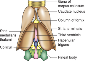
Pineal gland, also known as epiphysis, is a small structure (5 mm × 7 mm) shaped like a pine cone. It is situated in the groove between the two superior colliculi in diencephalic area of brain above the hypothalamus (Fig.8.7-1).
Structure. The pineal stroma has two types of cells: neuroglial and parenchymal. The parenchymal cells are large epithelial cells having secretory function.
Melatonin
The hormone melatonin is synthesized by the parenchymal cells of the pineal gland.
1. Role in circadian rhythm of the body. Melatonin secretion shows diurnal variation. It is secreted more during dark period of the day than during the day light hours. This correlates with various internal activities in different periods of the day, i.e. circadian rhythm.
2. Effects on the gonads. Melatonin exerts both inhibitory and facilitatory effects on the gonads due to the diurnal change in the melatonin secretion. It inhibits onset of puberty.
3. Effect on MSH and ACTH secretion. An inhibitory effect of melatonin on MSH and ACTH secretion has been reported.
Thymus
Functional anatomy
Thymus is a small lymphoid structure located in the lower part of neck in front of the trachea, below the thyroid gland. At birth, it is small (weighing 10–12 g), gradually enlarges till puberty and then it starts decreasing in size in old age.
Histologically, thymus consists of the inner medulla and outer cortex.
Functions
1. Immunological functions of thymus
(i) Development of immunologically competent Tlymphocytes is an essential function of the thymus (see page 99).
(ii) Maintenance of adequate pool of T-lymphocyte. The hormone thymosin produced by the thymus also stimulates lymphopoiesis in the peripheral lymphoid tissue and thus plays a role in maintenance of an adequate pool of T-lymphocytes in adult life.
2. Endocrine function of thymus. Thymus tissue secretes two hormones, thymosin and thymin.
Local hormones
The local hormones are the substances which are produced in many tissues, and when activated in certain circumstances, execute their actions in the same area or in immediate neighbourhood. Commonly produced local hormones are discussed below.
Prostaglandins and related substances
Prostaglandins (PGs) and related substances include thromboxanes, prostacyclin, leukotriene and lipoxin. These substances are called eicosanoids, reflecting their origin from arachidonic acid, linoleic and linolenic acid.
Prostaglandins were so named by Von Euler in 1937 because they were first isolated from prostatic secretion in semen. However, now they are known to be synthesized in almost all tissues of the body. Presently, a variety of PGs are identified.
Actions of prostaglandins. Prostaglandins have multitudinous and varied actions on almost all tissues of the body. Many of them are discussed in the chapters on the systems in which they play an important role. Some important actions of PGs are:
1. Actions on cardiovascular system. Peripheral arteriolar dilatation, especially in splanchnic and muscular bed.
2. Actions on kidneys. PGA2 increases renal cortical blood flow and increases urinary excretion of sodium, potassium and water.
3. Actions on female reproductive system
• Initiate labour by stimulating contraction of gravid uterus.
• Responsible for painful uterine contractions during menstruation (dysmenorrhoea).
• Promote secretion of hypothalamic gonadotropin releasing hormone (GnRH).
4. Role of PGs in inflammation. Prostaglandins mediate following effects of inflammation:
• Increase vascular permeability and cellular infiltration.
• Pain-producing effect of bradykinin by sensitizing cutaneous nerves.
Actions of thromboxane A2. Thromboxane A2 is synthesized by platelets. It promotes:
Actions of prostacyclin. Prostacyclin is produced in vascular endothelium. It produces vasodilatation.
Actions of leukotrienes. Leukotrienes are mediators of allergic responses and inflammation. Their release is provoked when specific allergens combine with IgE antibodies on the surfaces of mast cells.
Actions of lipoxins. Physiological role of lipoxins is uncertain. Their actions include: