Chapter Seventeen Skin, hair and nails
PURPOSE
The skin is the largest organ in the body and performs a number of key functions. In this chapter you will learn the structure and function of the skin as well as the nails and hair. This will enable you to understand the rationale for and the methods of inspection and palpation of the skin, nails and hair; abnormalities that may arise and how to record the assessment accurately.
KEY CONCEPTS
• Structure and function of the skin, hair and nails
• Health history regarding skin, hair and nails
• Changes related to the ageing process
• Abnormalities of the hair, skin and nails
While you are completing your reading assignment, ensure you understand each of the key concepts listed above.
READING ASSIGNMENT
Jarvis, Forbes & Watt: (JF&W) Jarvis’s Physical Examination and Health Assessment, Chapter 17, pp 431–479.
GLOSSARY
After reading the corresponding chapter in the text, learn the following terms. You should be able to cover the definition on the right and state the associated definition in your own words.
Alopecia male-pattern balding; a significant loss of hair (baldness)
Anasarca bilateral oedema or oedema that is generalised over the whole body
Annular circular shape to skin lesion
Atrophic skin very thin, shiny skin that occurs with arterial insufficiency
Bulla elevated cavity larger than 1 cm diameter containing free fluid
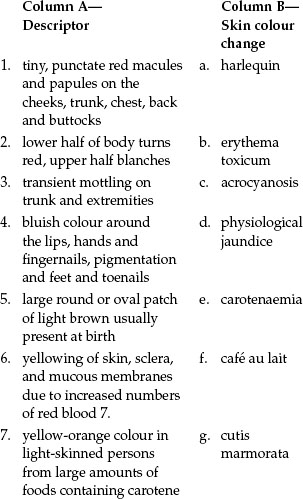
Café au lait spots large round or oval patch of light-brown pigmentation, usually present at birth; six or more larger than 1.5 cm are diagnostic of neurofibromatosis
Cherry (senile) angiomas small (1 to 5 mm), smooth, slightly raised bright red dots that appear on the trunk in all adults over 30 years old; increase in number with age; not significant
Chloasma irregular brown patch of hyperpigmentation on the face with changing hormone levels during pregnancy
Confluent skin lesions that run together
Crust thick, dried-out exudate left on skin when vesicles/pustules burst or dry up
Cyanosis dusky blue colour to skin or mucous membranes due to increased amount of deoxygenated haemoglobin
Diaphoresis profuse perspiration
Ephelides (freckles) small, flat macules of brown melanin pigment that occur on sun-exposed skin
Erosion scooped out, shallow depression in skin
Erythema intense redness of the skin due to excess blood in dilated superficial capillaries, as in fever or inflammation
Excoriation self-inflicted abrasion on skin due to scratching
Fissure linear crack in skin extending into dermis
Furuncle (boil) red, swollen, hard, tender, pus-filled lesion due to infected hair follicle
Haemangioma skin lesion due to benign proliferation of blood vessels in the dermis
Hirsutism excess body hair; in females a male pattern of hair distribution on the face and chest develops; indicates endocrine abnormalities
Iris target shape of skin lesion
Jaundice yellow colour to skin, palate and sclera due to excess bilirubin in the blood
Keloid hypertrophic scar, elevated beyond site of original injury
Lanugo the fine downy hair of the newborn infant
Lesion traumatic or pathological change in previously normal structure; called a primary lesion when it develops on previously unaltered skin; called a secondary lesion, when a lesion changes over time or changes because of a factor such as scratching or infection
Lichenification tightly packed set of papules that thickens skin, from prolonged intense scratching
Macule small pigmented spot on the skin that is neither raised nor depressed
Melanoma malignant skin lesion; usually brown; can be tan, black, pink-red, purple or mixed pigmentation; often with irregular or notched borders; may have scaling, flaking or oozing texture; half arise from preexisting naevi
Mongolian spot a common variation of hyperpigmentation in infants of South-East Asian, Pacific Island and African descent; is a blue-black to purple macular area at the sacrum or buttocks, sometimes it occurs on the abdomen, thighs, shoulders or arms; due to deep dermal melanocytes
Naevus (mole) a proliferation of melanocytes, tan to brown colour, flat or raised, circumscribed skin lesion due to excess melanocytes
Nodule Solid, elevated, hard or soft skin lesion; larger than 1 cm; may extend deeper into dermis than papule
Oedema fluid accumulating in the intercellular spaces
Pallor excessively pale, whitish-pink colour to lightly pigmented skin
Papule a small hard round protuberance on the skin <1 cm diameter
Plaque papules coalesce to form surface elevation wider than 1 cm; a plateau-like, disc-shaped lesion
Profile sign the angle of the nail base; should be about 160 degrees
Purpura red-purple skin lesion due to blood in tissues from breaks in blood vessels
Pustule elevated cavity containing thick turbid (pus) fluid
Scale compact desiccated flakes of skin from shedding of dead skin cells
Terminal hair the darker thicker hair that grows on the scalp and eyebrows and, after puberty, on the axillae, pubic area and the face and chest in the male
Turgor ability of skin to return to its normal position promptly when released after being pinched; reflects the elasticity of the skin
Ulcer sloughing of necrotic inflammatory tissue that causes a deep depression in skin, extending into dermis
Vellus hair fine, faint hair covering most of the body (except the palms and soles, the dorsa of the distal parts of the fingers, the umbilicus, the glans penis and inside the labia)
Vesicle elevated cavity containing free fluid up to 1 cm diameter
Vitiligo complete absence of melanin pigment in patchy areas of white or light skin on the face, neck, hands, feet, body folds and around orifices
Wheal superficial, raised, transient and erythematous; slightly irregular shape due to oedema
STUDY GUIDE
After completing the reading assignment, you should be able to answer the following questions in the spaces provided.
1. Describe the structure and contents of each of the following 3 layers associated with the skin
epidermis ________________________________________
_______________________________________________________________
_______________________________________________________________
_______________________________________________________________
_______________________________________________________________
_______________________________________________________________
dermis ________________________________________
_______________________________________________________________
_______________________________________________________________
_______________________________________________________________
subcutaneous layer ________________________________________
_______________________________________________________________
_______________________________________________________________
_______________________________________________________________
2. List 3 sources that influence skin colour.
3. Define ‘epidermal appendage’ and provide 3 examples.
4. Differentiate between sebaceous, eccrine and apocrine glands.
sebaceous glands ________________________________________
_______________________________________________________________
_______________________________________________________________
eccrine glands ________________________________________
_______________________________________________________________
_______________________________________________________________
apocrine glands ________________________________________
_______________________________________________________________
_______________________________________________________________
5. Fill in the labels indicated on the following illustration.
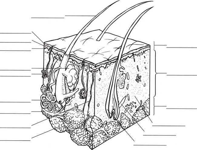
6. List the 9 functions of the skin and provide a brief description of each function.
7. Describe at least 7 changes that occur in the skin with ageing.
8. Complete the following sentences in relation to skin integrity:
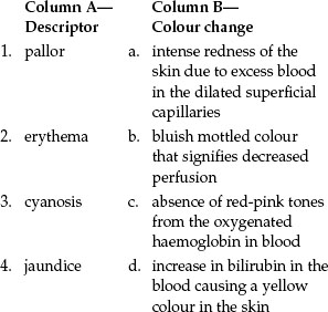
9. Define each of the following, describe recognition in various skin colours and identify conditions that may cause each:
pallor ________________________________________________________________
_______________________________________________________________
_______________________________________________________________
_______________________________________________________________
_______________________________________________________________
_______________________________________________________________
erythema ________________________________________
_______________________________________________________________
_______________________________________________________________
_______________________________________________________________
_______________________________________________________________
_______________________________________________________________
cyanosis ________________________________________
_______________________________________________________________
_______________________________________________________________
_______________________________________________________________
_______________________________________________________________
_______________________________________________________________
jaundice ________________________________________
_______________________________________________________________
_______________________________________________________________
_______________________________________________________________
_______________________________________________________________
_______________________________________________________________
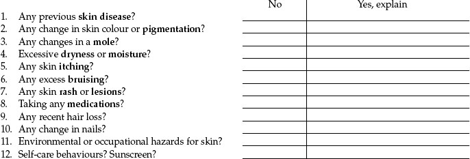
10. Rashes are a common reason for seeking healthcare. List specific questions you would ask to determine causation, progression and self-management.
11. Abnormal characteristics of pigmented lesions are summarised in the mnemonic ABCDE. Explain what each letter represents and what additional symptoms may indicate malignancy.
A. ___________________________________________________________________
_______________________________________________________________
_______________________________________________________________
B. ___________________________________________________________________
_______________________________________________________________
_______________________________________________________________
C. ___________________________________________________________________
_______________________________________________________________
_______________________________________________________________
D. ___________________________________________________________________
_______________________________________________________________
_______________________________________________________________
E. ___________________________________________________________________
_______________________________________________________________
_______________________________________________________________
Additional symptoms:___________________________________________________________________
_______________________________________________________________
_______________________________________________________________
_______________________________________________________________
_______________________________________________________________
_______________________________________________________________
12. Describe each of the 4 grades of pressure injury; identify those at risk and common sites of injury.
At risk:___________________________________________________________________
_______________________________________________________________
Common sites:___________________________________________________________________
_______________________________________________________________
13. List causes of changes in skin temperature, texture, moisture, mobility and turgor.
14. Describe each grade on the 4-point grading scale for pitting oedema.
15. When lesions are present list the 6 items that should be noted.
16. Differentiate between a primary and a secondary lesion.
17. Fill in the labels indicated on the following illustration.
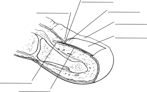
18. The white linear markings that normally are visible through the nail and on the pink nail bed are termed____________.
19. Describe the following findings that are common variations on the infant’s skin:
café au lait spot ________________________________________
_______________________________________________________________
erythema toxicum ________________________________________
_______________________________________________________________
cutis marmorata ________________________________________
_______________________________________________________________
physiological jaundice ________________________________________
_______________________________________________________________
milia ________________________________________
_______________________________________________________________
20. Describe the following findings that are common variations with the ageing adult’s skin:
lentigines ________________________________________
_______________________________________________________________
seborrhoeic keratosis ________________________________________
_______________________________________________________________
actinic keratosis ________________________________________
_______________________________________________________________
acrochordons (skin tags) ________________________________________
_______________________________________________________________
sebaceous hyperplasia ________________________________________
_______________________________________________________________
21. Describe each of the following vascular lesions:
haemangioma ________________________________________
_______________________________________________________________
spider or star angioma ________________________________________
_______________________________________________________________
purpuric lesion ________________________________________
_______________________________________________________________
petechiae ________________________________________
_______________________________________________________________
haematoma ________________________________________
_______________________________________________________________
contusion ________________________________________
_______________________________________________________________
22. Differentiate between the appearance of the skin rash of these childhood illnesses:
measles (rubeola) ________________________________________
_______________________________________________________________
_______________________________________________________________
German measles (rubella) ________________________________________
_______________________________________________________________
_______________________________________________________________
chickenpox (varicella) ________________________________________
_______________________________________________________________
_______________________________________________________________
23. Describe 3 skin lesions that are associated with HIV/AIDS.
24. Differentiate between a furuncle and an abscess.
25. State the causation and describe the appearance of the following nail disorders:
koilonychia (spoon nails) ________________________________________
_______________________________________________________________
paronychia ________________________________________
_______________________________________________________________
REVIEW QUESTIONS
This test is for you to check your own mastery of the content. Answers are provided in Appendix A.
1. Select the best description of the secretions of the eccrine glands.
2. Naevus is the medical term for:
3. To assess for early jaundice, you will assess:
4. Checking for skin temperature is best accomplished by using:
5. Skin turgor is assessed by picking up a large fold of skin on the anterior chest under the clavicle. This is done to determine the presence of:
6. You note a lesion during an examination. Select the description that is most complete.
7. You examine nail beds for clubbing. The normal angle between the nail base and the nails is:
8. The capillary beds should refill after being depressed in:
9. During a routine visit, Mr Bond, age 78, asks about small, round, flat, brown macules on the hands. Your best response after examining the areas is:
10. An area of smooth, rubbery, raised tissue near a scar is called:
11. Flattening of the angle between the nail and its base is:
12. The configuration for individual lesions arranged in circles or arcs, as occurs with ringworm, is called:
13. The ‘A’ in the ABCDE rule stands for:
14. A risk factor for melanoma is:
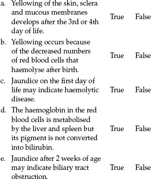
15. Physiological jaundice is a common newborn condition. Answer True or False to the following statements. If the answer is false, state the correct answer.
16. Place the name of the following 3 skin layers next to the structures which are contained within that specific layer. These items may be used more than once.
PRACTICAL SKILLS IN THE LABORATORY/CLINICAL SETTING
In clinical practice, skin assessment is integrated throughout the complete health assessment; it is not a separate isolated step. At times, you may need to perform a regional assessment if the patient enquires about a particular problem relating to their skin.
The skills you use when assessing the skin are inspection and palpation, as some skin changes have accompanying signs that can be felt. Remember to maintain patient privacy and ensure appropriate environment when performing your assessment.
Now you have completed the preparatory readings and exercises you are ready for the clinical component of the integumentary system.
The purpose of practising the steps of this examination separately is so that you begin to think of the skin and its appendages as a separate organ system, and so that you learn the components of skin examination. You will practise these skills on your peer in the laboratory or a patient in the clinical setting.
PROFESSIONAL PRACTICE NOTE
To accurately inspect all of the skin for lesions, your patient needs to disrobe, so remember to maintain patient privacy and ensure an appropriate environment when performing your assessment.
CLINICAL OBJECTIVES
At the completion of the clinical laboratory session, with further practice and self-directed learning you should be able to:
1. collect a health history that is related to the presenting signs and symptoms and problems as stated by the patient
2. inspect and palpate the skin, noting its colour, vascularity, oedema, moisture, temperature, texture, thickness, mobility and turgor, and any lesions
3. inspect the fingernails, noting colour, shape and any lesions
4. inspect the hair, noting texture, distribution and any lesions
5. record the history and physical examination findings accurately, reach an assessment of the health state and develop an appropriate plan of care.
INSTRUCTIONS
1. Form pairs. If possible, choose a peer from an ethnic background other than your own, as it will further heighten your recognition of the range of normal skin tones.
2. Prepare the examination setting and gather your equipment. Ensure you try to control external variables that may influence skin colour and confuse your findings. Don’t forget you may need additional equipment in addition to a strong direct light (natural daylight is ideal to evaluate skin characteristics but is usually not available in the clinical/laboratory area). Additional equipment may include a small centimetre ruler, penlight and gloves. A magnifying glass for close examination of suspicious lesions may also be needed.
4. Gain consent to perform the examination from either your peer or the patient.
5. Practise the health history interview and the steps of the examination of the skin, hair and nails on a peer in the skills laboratory or a patient in the clinical setting, providing appropriate instructions as you proceed.
6. Record your findings using the regional write-up worksheet.
7. Swap roles and repeat steps 2–6.
8. Discuss your assessment techniques, findings and performance with your peer to develop a complete understanding of the process.
REGIONAL WRITE-UP WORKSHEET–SKIN, HAIR AND NAILS
Interview conducted by _____________
Patient _____________Age_____________Gender_____________
Occupation _____________Medical Record Number_____________
REGIONAL DOCUMENTATION—SKIN, HAIR AND NAILS
Summarise your findings using the SOAP format.
Subjective (Reason for seeking care, health history)
Objective (Physical exam findings)
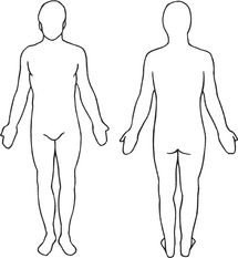
Record findings on diagram below
Assessment (Assessment of health state or problem, diagnosis)
Plan (Diagnostic evaluation, follow-up care, patient teaching)
PATIENT EDUCATION: SKIN SELF-EXAMINATION (SSE)