Mobile
Chest
AP
Patient position
Part position
• Center midsagittal plane to IR.
• Position IR under patient with top about 2 inches (5 cm) above relaxed shoulders.
• Internally rotate patient’s arms to prevent scapular superimposition of lung field, if not contraindicated.
Respiration:
Inspiration.
Central ray
Collimation:
kVp: 90 (40˝ SID, non-grid) 105 (40˝ grid)
Reference: 13th edition ATLAS p. 3:192-193.

Manual Factors
| Part Thickness (cm) | mA | kVp | Time | mAs | SID | Image Receptor Size | CR, DR Exposure Indicator | Grid | HF, 1Ø or 3Ø |

Notes: _________________________________________________________________________ Competency: ________/____/______
_______________________________________________________________________________ Instructor: _______________________
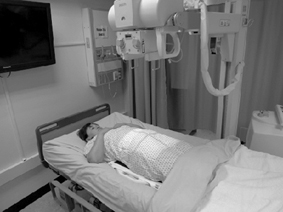
Chest
AP or PA (right or left lateral decubitus)
Patient position
Part position
• Perform AP projection whenever possible.
• Adjust patient to ensure true lateral position.
• Place IR behind patient and below support.
• Adjust grid so that it extends 2 inches (5 cm) above shoulders.
Respiration:
Inspiration.
Central ray
Collimation:
kVp: 105 (40˝ grid)
Reference: 13th edition ATLAS p. 3:180-181.


Manual Factors
| Part Thickness (cm) | mA | kVp | Time | mAs | SID | Image Receptor Size | CR, DR Exposure Indicator | Grid | HF, 1Ø or 3Ø |

Notes: _________________________________________________________________________ Competency: ________/____/______
_______________________________________________________________________________ Instructor: _______________________
Abdomen
AP
Patient position
Part position
• Position grid under patient.
• Keep grid from tipping side to side by placing it in center of bed and stabilizing with blankets if necessary.
• Center midsagittal plane to grid.
• If emphasis is on upper abdomen, center grid 2 inches (5 cm) above iliac crests or high enough to include diaphragm.
Respiration:
Suspended.
Central ray
Collimation:
kVp: 85
Reference: 13th edition ATLAS p. 3:196-197.

Manual Factors
| Part Thickness (cm) | mA | kVp | Time | mAs | SID | Image Receptor Size | CR, DR Exposure Indicator | Grid | HF, 1Ø or 3Ø |

Notes: _________________________________________________________________________ Competency: ________/____/______
_______________________________________________________________________________ Instructor: _______________________
Abdomen
AP or PA (left lateral decubitus)
Patient position
Part position
• Adjust patient to ensure true lateral position.
• Place grid vertically in front of patient for PA or behind patient for AP. Support grid to prevent grid cutoff.
• Position grid so that its center is 2 inches (5 cm) above iliac crests to ensure that diaphragm is included.
Respiration:
Suspended.
Central ray
Collimation:
Adjust to 14 × 17 inches (35 × 43 cm).
kVp: 85
Reference: 13th edition ATLAS p. 3:198-199.


Manual Factors
| Part Thickness (cm) | mA | kVp | Time | mAs | SID | Image Receptor Size | CR, DR Exposure Indicator | Grid | HF, 1Ø or 3Ø |

Notes: _________________________________________________________________________ Competency: ________/____/______
_______________________________________________________________________________ Instructor: _______________________
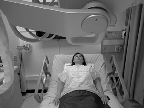
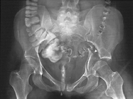
Pelvis
AP
Patient position
Part position
• Position grid under pelvis so that center is midway between ASIS and pubic symphysis (about 2 inches [5 cm] inferior to ASIS).
• Center midsagittal plane to midline of grid. Pelvis should not be rotated.
• Rotate patient’s legs medially 15 degrees when not contraindicated.
Respiration:
Suspend.
Central ray
• Perpendicular to midpoint of grid. Central ray should enter patient 2 inches (5 cm) above pubic symphysis and 2 inches (5 cm) below ASIS.
Collimation:
kVp: 85
Reference: 13th edition ATLAS p. 3:200-201.

Manual Factors
| Part Thickness (cm) | mA | kVp | Time | mAs | SID | Image Receptor Size | CR, DR Exposure Indicator | Grid | HF, 1Ø or 3Ø |

Notes: _________________________________________________________________________ Competency: ________/____/______
_______________________________________________________________________________ Instructor: _______________________
Femur
AP
Patient position
Part position
• Cautiously place grid lengthwise under patient’s femur, with distal edge of grid low enough to include fracture site and knee joint.
• Elevate grid with towels under each side to ensure proper grid alignment with x-ray tube.
Respiration:
Suspend.
Central ray
• Perpendicular to long axis of femur; center to grid
• Ensure that central ray and grid are aligned to prevent grid cutoff.
Collimation:
Adjust to 1 inch (2.5 cm) on sides of shadow of femur and 17 inches (43 cm) in length.
kVp: 85
Reference: 13th edition ATLAS p. 3:202-203.


Manual Factors
| Part Thickness (cm) | mA | kVp | Time | mAs | SID | Image Receptor Size | CR, DR Exposure Indicator | Grid | HF, 1Ø or 3Ø |

Notes: _________________________________________________________________________ Competency: ________/____/______
_______________________________________________________________________________ Instructor: _______________________
Femur
Lateral (dorsal decubitus)
Patient position
Part position
• Determine whether mediolateral or lateromedial projection is to be performed.
• Place grid in vertical position next to lateral aspect of femur.
• Place distal edge of grid low enough to include knee joint.
• Stabilize grid firmly in position (patient may hold).
• Support and elevate unaffected leg.
• Ensure that grid is placed perpendicular to epicondylar plane.
Respiration:
Suspend.
Central ray
Collimation:
Adjust to 1 inch (2.5 cm) on sides of shadow of femur and 17 inches (43 cm) in length.
kVp: 85
Reference: 13th edition ATLAS p. 3:204-205.


Manual Factors
| Part Thickness (cm) | mA | kVp | Time | mAs | SID | Image Receptor Size | CR, DR Exposure Indicator | Grid | HF, 1Ø or 3Ø |

Notes: _________________________________________________________________________ Competency: ________/____/______
_______________________________________________________________________________ Instructor: _______________________
Cervical spine
Lateral (right or left dorsal decubitus)
Patient position
Part position
• Ensure that upper torso and head are not rotated.
• Place grid lengthwise on right or left side, parallel to neck.
• Place top of grid 1 to 2 inches (2.5 to 5 cm) above EAM.
• Immobilize grid in vertical position.
• Have patient relax shoulders and reach for feet if possible.
Respiration:
Full expiration.
Central ray
Collimation:
kVp: 85
Reference: 13th edition ATLAS p. e3:206-207.


Manual Factors
| Part Thickness (cm) | mA | kVp | Time | mAs | SID | Image Receptor Size | CR, DR Exposure Indicator | Grid | HF, 1Ø or 3Ø |

Notes: _________________________________________________________________________ Competency: ________/____/______
_______________________________________________________________________________ Instructor: _______________________
Chest and abdomen: Neonate
AP
Patient position
Part position
• Carefully position x-ray tube over infant.
• Ensure that chest and abdomen are not rotated.
• Move infant’s arms away from body, and bring legs down and away from abdomen.
Respiration:
Inspiration
Central ray
Collimation:
Adjust to 1 inch (2.5 cm) on all sides of chest and abdomen.
kVp: 64
Reference: 13th edition ATLAS pp. 3:209-210.


Manual Factors
| Part Thickness (cm) | mA | kVp | Time | mAs | SID | Image Receptor Size | CR, DR Exposure Indicator | Grid | HF, 1Ø or 3Ø |

Notes: _________________________________________________________________________ Competency: ________/____/______
_______________________________________________________________________________ Instructor: _______________________
Chest and abdomen: Neonate
Lateral (right or left dorsal decubitus)
Patient position
Part position
• Ensure that infant’s chest and abdomen are centered to IR and not rotated.
• Move infant’s arms above head.
• Place IR lengthwise and vertical beside infant, then immobilize IR.
Respiration:
Inspiration
Central ray
Collimation:
Adjust to length of chest and abdomen and 1 inch (2.5 cm) above abdomen.
kVp: 72
Reference: 13th edition ATLAS pp. 3:211-212.


Manual Factors
| Part Thickness (cm) | mA | kVp | Time | mAs | SID | Image Receptor Size | CR, DR Exposure Indicator | Grid | HF, 1Ø or 3Ø |

Notes: _________________________________________________________________________ Competency: ________/____/______
_______________________________________________________________________________ Instructor: _______________________