B
Appendix: Anatomic Positions and Directions
In orthopaedics, anatomic positions and directions are routinely used to provide an accurate description of specific anatomic locations. These basic directions may be compared to looking at a map to determine the longitude and latitude of an area. Humans are three-dimensional subjects with points of reference made in the orthograde (upright) position. The surface locations and anatomic planes are described as follows.
Surface Location
The location of a structure is described in reference to a standing person facing the examiner with outstretched hands in a palms-up position. The basic directional terms are:
anterior (ventral): forward or front surface (Fig. B-1)
inferior: lower area, below, toward the tail end
lateral: sides, away from midline
medial: middle, toward the midline
posterior (dorsal): back surface
superior: upper area, above, toward the head
These reference points may be combined to give a more precise location to a specific region on a surface or in structures deep within the body, for example, the exposed hip in a surgical procedure. When these terms are compounded and hyphen omitted, the combining forms may be:
anteroinferior: front and below
anterolateral: front and to outer side
anteromedial: front and to inner side
anteroposterior: front and toward back
anterosuperior: front and above
These can be used in combination with locations in reverse order, such as:
posteroinferior: back to below
posterolateral: back to outer side
posteromedial: back to inner side
posterosuperior: back to front toward head
In some cases, dorsal (back) may be used for posterior (e.g., dorsolateral).
Anatomic Planes
The term plane comes from the Latin word planus, meaning a flat, level surface. There are three directions of planes, all in reference to a standing person facing the examiner: vertical anterior to posterior (sagittal), vertical side to side (coronal or longitudinal), and horizontal (transverse).
anteroposterior planes
median (midsagittal) plane: vertical plane directly through the midline of the body, transecting the nose, navel, and spine, and dividing the body into left and right halves.
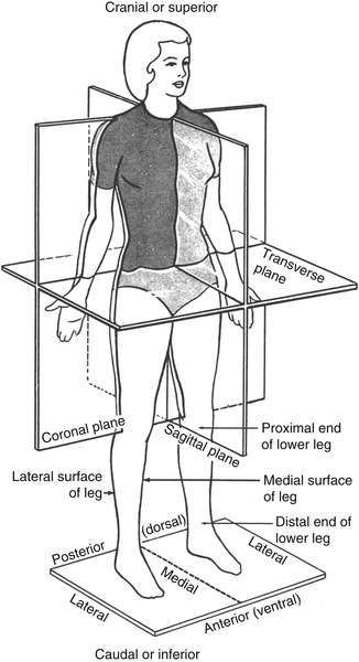
horizontal (transverse) plane: any of the horizontal planes across the body at right angles to coronal and sagittal planes parallel to baseline.
neutral plane: the plane of a structure around which bending occurs.
vertical side to side planes
coronal (frontal plane): a plane parallel with long axis of body at right angles to median sagittal plane going through coronal sutures of the skull (approximately center of body) and dividing body into front and back parts.
longitudinal: lengthwise and parallel with long axis of body or part; any of the vertical side to side planes. Coronal and longitudinal planes have been used interchangeably to describe each other. In this case, the context of the sentence will indicate the reference point of the plane.
Specific Locations
When describing limb anatomy, the nomenclature is very specific. The four appendages are correctly referred to as the upper and lower limbs, two forelimbs, and two hind limbs. The thigh indicates that portion above the knee and the shank the portion between the knee and ankle. The calf is the posterior aspect and the shin the anterior aspect of the leg. The sole (bottom) of the foot is called the plantar surface, and the top is the dorsal surface. The brachium refers to that portion of the arm above the elbow and below the shoulder. The antebrachium refers to the portion of the arm below the elbow but above the wrist. The ventral side of the hand is the palm or volar surface, with the opposite side being the dorsum or dorsal surface. The forearm is similarly divided into volar and dorsal aspects.
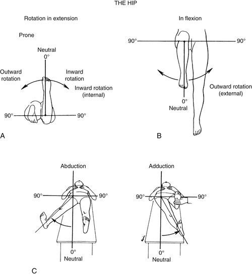
Joint Motions∗ (Fig. B-2)
Ranges of joint motion refer to the extent of movement within a given joint. Joint motion may be active, passive, or active assistive. The major joint areas involve the shoulder (glenoid), elbow (cubitus), hip (coxa) (Fig. B-3), and knee (genu).
All the hinge joints have motion described in terms of flexion and extension. Except for the ankle, the 0-degree position occurs when the limb is held out straight, and the degree of flexion is then stated in terms of degrees from the 0-degree extended position. The knee and elbow will occasionally extend beyond the 0-degree limit, and this motion is expressed in degrees of hyperextension. The wrist has approximately 90 degrees of extension and 90 degrees of flexion (dorsiflexion and palmar flexion).
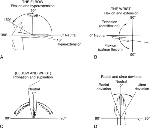
The Neutral Zone Method is used for measuring joint motion. The anatomic position of the joint defines the starting position at zero, and motion is measured in degrees of a circle. Other measured motions include:
circumduction: a maneuver or movement of a ball-and-socket joint in a circular motion; for example, the shoulder can circumduct 180 degrees with six movements possible.
flexion: to bend from the joint as in flexion movements of the spine at the waist (anterior or lateral). In the foot or hand is expressed as:
dorsiflexion: the toe-up motion of the ankle expressed in degrees from the 0-degree position of the foot at rest on the ground in standing position.
plantar flexion: the toe-down motion of the foot at the ankle expressed in degrees from 0-degree position of the foot at rest on the ground in standing position.
palmar flexion: of the wrist with palm up in flexion.
extension: extending distally away from body as in the limbs, or bending back posteriorly as in the spine.
pronation: palm-down position of hand with elbow at a 90-degree angle, brought about by the motion of the radius around the ulna (posterior rotation). In the foot, the plantar surface is turned down.
ulnar deviation: of the hand at the wrist such that the hand is directed in an ulnar direction; measured in degrees from 0 with hand in midline.
radial deviation: of the hand at the wrist such that the hand is directed radially; measured in degrees from 0 with hand in midline.
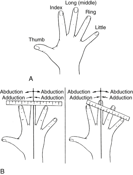
adduction: movement toward the midline in frontal plane as in abduction. On verbal transcription in clinical notes the person dictating will sometimes say 8220a-b-duction8221 or 8220a-d-duction8221 to clarify distinction between abduction and adduction (see Fig. B-4).
anteversion: to lean forward at an angle; in reference to the neck of humerus or femur, an anterior rotation.
anteroflexion: bending forward.
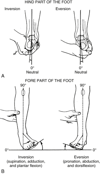
eversion: turning outward, when applied to the heel, describes the degree of motion of the heel pushed outward with ankle in neutral position; when applied to the foot, describes the combined motions of dorsiflexion, pronation, and abduction (Fig. B-6).
inversion: when applied to the heel, describes the degree of motion of the heel pushed inward with ankle in neutral position; when applied to the foot, describes the combined motions of plantar flexion, supination, and adduction (see Fig. B-6).
retroflexion: bending backward.
retroversion: turned toward the back; in reference to the neck of femur or humerus, a posterior rotation.
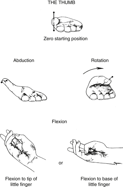
apposition: contact of two adjacent parts; bringing together as in a finger movement, the thumb to index finger.
opposition: applied mostly to the thumb but also to little finger; describes the motion required to bring about opposition, or the setting opposite, of thumb against little finger (pulp surfaces). For the thumb, opposition is the combined action of abduction, rotation, and flexion.
external rotation: in frontal plane is away from midline.
internal rotation: in a frontal plane is toward the midline.
valgus: turned outward; the distal part is bent away from the midline; for example, genu valgus (knock-kneed).
varus: turned inward; the distal part is toward the midline; for example, genu varus (bow-legged).
Anatomic Associated Terms
The following are other associated terms referring to directions of anatomy or physical signs.
a, an: no, not, against.
AAL: anterior axillary line.
ab: away from (abduct).
abducens: drawing away from median line of the body; also called abductor m.
ad: toward (adduct).
adjacent: close to, beside
AGE: angle of greatest extension.
AGF: angle of greatest flexion.
alignment: linear position of one part of an extremity compared with another; to bring into a straight line.
ambi: on both sides.
ambidextrous: using both right and left hands effectively.
amph-, amphi-: both ways, all around, both sides.
anatomic axis: the true axis of an extremity measured by lines.
angle: the figure or space outlined by the diverging of two lines from a common point or by the meeting of two planes; a projecting or sharp corner.
angulation: deviation from the norm; sharp bend of a structure to form an angle.
aniso: unequal, asymmetrical, dissimilar.
annulus: any circular structure, ring-shaped.
ante-: forward, before.
antecedent: to precede, or go before.
anteflexion: to bend forward.
anterior: front view, ventral side, face surface, superior.
antero-: before, in front of.
anteversion: tipping forward.
anticus: foremost, in the front.
apex: top, tip, point of activity, summit, vertex (refers to C2).
apical: pertaining to apex; situated near point of reference.
apo-: away from.
asymmetric: lacking symmetry; uneven, as one limb to another.
axis: line of symmetry, rotation, or revolution; pivot dividing line; also second vertebra.
axis of rotation: circular arcs of limb segments that move in a line of right angles to the plane.
basal, basilar: base of a part.
bilateral: both sides.
cata-: prefix meaning down, against, or according to.
caudal, caudad: toward the lower end of the erect trunk or tail, inferior to or bottom point of reference.
centri-: center.
cephalic: toward the head; uppermost point of reference.
circumference: around, outer circular boundary.
circumflex: describes an arc of a circle; winding around.
co-, com-, con-: together, with.
concave: rounded, depressed surface.
contralateral: opposite side.
convex: rounded, elevated surface.
coronal: a plane dividing the body with front and back portions; in the direction of the coronal suture; a longitudinal plane passing through the body at right angles to the median plane.
craniad: toward the head.
curvilinear: curved away from straight line.
de-: away, from, down.
deep: depth from surface.
delta: triangle (deltoid).
dexter: right.
dia-: between, through, across, apart.
diffuse: widely distributed.
dis-: apart from.
distal: away from, furthest point of reference.
dorsal: back or posterior aspect.
ec-: out, out from.
ectopic: located away from normal position; out of place.
em-, en-: in, within.
endo-: within, in.
epi-: on, above, over.
eso-: in, inward, inside.
ex-, exo-: out, outside.
external: outside, describing walls, cavities, or hollow viscera.
extra-: beyond, outside, without.
facet: flat surface.
flexure: curved or bent.
fore: in front of, before.
fusiform: spindle-shaped, tapered.
geniculum: an abrupt bend or angle in a small structure (knee).
hyper-: over, above, excessive.
hypo-: under, below, deficient.
in-, ino-: into, within.
inferior: below point of reference, underneath.
infra: below or under.
in situ: in its natural place or position.
inter-: between.
intercalary: middle.
internal: inside; describing walls, cavities, or hollow viscera.
interspace: the space between two similar parts (e.g., the vertebrae, ribs).
interstitial: spaces within a structure.
intra-: within.
intro-: into, beginning.
ipsilateral: same side.
iso-: equal, symmetrical.
juxta-: prefix for close to, near, in apposition, side by side (e.g., juxtaposition).
lat, latero-, lateral: sides, right and left; away from median plane, outer surface.
latus: broad.
length: the linear distance between two joints. The International System unit of length is measured in meters.
levo: left.
linea aspera: linea (line) and aspera (rough).
linear: elongated, straight line.
longitudinal: lengthwise, parallel to the long axis of a part (coronal or frontal planes).
MAL: midaxillary line.
medi-, medial: middle or median plane, toward midline, inner surface, link making halves.
megalo-: large.
met-, meta-: beyond, from one place to another, point of change.
multiangular: many angles.
neutral axis: the longitudinal line of a structure around which torsion occurs; the longitudinal line in a long structure where normal axial stresses are zero when structure is subjected to bending.
oblique: slanting diagonal; inclined.
oblongata: oblong.
orthograde: upright position, as in a standing person.
palmar: side of hand surface, face up.
para-, par-: beyond, beside.
parallel: equal in lines or surface; in the same direction.
pars: a division or part; a particular part of a greater structure (e.g., pars interarticularis).
patent: open, unobstructed, apparent, evident.
peri-, peripheral: immediately around, sphere.
perpendicular: exactly upright; being at right angles to a given line or plane.
pivot: to turn as in a circular motion.
plantar: sole, bottom of the foot.
posterior, postero-, post-: after, behind, tail end, back, inferior to surface.
prone: face down (lying face down).
protract: pull forward.
proximal: close to nearest point of reference.
quadratus: four-sided.
re-: back, again.
recurvatum: bending backward; a flexure or hyperextension.
residual: left behind.
retract: pull back.
retrad: backward, toward back part.
retro-, retrograde, retroflex: bending backward, behind.
rotation: turning in a circular motion.
scalene: having unequal sides; said of a triangle (e.g., scalenus muscle).
scalenus: uneven.
sinister: left.
striate: having transverse lines (muscles).
sub-: under.
subjacent: lying underneath (sub-: under + -jacent: to lie).
summit: top.
super-, supra-: above, beyond, upon, over.
superficial: near the surface.
superior, sup.-: uppermost side, above point of reference, toward head (cephalic).
supine: lying on back, face up.
sym-, syn-: together.
symmetric: exhibiting symmetry, even, alike.
terminal: end.
trans-, transverse: through, across, horizontal.
ultra: beyond.
unilateral: one side.
ventral: belly side (abdomen), anterior, front, face up.
vertex: the top or summit, apex.
vertical: perpendicular to the plane of the horizon (vertex).
volar: underneath surface, palm or sole side up.