Benign Melanocytic Neoplasms
Benign Pigmented Cutaneous Lesions Other Than Melanocytic Nevi
• This group of lesions can further be divided into: (1) predominantly epidermal lesions (Table 92.1; Figs. 92.1–92.5); and (2) dermal melanocytoses (Table 92.2; Figs. 92.6 and 92.7).
Table 92.1
Benign pigmented lesions other than melanocytic nevi (predominantly epidermal lesions).
PUVA, psoralens plus ultraviolet A phototherapy.
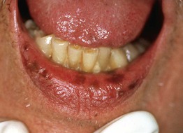
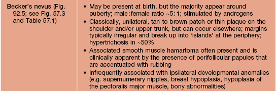
Fig. 92.1 Oral melanotic macules (labial lentigines). Multiple hyperpigmented macules on the lower lip in a patient with Laugier–Hunziker syndrome. Additional lesions are present on the tongue. Peutz–Jeghers syndrome can have a similar clinical appearance.
Fig. 92.2 Anogenital melanotic macules (anogenital lentiginosis). Biopsy of the darker lesion (at 7 o'clock) showed no cellular atypia. Over a period of 10 years, several of the macules faded. Courtesy, Jean L. Bolognia, MD.
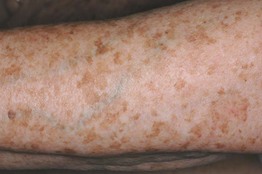
Fig. 92.3 Solar lentigines. Numerous light brown macules, some of which have an irregular border, on chronically sun-exposed skin. Courtesy, Raymond Barnhill, MD.
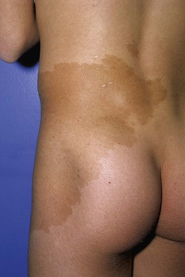
Fig. 92.4 Café-au-lait macule. Large tan patch in a geographic pattern on the lateral trunk. The patient did not have McCune–Albright syndrome.
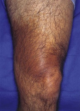
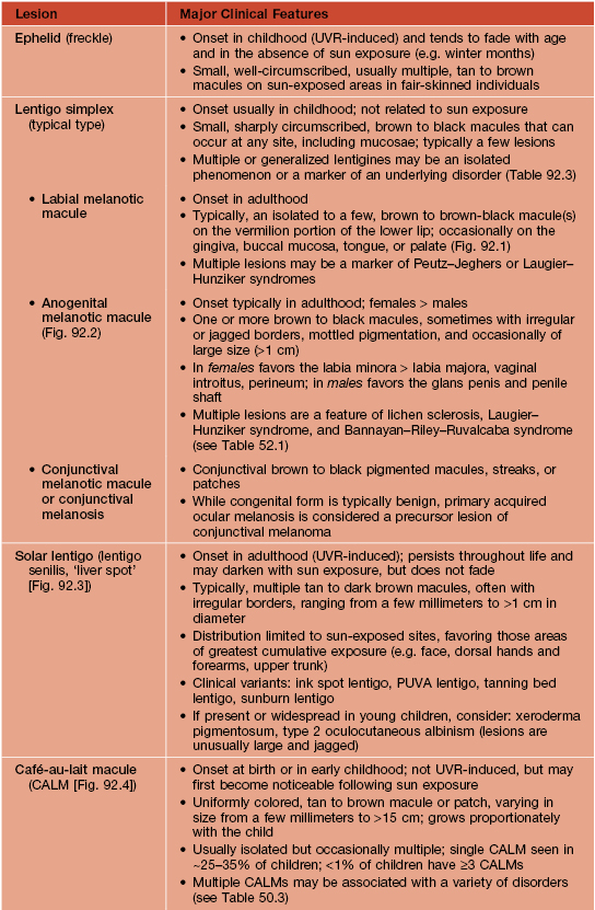
Fig. 92.5 Becker's nevus. Large patch of hyperpigmentation on the leg, which is medium brown in color. These lesions may be misdiagnosed as café-au-lait macules or congenital melanocytic nevi, especially when they do not occur on the upper trunk. Courtesy, Jean L. Bolognia, MD.
Table 92.2
The spectrum and clinical features of dermal melanocytoses.
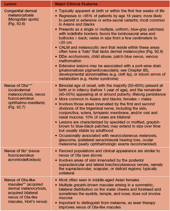
* DDx for nevus of Ota or Ito: patch or plaque blue nevus, ecchymosis, venous malformation; for nevus of Ito: extra-sacral Mongolian spot.
** Rx (if desired): pulsed Q-switched lasers (e.g. Q-switched ruby, alexandrite, or Nd:YAG lasers) beneficial but often requires multiple sessions.
CALM, café-au-lait macule.
Table 92.3
Disorders associated with multiple lentigines.
† May fade.
‡ Persists.
AD, autosomal dominant; AR, autosomal recessive; ECG, electrocardiogram; GI, gastrointestinal; LEOPARD, lentigines/ECG abnormalities/ocular hypertelorism/pulmonary stenosis/abnormalities of genitalia/retardation of growth/deafness syndrome; NAME, nevi/atrial myxoma/myxoid neurofibroma/ephelides syndrome; LAMB, lentigines/atrial myxoma/mucocutaneous myxoma/blue nevi syndrome.
Adapted from Bolognia JL. Disorders of hypopigmentation and hyperpigmentation. In Harper J, Oranje A, Prose N (eds.), Textbook of Pediatric Dermatology, 2nd edn. Oxford: Blackwell, 2006;997–1040.
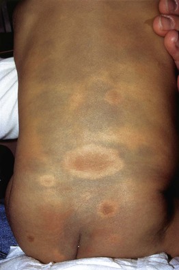
Fig. 92.6 Dermal melanocytosis (Mongolian spots) in a child with neurofibromatosis 1. Surrounding each café-au-lait macule, there is an absence of the characteristic blue discoloration.
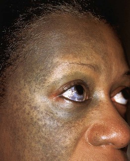
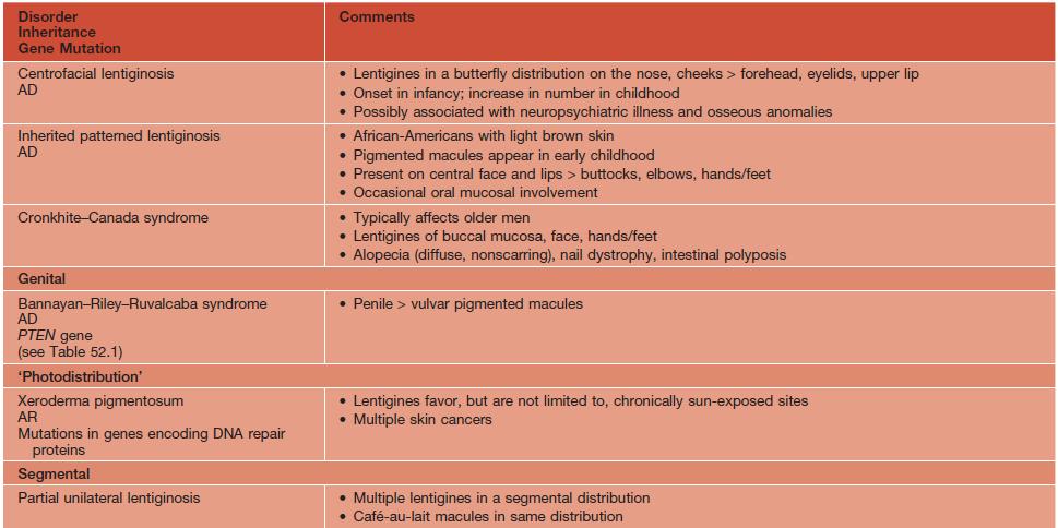
Fig. 92.7 Nevus of Ota (oculodermal melanocytosis). Unilateral blue-gray discoloration of the face, which is either mottled or confluent. There is also involvement of the sclera.
• In the predominantly epidermal lesions, the tan to brown color can result from a variety of mechanisms – e.g. increased melanocyte activity (melanogenesis), increased melanin content in keratinocytes, and a mild increase in the number of melanocytes.
• In dermal melanocytoses, the skin is blue to blue-gray in color (ceruloderma) due to the presence of melanin-producing melanocytes in the mid to lower dermis and the resultant Tyndall phenomenon (the preferential scattering of shorter wavelengths of light by the dermal melanin).
Acquired Melanocytic Nevi (Moles)
• Benign proliferations of a type of melanocyte called a ‘nevus cell’.
• Nevus cells differ from ‘ordinary’ melanocytes, which typically reside as single units in the basal layer of the epidermis, in that they: (1) usually cluster as nests in the lower epidermis and/or dermis; and (2) do not have dendritic processes (except when found in a blue nevus).
• Both ‘ordinary’ melanocytes and nevus cells can produce melanin.
• Acquired melanocytic nevi can be categorized as common (banal) or atypical (dysplastic), and they are further named based on the histologic location of the collections of nevus cells (Fig. 92.8):
– Junctional melanocytic nevus: dermal-epidermal junction.
– Compound melanocytic nevus: dermal–epidermal junction plus dermis.
– Intradermal melanocytic nevus: dermis.
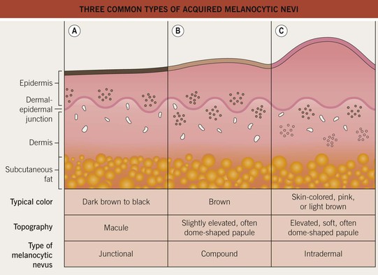
Fig. 92.8 Three common types of acquired melanocytic nevi. A Junctional, B compound, and C intradermal. The latter may also be pedunculated or papillomatous (see Chapter 1).
• Variants include halo, blue, Spitz, and ‘special site’ nevi (Fig. 92.9).
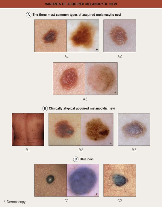
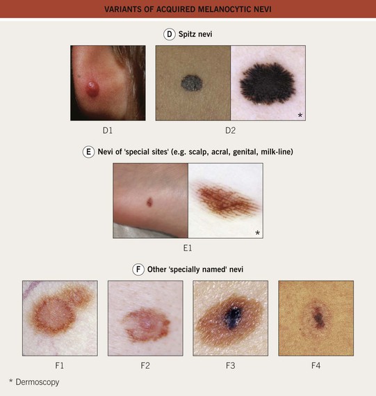
Fig. 92.9 Variants of acquired melanocytic nevi. A The three most common types of acquired melanocytic nevi. A1 Junctional nevus. Clinically, a brown macule with central hyperpigmentation. Dermoscopically, a uniform pigment network. A2 Compound nevus. Light to medium brown papule. A3 Intradermal nevus. A light tan, soft, raised papule. Dermoscopically, focal globular-like structures, whitish structureless areas, and fine comma vessels. B Clinically atypical acquired melanocytic nevi. B1 Multiple pigmented macules and papules of varying sizes on the back. B2 Close-up photo; the dermoscopy pattern is reticular-disorganized and can be seen with uncertain lesions. B3 ‘Fried egg’ appearance, with a central elevated soft papule and macular rim. C Blue nevi. C1 Common blue nevus. By dermoscopy, blue homogeneous color typically found in blue nevi. C2 Cellular blue nevus. A firm blue plaque is a common presentation. D Spitz nevi. D1 Classic Spitz nevus. Red dome-shaped papule on the ear of a child. D2 Reed nevus, typified dermoscopically by the classic starburst pattern (regular streaks at the periphery of a heavily pigmented and symmetric small macule). E Nevi of ‘special sites’ (e.g. scalp, acral, genital, milk-line). E1 Acral nevus. A brown macule on the sole of the foot. Dermoscopically, a lattice-like pattern is seen. F Other ‘specially named’ nevi. F1 Eclipse nevus. A tan center and thinner brown rim; note the stellate appearance of the brown rim. F2 Cockade or target nevus. Central lightly pigmented papule surrounded by a tan annulus then a brown ring. F3 One variant of combined melanocytic nevus. Dark brown to black papule within an otherwise uniformly pigmented light brown nevus. The differential diagnosis includes the possibility of a melanoma developing in a nevus. F4 Recurrent nevus. Dark brown pigmentation within the center of a circular scar; the pigmentation reflects the proliferation of melanocytes within the epidermis. Courtesy, Giuseppe Argenziano, MD, Raymond L. Barnhill, MD, Jean L. Bolognia, MD, Lorenzo Cerroni, MD, Harold S. Rabinovitz, MD, Ronald P. Rapini, MD, and Iris Zalaudek, MD.
• Risk factors for developing acquired melanocytic nevi: (1) a family history of numerous nevi; (2) a greater degree of sun exposure during childhood, especially intermittent and intense; and (3) lightly pigmented skin (individuals with phototype II have the greatest number of nevi).
• The vast majority of acquired melanocytic nevi remain as benign neoplasms throughout one's life and do not require treatment.
• Having >100 melanocytic nevi (8- to 10-fold increased relative risk) or multiple atypical nevi (>5 = 4- to 6-fold increased relative risk) are phenotypic markers for an entire skin surface at risk for developing cutaneous melanoma; such persons should have lifelong surveillance with periodic total body skin examinations (beginning around puberty) and counseling regarding home self-skin examinations and sun protective measures.
• Cutaneous melanoma may arise within a pre-existing nevus, but more than half of cutaneous melanomas arise de novo – i.e. in previously normal-appearing skin.
• Most patients with numerous nevi and atypical nevi will have a prominent morphologic type of nevus (‘signature nevus’); by recognizing signature nevi, the ‘ugly duckling’ can be identified and closely examined.
• Persons with more darkly pigmented skin will typically have darker colored nevi.
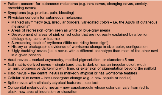
• Rx: it is not necessary to remove clinically atypical nevi prophylactically in order to confirm the presence of histological atypia; biopsies of nevi are indicated primarily when a severely atypical nevus or cutaneous melanoma is in the DDx (Table 92.4); if banal nevi become irritated (e.g. by clothing or jewelry), shave removal can be done.
Table 92.4
Potential indications for biopsy of melanocytic nevi.
Patients with numerous nevi can continue to develop new nevi, and banal nevi can increase in size over time.
* D and E represent large diameter and evolution.
• When cutaneous melanoma is in the DDx, complete removal of the nevus is recommended as well as submission to a dermatopathologist; removal can be accomplished by several methods (see Fig 1.6); providing additional information (e.g. eccentric hyperpigmented area) is also helpful.
Common (Banal) Acquired Melanocytic Nevi
• Benign lesions with varied clinical appearance, but typically ≤6 mm in diameter, symmetric, well-circumscribed, evenly pigmented, and round or oval in shape; perifollicular hypopigmentation or stippled pigmentation is often present upon closer inspection.
• Distribution favors intermittently sun-exposed areas of the trunk and extremities and, less frequently, acral sites (palms, soles, nail matrix); in patients who eventually develop many nevi, the scalp may be one of the first sites of involvement.
• Classically appear after the first 6 months of life, increase in number during childhood and adolescence, peak during the third decade, and then slowly regress with age.
• As nevi ‘age’ over time, they often become more elevated, softer, and less pigmented.
Atypical (Dysplastic or Clark's) Acquired Melanocytic Nevi
• Benign lesions that share, to a lesser extent, many clinical features of cutaneous melanoma (e.g. asymmetry, border irregularity, color variegation, and diameter >6 mm) but usually they ‘age’ in a manner similar to banal nevi and only a tiny proportion may develop melanoma within them.
• Histologically, architectural disorder is seen and cytologic atypia may be present; sometimes the latter is categorized as mild, moderate, or severe.
• The major risk factor for developing atypical nevi is genetic predisposition.
• The ‘atypical mole syndrome’ has been variably described in the literature, ranging from individuals with atypical nevi but no personal or family history of melanoma to the familial atypical multiple mole and melanoma syndrome (FAMMM syndrome), in which an individual with numerous nevi and ≥2 first-degree relatives with cutaneous melanoma has a very high risk of developing melanoma.
• Atypical nevi often appear around puberty and are thought to develop throughout life.
• Although the majority of atypical nevi are located in intermittently sun-exposed sites (e.g. trunk and extremities [lower > upper in females]), often they can be seen on the breasts and buttocks.
• Clinically, atypical nevi are recognized by various features – e.g. varied colors (pink, brown, tan), a ‘fried-egg’ appearance with a papular component and a macular rim, a larger size than banal nevi, and borders that are ill-defined, ‘fuzzy,’ and sometimes notched or irregular (see Fig. 92.9).
• Adjuncts to monitoring these patients include baseline and follow-up nevus and total body photography; dermoscopy; patient/partner education on skin self-examination and signs of melanoma; and possibly alternating examinations between two dermatologists.
• Nevi with mild cytologic atypia histologically and most with moderate cytologic atypia do not require re-excision, especially when the biopsy attempted to remove the entire nevus; it is generally recommended, however, that severely atypical nevi be completely excised or re-excised with conservative margins (e.g. 3–5 mm).
Halo Nevus
• A melanocytic nevus that is surrounded by a round or oval, usually symmetric, halo of complete depigmentation (i.e. white color).
• The central nevus is most often a common acquired melanocytic nevus, but it can also be other nevus subtypes.
• The halo of depigmentation is believed to represent a T-cell-mediated immune response against nevus antigens, analogous to vitiligo (see Chapter 54).
• Halo nevi are seen in up to 5% of Caucasian children aged 6–15 years; they are more common in patients with an increased number of nevi and a personal or family history of vitiligo.
• The most common location is the back; multiple halo nevi are seen in ~50% of cases.
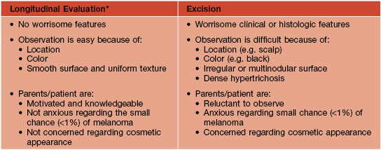
• There are four clinical stages in the life of a halo nevus, and the evolution usually occurs over years to decades (Fig. 92.10).
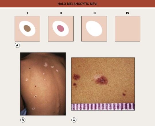
Fig. 92.10 Halo melanocytic nevi. A Four potential clinical stages in the life of a halo nevus. Stage I: central pigmented melanocytic nevus with a halo of depigmentation; Stage II: central nevus is pink with a halo of depigmentation; Stage III: no central nevus, just a depigmented macule; Stage IV: repigmentation (partial or complete). B Multiple halo nevi (seen here in all four of the various stages) are seen most commonly in children and young adults with numerous nevi. C A benign compound melanocytic nevus with an asymmetric halo. C, Courtesy, Jean L. Bolognia, MD.
• The central nevus should be assessed for suspicious or clinically atypical features; if none are present, then no treatment is necessary (vast majority of lesions); if present, then biopsy of just the central nevus should be performed.
• A new onset of multiple halo nevi is unusual in middle-aged and older adults; this clinical scenario should raise the possibility of the halo nevi representing an autoimmune reaction against a cutaneous or ocular melanoma.
• DDx: other pigmented lesions that can have halos (e.g. solar lentigo, seborrheic keratosis), halo primary cutaneous melanoma, halo melanoma metastasis.
Blue Nevi
• Benign lesions due to a proliferation of dendritic melanocytes within the dermis; blue in color because of the Tyndall effect (see above).
• Sites of predilection are those areas in which active dermal melanocytes are still present at the time of birth (e.g. head, neck, dorsal hands and feet, sacral region).
• Most blue nevi have a somatic activating mutation in GNAQ, which encodes the Q-class of G-protein α subunits.
• The common blue nevus typically presents as a solitary, blue to blue-black, dome-shaped papule, usually <1 cm in diameter and most often occurring on the dorsal hands or feet (see Fig. 92.9); frequently arises in adolescence; no malignant potential.
• The cellular blue nevus tends to be larger in size (i.e. more plaque-like), and it can be congenital or acquired (see Fig. 92.9); favors the head, sacral region, and buttocks; melanoma (malignant blue nevus) can develop within cellular blue nevi (see Chapter 93); if location (e.g. scalp) or patient awareness prevents satisfactory observation, then complete excision can be performed.
• The combined blue nevus typically presents as a blue to gray-brown macule or papule with a subtle brown rim; it usually represents a combination of a blue nevus with a banal nevus, but in theory can be a combination of any two types of nevi.
• DDx: traumatic tattoo (e.g. carbon from a pencil), which typically can be distinguished by history, or a venous lake (compressible) > melanoma metastasis, primary cutaneous melanoma.
• Rx: none for common blue nevi and as noted above for cellular blue nevi; lesions with a history of recent growth or change should be biopsied.
• Multiple blue nevi (normal in Asians) may signify an underlying syndrome, e.g. Carney complex (see Table 92.3).
Spitz Nevi (Spindle and Epithelioid Cell Nevi)
• Benign proliferations of epithelioid and/or spindled melanocytes.
• Majority appear during childhood or young adulthood and most commonly occur on the face and lower extremities.
• Classically, a Spitz nevus is symmetric, well-circumscribed, <1 cm in diameter, uniformly pink, tan, red, or red-brown, smooth, and dome-shaped (see Fig. 92.9); occasionally, lesions are verrucous or darkly pigmented.
• Pigmented spindle cell nevus of Reed is a variant that typically presents in young women as a very dark brown to black minimally elevated papule, most often on the thigh; characteristic ‘starburst pattern’ on dermoscopy (see Fig. 92.9).
• DDx: clinical: intradermal melanocytic nevus, juvenile xanthogranuloma, molluscum contagiosum, pyogenic granuloma, verruca vulgaris, dermatofibroma, cutaneous melanoma; histologic: when atypical features, cutaneous melanoma.
• Molecular techniques (e.g. FISH or CGH analysis, BRAFV600E and H-RAS mutational status) may help better categorize difficult lesions.
• Spitz nevi have a tendency to involute over time, and monitoring is an option for a presumed Spitz nevus that develops during childhood and has classic clinical and dermoscopic features.
• Any presumed Spitz nevus with clinically atypical features (e.g. size >1 cm, ulceration, asymmetry) should be biopsied.
• Any Spitz nevus with unusual features histologically should be completely excised.
Nevi of Special Sites
• Nevi in certain anatomic locations (e.g. scalp) may exhibit atypical clinical features, whereas those in several specific sites (e.g. acral, genital, auricular, milk line, flexural, and scalp nevi) may have atypical histologic features that simulate cutaneous melanoma (see Fig. 92.9).
• Recognition of this phenomenon prevents overdiagnosis of cutaneous melanoma.
Other ‘Specially Named’ Nevi
• Meyerson or eczematous nevus: a melanocytic nevus with an eczematous halo; eczematous reaction typically resolves spontaneously or with the application of a topical CS.
• Eclipse nevus: benign melanocytic nevus most often found on the scalp of children; presents as a tan, occasionally pink, central macule or papule with a brown, often stellate rim (see Fig. 92.9); ages into an intradermal nevus; when present on the scalp, may be a marker for developing numerous nevi elsewhere over time.
• Cockade nevus: a benign melanocytic nevus with a target configuration, namely a central pigmented papule surrounded by a concentric tan rim, which is then surrounded by another pigmented annulus (see Fig. 92.9); occurs in patients with eclipse nevi.
• Recurrent nevus: the reappearance of pigment within the scar of a previously biopsied or excised nevus (see Fig. 92.9); if symmetric, re-evaluation of the original histology and observation is generally all that is required; if asymmetric or continues to enlarge after its initial appearance, then biopsy is prudent.
• Agminated nevus: a clustering of melanocytic nevi within normal-appearing skin.
Congenital Melanocytic Nevi (CMN)
• Classically defined as a melanocytic nevus present at birth; may be subtle at birth and not readily apparent for a few months.
• Melanocytic nevi that become apparent after 3 months of age, but before 2 years of age, have been termed ‘tardive CMN,’ ‘congenital nevus-like nevi,’ or ‘early onset nevi’.
• CMN are due to a proliferation of melanocytes that arise during embryogenesis; melanocytes extend deep into the dermis and subcutaneous tissues and often follow follicular and neurovascular structures; NRAS mutations have been detected in CMN and associated neurologic lesions (see below).
• Incidence of small CMN is estimated at 1–2%, with large CMN having an incidence of ~1 in 20 000 individuals.
• CMN are classified into four groups, based on their final adult size:
– Small CMN: <1.5 cm (Fig. 92.11A).
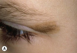
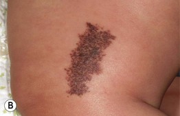
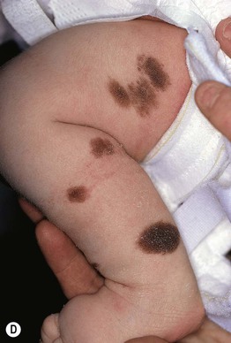
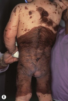
Fig. 92.11 Congenital melanocytic nevi (CMN). A Small CMN. Note the hypertrichosis and slight heterogeneity in pigmentation, reflecting its hamartomatous nature. B Medium-sized CMN. Note the pebbly appearance on clinical examination and the globular pattern with hyphae-like structures on dermoscopy (C). D Multiple medium-sized CMN. This patient had 25–30 such lesions. This presentation can be associated with neurocutaneous melanosis (NCM). E Giant CMN, also called a ‘bathing suit’ nevus. It is situated in a posterior axial location with numerous satellite nevi, some of which have hypertrichosis. This patient died of intractable ascites due to the migration of benign melanocytes from the brain to the peritoneal cavity via his VP shunt. In the perianal region, the nevus was softer and somewhat ‘boggy’ due to neurotization. A, Courtesy, Lorenzo Cerroni, MD; B, C, Courtesy, Raymond L. Barnhill, MD, and Harold S. Rabinovitz, MD; D, E, Courtesy, Jean L. Bolognia, MD.
– Medium CMN: 1.5–20 cm (Fig. 92.11B–D).
– Large CMN: >20–40 cm (in a neonate, large CMN are >9 cm on the head or >6 cm on the body).
– Giant CMN: >40 cm (Fig. 92.11E).
• Small or medium-sized CMN are usually solitary, can occur anywhere on the body, range in color from tan to black, and may have increased terminal hair growth (hypertrichosis).
• Giant CMN are sometimes referred to as ‘bathing suit’ or ‘garment’ nevi because of their distribution pattern; multiple smaller, widely disseminated ‘satellite’ nevi are also commonly associated with these giant CMN.
• CMN grow proportionately with the child and over time can change from initially flat, evenly pigmented patches to elevated, mottled, pebbly or verrucous plaques; scalp CMN may lighten in color and gradually regress; papules or nodules can develop within the CMN, but signs of rapid growth, induration, or ulceration should raise suspicion for the development of melanoma and prompt a biopsy.
• CMN are a known risk factor for melanoma and neurocutaneous melanosis (NCM), with the absolute risk being associated with the severity of the cutaneous phenotype.
• The risk of developing melanoma within a small or medium-sized CMN is thought to be <1% over a lifetime; the risk for giant CMN is ~5%, with greater risk being a function of nevus size and number of satellite lesions; in giant CMN, the majority of melanomas develop in childhood (~50% in the first 5 years of life).
• Cutaneous melanomas can arise within the dermis or subcutaneous tissues of a CMN, making clinical diagnosis difficult.
• Patients with a giant CMN in a posterior axial location that is associated with multiple satellite nevi (≥20) have the greatest risk of developing melanoma; although the melanoma can develop within the CMN itself (not the satellite lesions), the CNS, or the retroperitoneum, the primary site may remain unknown.
• In neurocutaneous melanosis (NCM), also referred to as leptomeningeal melanocytosis, there is a proliferation of melanocytes in the CNS (as well as the skin); individuals with multiple (≥3, but usually 10–20+), disseminated medium-sized CMN are at greatest risk of NCM, followed by those with a giant CMN in a posterior axial location with multiple satellites.
• NCM-associated signs – e.g. hydrocephalus, seizures, developmental delay, increased intracranial pressure, cranial nerve palsies, sensorimotor defects, and hypotonia – tend to manifest by 2–3 years of age.
• An approach to the management of CMN is presented in Table 92.5 and Fig. 92.12.
Table 92.5
Approach to the patient with a small or medium-sized congenital melanocytic nevus (CMN).
* Includes baseline photography, measurements, and periodic skin examinations.
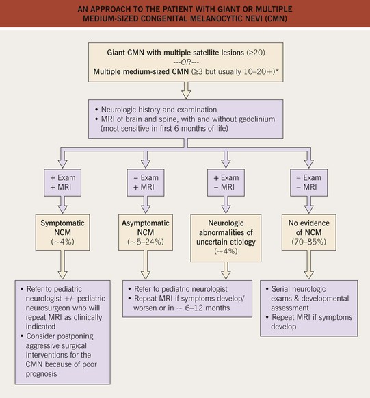
Fig. 92.12 An approach to the patient with giant or multiple medium-sized congenital melanocytic nevi (CMN). For large lesions that are on an extremity or the abdomen, with few, if any, satellite lesions, the risk of neurocutaneous melanosis (NCM) is low. *A higher proportion of these patients may have NCM compared to those with a giant CMN with multiple satellite lesions.
Speckled Lentiginous Nevus (SLN, Nevus Spilus)
• A subtype of CMN with a prevalence of ~2% in the general population.
• Often presents at or around birth as a tan patch; over time numerous macules and papules develop within the tan patch, ranging from lentigines to junctional, compound and intradermal nevi to Spitz and blue nevi (Fig. 92.13).
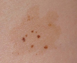
Fig. 92.13 Speckled lentiginous nevus (nevus spilus). Multiple brown macules and papules superimposed upon a tan patch. Courtesy, Raymond L. Barnhill, MD, and Harold S. Rabinovitz, MD.
• The risk of developing melanoma within the SLN is thought to be similar to classic CMN of the same size range, but may be related to the type of nevi that develop within it.
• DDx: agminated nevi (see above); partial unilateral lentiginosis.
• SLN should be followed with periodic examinations, photography, and biopsies as clinically indicated.
For further information see Ch. 112. From Dermatology, Third Edition.