The skeletal system*
Learning objectives
When you finish this chapter, you will be familiar with bone shapes, common types of bones, bone features, and clinically significant bones. You will not be asked to learn all the bones in the body, but you will be able to name most of them. Luckily the common domestic animal species generally have the same bones so you’ll have to learn them only once.
Terms to be identified
Bone shapes
Flat bones
Irregular bones
Sesamoid bones
Long bones
Short bones
Types of bone
Cancellous bone
Compact bone
General bone features
Articular surface
Condyle
Head
Facet
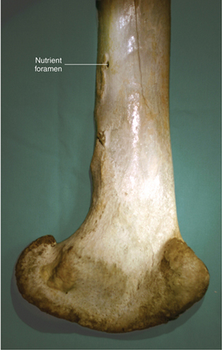
Foramen
Nutrient foramen
Fossa
Processes
Bones of the axial skeleton
Skull bones (external)
Frontal bones
Cornual process
Incisive bones
Interparietal bones
Lacrimal bones
Mandible
Ramus
Shaft
Maxillary bones
Nasal bones
Occipital bone
Foramen magnum
Occipital condyles
Parietal bones
Temporal bones
External acoustic meatus
Tympanic bullae
Zygomatic bones
Skull bones (internal)
Turbinates
Ribs
Costal cartilage
Costochondral junction
Head
Sternum
Manubrium (manubrium sterni)
Xiphoid (xiphoid process)
Vertebrae
Arch
Body
Processes
Articular processes
Spinous process
Transverse processes
Cervical vertebrae
Atlas
Axis
Thoracic vertebrae
Lumbar vertebrae
Sacral vertebrae (sacrum)
Coccygeal vertebrae
Bones of the appendicular skeleton
Thoracic limb
Scapula
Glenoid cavity
Neck
Spine
Humerus
Condyle
Epicondyles
Greater tubercle
Head
Neck
Olecranon fossa
Radius
Head
Neck
Styloid process
Ulna
Anconeal process
Coronoid processes
Olecranon process
Radial notch
Styloid process
Trochlear notch
Carpal bones (carpus)
Accessory carpal bone
Metacarpal bones
Cannon bone (hoofed animals)
Splint bones (horse)
Phalanges
Proximal sesamoid bones (hoofed animals)
Distal sesamoid bones (hoofed animals)
Pelvic limb
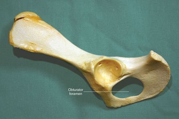
Pelvis
Acetabulum
Ilium
Ischium
Obturator foramen
Pubis
Femur
Condyles
Epicondyles
Greater trochanter
Head
Neck
Trochlea
Patella
Fabellae
Tibia
Condyles
Tibial crest
Tibial tuberosity
Medial malleolus
Fibula
Lateral malleolus
Tarsal bones (tarsus, hock)
Calcaneus
Metatarsal bones
Cannon bone (hoofed animals)
Splint bones (horse)
Phalanges
Proximal sesamoid bones (hoofed animals)
Distal sesamoid bones (hoofed animals)
Joints
Cartilaginous
Fibrous
Synovial
Clinical significance
Suppose you have been asked to take two radiographs of a patient’s left femur—a lateral (side-to-side) view and a cranial–caudal view. To take the shots, you need to know a few things:
1. Which bone is the femur? (thigh bone)
2. What externally palpable (feelable) landmark can be used to identify the proximal end of the bone? (greater trochanter)
3. What externally palpable landmarks can be used to identify the distal end of the bone? (patella, medial and lateral condyles and epicondyles)
Suppose you are doing a physical exam on a cat and as you palpate the hind legs, one leg seems to have a bump on a bone and the other leg doesn’t have the same bump on the same bone. Is the bone supposed to have the bump or not? What do you write on the physical exam sheet?
When you listen to heart and lung sounds in animals, you place the stethoscope at various places on the animals’ chests. Sometimes it’s next to the costochondral junction and other times it’s placed between the ribs more cranially or caudally. Specific sounds are heard at specific places. You will need to be able to locate these specific places by counting ribs and locating the area where the ribs attach to the cartilages that anchor them to the sternum.
Many other structures are named by which bones they are near. For example, the radial nerve is found by the radius bone in the front leg. The ulnar nerve is found by the ulna bone in the front leg. The femoral artery and vein run alongside the femur in the hind leg. If you know where the bone is located, you will have an easier time locating and identifying structures near it.
Bones have many physical characteristics. You will need to know their normal anatomy before you can detect abnormalities.
As you study the bones, feel them on live animals. Figure out which parts can be palpated on the outside of various animals’ bodies. Some things that are easily felt on a cat may be difficult or impossible to feel on a horse, and vice versa.
Introduction
The bones that make up an animal’s skeleton (1) support and (2) protect the soft tissues of the body, and (3) act as levers that the skeletal muscles use to move the body. They also (4) store minerals, particularly calcium.
• Bones providing support. The support function is often easy to visualize. The bones of the limbs support the rest of the body. Other bones perform their supporting roles more subtly. The ribs, for example, help maintain the size and shape of the thoracic (chest) cavity. They help prevent the lateral walls of the thorax from collapsing in response to gravity. The lumbar (abdominal) vertebrae support the weight of the abdominal organs by serving as the attachment sites for the slinglike abdominal muscles.
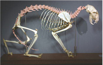
• Bones providing protection. The best examples of bones playing protective roles are the bones of the cranium (the portion of the skull that surrounds the brain) and the vertebrae that make up the spinal column. The very important brain and spinal cord are soft and fragile. If you held an animal’s brain in your hand, you could easily crush it between your fingers. The spinal cord has the same soft consistency. The bones of the cranium form a rigid case for the delicate brain. The arches of the vertebrae combine to form a bony canal (the spinal canal) that protects the fragile spinal cord. The individual joints between the vertebrae allow limited movement. These joints combine to allow the spine as a whole a considerable range of motion while still protecting the enclosed spinal column. Cats in particular give excellent demonstrations of spinal flexibility as they contort themselves.
• Bones as levers. The role of bones as levers is easy to visualize. The articular (joint) surfaces are the fulcrums for the levers, and the processes where the tendons attach muscles to bones are where the forces are applied. Note that the larger the process, generally the greater the force that can be applied there. Note also that, as a result of the body being sleek and compact, some bony levers have very poor mechanical advantage. This results in some muscles, such as the gluteals, having to be fairly large and powerful.
• Bones as storage sites. Bones act as reservoirs for important minerals, such as calcium. This storage capacity enables the body to deposit and withdraw vital minerals precisely as needed to maintain health.
As you examine skeletons and bones, try to imagine how the bones, joints, and muscles work together to make the body move. Look at the various bony processes and try to infer what the muscles attached to them would do when they contracted. That’s the kind of detective work paleontologists perform when they examine the fossilized bones of dinosaurs and other extinct beasts. They can make educated guesses about things such as muscle sizes, shapes, and strengths, body shapes, and even how the animal moved.
Medical word parts
The skeletal system
Types of bone
Virtually all bones are made up of two types of bone: cancellous bone, which is light and spongy; and compact bone, which is heavy and dense (Figures 6-1 and 6-2).


Bone shapes
Long bones
Long bones are so named because they are longer than they are wide (Figure 6-3). Most of the bones of the limbs are long bones. The two bones in Figure 6-3 are a feline femur (A) from the thigh area of the hind leg, and a canine humerus (B) from the upper front leg. The ends of long bones are called epiphyses. Each long bone has a proximal epiphysis and a distal epiphysis. The epiphyses are made of cancellous bone covered with a thin layer of compact bone. The long part of a long bone is the diaphysis. It is composed primarily of compact bone. Between the epiphyses and diaphysis, there are the areas where the bone grows longer in young animals. These growth plates are composed mainly of cartilage and are called the epiphyseal plates. The epiphyseal plates are the weakest part of the bone in young animals and are prone to fractures. Once an animal reaches its full size, the epiphyseal plates are replaced by solid bone through a process called ossification.

Short bones
This equine carpus shows short bones that are shaped like cubes or marshmallows (Figure 6-4). They are composed of an inner core of cancellous bone covered by a thin layer of compact bone. Carpal and tarsal bones are the most common short bones in the body (Figure 6-5).


Flat bones
Flat bones are mostly flat and thin (Figure 6-6, and see Figure 6-10). Their structure is like a “cancellous bone sandwich”—a central layer of cancellous bone covered on both sides by thin layers of compact bone. The pelvic bones and the scapula (shoulder blade) are prominent flat bones as are some of the skull bones (Figure 6-7).


Irregular bones
Irregular bones are odd-shaped and don’t fit into any of the other three categories (Figures 6-8, 6-9, 6-10, and 6-11).


Common bone features (lumps, bumps, grooves, and holes)
Articular surfaces
Articular surfaces are smooth areas of compact bone that come in contact with smooth surfaces of another bone to form a joint. The articular surfaces are covered with hyaline cartilage (Figure 6-12).

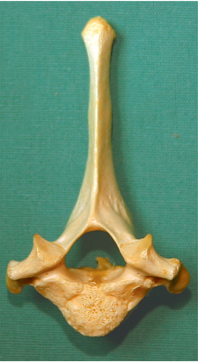
Condyles
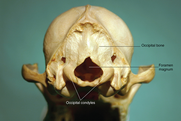
A condyle is usually a large, round articular surface. The distal ends of the femur and humerus, and the occipital bone have the most prominent condyles (Figures 6-13 to 6-15).


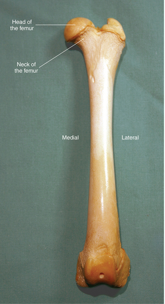
Head
The head of a bone is found at the proximal end of a long bone. It is mostly spherical in shape. The proximal ends of the femur, humerus, and ribs have heads (Figures 6-16 to 6-18). The head is usually joined to the rest of the bone by a narrowed region called the neck.


Facet
A facet is a flat articular surface. It is found on carpal bones, tarsal bones, vertebrae, and some long bones, such as the radius and ulna (Figures 6-19 and 6-20).


Processes
Processes are the lumps and bumps on bones. Condyles and heads on long bones are considered processes, but they have a specific articular function so they are classified as articular surfaces. Most of the other processes on bones are places where the tendons of muscles attach to the bone. Larger processes are where more powerful muscles attach. Processes are given different names on different bones. Some of the names used are trochanter (femur), tubercle (humerus), tuber (ischium), crest (tibia), olecranon (ulna), spine (scapula), and wing (atlas) (Figures 6-21 to 6-27).


Holes and depressed areas
A hole in a bone is called a foramen. Usually it is a passageway for blood vessels or nerves to enter and leave the bone (Figures 6-28 and 6-29).
A fossa is a depressed, sunken area on the surface of a bone. Bone fossae are usually occupied by muscles or tendons (Figures 6-30 and 6-31).


The skeleton
Even though an animal has only one complete skeleton, we are going to divide it into two main parts (skeletons) for the purpose of studying the bones. The axial skeleton is made up of the bones located on or near the central cranial–caudal axis of the body—the skull, hyoid bone, spinal column, ribs, and sternum. The appendicular skeleton is made up of the main “appendages” of the body: the thoracic limbs and the pelvic limbs.
Axial skeleton
The axial skeleton is made up of the skull, hyoid bone, spinal column, ribs, and sternum. All the bones of the axial skeleton are located at or near the median plane of the animal’s body (Box 6-1 and Figure 6-32).

Skull
The skull is the most complex part of the skeleton. It is made up of bones that, with one exception, are united by jagged, immovable, fibrous joints called sutures. The only freely movable joint is the one between the mandible and the temporal bone. This joint is called the temporomandibular joint or TMJ.
The bones of the skull can be conveniently grouped into the bones of the cranium, which surround the brain, and the bones of the face. We will restrict our examinations to the external bones in each group, which are at least partly visible on the outside of the skull (Box 6-2). The names of these external skull bones are often used to describe locations on animals’ heads (e.g., left parietal region, right maxillary area) in medical records. The internal skull bones are hidden within the skull. The only internal bones we will cover are the nasal turbinates, which play important roles in the conditioning of inhaled air on its way to the lungs (Figures 6-33 to 6-38).


External bones of the cranium
Occipital bone
Interparietal bones
Parietal bones
Temporal bones
• Two bones that form the ventrolateral portion of the cranium
• Contain the middle and inner ear structures
• Form a portion of the zygomatic arch
• Form the temporomandibular joint with the mandible
• The external acoustic meatus opening leads to the middle and inner ear cavities
• The tympanic bullae, “egg-shaped” swellings on the ventral surface, contain the middle ear structures
Frontal bones
External bones of the face
Incisive bones
Nasal bones
Maxillary bones
Lacrimal bones
Zygomatic bones
Note: The zygomatic arches are easily palpated on each side of an animal’s head just below and behind the eyes. They form the widest part of dog and cat skulls.
Mandible
• Two bones united rostrally by the mandibular symphysis in dogs, cats, and cattle
• One solid bone in adult horses and swine
• The shaft is the horizontal part that houses all the lower teeth
• The ramus is the vertical part at the caudal end that forms the temporomandibular joint with the temporal bone. This is also where the powerful jaw muscles attach
Internal bones of the face
Turbinates
Hyoid bone
The hyoid bone (also known as the hyoid apparatus) attaches to the temporal bones and supports the base of the tongue, the pharynx, and the larynx. It is made up of several individual parts united by cartilage, but is usually referred to as a single bone (Figure 6-39).

Spinal column
The spinal column, also known as the vertebral column, is made up of a series of irregular bones called vertebrae that extend from the skull to the tip of the tail. A typical vertebra consists of a ventral body, a dorsal arch, and a group of processes. The body is the heaviest, most dense part of the bone. The bodies of adjacent vertebrae are separated by cartilaginous intervertebral discs. The arch dorsal to the vertebral body houses the spinal cord in the living animal. Three kinds of process are commonly found on vertebrae: a single spinous process that projects dorsally, two transverse processes that project laterally, and articular processes on the cranial and caudal ends of the vertebra (Figure 6-40).

Vertebrae are grouped into five regions—cervical (neck region, abbreviated “C”), thoracic (chest region, abbreviated “T”), lumbar (abdominal region, abbreviated “L”), sacral (pelvic region, abbreviated “S”), and coccygeal (tail region, abbreviated “Cy”). Most vertebrae do not have specific names, but are identified by numbers within each region from cranial to caudal. A shorthand method of identifying vertebrae uses the abbreviation for the vertebral group followed by the number of the particular vertebra. For example vertebra C5 is the fifth cervical vertebra and T10 is the tenth thoracic vertebra (Box 6-3 and Figure 6-41).
The first two cervical vertebrae are unusual in shape compared with the rest of the vertebrae, and they have specific names. The first cervical vertebra (C1) is called the atlas. It does not have a vertebral body, but consists of a bony ring that the spinal cord passes through and two large transverse processes called the “wings” of the atlas. The occipital bone of the skull forms a joint with the atlas—the atlantooccipital joint. The second cervical vertebra (C2) is called the axis. Its main characteristics are a large, bladelike spinous process and the peglike dens on its cranial end that tucks into the caudal end of the atlas to help form and stabilize the atlantoaxial (C1–C2) joint. The rest of the cervical vertebrae are fairly normal in appearance, and are just identified by number, like the rest of the vertebrae (Figure 6-42).

The number of thoracic vertebrae is usually equal to the numbers of pairs of ribs the animal has. The main characteristics of thoracic vertebrae are their tall spinous processes and their lateral articular facets, which form joints with the heads of the ribs.
The lumbar vertebrae are the most massive-looking vertebrae of the spinal column. They have to support the weight of the abdominal organs without the aid of the ribs, which help support the organs in the chest.
The sacral vertebrae are fused into a single solid structure called the sacrum. The sacrum forms a joint with the ilium of the pelvis—the sacroiliac joint.
The coccygeal vertebrae are the bones of the tail. At the cranial end, the first few coccygeal vertebrae look like small versions of normal vertebrae. They have bodies, arches, and processes. Further caudally, however, they gradually turn into simple little rods of bone.
Ribs
Ribs are flat bones that form the lateral sides of the thorax. They articulate with the thoracic vertebrae dorsally. The ventral part of a rib is composed of costal (rib) cartilage. Where the cartilaginous part meets the bony part is the costochondral junction. The costal cartilages join either the sternum or the costal cartilage of the ribs ahead of them (Figures 6-43 to 6-46).

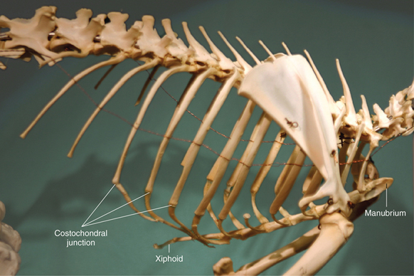


Sternum
The sternum (breastbone) is made up of bones called sternebrae. The first sternebra is named the manubrium (full name manubrium sterni). The last sternebra is named the xiphoid (full name xiphoid process). A piece of cartilage that extends off the caudal end of the xiphoid process is the xiphoid cartilage, which can be palpated at the caudal end of the sternum in most animals (Figure 6-47).

Appendicular skeleton
The appendicular skeleton is made up of the thoracic (front) and pelvic (hind) limb bones of the animal. They make up the main appendages of the body, hence the name (Box 6-4 and Figure 6-48).

Thoracic limb
The thoracic limb is the front leg. In most domestic animals it has no bony connection to the axial skeleton. Instead, the weight of the front part of the body is supported by a slinglike arrangement of muscles and tendons. From proximal to distal, the bones of the thoracic limb are the scapula, humerus, radius and ulna, carpal bones, metacarpal bones, and phalanges.
Scapula
• Commonly called the shoulder blade
• Flat and somewhat triangular in shape (Figure 6-49)
• The scapular spine is a ridge that projects laterally
• The concave glenoid cavity is the socket part of the ball-and-socket shoulder joint
• The neck joins the glenoid cavity to the main part of the bone

Humerus
• Long bone of the brachium (“upper arm”) (Figure 6-50)
• The rounded head on proximal end is the ball part of the ball-and-socket shoulder joint
• The neck joins the head to the shaft (not as obvious as the neck of the femur)
• The greater tubercle on proximal end is a large process to which shoulder muscles attach
• The distal articular surface is collectively called the condyle (the medial part is the trochlea and the lateral part is the capitulum)
• Medial and lateral epicondyles are the “knobs” on the medial and lateral sides of the condyle
• The olecranon fossa is the indentation on the caudal surface just proximal to the condyle, which the anconeal process of the ulna tucks into when the elbow is extended

Ulna
• Long bone of the antebrachium (“forearm”) (Figure 6-51)
• Along with the radius, forms the elbow joint with the humerus
• The large olecranon process on the proximal end is the attachment site for the triceps brachii muscle
• The trochlear notch is a half-moon–shaped, concave articular surface that wraps around the trochlea of the humeral condyle to help make the elbow joint a very tight, secure joint
• The anconeal process is a beak-shaped process at the proximal end of the trochlear notch
• Medial and lateral coronoid processes on distal end of trochlear notch are located on the ends of the horizontal, concave radial notch where the proximal end of the radius articulates with the ulna
• The shaft of the ulna extends down to the carpus in all common domestic species except the horse
• The equine ulna consists only of the proximal structures and joins the radius about midshaft
• The styloid process on distal end articulates with the carpus

Radius
• Main weight-bearing bone of the antebrachium (“forearm”) (Figure 6-52)
• The head on the proximal end has a large, concave articular surface that articulates with the capitulum of the humeral condyle to form part of the elbow joint
• The neck on the proximal end connects the head with the shaft of the bone
• The styloid process on the distal end articulates with the carpus

Carpal bones (carpus)
• Located immediately distal to the radius and ulna (Figures 6-53 to 6-55)
• Equivalent to the human wrist
• Consist of two parallel rows of short bones
• Bones in the proximal row are named the radial carpal, ulnar carpal, accessory carpal, and, in some species, an intermediate carpal
• The accessory carpal bone protrudes backward on the lateral side of the carpus (useful landmark for radiography)
• Bones in the distal row are numbered starting at the medial side

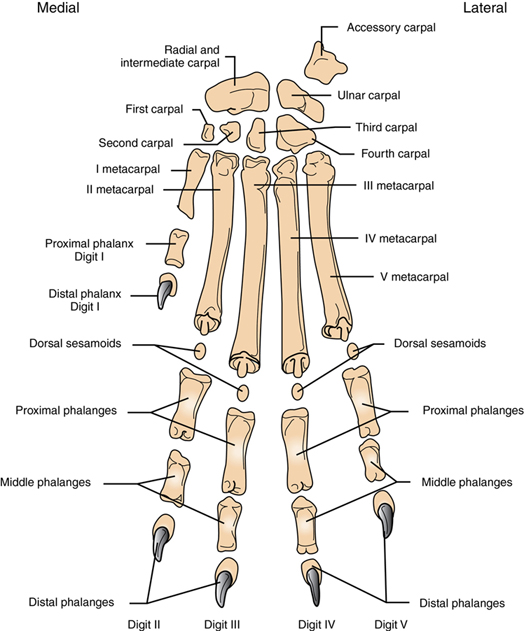

Metacarpal bones
• Extend distally from distal row of carpal bones to the proximal phalanges of the digit
• Dogs and cats typically have five metacarpal bones, numbered from medial (I) to lateral (V)
• Ruminants have a large metacarpal bone (commonly called the cannon bone) formed from two fused bones—metacarpals III and IV. A longitudinal groove shows its two-bone origin
• Horses have a single large metacarpal bone (metacarpal III, commonly called the cannon bone) with two small, incomplete metacarpal bones on either side (metacarpals II and IV, commonly called the splint bones)
Phalanges
• Phalanges (singular: phalanx) are the individual bones that make up the digits (toes)
• Dog and cat forepaws typically have four or five digits numbered from medial to lateral:
Digit I, if present, is commonly called the dewclaw and has two phalanges—proximal and distal
Digits II–V each contain three phalanges—proximal, middle, and distal
The distal phalanx contains the ungual process that is surrounded by the claw
• Ruminants have four digits on each limb:
Two support weight (III and IV)
Two are smaller non–weight-bearing “dewclaws” (II and V)
Weight-bearing digits each contain three phalanges—proximal, middle, and distal
Weight-bearing digits each contain two proximal sesamoid bones on the palmar surface of the joint between the metacarpal and the proximal phalanx, and one distal sesamoid bone on the palmar surface of the joint between the middle and distal phalanges
• Horses have one digit on each limb, composed of three phalanges and three sesamoid bones:
The phalanges are the proximal phalanx (common name: long pastern bone), middle phalanx (common name: short pastern bone), and distal phalanx (common name: coffin bone)
Two proximal sesamoid bones are located on the palmar surface of the joint between the metacarpal and the proximal phalanx
The single distal sesamoid bone (common name: navicular bone) is located on the palmar surface of the joint between the middle and distal phalanges
Pelvic limb
The pelvic limb is the hind leg. Unlike the thoracic limb, the pelvic limb is connected to the axial skeleton through the sacroiliac joint that unites the ilium of the pelvis with the sacrum of the spinal column. From proximal to distal, the bones of the pelvic limb are the pelvis, femur, tibia and fibula, tarsal bones, metatarsal bones, and phalanges.
Pelvis
• Develops from three separate bones (ilium, ischium, and pubis) on each side that eventually fuse into a solid structure (Figure 6-56)
• The names of the separate bones are used to indicate the main regions of the pelvis
• The ilium is the cranial-most area of the pelvis
• The ischium is the caudal-most area of the pelvis
• The pubis is located medially and forms the cranial part of the pelvic floor (the ischium forms the caudal part)
• The concave acetabulum on the lateral surface receives the head of the femur to form the hip joint
• The two halves of the pelvis are joined ventrally by a cartilaginous joint—the pelvic symphysis
• The obturator foramina are two large holes on either side of the pelvic symphysis that serve to reduce the weight of the pelvis

Femur
• Long bone of the “thigh” (Figure 6-57)
• The rounded head on proximal end is the ball part of the ball-and-socket hip joint
• The neck joins the head to the shaft
• The greater trochanter on proximal end is the large process to which the gluteal muscles attach
• The distal articular surfaces are rounded medial and lateral condyles
• The medial and lateral epicondyles are the “knobs” on the medial and lateral sides of the condyles
• The trochlea is a smooth articular surface on the cranial surface of the distal end in which the patella (kneecap) rides

Patella
• Largest sesamoid bone in the body
• Located in the distal tendon of the large quadriceps femoris muscle and helps protect it as it passes down over the trochlea of the femur to insert on the tibial crest (Figure 6-58)
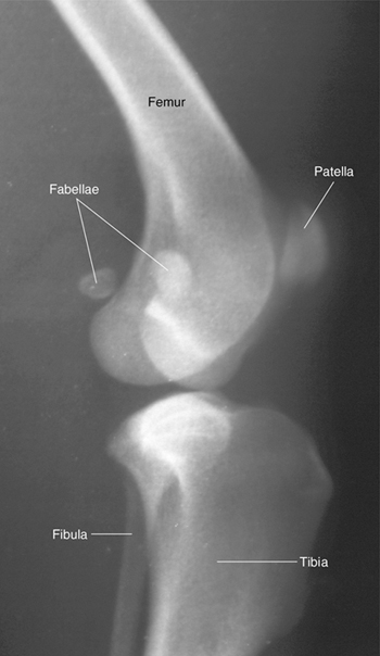
Fabellae
• The medial and lateral fabellae (see Figure 6-58) are two small sesamoid bones in the proximal gastrocnemius (calf) muscle tendons of dogs and cats
• Located just proximal to (above) and caudal to (behind) the femoral condyles
Tibia
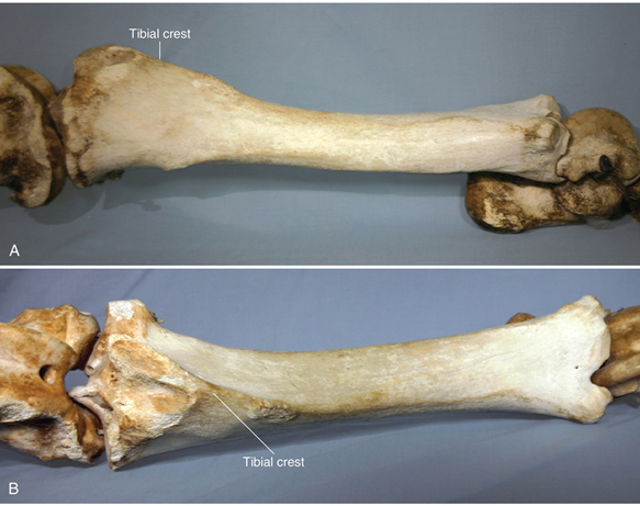
• Main weight-bearing bone of the lower leg
• The proximal end forms the stifle joint with the femur and the distal end forms the hock joint with the tarsus
• Concave tibial condyles on the proximal end articulate with the condyles of the femur
• Proximal end looks triangular when viewed from above, with the apex, the tibial tuberosity, pointed forward; the patellar tendon attaches to the tibial tuberosity
• The tibial crest is a ridge of bone that continues distally from the tibial tuberosity
• The medial malleolus is a medially facing rounded process on the distal end of the tibia (the “knob” on the medial side of our ankle is our medial malleolus)
Fibula
• Thin but complete bone in dog and cat (Figure 6-59)
• Incomplete in horses and cattle—only the proximal and distal ends are present
• Does not support significant weight
• Serves as muscle attachment site and helps form a stable joint distally with tarsus
• The lateral malleolus is the laterally facing rounded process on the distal end of the fibula (the lateral “knob” on our ankle is our lateral malleolus)

Tarsal bones (tarsus, hock)
• Located immediately distal to the tibia and fibula
• Equivalent to the human ankle
• Consist of two rows of short bones
• Bones in the proximal row are named—large tibial tarsal and fibular tarsal, and small central tarsal
• The tibial tarsal bone has a large rounded trochlea that articulates with the distal end of the tibia to form the most movable part of the hock joint
• The large calcaneus (calcaneal tuberosity) projects upward and backward to form the point of the hock; this is the attachment site for the tendon of the large gastrocnemius muscle and corresponds to our heel
• Bones in the distal row are numbered from medial to lateral, in a similar fashion to the distal row of carpal bones
Metatarsal bones
• Extend distally from the distal row of tarsal bones to the proximal phalanges of the digits
• Dogs and cats typically have four metatarsal bones, numbered from medial (II) to lateral (V)
• Ruminants have a large metatarsal bone (commonly called the cannon bone) formed from two fused bones—metatarsals III and IV. A longitudinal groove shows its two-bone origin
• Horses have a single large metatarsal bone (metatarsal III, commonly called the cannon bone) with two small, incomplete metatarsal bones on either side (metatarsals II and IV, commonly called the splint bones)
Phalanges
• Similar to the phalanges of the thoracic limb (Figures 6-60 and 6-61)
• Dog and cat hind paws typically have four digits, numbered from medial to lateral
If digit I is present, it is commonly called the dewclaw and has two phalanges—proximal and distal
Digits II–V each contain three phalanges—proximal, middle, and distal
The distal phalanx contains the ungual process that is surrounded by the claw
• Ruminants have four digits on each limb
Two support weight (III and IV)
Two are smaller non–weight-bearing “dewclaws” (II and V)
Weight-bearing digits each contain three phalanges—proximal, middle, and distal
Weight-bearing digits each contain two proximal sesamoid bones on the plantar surface of the joint between the metatarsal and the proximal phalanx, and one distal sesamoid bone on the plantar surface of the joint between the middle and distal phalanges
• Horses have one digit on each limb, composed of three phalanges and three sesamoid bones
The phalanges are the proximal phalanx (common name: long pastern bone), middle phalanx (common name: short pastern bone), and distal phalanx (common name: coffin bone)
Two proximal sesamoid bones are located on the plantar surface of the joint between the metatarsal and the proximal phalanx
A single distal sesamoid bone (common name: navicular bone) is located on the plantar surface of the joint between the middle and distal phalanges


Joints
Joints are where bones connect with each other. The three types of joint in the animal body are immovable fibrous joints, slightly movable cartilaginous joints, and freely movable synovial joints.
Fibrous joints
• Bones are firmly united by fibrous tissue
• Examples: sutures uniting most of the skull bones (Figure 6-62)

Cartilaginous joints
• Also known as amphiarthroses
• Bones are united by fibrocartilage
• Allow slight rocking movement
• Examples: pelvic symphysis, mandibular symphysis (Figure 6-63)

Synovial joints
Smooth articular surfaces covered with smooth articular cartilage
Joint capsule surrounds joint cavity that contains synovial fluid
Ligaments (fibrous connective tissue) may connect the bones together
(Note: ligaments connect bones to other bones; tendons connect muscles to bones)
Hinge joint—e.g., elbow, joints of digits (Figure 6-64)
Gliding joint—e.g., carpus (Figure 6-65)
Pivot joint—e.g., atlantoaxial joint (joint between C1 and C2 vertebrae) (Figure 6-66)
Ball and socket joint—e.g., shoulder and hip (Figure 6-67)
Flexion—decreased angle between the bones
Extension—increased angle between the bones
Adduction—movement of an extremity toward the median plane (inward)
Abduction—movement of an extremity away from the median plane (outward)
Rotation—twisting (rotational) movement
Circumduction—movement of an extremity so the distal end moves in a circle


Suggested in-class activities
Bone shapes and features
Bone shape and feature identification
Find bones that represent the various types of bone and the processes on them.
Supplies needed: disarticulated bones.
Bone shape and feature hunt
Students draw a shape or feature name out of a hat. They then must locate a bone on a skeleton that represents that shape or contains that feature. The student must also identify the bone.
OR
Students draw the name of a bone out of a hat. They must then locate the bone on the skeleton and identify the shape and what features the bone may possess.
Supplies needed: articulated skeleton.
The axial skeleton
Skull bone identification
Students examine various skulls from different species of animal, identifying the bones of the skull.
Supplies needed: various skulls.
Live animal palpation
Students palpate and identify the bones of the skull on live animals.
Skull bone hunt
Students draw the name of a skull bone out of a hat. They then must locate that bone on a live animal.
Supplies needed: live animals.
Vertebral process identification
Various vertebrae are set out. Students are to identify each vertebra by identifying the specific process types.
Supplies needed: vertebrae.
Axial assembly
Students assemble the bones of the axial skeleton in order.
Supplies needed: disarticulated skeleton.
Radiographic identification
Students identify bones of the axial skeleton on radiographs.
Supplies needed: radiographs.
Live animal palpation
Students palpate the bones of the axial skeleton on live animals.
Supplies needed: live animals.
The appendicular skeleton
Appendicular limb assembly
Students assemble, in order, both the thoracic and pelvic limbs.
Supplies needed: disarticulated skeleton.
Radiographic identification
Students identify bones of the appendicular skeleton on radiographs.
Supplies needed: radiographs.
Live animal palpation
Students palpate the bones of the appendicular skeleton on live animals.
Supplies needed: live animals.
Animals in motion
Each student is given a laminated drawing or photograph of an animal in motion. The student is to draw the location of the bones of the animal’s skeleton on the picture with an erasable marker. It is helpful to have live animals in the room for the students to position similar to their picture, then palpate for the bone locations and directions.
Supplies needed: laminated drawings and photographs of animals, erasable markers, and live animals.
Appendicular bone hunt
Students draw the name of a bone out of a hat. They then must locate that bone on an animal skeleton and/or a live animal.
Exercises
The skeletal system
Exercise 1
Identify Structures of Compact and Cancellous Bone
Identify each of the compact and cancellous bone structures by labeling where indicated.

1. _________________________________________________
2. _________________________________________________
3. _________________________________________________
4. _________________________________________________
5. _________________________________________________
6. _________________________________________________
7. _________________________________________________
8. _________________________________________________
9. _________________________________________________
10. _______________________________________________
11. _______________________________________________
12. _______________________________________________
13. _______________________________________________
14. _______________________________________________
15. _______________________________________________
Exercise 2
Identify Structures of Long Bone
______________________ 1. (What area of the bone)
______________________ 2. (What area of the bone)
______________________ 3. (What area of the bone)
______________________ 4. (What type of bone)
______________________ 5. (What normally fills this cavity)
______________________ 6. (What type of bone)

Exercise 3
Identify Bone Shape
For each of the bones pictured, identify the shape of the bone.


The skeleton
Exercise 4
Identify Rabbit Skeletal Structures

1. _______________________________________________
2. _______________________________________________
3. _______________________________________________
4. _______________________________________________
5. _______________________________________________
6. _______________________________________________
7. _______________________________________________
8. _______________________________________________
9. _______________________________________________
10. _______________________________________________
11. _______________________________________________
12. _______________________________________________
13. _______________________________________________
14. _______________________________________________
15. _______________________________________________
16. _______________________________________________
17. _______________________________________________
18. _______________________________________________
19. _______________________________________________
20. _______________________________________________
21. _______________________________________________
22. _______________________________________________
23. _______________________________________________
Exercise 5
Identify Rat Skeletal Structures

1. _______________________________________________
2. _______________________________________________
3. _______________________________________________
4. _______________________________________________
5. _______________________________________________
6. _______________________________________________
7. _______________________________________________
8. _______________________________________________
9. _______________________________________________
10. _______________________________________________
11. _______________________________________________
12. _______________________________________________
13. _______________________________________________
14. _______________________________________________
15. _______________________________________________
16. _______________________________________________
17. _______________________________________________
18. _______________________________________________
19. _______________________________________________
20. _______________________________________________
21. _______________________________________________
Exercise 6
Identify Skeletal Features of the Cat Skull
Label the bones of the skull using the following terms:
Label the bones of the skull using the following terms:

Exercise 7
Identify Bones and Processes of the Equine Thoracic Limb

Exercise 8
Identify Bones, Joints, and Processes of the Equine Pelvic Limb


