References

1. Bevilacqua MP. Endothelial-leukocyte cellular adhesion molecules. Ann Rev Immunol. 1993;11:767–804.

2. Panza JA, Quyyumi AA, Brush JE, et al. Abnormal endothelium-dependent vascular relaxation in patients with essential hypertension. N Engl J Med. 1990;323:22–27.

3. Achike F, Kwan C. Nitric oxide, human diseases and the herbal products that affect the nitric oxide signalling pathway. Clin Exp Pharmacol Physiol. 2003;30:605–615.

4. Aggarwal BB, Van Kuiken ME, Iyer LH, et al. Molecular targets of nutraceuticals derived from dietary spices: potential role in suppression of inflammation and tumorigenesis. Exp Biol Med. 2009;234(8):825–849.

5. Ghayur MN, Gilani AH, Afridi MB, et al. Cardiovascular effects of ginger aqueous extract and its phenolic constituents are mediated through multiple pathways. Vasc Pharmacol. 2005;43:234–241.

6. Yanaga A, Goto H, Nakagawa T, et al. Cinnamaldehyde induces endothelium-dependent and -independent vasorelaxant action on isolated rat aorta. Biol Pharm Bull. 2006;29(12):2415–2418.

7. Goto H, Sasaki Y, Fushimi H, et al. Effect of curcuma herbs on vasomotion and hemorheology in spontaneously hypertensive rat. Am J Chin Med. 2005;33(3):449–457.

8. Williams M, Sutherland W, McCormick M, et al. Aged garlic extract improves endothelial function in men with coronary artery disease. Phytother Res. 2005;19:314–319.

9. Pajonk F, Riedisser A, Henke M, et al. The effects of tea extracts on proinflammatory signaling. BMC Med. 2006;28:1–12.

10. Steptoe A, Gibson EL, Vuononvirta R, et al. The effects of chronic tea intake on platelet activation and inflammation: a double blind placebo controlled trial. Atherosclerosis. 2007;193:262–277.

11. Kempf K, Herder C, Erlund I, et al. Effects of coffee consumption on subclinical inflammation and other risk factors for type 2 diabetes: a clinical trial. Am J Clin Nutr. 2010;91:950–957.

12. Grassi D, Necozione S, Lippi C, et al. Cocoa reduces blood pressure and insulin resistance and improves endothelium-dependent vasodilation in hypertensives. Hypertension. 2005;46:398–405.

13. di Giuseppe R, Di Castelnuovo A, Centritto F, et al. Regular consumption of dark chocolate is associated with low serum concentrations of C-reactive protein in a healthy Italian population. J Nutr. 2008;138:1939–1945.

14. Taylor P, Noriega R, Farah C, et al. Ajoene inhibits both primary tumor growth and metastasis of B16/BL6 melanoma cells in C57BL/6 mice. Cancer Lett. 2006;239:298–304.

15. Lang A, Lahav M, Sakhnini E, et al. Allicin inhibits spontaneous and TNF-alpha induced secretion of proinflammatory cytokines and chemokines from intestinal epithelial cells. Clin Nutr. 2004;23:1199–1208.

16. Woo HM, Kang JH, Kawada T, et al. Active spice-derived components can inhibit inflammatory responses of adipose tissue in obesity by suppressing inflammatory actions of macrophages and release of monocyte chemoattractant protein-1 from adipocytes. Life Sci. 2007;80:926–931.

17. Chainy GB, Manna SK, Chaturvedi MM, et al. Anethole blocks both early and late cellular responses transduced by tumor necrosis factor: effect on NF-kappaB, AP-1, JNK, MAPKK and apoptosis. Oncogene. 2000;19:2943–2950.

18. Kowalski J, Samojedny A, Paul M, et al. Effect of apigenin, kaempferol and resveratrol on the expression of interleukin-1beta and tumor necrosis factor-alpha genes in J774.2 macrophages. Pharmacol Rep. 2005;57:390–394.

19. Bhutani M, Pathak AK, Nair AS, et al. Capsaicin is a novel blocker of constitutive and interleukin-6-inducible STAT3 activation. Clin Cancer Res. 2007;13:3024–3032.

20. Lo AH, Liang YC, Lin-Shiau SY, et al. Carnosol, an antioxidant in rosemary, suppresses inducible nitric oxide synthase through down-regulating nuclear factor-kappaB in mouse macrophages. Carcinogenesis. 2002;23:983–991.

21. Medeiros R, Passos GF, Vitor CE, et al. Effect of two active compounds obtained from the essential oil of Cordia verbenacea on the acute inflammatory responses elicited by LPS in the rat paw. Br J Pharmacol. 2007;151:618–627.

22. Kim DH, Kim CH, Kim MS, et al. Suppression of age-related inflammatory NF-kappaB activation by cinnamaldehyde. Biogerontology. 2007;8:545–554.

23. Marin YE, Wall BA, Wang S, et al. Curcumin downregulates the constitutive activity of NF-kappaB and induces apoptosis in novel mouse melanoma cells. Melanoma Res. 2007;17:274–283.

24. Thejass P, Kuttan G. Antiangiogenic activity of iallyl sulfide (DAS). Int Immunopharmacol. 2007;7:295–305.

25. Lee YY, Hung SL, Pai SF, et al. Eugenol suppressed the expression of lipopolysaccharide-induced proinflammatory mediators in human macrophages. J Endod. 2007;33:698–702.

26. Tripathi S, Maier KG, Bruch D, et al. Effect of 6-gingerol on pro-inflammatory cytokine production and costimulatory molecule expression in murine peritoneal macrophages. J Surg Res. 2007;138:209–213.

27. Souza MC, Siani AC, Ramos MF, et al. Evaluation of anti-inflammatory activity of essential oils from two Asteraceae species. Pharmazie. 2003;58:582–586.

28. Berchtold CM, Chen KS, Miyamoto S, et al. Perillyl alcohol inhibits a calcium-dependent constitutive nuclear factor-kappaB pathway. Cancer Res. 2005;65:8558–8566.

29. Weglarz L, Wawszczyk J, Orchel A, et al. Phytic acid modulates in vitro IL-8 and IL-6 release from colonic epithelial cells stimulated with LPS and IL-1beta. Dig Dis Sci. 2007;52:93–102.

30. Pradeep CR, Kuttan G. Piperine is a potent inhibitor of nuclear factor-kappaB (NF-kappaB), c-Fos, CREB, ATF-2 and proinflammatory cytokine gene expression in B16F-10 melanoma cells. Int Immunopharmacol. 2004;4:1795–1803.

31. Min YD, Choi CH, Bark H, et al. Quercetin inhibits expression of inflammatory cytokines through attenuation of NF-kappaB and p38 MAPK in HMC-1 human mast cell line. Inflamm Res. 2007;56:210–215.

32. Manjeet KR, Ghosh B. Quercetin inhibits LPS-induced nitric oxide and tumor necrosis factor-alpha production in murine macrophages. Int J Immunopharmacol. 1999;21:435–443.

33. Shishodia S, Majumdar S, Banerjee S, et al. Ursolic acid inhibits nuclear factor-kappaB activation induced by carcinogenic agents through suppression of IkappaBalpha kinase and p65 phosphorylation: correlation with down-regulation of cyclooxygenase 2, matrix metalloproteinase 9, and cyclin D1. Cancer Res. 2003;63:4375–4383.

34. Woo HM, Kang JH, Kawada T, et al. Active spice-derived components can inhibit inflammatory responses of adipose tissue in obesity by suppressing inflammatory actions of macrophages and release of monocyte chemoattractant protein-1 from adipocytes. Life Sci. 2007;80:926–931.

35. Mo SJ, Son EW, Rhee DK, et al. Modulation of TNF-alpha-induced ICAM-1 expression, NO and H2O2 production by alginate, allicin and ascorbic acid in human endothelial cells. Arch Pharm Res. 2003;26:244–251.

36. Hwang ES, Lee HJ. Allyl isothiocyanate and its N-acetylcysteine conjugate suppress metastasis via inhibition of invasion, migration, and matrix metalloproteinase-2/-9 activities in SK-Hep 1 human hepatoma cells. Exp Biol Med (Maywood). 2006;231:421–430.

37. Chen CC, Chow MP, Huang WC, et al. Flavonoids inhibit tumor necrosis factor-alpha-induced up-regulation of intercellular adhesion molecule-1 (ICAM-1) in respiratory epithelial cells through activator protein-1 and nuclear factor-kappaB: structure-activity relationships. Mol Pharmacol. 2004;66:683–693.

38. Park WH, Kim SH, Kim CH. A new matrix metalloproteinase-9 inhibitor 3,4-dihydroxycinnamic acid (caffeic acid) from methanol extract of Euonymus alatus: isolation and structure determination. Toxicology. 2005;207:383–390.

39. Mitra A, Chakrabarti J, Banerji A, et al. Curcumin, a potential inhibitor of MMP-2 in human laryngeal squamous carcinoma cells HEp2. J Environ Pathol Toxicol Oncol. 2006;25:679–690.

40. Lee HS, Seo EY, Kang NE, et al. [6]-Gingerol inhibits metastasis of MDA-MB-231 human breast cancer cells. J Nutr Biochem. 2008;19:313–319.

41. Ko CH, Shen SC, Lee TJ, et al. Myricetin inhibits matrix metalloproteinase 2 protein expression and enzyme activity in colorectal carcinoma cells. Mol Cancer Ther. 2005;4:281–290.

42. Vijayababu MR, Arunkumar A, Kanagaraj P, et al. Quercetin downregulates matrix metalloproteinases 2 and 9 proteins expression in prostate cancer cells (PC-3). Mol Cell Biochem. 2006;287:109–116.

43. Venkatesha SH, Berman BM, Moudgil KD. Herbal medicinal products target defined biochemical and molecular mediators of inflammatory autoimmune arthritis. Bioorg Med Chem. 2011;19(1):21–29.

44. Anonymous. Respiratory complications of rheumatoid disease [editorial]. BMJ. 1978;6125:1437–1438.

45. Krause A, Kamradt T, Burmester GR. Potential infectious agents in the induction of arthritides. Curr Opin Rheumatol. 1996;8(3):203–209.

46. Aoki S, Yoshikawa K, Yokoyama T, et al. Role of enteric bacteria in the pathogenesis of rheumatoid arthritis: evidence for antibodies to enterobacterial common antigens in rheumatoid sera and synovial fluids. Ann Rheum Dis. 1996;55(6):363–369.

47. Eerola E, Mottonen T, Hannonen P, et al. Intestinal flora in early rheumatoid arthritis. Br J Rheumatol. 1994;33(11):1030–1038.

48. Melief MJ, Hoijer MA, Van Paassen HC, et al. Presence of bacterial flora-derived antigen in synovial tissue macrophages and dendritic cells. Br J Rheumatol. 1995;34(12):1112–1116.

49. Amital H, Govoni M, Maya R, et al. Role of infectious agents in systemic rheumatic diseases. Clin Exp Rheumatol. 2008;26(1 suppl 48):27–32.

50. Rashid T, Ebringer A. Ankylosing spondylitis is linked to Klebsiella – the evidence. Clin Rheumatol. 2007;26(6):858–864.

51. Tiwana H, Wilson C, Cunningham P, et al. Antibodies to four gram-negative bacteria in rheumatoid arthritis which share sequences with the rheumatoid arthritis susceptibility motif. Br J Rheumatol. 1996;35(6):592–594.

52. Ebringer A, Rashid T. B27 disease is a new autoimmune disease that affects millions of people. Ann NY Acad Sci. 2007;1110:112–120.

53. Ogrendik M. Rheumatoid arthritis is linked to oral bacteria: etiological association. Mod Rheumatol. 2009;19(5):453–456.

54. Lundberg K, Wegner N, Yucel-Lindberg T, et al. Periodontitis in RA – the citrullinated enolase connection. Nat Rev Rheumatol. 2010;6(12):727–730.

55. Ebringer A, Rashid T. Rheumatoid arthritis is an autoimmune disease triggered by Proteus urinary tract infection. Clin Dev Immunol. 2006;13(1):41–48.

56. Albani S, Carson DA. A multistep molecular mimicry hypothesis for the pathogenesis of rheumatoid arthritis. Immunol Today. 1996;17(10):466–470.

57. Firestein GS. Evolving concepts of rheumatoid arthritis. Nature. 2003;423(6927):356–361.

58. Mason RM, Murray RS, Oates JK, et al. Prostatitis and ankylosing spondylitis. Br Med J. 1958;1(5073):748–751.

59. Mason RM, Murray RS, Oates JK, et al. Prostatitis and ankylosing spondylitis. Rheum Phys Med. 1971;1:78.

60. Lange U, Teichmann J. Ankylosing spondylitis and genitourinary infection. Eur J Med Res. 1999;4(1):1–7.

61. Chiodini RJ, Van Kruiningen HJ, Thayer WR, et al. Possible role of mycobacteria in inflammatory bowel disease. I. An unclassified Mycobacterium species isolated from patients with Crohn’s disease. Dig Dis Sci. 1984;29(12):1073–1079.

62. Greenstein RJ, Collins MT. Emerging pathogens: is Mycobacterium avium subspecies paratuberculosis zoonotic? Lancet. 2004;364(9432):396–397.

63. Rashid T, Ebringer A, Tiwana H, et al. Role of Klebsiella and collagens in Crohn’s disease: a new prospect in the use of low-starch diet. Eur J Gastroenterol Hepatol. 2009;21(8):843–849.

64. Chamberlin W, Graham DY, Hulten K, et al. Review article: mycobacterium avium subsp. paratuberculosis as one cause of Crohn’s disease. Aliment Pharmacol Ther. 2001;15(3):337–346.

65. Balandraud N, Roudier J, Roudier C. Epstein-Barr virus and rheumatoid arthritis. Autoimmun Rev. 2004;3(5):362–367.

66. Farmer GW, Vincent MM, Fuccillo DA, et al. Viral investigations in ulcerative colitis and regional enteritis. Gastroenterology. 1973;65(1):8–18.

67. Diepersloot RJ, Kroes AC, Visser W, et al. Acute ulcerative proctocolitis associated with primary cytomegalovirus infection. Arch Intern Med. 1990;150(8):1749–1751.

68. Lortholary O, et al. Primary cytomegalovirus infection associated with the onset of ulcerative colitis. Eur J Clin Microbiol Infect Dis. 1993;12(7):570–571.

69. Wakefield AJ, Fox JD, Sawyerr AM, et al. Detection of herpesvirus DNA in the large intestine of patients with ulcerative colitis and Crohn’s disease using the nested polymerase chain reaction. J Med Virol. 1992;38(3):183–190.

70. Hommes DW, Sterringa G, van Deventer SJ, et al. The pathogenicity of cytomegalovirus in inflammatory bowel disease: a systematic review and evidence-based recommendations for future research. Inflamm Bowel Dis. 2004;10(3):245–250.

71. Mielants H, Veys EM. Significance of intestinal inflammation in the pathogenesis of spondylarthropathies. Verh K Acad Geneeskd Belg. 1996;58(2):93–116.

72. Mielants H, Veys EM, De Vos M, et al. The evolution of spondyloarthropathies in relation to gut histology. I. Clinical aspects. J Rheumatol. 1995;22(12):2266–2272.

73. De Keyser F, Mielants H. The gut in ankylosing spondylitis and other spondyloarthropathies: inflammation beneath the surface. J Rheumatol. 2003;30(11):2306–2307.

74. Altomonte L, Zoli A, Veneziani A, et al. Clinically silent inflammatory gut lesions in undifferentiated spondyloarthropathies. Clin Rheumatol. 1994;13(4):565–570.

75. Jacques P, Mielants H, Coppieters K, et al. The intimate relationship between gut and joint in spondyloarthropathies. Curr Opin Rheumatol. 2007;19(4):353–357.

76. Mielants H, De Keyser F, Baeten D, et al. Gut inflammation in the spondyloarthropathies. Curr Rheumatol Rep. 2005;7(3):188–194.

77. Nissila M, Lahesmaa R, Leirisalo-Repo M, et al. Antibodies to Klebsiella pneumoniae, Escherichia coli, and Proteus mirabilis in ankylosing spondylitis: effect of sulfasalazine treatment. J Rheumatol. 1994;21(11):2082–2087.

78. Burke DA, Axon AT. Adhesive Escherichia coli in inflammatory bowel disease and infective diarrhoea. BMJ. 1988;297(6641):102–104.

79. Darfeuille-Michaud A, Boudeau J, Bulois P, et al. High prevalence of adherent-invasive Escherichia coli associated with ileal mucosa in Crohn’s disease. Gastroenterology. 2004;127(2):412–421.

80. Mow W, Vasiliauskas EA, Lin YC, et al. Association of antibody responses to microbial antigens and complications of small bowel Crohn’s disease. Gastroenterology. 2004;126(2):414–424.

81. Darfeuille-Michaud A. Adherent-invasive Escherichia coli: a putative new E. coli pathotype associated with Crohn’s disease. Int J Med Microbiol. 2002;292(3–4):185–193.

82. Olusanya O, Steinrück H, Aleljung P, et al. Surface properties, connective tissue protein binding and Shiga-like toxin production of Escherichia coli isolated from patients with ulcerative colitis. Zentralbl Bakteriol. 1992;276(2):254–263.

83. Cummings JH, Macfarlane GT, Macfarlane S. Intestinal bacteria and ulcerative colitis. Curr Issues Intest Microbiol. 2003;4(1):9–20.

84. Bullock NR, Booth JC, Gibson GR. Comparative composition of bacteria in the human intestinal microflora during remission and active ulcerative colitis. Curr Issues Intest Microbiol. 2004;5(2):59–64.

85. Ott SJ, Plamondon S, Hart A, et al. Dynamics of the mucosa-associated flora in ulcerative colitis patients during remission and clinical relapse. J Clin Microbiol. 2008;46(10):3510–3513.

86. Campieri M, Gionchetti P. Bacteria as the cause of ulcerative colitis. Gut. 2001;48(1):132–135.

87. Roediger WE. Decreased sulphur aminoacid intake in ulcerative colitis. Lancet. 1998;351(9115):1555.

88. Rowan FE, Docherty NG, Coffey JC, et al. Sulphate-reducing bacteria and hydrogen sulphide in the aetiology of ulcerative colitis. Br J Surg. 2009;96(2):151–158.

89. Farrell RJ, Peppercorn MA. Ulcerative colitis. Lancet. 2002;359(9303):331–340.

90. Fojtíková M, Tomasová Studýnková J, Filková M, et al. Elevated prolactin levels in patients with rheumatoid arthritis: association with disease activity and structural damage. Clin Exp Rheumatol. 2010;28(6):849–854.

91. Vera-Lastra O, Mendez C, Jara LJ, et al. Correlation of prolactin serum concentrations with clinical activity and remission in patients with systemic lupus erythematosus. Effect of conventional treatment. J Rheumatol. 2003;30(10):2140–2146.

92. Ram S, Blumberg D, Newton P, et al. Raised serum prolactin in rheumatoid arthritis: genuine or laboratory artefact? Rheumatology (Oxford). 2004;43(10):1272–1274.

93. El Meidany YM, Ahmed I, Mooustafa H, et al. Hyperprolactinemia in Sjogren’s syndrome: a patient subset or a disease manifestation? Joint Bone Spine. 2004;71(3):203–208.

94. Picco P, Gattorno M, Buoncompagni A, et al. Interactions between prolactin and the proinflammatory cytokine network in juvenile chronic arthritis. Ann N Y Acad Sci. 1999;876:262–265.

95. Pacilio M, Migliaresi S, Meli R, et al. Elevated bioactive prolactin levels in systemic lupus erythematosus – association with disease activity. J Rheumatol. 2001;28(10):2216–2221.

96. Polednak AP. Pre-eclampsia, autoimmune disease and breast cancer etiology. Med Hypotheses. 1995;44(5):414–418.

97. Panayi GS. Hormonal control of rheumatoid inflammation. Br Med Bull. 1995;51(2):462–471.

98. Bingol F, Sener B. A review of terrestrial plants and marine organisms having antiinflammatory activity. Int J Pharmacog. 1995;33(2):81–97.

99. Larmo P, Alin J, Salminen E, et al. Effects of sea buckthorn berries on infections and inflammation: a double blind, randomized, placebo-controlled trial. Eur J Clin Nutr. 2008;62(9):1123–1130.

100. Grant L, McBean DE, Fyfe L, et al. A review of the biological and potential therapeutic actions of Harpagophytum procumbens. Phytother Res. 2007;21(3):199–209.

101. Winther K, Apel K, Thamsborg G. A powder made from seeds and shells of a rose hip subspecies (Rosa canina) reduces symptoms of knee and hip osteoarthritis: a randomized, double blind, placebo-controlled trial. Scand J Rheumatol. 2005;34:302–308.

102. Hardin S. Cat’s claw: an Amazonian vine decreases inflammation in osteoarthritis. Complement Ther Clin Pract. 2007;13:25–28.

103. Etzel R. Special extract of Boswellia serrata (H 15) in the treatment of rheumatoid arthritis. Phytomedicine. 1996;3(1):91–94.

104. Tao X, Davis LS, Lipsky PE. Effect of an extract of the Chinese herbal remedy Tripterygium wilfordii Hook F on human immune responsiveness. Arthritis Rheum. 1991;34(10):1274–1281.

105. Li XW, Weir MR. Radix Tripterygium wilfordii – a Chinese herbal medicine with potent immunosuppressive properties. Transplantation. 1990;50(1):82–86.

106. Cameron M, Gagnier JJ, Chrubasik S. Herbal therapy for treating rheumatoid arthritis. Cochrane Database Syst Rev. 2011;2:CD002948.

107. Latchman Y, Banerjee P, Poulter LW, et al. Association of immunological changes with clinical efficacy in atopic eczema patients treated with traditional Chinese herbal therapy (Zemaphyte). Int Arch Allergy Immunol. 1996;109(3):243–249.

108. Mazokopakis EE, Vrentzos GE, Papadakis JA, et al. Wild chamomile (Matricaria recutita L.) mouthwashes in methotrexate-induced oral mucositis. Phytomedicine. 2005;12:25–27.

109. Koh K, Pearce, Marshman G, et al. Tea tree oil reduces histamine-induced skin inflammation. Br J Dermatol. 2002;147:1212–1217.

110. Fukuhara Y, Kaneko T, Orita Y. Drug-induced acid-base disorders. Nippon Rinsho. 1992;50(9):2231–2236.

111. Kjeldsen-Kragh J, Kvaavik E, Bottolfs M, et al. Inhibition of growth of Proteus mirabilis and Escherichia coli in urine in response to fasting and vegetarian diet. APMIS. 1995;103(11):818–822.

112. Zurier RB, Rosetti EW, Jacobsen DM, et al. Gamma-linolenic acid treatment of rheumatoid arthritis: a randomised placebo-controlled trial. Arthritis Rheum. 1996;39(11):1808–1817.

113. Terato K, DeArmey DA, Ye XJ, et al. The mechanism of autoantibody formation to cartilage in rheumatoid arthritis: possible cross-reaction of antibodies to dietary collagens with autologous type II collagen. Clin Immunol Immunopathol. 1996;79(2):142–154.

114. Mitchison A, Sieper J. Immunological basis of oral tolerance. Z Rheumatol. 1994;54(3):141–144.

115. Ebringer A, Wilson C. The use of a low starch diet in the treatment of patients suffering from ankylosing spondylitis. Clin Rheumatol. 1996;15(suppl 1):62–66.

116. Hagen KB, Byfuglien MG, Falzon L, et al. Dietary interventions for rheumatoid arthritis. Cochrane Database Syst Rev. 2009;21(1):CD006400.

117. Von Kruedener S, Schneider W, Elstner EF. A combination of populus tremula, Solidago virgaurea and Fraxinus excelsior as an anti-inflammatory and antirheumatic drug. A short review. Arzneimittelforschung. 1995;45(2):169–171.

Fatigue and debility

Scope

Apart from their use to provide non-specific support for recuperation and repair, specific phytotherapeutic strategies include the following.

Treatment of:

• chronic fatigue syndrome and related conditions such as fibromyalgia

• fatigue and debility after illness, prolonged stress, injury or trauma (convalescence).

Management of:

• fatigue linked to clinical depression

• fatigue due to untreatable or terminal illness.

Because of the use of secondary plant products, caution is necessary in applying phytotherapy in cases of:

• severe digestive depletion

• renal or hepatic failure.

Orientation

A debilitating symptom

Phytotherapists increasingly find that a major indication for treatment is a degenerative or debilitating illness. Unlike their forebears, for whom acute diseases were the norm and recuperative support of debility was usually convalescent aftercare, the modern practitioner will be less often involved in first-line treatment. Patients will more often report for help after years of ill health or when conventional medicine has run out of options.

There are many diseases that can lead to such signs of debility as tiredness, inability to rest, weakness, depression, wasting and anorexia. Indeed, any illness of sufficient duration or severity can lead to such symptoms; chronic low-grade infections, especially viral infections, are particular precursors in modern times. In some cases severe or traumatic diseases from the distant past can lead to a legacy of weaknesses of this type. A few are constitutionally enfeebled and are prone to debilitating responses to a range of stressors. A good practitioner will obviously seek to address current problems as far as possible. However, one of the prominent elements of a debilitating condition is that the weakness imposes its own limitations on any treatment. It is often impractical to embark upon the usual treatment strategy while the patient is at a low ebb, as even the gentlest remedies can provoke uncomfortable responses.

Finding a regime of treatment that simply addresses the debility with little consideration of the causes or background factors might be the only strategy feasible if the condition is especially severe. The principles involved in such approaches can best be reviewed for a classic modern syndrome of debility – chronic fatigue syndrome.

Chronic fatigue syndrome

Although the name might be relatively new, chronic fatigue syndrome (CFS) is not a new disorder. While the affliction, described as ‘neurasthenia’ in Victorian times, does not necessarily represent an early forerunner, the ‘bed cases’ or ‘sofa cases’ reported among middle-class women in the period from 1860 to 1910 probably were CFS and, by the time of World War I, a syndrome resembling CFS was a common complaint in Europe and North America.1 CFS is also known as postviral fatigue syndrome or myalgic encephalomyelitis (ME). Although the medical profession was reluctant at first to recognise CFS as a physical disorder rather than a variant of depression or neurosis, this opinion is changing. Nonetheless, treatment of CFS as a psychiatric problem is still relatively widespread, with a conventional treatment preference for antidepressants. More enlightened thinking is to see the disturbance in biopsychosocial terms, as a complex disruption of a psychoneuroimmunoendocrine information network which, when dysregulated, leads to both psychological and somatic symptoms.2 The basic principles applied here in the discussion of phytotherapy of CFS will be relevant to the management of patients suffering from most types of fatigue.

CFS was formally defined in 1988 as disabling fatigue of at least 6 months duration of uncertain aetiology. Additional symptoms can include mild fever, sore throat, painful lymph nodes, weight gain, exertional malaise, muscle weakness, muscle and joint pain, headaches, depression, light-headedness, anxiety, visual and cognitive impairment and disturbed sleep patterns. It usually has a relatively definite onset that resembles influenza. Six of these additional symptoms must be present, plus two or more of the following signs: low-grade fever, non-exudative pharyngitis and palpable or tender lymph nodes.3

This 1988 definition was amended in 1994 by the United States Centers for Disease Control and Prevention (CDC) to the following:4

1. Having severe chronic fatigue for at least 6 months or longer with other known medical conditions (that can cause fatigue) excluded by clinical diagnosis.

2. Concurrently having four or more of the following symptoms:

image post-exertional malaise
image impaired memory or concentration
image unrefreshing sleep
image muscle pain
image multi-joint pain without redness or swelling
image tender cervical or axillary lymph nodes
image sore throat
image headache.

3. The symptoms must have persisted or recurred during 6 or more consecutive months of illness and must not have predated the fatigue.

Currently there is no accepted biochemical test for the condition. Another problem is that the definitions are somewhat restrictive. Many patients with chronic, unexplained fatigue and typical symptoms of CFS may not exactly fulfil the above criteria.

Possible causes of chronic fatigue syndrome

Viruses

The fact that CFS can occur in epidemics has always pointed to an infectious origin. However, despite the fact that various researchers have implicated a number of viruses, a clear association with a single viral infection has not been established. Originally, Epstein-Barr (EBV) virus was thought to be the cause, since CFS can follow glandular fever. Human herpesvirus-6 (HHV-6) is another virus that has been plausibly lined to CFS outbreaks and prevalence.5

The link between CFS and some kind of enterovirus, possibly a Coxsackie B virus, is particularly interesting. Coxsackie B viruses are related to the polio virus, which can infect and weaken muscle tissue. There was evidence from British research that enteroviral RNA occurs in the muscle tissue of CFS patients6 and this may lead to mitochondrial injury.7 However, a Spanish investigation found only minor changes in muscle tissue, which did not support the hypothesis that viral infection is a cause of muscle fatigue.8 Also the British research group appears to have abandoned their stance on enteroviruses, concluding it was unlikely that a persistent enterovirus plays a pathogenic role in CFS.9 Nonetheless, an effect in initiating the disease process should not be excluded,9 and other groups have certainly pursued the enterovirus connection.10,11 More recently, the human parvovirus (HPV)-B19 has been the most reported CFS-associated virus,12 and a new infectious human gamma-retrovirus, xenotropic murine leukaemia virus-related virus, has also been detected at high levels in CFS patients.13,14

The only sense which can be made of this research is that:

1. Either a number of viruses are capable of triggering CFS, in which case CFS is not an infection in the strict meaning of the term because there is no single causative agent

or

2. CFS may involve the reactivation of the immune response to previous viral infections. In other words, the immune system may be fighting the ghosts of past viral infections.

On the latter point it is interesting to note that several investigators have reported increased 2′,5′-oligoadenylate synthetase activity in the mononuclear cells of patients with CFS, with levels correlating with disease severity. This protein is induced by interferons and is an important defence against viral proliferation.10 It has been suggested that stress-induced EBV reactivation may represent the initial event that leads to a disturbance of immune memory, which in turn leads to a prolongation and accentuation of viral symptoms.

It is worthwhile to examine the implications of CFS epidemics. The Royal Free Hospital epidemic in London was a famous epidemic where a polio-like illness struck down many people. The illness also affected cranial nerves, which is not a feature of CFS. The majority recovered in a matter of weeks to months, but a significant number went on to develop CFS. These susceptible people among the staff at the Royal Free Hospital were left with CFS, and the original epidemic was probably a Coxsackie viral infection.15 Hence the CFS was probably caused by this viral trigger. The viral infection occurred in an epidemic and created a related epidemic of CFS.15 It is likely that the same conclusion could be drawn from studying other CFS epidemics.

Other microorganisms

Other microorganisms have also been linked to the incidence of CFS. Polymerase chain reaction (PCR) techniques have established a connection between possible mycoplasmal blood infections and CFS in 50% to 60% of patients.1618 However, until these organisms are isolated and cultured from the blood of CFS sufferers, such a link must be regarded as tenuous. Mycoplasmal DNA was not detected in the plasma of 34 sufferers of CFS.19

Infection with coagulase negative Staphylococcus has been described as another indirect connection. The pattern of muscle catabolism seen in CFS corresponds to that produced by this organism.20 Also CFS and fibromyalgia patients treated with staphylococcus toxoid vaccine do show some clinical improvement.21

Infection with yeast, possibly Candida albicans, has been hypothesised.22 Since there is a high prevalence in CFS sufferers of non-allergic sinusitis, and this condition is associated with fungal infection and fungal allergy, it was suggested that the upper respiratory tract could harbour a chronic yeast infection in CFS.23

Chronic Lyme disease due to past or current infection with Borrelia burgdorferi has been described as a possible variant of CFS, although this connection is controversial.24

Inflammatory disease and immune abnormalities

Chronically elevated levels of proinflammatory cytokines are associated with inflammatory diseases and psychological symptoms of depression and tiredness. There is now evidence that fatigue correlates closely with inflammatory symptoms (relating to allergy, gastrointestinal upset and to pain) in otherwise healthy populations. It has been suggested that immune dysregulation may explain the existence and covariation of psychological and physical symptoms in the healthy population, including people with medically unexplained symptoms.25 The immune abnormalities that occur in CFS are, however, inconsistent, perhaps because different viral triggers might cause different malfunctions. One important study found no difference between CFS patients and controls for any white blood cell counts, save the CD8 T-lymphocytes.26 These cells were activated as in a viral infection and the cytotoxic cell subset was increased. These differences were significant (p=0.01) in patients with major symptoms of CFS. The study was noteworthy because of the large number of patients involved and also because the degree of these changes corresponded to the severity of the CFS. The authors concluded that immune activation in CFS leads to increased secretion of cytokines causing the observed symptoms. Their findings were consistent with chronic stimulation of the immune system, perhaps by a virus. If this is correct, the feeling of malaise experienced in the early stages of influenza, when cytokine output is increased, is similar to the way CFS sufferers must feel most of the time. Reviews have supported findings that proinflammatory cytokines such as IL-1, IL-6 and TNF-alpha are raised in CFS, with impaired natural killer (NK) cell function as well. Cancer patients treated with the cytokine IL-2 to boost immunity experience side effects remarkably similar to CFS. Serum levels of some cytokines are often raised in CFS. For example, levels of IL-1 alpha,27 TNF-alpha10 and TNF-beta28 were significantly more often increased in CFS patients. A recent review concluded that, despite the heterogeneity in CFS, there is growing evidence that immune dysfunction plays an important role, with cytokine dysregulation a key feature.10

Reduced NK cell activity has been reported in several studies. For example, a correlation between low levels of NK cell activity and severity of CFS was found in 20 CFS patients.29 Also, a marked decrease in NK cell activity was found in almost all patients with CFS, as compared with healthy individuals.30 However, a Danish study found that NK cell activity in CFS patients was no different from healthy controls.31 Another relatively common, but inconsistent, finding is the reduced response of lymphocytes to stimulation by mitogens.32,33

An increased occurrence of autoantibodies such as rheumatoid factor, thyroid antibodies and antinuclear antibodies (ANA) can be found in CFS patients.34 This, together with an observed high incidence of circulating immune complexes, led a German research team to conclude that CFS is associated with, or is the beginning of, manifest autoimmune disease.35 These findings were somewhat supported by a large study on 579 patients from Boston and Seattle that found levels of immune complexes were abnormal in 35% of CFS patients compared with 2% of controls (p=0.0001), and ANA was abnormally high in 15% of CFS patients compared with 0% of controls (p=0.003).36 The same study found that serum cholesterol and IgG levels were also significantly raised in CFS. Immunodeficiency and disturbed immunological memory have also been explored as contributors to the pathophysiology of CFS.10

Gut dysfunction

In any condition marked by immunological disturbances, the digestive system is likely to be implicated. There is increasing evidence of this. CFS is marked by lower levels in the bowel of Bifidobacteria and higher levels of aerobic bacteria and higher prevalence and median values for serum IgA against enterobacteria lipopolysaccharides (pointing also to increased leakiness of the gut wall).37 These findings resonate with clinical experience that symptoms of gut dysbiosis, irritable bowel and food intolerances are widely encountered in chronically fatigued patients.

Circulatory abnormalities

A study of 24 CFS patients who were 50 years or younger found that 100% had slightly abnormal ECG readings, compared with only 22.4% of controls (p<0.01).38 Mild left ventricular dysfunction was found in 8 of 60 patients with CFS, and gross dysfunction occurred with increasing workloads.38 Some studies suggest that CFS patients have a low cardiac output due to a small heart39,40 or perhaps a comorbid hypovolaemic condition.41 Lower blood pressure and abnormal diurnal blood pressure can be associated with CFS.42

A significant breakthrough came with the study by Peckerman and co-workers published in 2003.43 Impedance cardiography and symptom data were collected from 38 patients with CFS grouped into cases with severe (n=18) and less severe (n=20) illness and compared with those from 27 matched, sedentary control subjects. The patients with severe CFS had significantly lower stroke volume and cardiac output than the controls and less ill patients. Post-exertional fatigue and flu-like symptoms of infection differentiated the patients with severe CFS from those with less severe CFS (88.5% concordance) and were predictive (p<0.0002) of lower cardiac output.

Simpson and co-workers found that subjects complaining of chronic fatigue were more likely to have abnormally shaped (nondiscocytic) red blood cells.44 They concluded that this association between increased nondiscocytes and impaired muscle function could indicate a cause and effect relationship, which would be in agreement with the physiological concept of fatigue resulting from inadequate oxygen delivery.44 Simpson advocated the use of evening primrose oil and fish oil to decrease nondiscocytes, and given the favourable influence of Ginkgo biloba on red blood cell fragility and blood rheology, it might also be indicated.

Regional cerebral blood flows to the cortex and basal ganglia were significantly reduced in a majority of CFS patients.45,46 This finding of reduced regional cerebral blood flow in CFS is supported by a study in older patients, which found that the abnormal blood flow in CFS was different to that observed in depression.47

Delayed orthostatic hypotension caused by excessive venous pooling (and also linked to a subnormal circulating erythrocyte volume) is a frequent finding in CFS that appears to be linked to fatigue.4851 This can be associated with orthostatic hypocapnia.52

An autonomic imbalance with a sympathetic dominance expressed as dysregulated cardiac function has been observed in several studies. For example, heart rate during sleep and mean arterial blood pressure were significantly higher in CFS patients,53 and another study also observed a higher heart rate together with reduced heart rate variability (a sign of sympathetic dominance) during sleep.54 Reduced heart rate variability also predicts poor sleep quality in CFS55 and is associated with orthostatic stress in CFS patients.56

Brain and cognitive abnormalities

Magnetic resonance imaging (MRI) scans of the brains of CFS sufferers found a high incidence of inflammation (oedema and demyelination) in association with serological evidence of active HHV-6 infection.57 This controversial finding of brain abnormalities in CFS has been somewhat supported by a study which observed that CFS patients had significantly more abnormal scans than controls: 27% versus 2%.58 However, the authors felt that this might instead indicate that some patients labelled with CFS could actually be suffering from other medical conditions. Abnormal MRI and single-photon emission computed tomography (SPECT) scans were found with far greater frequency in CFS patients compared to normal controls.59 SPECT abnormalities were present in 81% of CFS patients versus 21% of control subjects (p<0.01).59

The presence of brain abnormalities in CFS, as assessed by MRI, was related to subjective reports of poor physical function60 and mental fatigue.61 A strong correlation in CFS between brainstem grey matter volume and pulse pressure suggested impaired cerebrovascular autoregulation.62 However, abnormal MRI findings have not always been observed in CFS.63

It should also be stressed that modern techniques of brain imaging are highly sensitive, and these findings do not necessarily indicate gross organic brain defects. They are probably more indicative of chronic encephalitis, which is possibly either viral or immunological in origin.

A number of objective tests have revealed memory deterioration in CFS patients compared with healthy controls, but findings between studies have not been consistent. Short-term memory,64 general memory,65 retrieval from semantic memory66 and memory requiring cognitive effort67 have been found to be impaired. Attention can also be impaired.68 However, other studies have found that memory was not affected,69 or was only mildly impacted.70 It has been suggested that impaired information processing, rather than a primary memory dysfunction, may underlie the cognitive problems that afflict so many patients with CFS.71 A 2010 meta-analysis that included 50 eligible studies concluded that patients with CFS demonstrate moderate to large impairments in simple and complex information processing speed and in tasks requiring working memory over a sustained period of time.72

Pituitary and hypothalamic abnormalities

Patients with CFS have a mild central adrenal insufficiency secondary to either a deficiency of corticotropin-releasing hormone or some other central stimulus to the pituitary-adrenal axis.73 This leads to a decreased response of the adrenal cortex. Abnormalities in the regulation of the hypothalamic-pituitary-adrenal (HPA) axis are also a well-recognised feature of endogenous depression. It has been suggested that, since cytokines potently influence the HPA axis, their activation may underlie many of the features found in CFS and depression.74

A comprehensive 2007 review of this topic concluded that there is distinct evidence for a hypofunction of the HPA axis in a proportion of patients with CFS, despite negative studies and methodological difficulties.75 About half the reviewed studies indicated this finding; the others found no significant changes. The following mechanisms underlying the hypocortisolism were discussed:

• Reduced biosynthesis of releasing factors (CRH, ACTH)

• Downregulation of central receptors

• Increased negative feedback sensitivity to endogenous glucocorticoids

• Decreased availability of free cortisol

• Reduced effects of cortisol on target tissues (relative cortisol resistance).

The above findings suggest that a focus on regulating and restoring normal HPA function in CFS should be a priority for herbal clinicians, one that they are well equipped to deal with. Most patients with CFS were found to have sleep disorders that are likely to contribute to the daytime fatigue and may also be important in the aetiology of the syndrome.76,77 CFS patients exhibited significant elevations in fatigue, subjective sleep disturbance and objective sleep disorders compared to MS patients and a healthy control group.78

A 2008 study confirmed these observations, finding that CFS patients had significant differences in polysomnographic recordings compared with healthy controls and felt sleepier and more fatigued than controls after a night’s sleep.79 CFS patients also had less total sleep time, lower sleep efficiency and less rapid eye movement (REM) sleep than controls.

Biochemical abnormalities

It has been hypothesised that the imbalances in immune function, the HPA axis and the sympathetic nervous system in CFS can be explained by changes in essential fatty acid (EFA) metabolism. Dietary EFA modulation afforded substantial improvement in a majority of cases.80 A Japanese study did find that serum concentrations of EFAs were depleted in CFS sufferers81 and controlled clinical trials of evening primrose oil82 and fish oil demonstrated significant symptom reduction.83 Japanese scientists have found lower levels of serum acylcarnitine in CFS, which they proposed might explain the fatigue and muscle weakness.84 Also, the concentration of serum acylcarnitine tended to increase to normal with recovery from fatigue in CFS.65 However, an open clinical study in 20 CFS patients found no improvement after 3 months of L-carnitine therapy.8

Studies on the magnesium status of CFS patients have not resolved this issue. A report on one patient found considerable improvement after 6 weeks of therapy with intravenous magnesium sulphate,85 but a study of 89 patients with CFS found no evidence of magnesium deficiency in any patient.86

When serum folate levels of 60 patients with CFS were assayed it was found that 50% had values below 3.0 µg/L.87 The authors concluded that some CFS patients are deficient in folic acid.

Clinical trials

There have been a few studies of herbal interventions for CFS or chronic fatigue (not necessarily CFS) in randomised, controlled trials. A randomised, double blind, placebo-controlled clinical study of a standardised Lycium barbarum juice product was conducted (by the manufacturer) with 60 older healthy adults (55 to 72 years old). Participants either took 120 mL/day of the juice, equivalent to at least 150 g of fresh fruit, or placebo for 30 days. The Lycium group showed significantly improved measures of fatigue and sleep, and a statistically significant increase in the number of lymphocytes and levels of IL-2 and immunoglobulin G compared to the placebo group. The number of CD4, CD8 and NK cells and levels of IL-4 and immunoglobulin A were not significantly altered.88

Consuming high-cocoa liquor/polyphenol-rich chocolate 45 g/day for 8 weeks was beneficial in improving fatigue and residual function in CFS, compared with the consumption of simulated isocaloric low polyphenol chocolate.89 In a separate study, flavanol-rich cocoa in drinks at 520 mg and 994 mg were compared to matched controls in a randomised, controlled, double blinded, three period crossover trial in 30 healthy adults undergoing the sort of sustained mental demands often leading to fatigue. Various assessments, psychomotor tests and measures of cognitive performance showed the highest dose of cocoa flavanols had the clearest benefit on mood and psychomotor performance in these circumstances.90 While the mechanisms underlying the effects are unknown, they are thought to be related to known effects of cocoa flavanols on endothelial function and blood flow, a further link between inflammatory processes and the aetiology of fatigue that picks up discussions earlier in this chapter.

A randomised, double blind, controlled trial found that a combination of Astragalus and Salvia miltiorrhiza ameliorated chronic fatigue.91 (See the Astragalus monograph for more details.) There is also the trial assessing Rhodiola in chronic fatigue mentioned later in this chapter.

Phytotherapeutics

Clinical impressions of fatigue

CFS appears to involve a complex interaction between emotional, infectious and environmental stressors leading to subtle immune dysfunction. The extreme debility sometimes encountered has the unfortunate effect of blocking many treatment approaches: rest may be disrupted, exercise may be debilitating and even the simplest foods may seem to be too demanding. Many otherwise useful remedies may be too stimulating or unsettling.

Fatigue may take different forms and arise from different stresses. There may be a deficiency condition, there may be an obstruction to normal functions (such as the effects of clinical depression) or fatigue may follow excessive activity, perhaps marked by anxiety and nervous stress. In other cases it may predispose to recurrent infections, creating a vicious cycle. The therapeutic approach in each case will be different. In the first instance, nutritional and supportive therapies will dominate. In the second, there may be the need to embark upon substantial constitutional and metabolic strategies. Where tension is the predominant factor then repair will be difficult if there is not some relaxant or even sedative relief. If poor immune function is evident, then emphasis needs to be placed here.

The majority of CFS patients were probably devitalised before they contracted the disorder. This might have been due to emotional pressures, work pressures, family pressures, ambition, toxins, pregnancy, or even a bad diet, but the end result is the same. This observation is supported by the finding that stress is a significant predisposing factor in CFS.92 Any stressor, be it chemical, physical, biological or emotional, then acts to aggravate this condition. This reduced capacity to cope with stress is a key factor in creating the vicious cycle that perpetuates the syndrome.

The devitalisation leads to weakened immunity and finally to an abnormal immune response to a viral infection. A stalemate is reached where the resultant hyperimmune state causes autotoxicity, but is not sufficiently focused to resolve a viral presence, or any other cause, and restore health. It is a curious state where some compartments of the immune system are overactive, but other compartments are deficient.93Figure 8.1 summarises the interplay between psychosocial, immune and viral factors in the initiation and perpetuation of CFS.10

image

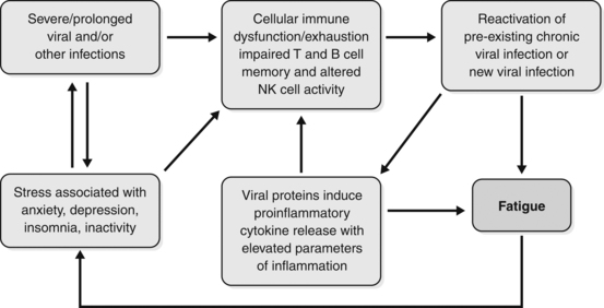
Figure 8.1 The inter-relationship between psychosocial, immune and viral factors in the initiation and perpetuation of chronic fatigue.10

Reproduced from Bansal AS, Bradley AS, Bishop KN, et al. Chronic fatigue syndrome, the immune system and viral infection. Brain Behav Immun 2011, with permission from Elsevier.

Sometimes benefits will follow a focus on what could be exacerbating or even causative factors. These might include:

• intestinal dysbiosis, endotoxaemia or similar syndromes, other syndromes involving autotoxicity, chronic inflammation

• allergies or food intolerances

• toxins, e.g. dental amalgam, hair dyes, pesticides

• recurrent fungal, viral or bacterial infection.

Above all else, it is important to ensure that sleep and rest are adequate and much useful treatment effort can be directed to this end as a first priority. Whatever the initiating disturbance, the treatment of fatigue must be marked by extreme gentleness and patience.

Convalescence

With any fatigue syndrome the fundamental principle of treatment is to set up an appropriate recuperative regime. Remedies should be set against a wider programme of convalescence. Convalescence as a strategy is outlined in Chapter 3. Extensive and appropriate rest is essential and may need to be supported by treatment to help with sleep and relaxation: exercise, even if minimal, will help engage adrenosympathetic disturbances; the diet should be based on the most easily assimilable foods possible. Only when such a regime is in place can herbal treatment have a chance of facilitating further improvements.

Herbal remedies useful in CFS include the following.

Tonic and adaptogenic herbs

Tonics help revitalise the patient and adaptogens improve the response to stress. Tonics were traditionally used to help build strength after illness and trauma and may also build immune function.

Major herbs in this group are:

• Astragalus membranaceus (Astragalus): tonic and immune enhancing (see monograph)

• Eleutherococcus senticosus (Siberian ginseng): adaptogenic, stimulates T-lymphocyte function (see monograph)

• Lycium spp. (goji): a tonic remedy in China with some evidence as an adaptogen

• Panax ginseng (Ginseng): tonic, adaptogenic, stimulates hypothalamic output and ACTH and hence adrenal cortex function, increases stamina, spares muscle use of carbohydrate (see monograph)

• Rhodiola rosea: a herb with adaptogenic and ergogenic reputations

• Schisandra chinensis: a herb with adaptogenic and hepatoprotective reputations94

• Withania somnifera (Withania): a tonic herb which is not stimulating and facilitates sleep (as its name implies), hence is ideal in situations of sympathetic dominance (see monograph).

Of this group, most interest in recent years has been in Rhodiola, with a wide range of clinical studies reported. These have been unsympathetically reviewed95 and defended.96 However, in one case a substantial positive study has been reported: 60 men and women with stress-related fatigue were randomised into two equal groups, one received four tablets daily of standardised Rhodiola extract (576 mg/day), and the other placebo. After comparing results for various psychomotor and quality of life tests and serum cortisol levels, it was concluded that, compared with placebo, repeated administration of this particular extract exerts an anti-fatigue effect with increased mental performance, ability to concentrate and decreased cortisol response to awakening stress.97 This probably also has relevance to the use of this herb in CFS. Rhodiola has the additional advantage that its efficacy in depression has been demonstrated in a randomised, placebo-controlled trial and also has considerable pharmacological support.98

Adrenal supportive herbs

The main herbs for this purpose are Glycyrrhiza (licorice) and Rehmannia glutinosa (see also the relevant monographs). There is one case report of recovery from CFS with the use of licorice that speculates a role in adrenocortical function.99 Another case report from Japan observed that a chronic fatigue patient went into remission when she developed hyperaldosteronism due to an adrenal tumour. When the adrenal tumour was removed the fatigue returned.100 Licorice in high doses can cause pseudoaldosteronism due to its aldosterone-like action, but obviously should not be used to this level. A high-potassium, low-sodium diet, as in the Gerson therapy, can also raise plasma aldosterone.

Immune-enhancing herbs

Although these may seem contraindicated, immune-enhancing herbs are often needed to help prevent the recurrent viral infections that can plague patients with CFS. In cases of infection, treatment with tonic herbs may need to be discontinued so that defensive measures can be applied. Echinacea angustifolia and E. purpurea root are safe to use since, on current knowledge, they mainly enhance innate immunity. This can improve antigen recognition, which leads to better immune responsiveness. Picrorrhiza kurroa should be used with caution as it is a potent promoter of all aspects of immune function, but it may be indicated for patients who have frequent viral infections. Andrographis and Astragalus also have a role, especially the latter where there is chronic immune depletion.

Antiviral herbs

Although the viral association is not always clear, these herbs can be useful in many cases. Hypericum perforatum (St John’s wort) may contribute to antiviral and antidepressant activity. Hypericum is probably active against enveloped viruses such as EBV and HHV-6. Thuja occidentalis is also active against enveloped viruses as well as naked viruses, such as the wart virus and enteroviruses.

Immune-depressing and anti-inflammatory herbs

There are a few non-toxic herbs that can depress immune function and may be useful at some stages of treatment. The safest to use is the Indian herb Hemidesmus indicus (Indian sarsaparilla). Rehmannia, Bupleurum and Boswellia can be useful anti-inflammatories and may downregulate cytokine production and responses (see monographs).

Others

Ginkgo biloba, Salvia miltiorrhiza and Zanthoxylum (prickly ash) can improve blood flow. Ginkgo decreases erythrocyte fragility and improves blood rheology and short-term memory. Valeriana (valerian), Passiflora incarnata (passionflower), Piper methysticum (kava) and other such herbs will help the disordered sleep pattern and sympathetic dominance. Crataegus (hawthorn) may help any cardiac and circulatory abnormalities and the sympathetic dominance. Butcher’s broom (Ruscus aculeatus) and horsechestnut (Aesculus hippocastanum), being venous toning, should be considered for orthostatic symptoms (see monographs). EFA therapy with evening primrose oil and fish oil is also recommended.

Notes about treatment

Deep-seated devitalisation is a major part of CFS, so results must be measured in months not weeks. If results are slow to come about the patient should be encouraged, since improvement usually does occur with consistent use of herbs. Patients must be instructed not to overexert themselves when they begin to feel improvement, as this can cause a relapse. They should only partake in mild exercise and not exercise beyond the point of fatigue. Sleep helps to restore the immune system and CFS patients will not improve unless they have adequate sleep, which may be more than they needed previously. They must avoid stress and emotional crises as much as possible. The importance of stress as a cause and sustaining factor of CFS should not be underestimated and appropriate lifestyle measures must be incorporated into the treatment plan.

Case History

‘William’, aged 48, sought treatment for CFS, from which he had suffered for 3 years after exposure to Ross River virus. Symptoms included aches and pains, especially in the first 6 months after the infection, malaise, mild fever, physical weakness, lack of stamina and poor sleep. He worked as a manager at a cemetery and was still able to manage (barely) his job, despite the CFS. He was previously very active. Current medication was salbutamol for asthma and the occasional use of a benzodiazepine to help his sleep

He was placed on the following herbal treatment:

A. Liquid formula:

Echinacea angustifolia and E. purpurea (root) 1:2 35 mL
Glycyrrhiza glabra (high in glycyrrhizin) 1:1 15 mL
Astragalus membranaceus 1:2 20 mL
Hypericum perforatum (high in hypericin) 1:2 20 mL
Withania somnifera 1:1 20 mL
  TOTAL 110 mL

Dose: 8 mL with water twice a day.

B. Withania (600 mg) and Korean ginseng (125 mg) tablets 3/day

Eleutherococcus (Siberian ginseng) tablets 1.25 g 3/day
Valeriana edulis (Mexican valerian) tablets 1.0 g 3/day (as required)

There was not much improvement over the first 8 to 12 weeks, but after that he felt the herbs were helping, especially the feeling of malaise and low energy. He then changed to a more stressful job, but was able to cope well with this, relying on the support of the herbal treatment. After another 8 weeks he reported a noticeable improvement all round and was able to undertake a regular programme of exercise.

Case History

‘Kylie’, aged 17, had glandular fever 6 years previously. She had suffered from fatigue ever since, but after beginning Year 12 at school her fatigue was particularly severe. Often catching colds and influenza, her attendance was less than 50%. Even on ‘well’ days, she often did not have the energy to attend school. Herbal treatment consisted of the following basic formula (written for 1 week):

Astragalus membranaceus 1:2 30 mL
Panax ginseng (standardised extract) 1:2 15 mL
Ginkgo biloba (standardised extract) 2:1 20 mL
Echinacea angustifolia root 1:2 35 mL
  Total: 100 mL

Dose: 5 mL with water three times a day.

In addition, Withania 1:2 5 mL with water once a day was prescribed.

If a cold came on, the above treatment was stopped for 3 to 4 days and the following formula was taken:

Zingiber officinale 1:2 10 mL
Echinacea angustifolia root 1:2 40 mL
Euphrasia officinalis 1:2 20 mL
Achillea millefolium 1:2 30 mL
  Total: 100 mL

Dose: 5 mL with warm water up to five times a day.

Initially, Kylie had difficulty taking the herbal formula because it was too stimulating. So the dose was reduced to half and gradually increased. There was only slight improvement in her condition for 3 months, but then gradual and steady progress was made. While she received herbal treatment from time to time, she was free of CFS for more than 2 years.

Case History

‘John’, was 35 and had not worked for 3 years. By the time he sought herbal treatment he complained that he was getting sicker and sicker. He experienced constant headaches and had suffered from chronic sinusitis for about 10 years. His history showed a previous high exposure to insecticides and years of overwork due to family pressures. His wife could not cope with his not working and his marriage was strained. Various formulas were given but the treatment settled at the following (for 1 week):

Panax ginseng (standardised extract) 1:2 15 mL
Astragalus membranaceus 1:2 30 mL
Crataegus spp. fol. 1:2 20 mL
Ginkgo biloba (standardised extract)   20 mL
Picrorrhiza kurroa 1:2 15 mL
Glycyrrhiza glabra (high in glycyrrhizin) 1:1 20 mL
Scutellaria baicalensis 1:2 30 mL
  Total: 150 mL

Dose: 8 mL with water three times a day.

In addition, Echinacea angustifolia root 1:2 5 mL once a day was prescribed from time to time. Also an ‘acute formula’, similar to the one for ‘Kylie’, was taken during colds and influenza instead of the basic formula. The Crataegus was for the headaches and circulation and the Baical skullcap for the sinusitis. ‘John’ actually worsened in the first 3 months of treatment, probably because of the natural progression of the disorder. However, after 5 months he thanked the friend who recommended herbal treatment because it was ‘the best thing I could have done’. He gradually improved over a period of several more months.

Tonics

Plant remedies traditionally used as tonics

• Astragalus membranaceus

• Avena sativa (oatstraw)

• Glycyrrhiza glabra (licorice)

• Hypericum perforatum (St John’s wort)

• Medicago sativa (alfalfa)

• Panax ginseng (ginseng)

• Rhodiola rosea

• Serenoa repens (saw palmetto)

• Swertia chirata (chiretta)

• Trigonella foenum-graecum (fenugreek)

• Turnera diffusa (damiana)

• Verbena officinalis (vervain)

• Withania somnifera.

Indications for tonics

• Convalescence

• Debilitating conditions with or without anorexia

• Chronic fatigue syndrome.

Cautions in the use of tonics

The use of tonic herbs may be difficult in the following circumstances:

• Very severe debility especially if associated with immune or digestive collapse

• Renal or hepatic failure

• Rampant cancer or strong regimes of chemotherapy.

Application

The state of digestion is the main determinant of dosage times. Tonics may be best taken with or after meals if the stomach and digestive system are weakened; in severe cases, they may need to be taken with fluid nourishment. Dosage should be rather more than less frequent: ‘little and often’ might be a useful axiom. Tonics taken immediately before bed can help when sleep is non-restorative. Withania is ideal for this.

Long-term therapy with tonics is generally indicated and often advisable.

Chinese tonics

In Chinese medicine, tonic remedies are generally divided into four groups, depending on whether they are seen to particularly support qi, yang, xue or yin. The first two groups tend to be warming, the last two cooling. They are often more dynamic than the tonics listed above and may thus be more likely to generate adverse reactions. It is more important, therefore, to be careful in their prescription and to take close account of the interpretation of debilitated conditions that Chinese practitioners use. However, they reflect a perspective on debility and its treatment that is not well articulated in the Western traditions. This works both ways: unfortunately methodological limitations have also undermined the evidence for efficacy in the West so far published.101

In the following review, Chinese terms will therefore be used. They are briefly introduced in the summary of Chinese herbal medicine in this text (see p. 5) but any practitioner wishing to apply them should be well trained in that tradition. Nevertheless, there is some overlap with Western remedies and some useful insights are possible for a Western phytotherapist.

Qi tonics

These support active energies; they are used for depletion of qi, particularly in the Spleen and Lungs.

In the first case, possibly as a result of prolonged illness or constitutional weakness, debility may affect the functions of assimilation and distribution and be associated with such symptoms as fatigue and depression with depressed digestion, diarrhoea, abdominal pain or tension, visceral prolapse, pale yellow complexion with a tinge of red or purple, pale tongue with white coating and/or languid, frail or indistinct pulses. This may lead in turn to a ‘damp’ condition developing.

In the second case, extreme or prolonged stress or disease, or chronic pulmonary disease, leads to depletion or cold in the Lungs, with easy fatigue and prostration associated with disturbances of regulation, shortness of breath or shallow breathing, rapid, slow or little speech, spontaneous perspiration, pallid complexion, dry skin, pale tongue with thin white coating, weak and depleted pulses.

Plant remedies traditionally used as Qi tonics:

• Astragalus membranaceus (huang qi)

• Atractylodes macrocephela (bai zhu)

• Codonopsis pilulosa (dang shen)

• Glycyrrhiza uralensis (Chinese liquorice – gan cao)

• Panax ginseng (Asiatic ginseng – ren shen)

• Zizyphus jujuba (Jujube – da zao).

Yang tonics

These remedies support the active energies, particularly those of the Kidneys (but also Heart and Spleen).

Deficient Kidney yang leads to listlessness with a feeling of cold and cold extremities, back and loins; there may be weak legs, poor reproductive function, frequency of micturition, nocturia, diarrhoea (especially early in the morning), pale complexion and submerged weak pulses.

Deficiency affecting the Heart involves poor performance and coordination associated with profuse cold sweating, asthmatic states, thoracic or anginal pain on exertion, palpitations and fear attacks, cyanosis, white tongue coating and/or diminished, hesitant or intermittent pulses.

Plant remedies traditionally used as yang tonics:

• Eucommia ulmoides (du zhong)

• Juglans regia (walnut – hu tao ren)

• Morinda officinalis (ba ji)

• Trigonella foenum-graecum (fenugreek – hu lu ba).

Xue tonics

These are remedies that support more substantial energies, those manifesting in substantial disturbances or pathologies. By definition, such disturbances are serious and profound and treatment will need to be prolonged. Symptoms of depletion of xue may include cyanosis, pallor, vertigo or tinnitus, palpitations, loss of memory, insomnia or menstrual problems.

There is considerable overlap with yin tonics.

Plant remedies traditionally used as xue tonics:

• Angelica sinensis (dang gui)

• Mori alba (mulberry fruit – sang shen)

• Paeonia lactiflora (paeony root – bai shao)

• Rehmannia glutinosa (sheng di huang – fresh, and shu di huang – prepared).

Yin tonics

These are remedies for replenishing the body fluids and essence, supplying condensed energies and nourishment, for the most depleted conditions. Areas in most need of support are the Kidneys, Liver, Lungs and Stomach.

Deficient Kidney yin often follows very serious debilitating disease or, alternatively, extended sexual or alcohol abuse or overwhelming nervous stress. It may manifest as a deficient Fire condition, marked by a pale complexion with red cheeks, red lips, dry mouth, dry but deeply red tongue, dry throat, hot palms and soles, palpitations, vertigo or tinnitus, pains in the loins, night sweats, nocturnal emissions, nightmares, urinary retention, constipation, accelerated though weak pulses.

Deficient Liver yin, usually following the above, is often associated with dry eyes, poor vision and vertigo or tinnitus, deafness, muscle twitching, sleeplessness, hot flushed face with red cheeks, red dry tongue with little coating, diminished, stringy and accelerated pulses.

Deficient Stomach yin is marked by anorexia, regurgitation, thirst, abdominal rumbling, red lips and red tongue with no coating.

Deficient Lung yin, often following prolonged exposure to dryness or chronic pulmonary disease, is marked by dry cough, haemoptysis, hoarseness and loss of voice, strong thirst and/or restlessness and insomnia.

Plant remedies traditionally used as yin tonics:

• Asparagus cochinchinensis (tian men dong)

• Ligustrum lucidum (nu zhen zi)

• Lycium chinensis (gou qi zi)

• Ophiopogon japonicus (mai men dong)

• Panax quinquefolium (American ginseng)

• Sesamum indicum (sesame seeds – hei zhi ma).

References

1. Shorter E. Chronic fatigue in historical perspective. Ciba Found Symp. 1993;173:6–16.

2. Hyland ME. Network origins of anxiety and depression. Behav Brain Sci. 2010;33(2–3):161–162.

3. Holmes GP, Kaplan JE, Gantz NM, et al. Chronic fatigue syndrome: a working case definition. Ann Intern Med. 1988;108(3):387–389.

4. Centers for Disease Control and Prevention. CFS Case Definition. <http://www.cdc.gov/cfs/general/case_definition/index.html>. Accessed 28.08.2011.

5. Komaroff AL. Is human herpesvirus-6 a trigger for chronic fatigue syndrome? J Clin Virol. 2006;37(suppl 1):S39–S46.

6. Gow JW, Behan WM, Clements GB, et al. Enteroviral RNA sequences detected by polymerase chain reaction in muscle of patients with postviral fatigue syndrome. BMJ. 1991;302(6778):692–696.

7. Gow JW, Behan WM. Amplification and identification of enteroviral sequences in the postviral fatigue syndrome. Br Med Bull. 1991;47(4):872–885.

8. Grau JM, Casademont J, Pedrol E, et al. Chronic fatigue syndrome: studies on skeletal muscle. Clin Neuropathol. 1992;11(6):329–332.

9. Gow JW, Behan WM, Simpson K, et al. Studies on enterovirus in patients with chronic fatigue syndrome. Clin Infect Dis. 1994;18(suppl 1):S126–S129.

10. Bansal AS, Bradley AS, Bishop KN, et al. Chronic fatigue syndrome, the immune system and viral infection. Brain Behav Immun. 2012;26(1):24–31.

11. Chia JK. The role of enterovirus in chronic fatigue syndrome. J Clin Pathol. 2005;58(11):1126–1132.

12. Frémont M, Metzger K, Rady H. Detection of herpes viruses and parvovirus B19 in gastric and intestinal mucosa of chronic fatigue syndrome patients. In Vivo. 2009;23(2):209–213.

13. Lombardi VC, Ruscetti FW, Das Gupta J, et al. Detection of an infectious retrovirus, XMRV, in blood cells of patients with chronic fatigue syndrome. Science. 2009;326(5952):585–589.

14. Coffin JM, Stoye JP. Virology. A new virus for old diseases? Science. 2009;326(5952):530–531.

15. The Medical Staff of the Royal Free Hospital. An outbreak of encephalomyelitis in the Royal Free Hospital group London, in 1955. BMJ. 1957;2:895–904.

16. Nasralla M, Haier J, Nicolson GL. Multiple mycoplasmal infections detected in blood of patients with chronic fatigue syndrome and/or fibromyalgia syndrome. Eur J Clin Microbiol Infect Dis. 1999;18(12):859–865.

17. Vojdani A, Choppa PC, Tagle C, et al. Detection of mycoplasma genus and Mycoplasma fermentans by PCR in patients with chronic fatigue syndrome. FEMS Immunol Med Microbiol. 1998;22(4):355–365.

18. Choppa PC, Vojdani A, Tagle C, et al. Multiplex PCR for the detection of Mycoplasma fermentans, M. hominis and M. penetrans in cell cultures and blood samples of patients with chronic fatigue syndrome. Mol Cell Probes. 1998;12(5):301–308.

19. Vernon SD, Shukla SK, Reeves WC. Absence of Mycoplasma species DNA in chronic fatigue syndrome. J Med Microbiol. 2003;52(Pt 11):1027–1028.

20. Dunstan RH, McGregor NR, Roberts TK, et al. The development of laboratory-based tests in chronic pain and fatigue: (1) Muscle catabolism and coagulase negative staphylococci which produce membrane damaging toxins. JCFS. 2000;7:23–28.

21. Andersson M, Bagby JR, Dyrehag L, et al. Effects of staphylococcus toxoid vaccine on pain and fatigue in patients with fibromyalgia/chronic fatigue syndrome. Eur J Pain. 1998;2(2):133–142.

22. Levin AM. Chronic fatigue syndrome: the yeast concept. JCFS. 2001;8(2):71–76.

23. Chester AC. Yeast and chronic fatigue syndrome. JCFS. 2000;7(3):87–88.

24. Marques A. Chronic Lyme disease: a review. Infect Dis Clin North Am. 2008;22(2):341–360.

25. Whalley B, Jacobs PA, Hyland ME. Correlation of psychological and physical symptoms with chronically elevated cytokine levels associated with a common immune dysregulation. Ann Allergy Asthma Immunol. 2007;99(4):345–348.

26. Landay AL, Jessop C, Lennette ET, et al. Chronic fatigue syndrome: clinical condition associated with immune activation. Lancet. 1991;338(8769):707–712.

27. Linde A, Andersson B, Svenson SB, et al. Serum levels of lymphokines and soluble cellular receptors in primary Epstein-Barr virus infection and in patients with chronic fatigue syndrome. J Infect Dis. 1992;165(6):994–1000.

28. Patarca R, Klimas NG, Lugtendorf S, et al. Dysregulated expression of tumor necrosis factor in chronic fatigue syndrome: interrelations with cellular sources and patterns of soluble immune mediator expression. Clin Infect Dis. 1994;18(1):147–153.

29. Ojo-Amaize EA, Conley EJ, Peter JB. Decreased natural killer cell activity is associated with severity of chronic fatigue immune dysfunction syndrome. Clin Infect Dis. 1994;18(suppl 1):S157–S159.

30. Barker E, Fujimura SF, Fadem MB, et al. Immunologic abnormalities associated with chronic fatigue syndrome. Clin Infect Dis. 1994;18(suppl 1):S136–S141.

31. Rasmussen AK, Nielsen H, Andersen V, et al. Chronic fatigue syndrome–a controlled cross sectional study. J Rheumatol. 1994;21(8):1527–1531.

32. Straus SE, Fritz S, Dale JK, et al. Lymphocyte phenotype and function in the chronic fatigue syndrome. J Clin Immunol. 1993;13(1):30–40.

33. Lloyd A, Hickie I, Hickie C, et al. Cell-mediated immunity in patients with chronic fatigue syndrome, healthy control subjects and patients with major depression. Clin Exp Immunol. 1992;87(1):76–79.

34. Hashimoto N, Kuraishi Y, Yokose T, et al. Chronic fatigue syndrome-51 cases in the Jikei University School of Medicine. Nippon Rinsho. 1992;50(11):2653–2664.

35. Hilgers A, Frank J. Chronic fatigue syndrome: immune dysfunction, role of pathogens and toxic agents and neurological and cardial changes. Wien Med Wochenschr. 1994;144(16):399–406.

36. Bates DW, Buchwald D, Lee J, et al. Clinical laboratory test findings in patients with chronic fatigue syndrome. Arch Intern Med. 1995;155(1):97–103.

37. Lakhan SE, Kirchgessner A. Gut inflammation in chronic fatigue syndrome. Nutr Metab (Lond). 2010;7:79.

38. Lerner AM, Lawrie C, Dworkin HS. Repetitively negative changing T waves at 24-h electrocardiographic monitors in patients with the chronic fatigue syndrome. Left ventricular dysfunction in a cohort. Chest. 1993;104(5):1417–1421.

39. Miwa K, Fujita M. Cardiovascular dysfunction with low cardiac output due to a small heart in patients with chronic fatigue syndrome. Intern Med. 2009;48(21):1849–1854.

40. Miwa K, Fujita M. Cardiac function fluctuates during exacerbation and remission in young adults with chronic fatigue syndrome and ‘small heart’. J Cardiol. 2009;54(1):29–35.

41. Hurwitz BE, Coryell VT, Parker M, et al. Chronic fatigue syndrome: illness severity, sedentary lifestyle, blood volume and evidence of diminished cardiac function. Clin Sci (Lond). 2009;118(2):125–135.

42. Newton JL, Sheth A, Shin J, et al. Lower ambulatory blood pressure in chronic fatigue syndrome. Psychosom Med. 2009;71(3):361–365.

43. Peckerman A, Lamanca JJ, Dahl KA, et al. Abnormal impedance cardiography predicts symptom severity in chronic fatigue syndrome. Am J Med Sci. 2003;326(2):55–60.

44. Simpson LO, Murdoch JC, Herbison GP. Red cell shape changes following trigger finger fatigue in subjects with chronic tiredness and healthy controls. N Z Med J. 1993;106(952):104–107.

45. Ichise M, Salit IE, Abbey SE, et al. Assessment of regional cerebral perfusion by 99 Tcm–HMPAO SECT in chronic fatigue syndrome. Nucl Med Commun. 1992;10:767–772.

46. Biswal B, Kunwar P, Natelson BH. Cerebral blood flow is reduced in chronic fatigue syndrome as assessed by arterial spin labeling. J Neurol Sci. 2011;301(1–2):9–11.

47. Goldstein JA, Mena I, Jouanne E, et al. The assessment of vascular abnormalities in late life chronic fatigue syndrome by brain SPECT: comparison with late life major depressive disorder. JCFS. 1995;1(1):55–79.

48. Streeten DH, Thomas D, Bell DS. The roles of orthostatic hypotension, orthostatic tachycardia, and subnormal erythrocyte volume in the pathogenesis of the chronic fatigue syndrome. Am J Med Sci. 2000;320(1):1–8.

49. Schondorf R, Freeman R. The importance of orthostatic intolerance in the chronic fatigue syndrome. Am J Med Sci. 1999;317(2):117–123.

50. Streeten DH, Anderson GH, Jr. The role of delayed orthostatic hypotension in the pathogenesis of chronic fatigue. Clin Auton Res. 1998;8(2):119–124.

51. Wilke WS, Fouad-Tarazi FM, Cash JM, et al. The connection between chronic fatigue syndrome and neurally mediated hypotension. Cleve Clin J Med. 1998;65(5):261–266.

52. Natelson BH, Intriligator R, Cherniack NS, et al. Hypocapnia is a biological marker for orthostatic intolerance in some patients with chronic fatigue syndrome. Dyn Med. 2007;30(6):2.

53. Hurum H, Sulheim D, Thaulow E, et al. Elevated nocturnal blood pressure and heart rate in adolescent chronic fatigue syndrome. Acta Paediatr. 2011;100(2):289–292.

54. Boneva RS, Decker MJ, Maloney EM, et al. Higher heart rate and reduced heart rate variability persist during sleep in chronic fatigue syndrome: a population-based study. Auton Neurosci. 2007;137(1–2):94–101.

55. Burton AR, Rahman K, Kadota Y, et al. Reduced heart rate variability predicts poor sleep quality in a case-control study of chronic fatigue syndrome. Exp Brain Res. 2010;204(1):71–78.

56. Wyller VB, Barbieri R, Thaulow E, et al. Enhanced vagal withdrawal during mild orthostatic stress in adolescents with chronic fatigue. Ann Noninvasive Electrocardiol. 2008;13(1):67–73.

57. Buchwald D, Cheney PR, Peterson DL, et al. A chronic illness characterized by fatigue, neurologic and immunologic disorders, and active human herpesvirus type 6 infection. Ann Intern Med. 1992;116(2):103–113.

58. Natelson BH, Cohen JM, Brassloff I, et al. A controlled study of brain magnetic resonance imaging in patients with the chronic fatigue syndrome. J Neurol Sci. 1993;120(2):213–217.

59. Schwartz RB, Garada BM, Komaroff AL, et al. Detection of intracranial abnormalities in patients with chronic fatigue syndrome: comparison of MR imaging and SPECT. Am J Roentgenol. 1994;162(4):935–941.

60. Cook DB, Lange G, DeLuca J, et al. Relationship of brain MRI abnormalities and physical functional status in chronic fatigue syndrome. Int J Neurosci. 2001;107(1–2):1–6.

61. Cook DB, O’Connor PJ, Lange G, et al. Functional neuroimaging correlates of mental fatigue induced by cognition among chronic fatigue syndrome patients and controls. Neuroimage. 2007;36(1):108–122.

62. Barnden LR, Crouch B, Kwiatek R, et al. A brain MRI study of chronic fatigue syndrome: evidence of brainstem dysfunction and altered homeostasis. NMR Biomed. 2011;24(10):1302–1312.

63. Perrin R, Embleton K, Pentreath VW, et al. Longitudinal MRI shows no cerebral abnormality in chronic fatigue syndrome. Br J Radiol. 2010;83(989):419–423.

64. Riccio M, Thompson C, Wilson B, et al. Neuropsychological and psychiatric abnormalities in myalgic encephalomyelitis: a preliminary report. Br J Clin Psychol. 1992;31(Part 1):111–120.

65. Sandman CA, Barron JL, Nackoul K, et al. Memory deficits associated with chronic fatigue immune dysfunction syndrome. Biol Psychiatry. 1993;33(8–9):618–623.

66. Smith AP, Behan PO, Bell W, et al. Behavioural problems associated with the chronic fatigue syndrome. Br J Psychol. 1993;84(Pt 3):411–423.

67. McDonald E, Cope H, David A. Cognitive impairment in patients with chronic fatigue: a preliminary study. J Neurol Neurosurg Psychiatry. 1993;56(7):812–815.

68. Constant EL, Adam S, Gillain B, et al. Cognitive deficits in patients with chronic fatigue syndrome compared to those with major depressive disorder and healthy controls. Clin Neurol Neurosurg. 2011;113(4):295–302.

69. Scheffers MK, Johnson R, Jr., Grafman J, et al. Attention and short-term memory in chronic fatigue syndrome patients: an event-related potential analysis. Neurology. 1992;42(9):1667–1675.

70. Grafman J, Schwartz V, Dale JK, et al. Analysis of neuropsychological functioning in patients with chronic fatigue syndrome. J Neurol Neurosurg Psychiatry. 1993;56(6):684–689.

71. Johnson SK, DeLuca J, Fiedler N, et al. Cognitive functioning of patients with chronic fatigue syndrome. Clin Infect Dis. 1994;18(suppl 1):S84–S85.

72. Cockshell SJ, Mathias JL. Cognitive functioning in chronic fatigue syndrome: a meta-analysis. Psychol Med. 2010;40(8):1253–1267.

73. Demitrack MA, Dale JK, Straus SE, et al. Evidence for impaired activation of the hypothalamic-pituitary-adrenal axis in patients with chronic fatigue syndrome. J Clin Endocrinol Metab. 1991;73(6):1224–1234.

74. Ur E, White PD, Grossman A. Hypothesis: cytokines may be activated to cause depressive illness and chronic fatigue syndrome. Eur Arch Psychiatry Clin Neurosci. 1992;241(5):317–322.

75. Van Den Eede F, Moorkens G, Van Houdenhove B, et al. Hypothalamic-pituitary-adrenal axis function in chronic fatigue syndrome. Neuropsychobiology. 2007;55(2):112–120.

76. Morriss R, Sharpe M, Sharpley AL, et al. Abnormalities of sleep in patients with the chronic fatigue syndrome. BMJ. 1993;306(6886):1161–1164.

77. McCluskey DR. Pharmacological approaches to the therapy of chronic fatigue syndrome. Ciba Found Symp. 1993;173:280–287.

78. Krupp LB, Jandorf L, Coyle PK, et al. Sleep disturbance in chronic fatigue syndrome. J Psychosom Res. 1993;37(4):325–331.

79. Togo F, Natelson BH, Cherniack NS, et al. Sleep structure and sleepiness in chronic fatigue syndrome with or without coexisting fibromyalgia. Arthritis Res Ther. 2008;10(3):R56.

80. Gray JB, Martinovic AM, Horrobin D. Eicosanoids and essential fatty acid modulation in chronic disease and the chronic fatigue syndrome. Med Hypotheses. 1994;43(1):31–42.

81. Ogawa R, Toyama S, Matsumoto H. Chronic fatigue syndrome – cases in the Kanebo Memorial Hospital. Nippon Rinsho. 1992;50(11):2648–2652.

82. Nicolson GL. Lipid replacement as an adjunct to therapy for chronic fatigue, anti-aging and restoration of mitochondrial function. JANA. 2003;6(3):22–28.

83. Behan PO, Behan WM, Horrobin D. Effect of high doses of essential fatty acids on the postviral fatigue syndrome. Acta Neurol Scand. 1990;82(3):209–216.

84. Kuratsune H, Yamaguti K, Takahashi M, et al. Acylcarnitine deficiency in chronic fatigue syndrome. Clin Infect Dis. 1994;18(suppl 1):S62–S67.

85. Takahashi H, Imai K, Katanuma A, et al. A case of chronic fatigue syndrome who showed a beneficial effect by intravenous administration of magnesium sulphate. Aerugi. 1992;41(11):1605–1610.

86. Hinds G, Bell NP, McMaster D, et al. Normal red cell magnesium concentrations and magnesium loading tests in patients with chronic fatigue syndrome. Ann Clin Biochem. 1994;31(Pt 5):459–461.

87. Jacobson W, Saich T, Borysiewicz LK, et al. Serum folate and chronic fatigue syndrome. Neurology. 1993;43(12):2645–2647.

88. Amagase H, Sun B, Nance DM. Immunomodulatory effects of a standardized Lycium barbarum fruit juice in Chinese older healthy human subjects. J Med Food. 2009;12(5):1159–1165.

89. Sathyapalan T, Beckett S, Rigby AS, et al. High cocoa polyphenol rich chocolate may reduce the burden of the symptoms in chronic fatigue syndrome. Nutr J. 2010;9:55.

90. Scholey AB, French SJ, Morris PJ, et al. Consumption of cocoa flavonols results in acute improvements in mood and cognitive performance during sustained mental effort. J Psychopharmacol. 2010;24(10):1505–1514.

91. Cho JH, Cho CK, Shin JW, et al. Myelophil, an extract mix of Astragali Radix and Salviae Radix, ameliorates chronic fatigue: a randomised, double blind, controlled pilot study. Complement Ther Med. 2009;17(3):141–146.

92. Stricklin A, Sewell M, Austad C. Objective measurement of personality variables in epidemic neuromyasthenia patients. S Afr Med J. 1990;77(1):31–34.

93. Patarca-Montero R, Antoni M, Fletcher MA, et al. Cytokine and other immunologic markers in chronic fatigue syndrome and their relation to neuropsychological factors. Appl Neuropsychol. 2001;8(1):51–64.

94. Panossian A, Wikman G. Pharmacology of Schisandra chinensis Bail. An overview of Russian research and uses in medicine. J Ethnopharmacol. 2008;118(2):183–212.

95. Blomkvist J, Taube A, Larhammar D. Perspective on roseroot (Rhodiola rosea) studies. Planta Med. 2009;75(11):1187–1190.

96. Panossian A, Wikman G, Sarris J. Rosenroot (Rhodiola rosea): traditional use, chemical composition, pharmacology and clinical efficacy. Phytomedicine. 2010;17(7):481–493.

97. Olsson EM, von Schéele B, Panossian AG. A randomised, double blind, placebo-controlled, parallel-group study of the standardised extract shr-5 of the roots of Rhodiola rosea in the treatment of subjects with stress-related fatigue. Planta Med. 2009;75(2):105–112.

98. Sarris J, Panossian A, Schweitzer I, et al. Herbal medicine for depression, anxiety and insomnia: a review of psychopharmacology and clinical evidence. Eur Neuropsychopharmacol. 2011;21(12):841–860.

99. Baschetti R. Chronic fatigue syndrome and licorice. N Z Med J. 1995;108(998):156–157.

100. Kato Y, Kamijima S, Kashiwagi A, et al. Chronic fatigue syndrome, a case of high anti-HHV-6 antibody titer and one associated with primary hyperaldosteronism. Nippon Rinsho. 1992;50(11):2673–2678.

101. Adams D, Wu T, Yang X, et al. Traditional Chinese medicinal herbs for the treatment of idiopathic chronic fatigue and chronic fatigue syndrome. Cochrane Database Syst Rev. 2009;4:CD006348.

Malignant diseases

Scope

Apart from their use in providing non-specific support for recuperation and repair, specific phytotherapeutic strategies include the following.

Adjunctive treatment of:

• most cancers.

Management of:

• symptoms of cancer treatment, such as nausea/vomiting of chemotherapy

• recuperation from chemotherapy and radiotherapy.

Because of its use of secondary plant products, particular caution is necessary in applying phytotherapy in cases of cancers of the stomach, liver and kidneys.

Orientation

Background

Herbal practitioners are often asked to provide treatment for cancer patients. This is most often as a complement to conventional treatments. However, in a few cases they are asked to construct completely alternative treatment strategies. In the latter case especially, the practitioner can be put in a most difficult position. Cancer is undoubtedly one of the most serious and also one of the most complex and diverse diagnoses to receive. Modern oncology has confirmed that finding and attacking malignant targets remains most difficult, and that different forms of cancer present very different prospects. Modern treatments have, moreover, been powerful, indeed dangerous, assaults on cells that may be more vigorous than their normal neighbours. The efficacy and safety of these treatments depend on being able to focus on their target most selectively. Alternative approaches are usually derided by orthodox authorities because they apparently lack such potency, and reviews tend to reinforce the view that direct efficacy is lacking.1

Herbal clinicians respond that they are trying a different approach: mobilising the body’s own powerful defences against the proliferation of malignant cells by providing appropriate supportive therapies. They point out that endogenous anticancer defences are formidable in even modest health. It is statistically likely that a tiny fraction of the body’s cells (but this could mean thousands of cells) slip out of normal tissue constraints and become malignant each day; through quiet surveillance, the body’s defences normally clean up such vagaries without further harm. Against that background, the actual development of a tumour must represent the failure of an impressive protective mechanism. In theory, it may be possible to reactivate these defences, at least in conditions that are not too advanced or debilitating. In practice, there is only modest evidence to guide a practitioner who aims to work in this direction, mainly from in vitro and in vivo studies.

On the positive side, the cases of spontaneous remissions from cancer and unexpected delays in deterioration that all healthcare practitioners encounter are evidence that there are powerful defensive forces to be had. To be set against this, however, is the need for evidence for any strategy towards mobilising these forces. However, there are some pointers, at least to the possibility of improving protection against cancer development before it happens and, by implication (but only by implication), to guide supportive or adjunctive measures to improve the prospects of spontaneous recovery after the event.

Plants and cancer prevention

After a $20 million research campaign over many years and on the basis of in vitro and in vivo studies as well as epidemiological evidence, the American National Cancer Institute (NCI) identified a range of foods with cancer preventive potential in three categories:2,3

1. Highest anticancer activity was found in garlic, soya, ginger, licorice and the umbelliferous vegetables (e.g. carrots, celery, parsley and parsnips).

2. Moderate anticancer activity was found in onions, linseed (flaxseed), citrus, turmeric, cruciferous vegetables (e.g. broccoli, Brussels sprouts, cabbage and cauliflower), solanaceous vegetables (tomatoes and peppers), brown rice and whole wheat.

3. Modest anticancer activity has been demonstrated in oats and barley, cucumber and the kitchen herbs such as the mints, rosemary, thyme, oregano, sage and basil.

The chemical groups suggested by the NCI as conveying some of this activity include allyl sulphides in garlic and onions; phytates in grains and legumes; lycopene, limonoids, glucarates and related terpenes; carotenes and flavonoids in citrus; lignans in linseed4 and soya; isoflavones in soya; saponins in legumes; indoles, isothiocyanates and dithiolthione in cruciferous vegetables;5 ellagic acid in grapes and other fruit; and phthalides and polyacetylenes in umbelliferous vegetables. Most of these are relatively robust to food preparation and are likely to reach target tissues in the body after oral consumption. Interaction with hormone receptors and other metabolic pathways leading to tumour generation is implicated as a mechanism.

Epidemiological evidence so far supports these findings.6 For example, a study in Finland, following a cohort of 9959 cancer-free individuals (from a population of 62 440) for 24 years from 1967 found an inverse relationship between the development of lung and other cancers and the consumption of flavonoid-rich fruits and vegetables.7 In a study in The Netherlands on a randomly selected cohort of 3123 subjects (from a total study size of 120 852), researchers found an inverse relationship between onion consumption and the development of stomach cancers over 3 years.8 A review of this and other studies concluded that onions, garlic and other allium vegetables were likely to convey protective effects against cancer development, especially in the gastrointestinal tract.9 There is actually some evidence for similar protective effect on the incidence of breast cancer in women for the use of herbal medicines in general. The Mammary Carcinoma Risk Factor Investigation (MARIE) study, a German population-based case-control study of 10 121 postmenopausal women (3464 with histologically confirmed breast cancer compared to 6657 controls) between 2002 and 2005, included analysis of associations between patterns of herbal preparations use and invasive breast cancer. Accounting for lifestyle and other risk factors, the authors concluded that those who used herbal preparations – such as black cohosh (Actaea racemosa), St John’s wort (Hypericum perforatum), chaste tree (Vitex agnus-castus), pulsatilla (Pulsatilla pratensis), rhubarb (Rheum rhaponticum) and Asian ginseng (Panax ginseng) – to manage their menopausal symptoms reduced their risk of breast cancer by 4% for every year’s use of the herbal remedies and up to one-quarter, irrespective of histological type and receptor status.10

Some of the most persuasive data on the benefits of plants in at least reducing the incidence of cancer has been in epidemiological studies on the association with tea drinking. In addition to a number of studies demonstrating antitumour effects of extracts of Camellia sinensis (green tea) consumption and cancers of the bowel, stomach and breast, there have also been epidemiological studies supporting its protective effect in humans, and there are early studies under way to investigate its benefits in cancer patients.11

In the Shanghai Women’s Health Study, 69 710 women aged 40 to 70 years were selected for observations over 6 years between 1996 and 2000. Women who reported drinking green tea regularly had around two-thirds of the risk for colorectal cancer compared with non-regular tea drinkers, and there also appeared to be a dose-dependent effect.12 In a Japanese study, 26 464 men and 38 720 women between the ages of 40 and 80 were studied over 10 years. In all, 37 oral cancer cases were identified in this group. Subjects who consumed 5 or more cups per day had a reduced risk of oral cancer compared with those who consumed less than one cup per day; this was marked mainly in women. Interestingly, the authors proposed that the protective effects of green tea consumption may be through prevention of caries and periodontal disease.13 Another study also in Japan pooled the results of several studies to indicate that there was a significant decrease in gastric cancer risk in the highest category of green tea consumption (at least five cups per day), but again only in women.14

Potential benefit in women at least was also suggested in Swedish studies on the links between all tea consumption and epithelial ovarian cancer. In a large study, 61 057 women between the ages of 40 and 76 years (the Swedish Mammography Cohort) were observed over an average of 15 years. After controlling for potential confounders, an inverse association was observed between tea consumption and ovarian cancer, and in a dose-dependent manner.15

Although there is criticism of such conclusions being drawn from low incidences of cancer, inadequate descriptions of tea consumption and of confounding factors,16,17 there is an interesting pointer to the effect of a flavonoid-rich food and the incidence of cancer in women. This picks up on the associations already established with the consumption of flavonoid-rich diets referred to earlier. It also bridges to the interesting debate on the potential of phyto-oestrogen diet sources.

In a large controlled study in China reported in the British Medical Journal, data was consistent with the hypothesis that regular intake of soya foods is associated with a reduced risk of endometrial cancer, especially among overweight women.18 The authors of this report believed that soya foods exert an anti-oestrogenic effect in an oestrogen-rich environment. This view is widely held. It is also postulated as a basis for a protective effect of other plant remedies with oestrogenic properties. However the evidence so far on this point has not been compelling (see also Chapter 2 under Phyto-oestrogens).19

On the other hand there seems to be little risk of phyto-oestrogens worsening oestrogen-dependent tumours either. In one study 18 861 women were included in a single-blinded observational retrospective cohort study, on survival time from breast cancer and the use of an isopropanolic black cohosh extract. Only about 1000 of these women were taking the black cohosh, but there was no shift in relapse rates in this group compared to controls, even among patients with oestrogen-dependent tumours.20 In a substantial study conducted in Cambridge, UK, the effect of another phyto- oestrogen source, a red-clover-derived isoflavone extract, on oestrogen-linked mammographic breast density (this is associated with an increased risk for breast cancer) was investigated in 205 women aged 49 to 65 years and without a history of breast cancer. The women received either a placebo or an isoflavone tablet containing 26 mg biochanin A, 16 mg formononetin, 1 mg genistein and 0.5 mg daidzein, derived from a proprietary brand of red clover, daily for 1 year. Baseline and 12-month follow-up mammograms were evaluated for density. The supplement did not significantly increase mammographic breast density nor did it have any other effect on hormonal levels. There was no significant effect on menopausal symptoms or hot flushes. The authors conclude that the isoflavone supplement, at the dose given, was acting neither as an oestrogen nor as an anti-oestrogen.21

However, this is an area where promising leads rarely translate into firm recommendations: lycopene from tomatoes has inconsistent benefits in human studies,22 and the disappointing outcomes from the large study on the effects of beta-carotene as a food supplement have acted as a reminder that isolated plant-based solutions may be the wrong targets.23 There are even data suggesting that some plant beverages, for example maté tea,24 can have a negative effect on cancer incidence. On the other hand, it is noticeable that reviewers are less dismissive of natural approaches to cancer prevention and even management,25 with, for example, the role of ginger in treating the side effects of chemotherapy widely appreciated (see below and the ginger monograph).26 There is even evidence for positive combination effects: 5-fluorouracil is a common chemotherapy that can be limited over time. In one study, an improvement was observed in some patients with combined therapy with standardised Ginkgo biloba (see also later).27

There remain future prospects of genuine leads. Anti-inflammatory activity, for example by aspirin and indomethacin, has been shown to have tumour-inhibitory effects in some models (and aspirin consumption has been linked to reduced human colorectal incidence; for example, see the willow bark monograph). Cyclo-oxygenase inhibition by a polyphenol, resveratrol, has been cited as a mechanism for a range of in vitro and in vivo tumour inhibitory effects of red grapeskin products like red wine.28 There are many other plant mechanisms that may modulate inflammation mentioned in other parts of this text (see, for example, the feverfew and turmeric monographs) and including the flavonoids mentioned earlier. Some of the key phytochemicals with cancer preventative activity are also discussed in Chapter 2.

Plants and cancer treatment

The potential of plant remedies directly to correct established malignancies is apparently limited. In its intensive programme from 1955, screening plant extracts for anticancer activity, the NCI found less than four extracts in every thousand tested containing compounds that demonstrated efficacy. Of the approximately 114 000 extracts studied, 11 000 exhibited antitumour activity in the P338 pre-screen, but of these only 500 demonstrated such activity in panel testing and only 26 proceeded to secondary testing. Of the plant isolates that reached this stage, most were alkaloids or terpenoids, with few from plants used in traditional medicine (for example, a triterpene from Withania spp., a diterpene from Brucea spp., a lignan from Podophyllum spp., a quinone from Tabebuia spp., an alcohol from Aristolochia spp. and alkaloids from Camptotheca acuminata, Fagara macrophylla, Ficus spp., Heliotropium indicum, Ochorisia moorei and Zanthoxylum nitidum). Only the diterpene taxol from Taxus brevifolia eventually received marketing approval from the Food and Drug Administration.29 The anticancer benefits of the alkaloids vinblastine and vincristine from the Madagascar periwinkle, Catharantheus roseus, were identified separately from the NCI programme. The NCI has studied the tamoxifen-like properties of the monoterpene perillyl alcohol, a naturally occurring analogue of limonene in citrus fruits and extracted from oil of lavender.30

Other plant isolates from traditional remedies have been identified as having antitumour effects in vitro and in vivo in other research programmes.31 These include:

• the anthraquinones rhein and emodin from Cassia spp. (the sennas), Rhamnus spp. (cascara sagrada and alder buckthorn) and Rumex crispus (yellow dock)

• the furanocoumarin psoralen from Angelica spp. and other umbelliferous remedies

• the alkaloid berberine from Berberis spp. (barberry and Oregon grape), Hydrastis canadensis (golden seal) and the Chinese remedy Coptis chinensis (see monograph)

• the alkaloids matrine and oxymatrine from the Chinese remedies Sophora spp.

• boswellic acids from Boswellia serrata (see monograph).

There have been a few studies that suggest that plant preparations may have anticancer effects in experimental conditions. An extract of garlic was shown to have effects against bladder cancer in mice.32 A pectin fraction from citrus demonstrated inhibitory effects on metastasis also in mice,33 and curcumin has shown consistent laboratory effects (see the turmeric monograph for more details).34

Among plant-based treatments for cancer, the fresh plant mistletoe extract marketed as Iscador has been one widely prescribed by physicians in Europe. There is contention in the literature on the strength of the evidence for this. One meta-analysis found broadly in favour of benefit in combination with conventional cancer therapy, although it also called for more rigorous studies.35 Other reviewers have been more sceptical and deemed the positive studies to be methodologically compromised.36 It should be noted that this extract is administered by injection.

Phytotherapeutics

Patient expectations

There are several reasons why a patient with cancer might seek herbal treatment. They include:

• As a stand-alone alternative treatment aimed at controlling the tumour and alleviating symptoms. This is often the situation with terminal patients who have no conventional treatment options left: the focus here should be on extending life, improving quality of life (QOL), well-being and palliative care

• As a complementary treatment to enhance survival prospects, reduce side effects and improve QOL and well-being after or in conjunction with surgery and/or chemotherapy and/or radiotherapy

• As a post-conventional treatment to increase survival prospects (prevent recurrence) and improve QOL. In a sense this is tertiary prevention.

An improvement in survival prospects might occur via an enhancement of the effects of conventional therapy, a mitigation of the toxic effects of conventional therapy or a contribution via different pathways, such as promoting the immune response to the tumour.

Surveys tend to back these reasons. For example, an Australian survey of breast cancer patients identified the following key reasons for the use of vitamin and herbal remedies:37

• To improve physical well-being (major reason)

• To boost the immune system (major reason)

• To reduce side effects (major reason for herbs only)

• To improve emotional well-being

• To prevent recurrence

• To assist in treating the cancer

• To reduce symptoms.

A US survey across the 10 most common cancers (bladder, colorectal, breast, kidney, lung, lymphoma, ovarian, prostate, melanoma and uterine) assessed dietary supplement use by cancer survivors.38 Time between diagnosis and participating in the survey averaged 19 months. Over half had localised disease and most (78.22%) did not have recurrence, metastasis or multiple tumours. Almost all (97.3%) underwent conventional treatment and 69.3% had used supplements after diagnosis. Gender (female) and higher education strongly predicted such use. The supplements most commonly used after diagnosis were green tea, multivitamins and protein supplements. The main reasons for use were self-help, to boost immunity and increase energy, and the most common sources for information were doctors (47.3%) and friends or family (37.5%).

In reality the key drivers for patients with cancer seeking herbal treatment can be complex and varied. They might include:

• empowerment, taking control, being an active participant, wanting to do everything possible

• urging of friends and family

• fear of the cancer and the need to find hope and moral support

• fear of failure of conventional treatments

• fear of the side effects of conventional treatments.

Some cancer patients can hold unrealistic expectations about what phytotherapy might have to offer, often encouraged by their encounter with fad cures. Others come in hope or as a last option. In all these difficult circumstances, an evidence-based approach can help to manage expectations on both sides and enable the setting of realistic goals.

Complete eradication of a malignant tumour is not always achieved, even by aggressive mainstream treatments. As already discussed, natural approaches are generally not that active at killing tumour cells or controlling tumour growth. Often they work best in a complementary role in conjunction with the conventional approach. In this context, a highly relevant concept has been suggested by one research group: living with cancer.

Dr Harvey Schipper, Professor of Medicine at the University of Toronto, has proposed this interesting concept.39 It has profound implications for phytotherapy in the context of cancer. In particular, it challenges the notion that the tumour is the ‘disease’ in an otherwise healthy body and asserts that cancer is a process, not a specific lump of abnormal tissue. Schipper asserts that for more than 50 years the paradigm underlying cancer research and treatment has been that of irreversibly deranged cells, which would inevitably kill their host unless they were totally eradicated.

Experimentation procedures, evaluation criteria and therapies are all based on cell killing. Yet there is substantial evidence that the kill/cure paradigm has reached its limit, and may be blinding us to other mechanisms. Both old and new data lead to the conclusion that the derangements in cellular function, while profound, are in large measure derivatives of normal mechanisms for organ growth, repair and renewal. Further, these are dynamic processes, potentially capable of reversal.39

If cancer is viewed as a community of cells in which signalling and communication is deranged, but functional, it becomes clear that cytotoxic approaches may serve to worsen the very control and communication defects that permit growth, invasion and metastasis. In the new model, the cancer cell is seen as largely normal, but aberrantly regulated. Viewed from this perspective, any ‘cures’ achieved may not be the result of a total kill, but instead may result from a reassertion of control.

This model leads to an approach aimed at the re-institution of normal regulatory process. In particular, inducing apoptosis, redifferentiation and tumour regulation become specific priorities. In this context the seven strategies proposed by Boik become highly relevant (Box 8.1).40 However, at the same time the clinician should be guided by clinical outcome evidence and not be blinded by molecular biology theories and purely in vitro evidence.

Box 8.1 The seven strategies according to Boik

• Promote genetic stability

• Promote differentiation and apoptosis

• Inhibit abnormal signal transduction

• Promote normal cell-to-cell communication

• Inhibit tumour angiogenesis

• Inhibit tumour invasion and metastasis

• Promote immunity against the tumour

Phytotherapy

The traditional approach

All the above evidence suggests that in sufferers from cancer it would generally be wise to start supportive treatment with dietary measures to increase fruits, especially citrus fruits and grapes, and vegetables, particularly of the onion, cabbage, umbelliferous and nightshade families and edible fungi. Supplementation with garlic and green tea seems generally to be advisable. Particularly if there is a tendency to constipation (itself a strong naturopathic focus for correction in cancer patients), the use of linseed as a bulking agent for the bowel seems indicated.

So far, so good. However, all generalities are just that, statistical guides for the population as a whole. None of the above can take account of individual differences. Herbal treatment is most consistently predicated on the uniqueness of each patient and his or her story of illness. It is always possible that there will be some for whom the above advice will not be helpful. It is even more difficult to steer more active treatments, especially without rigorous observations for guidance.

Apart from such general supports, therefore, treatment will concentrate in the first place on supporting what are often depleted resources (see the section on Chronic fatigue syndrome, p. 164, for general principles). Only once everything has been done to bolster endogenous defences can gentle active remedies be considered, using only the mildest remedies until or unless the practitioner can be confident that the patient is strong enough to take anything more vigorous. Without any clear clinical evidence for this next step, the practitioner can only be guided by precedent.

The overwhelming instinct through history is to see cancer as an indication for cleansing. Manifestations of this have included strict dietary eliminations (raw food, grain and fruit only, even grape-only diets have been applied). In the nature cure clinics of central Europe, often alpine establishments dedicated originally to the treatment of tuberculosis but switching increasingly to cancer management as the former condition diminished, the emphasis was on strict diets, invigorating hydrotherapy treatments and alterative herbal formulations. The latter might include, depending on other indications:

• Arctium lappa (burdock)

• Calendula officinalis (marigold)

• Galium aparine (cleavers)

• Phytolacca decandra (poke root)

• Rumex acetosella (sheep’s sorrel)

• Rumex crispus (yellow dock root)

• Taraxacum officinale (dandelion)

• Thuja occidentalis (arbor-vitae)

• Trifolium pratense (red clover flowers)

• Urtica dioica (nettle)

• Viola odorata (sweet violet)

• Viola tricolor (heartsease).

Laxatives and cholagogues were often applied where indicated. The relatively toxic laxatives Podophyllum peltatum (American mandrake) and P. emodi from India were specifically used and have been found to have notable cytotoxic constituents that have featured in conventional chemotherapy; however, these are not part of the conservative strategy being discussed here. The full panoply of phytotherapy would additionally be directed at other factors seen to be contributing to the problem. The inclusion of Zingiber (ginger) in cases judged to be in need of circulatory stimulation seems to be supported by its status in the NCI rankings. The use of Glycyrrhiza (licorice) to modulate a prescription is similarly justified. European medicine also features the specific use of mistletoe extract (notably a product Iscador) as a non-toxic stimulant of endogenous defences (see earlier).

There were comparable strategies applied in traditional Chinese medicine, although diagnostic differentiation here is consistently more specific and application of such signs as pulse type and tongue appearance might lead to very individual treatments, particularly in replenishing deficient energies.41 Nevertheless, cleansing or clearing (of toxins, damp-heat phlegm or stagnant qi, for example) featured highly. The impression from other cultural traditions is that remedies generally considered as eliminatory are those most often used for cancer.

In a major review of traditional treatments for ‘tumours’, under the aforementioned NCI programme, researchers at the University of Illinois listed several thousand remedies from around the world.4244 Allowing for semantic confusion about what ‘tumours’ may have meant in early societies (widely encountered ‘tumours’, for example, are likely to have included faecal impaction, the result of other digestive obstructions or simple lymphadenopathies), it is still notable how many of the remedies listed could be classified as eliminatives.

In North America the theme has been extended into a number of popular approaches to cancer treatment. The controversial Harry Hoxsey treatments (derived from the formulations of a veterinary practitioner), the Essiac formula (based on sheep sorrel and burdock and supported by more commendations and professional and political support than other natural approaches) and many other examples usually emphasise the same or comparable remedies and themes. They usually support their theory with dramatic case reports – encouraging but not entirely persuasive. Whatever the doubts about efficacy, these strategies are at least generally consistent with historical approaches. What is less desirable is the occasional promotion of treatments based on whimsical rationale. The claims for apricot kernels and their constituent ‘laetrile’ or vitamin B17 obscured the fact that this active is a cyanogenic glycoside for which the rationale was in effect a selective toxicity against tumour cells. Apart from the fact that the claim was never adequately tested, it sat uneasily in the alternative culture by competing directly with conventional chemotherapy (see also Chapter 2).

The laetrile incident reminds practitioners that cancer patients are notably vulnerable to marketing hype. Claims for treating cancer are for that reason proscribed for over-the-counter medications in the European Union and have been the subject of proscriptive court judgments. A practitioner faced with a request to help an individual patient through cancer will approach the task with humility and a recognition that, in most cases, the treatment course will be set without a compass or a map. The most responsible practitioner will privately dread the burden.

Combination with conventional treatments

Far safer ground is to use phytotherapy to support the cancer patient undergoing conventional treatments. Phytotherapy may be given prior to conventional treatments to help the patient prepare for them (in order to minimise side effects). They may also be given during conventional treatments to improve treatment outcomes, QOL and reduce side effects. They may be given after conventional treatment for all the above reasons, but also to prevent cancer recurrence and improve survival prospects. This is the most active area of research for phytotherapy in cancer and some relevant clinical studies will be reviewed.

Twenty-five patients with a variety of very advanced cancers of the gastrointestinal tract undergoing palliative radio- or chemotherapy received up to 3 g/day of GLA as evening primrose oil (EPO). They were matched to 25 controls.45 The group receiving EPO exhibited highly statistically significant and clinically relevant survival differences (p=0.0001) and fewer adverse effects were noted from concurrent conventional treatment. Cancer markers also fell in some patients (see monograph).

In a long-term Korean study, the impact of Korean red ginseng (Panax ginseng) therapy on postoperative immunity and survival was investigated in patients with gastric cancer.46 Forty-nine patients who had undergone gastric resection with lymph node removal by the same surgeon for histologically proven AJCC (American Joint Committee on Cancer) stage III gastric adenocarcinoma were enrolled in the trial. After the application of predefined exclusion criteria, 22 patients were given ginseng (4.5 g/day) for the first 6 months after surgery and 20 acted as placebo controls. All patients were also treated with chemotherapy each month for 6 months after surgery. The study demonstrated 5-year disease-free survival and overall survival rates that were significantly higher in patients taking ginseng compared with controls (68.2% versus 33.3% and 76.4% versus 38.5%, respectively; p<0.05). Sun ginseng is a red ginseng extract manufactured under a patented process in Korea. A randomised, placebo-controlled, double blind trial in 53 cancer patients undergoing ‘usual medical treatment’ found that 12 weeks of 3 g/day of sun ginseng significantly improved QOL (p=0.02) and general health (p<0.01).47 A well-publicised study (n=282) on American ginseng root (Panax quinquefolium) found that 750 to 2000 mg/day for 8 weeks significantly reduced cancer-related fatigue.48

Nausea is a common side effect of chemotherapy and ginger has been well tested in that context (see the ginger monograph). For example, in a large clinical study funded by the NCI, 644 cancer patients (mostly with breast cancer) were included in a double blind trial49 and given either a placebo or three different doses of ginger root as 250 mg capsules for 6 days, starting 3 days before chemotherapy. On the day of chemotherapy patients were given a standard antiemetic drug. All the tested doses of ginger significantly reduced nausea compared with the placebo, and surprisingly the largest reduction occurred for the lower ginger doses (500 mg and 1000 mg).

The value of medicinal mushrooms has also been assessed in cancer patients. The results of a randomised, double blind, placebo-controlled trial indicate that reishi (Ganoderma lucidum) extract (equivalent to 90 g/day of mushroom) may have an adjunct role in the treatment of patients with advanced lung cancer. After 12 weeks of reishi treatment stable disease occurred in 35% of patients, compared to 22% in the control group.50 A palliative effect on cancer-related symptoms, and an increase in Karnofsky performance score occurred in a greater number of patients receiving reishi. In uncontrolled trials, reishi extract improved the immune function and stamina of debilitated patients and cancer patients undergoing chemo- and radiotherapies.51 Oral lentinan from Lentinula edodes has improved immune function in cancer patients, although it is usually given by injection for this application.52

PSK is an isolate rich in polysaccharides from the mushroom Coriolus versicolor approved as an adjunctive cancer treatment in Japan. It is usually administered orally at a dose of 3 g daily in conjunction with chemotherapy, but also with radiotherapy. Significant improvements in 5-year survival rates in breast, colorectal and gastric cancer have been demonstrated in several controlled clinical trials and in one meta-analysis that included 8009 patients from eight randomised controlled trials.53 However, differences are often relatively modest.

Chemotherapy and radiotherapy are obviously substantial stressors; hence the stress-adapting role of adaptogens is a strong reason for their consideration. Other compelling reasons for using adaptogens as adjunctive therapy are to help the patient survive the treatment, to support immunity and blood cell counts and to assist with well-being and improve QOL. However, choosing adaptogens that also have some additional benefits on specific cancer mechanisms is a useful parallel strategy. These are best given before conventional therapy for reasons outlined below.

Recently the concept of hormesis has been adopted by biology and medicine as the beneficial adaptive response of cells and organisms to moderate stress. In other words moderate stress promotes health, well-being and mental and physical performance, and increases resistance to toxic influences. There are countless examples: for example, mild 34°C shock protected fruit fly larvae against a long 0°C shock, mild hypoxia can increase lifespan of the roundworm (Caenorhabditis elegans) and induce cross-tolerance to other types of stress.54

How can exposure to low levels of toxins or other stressors have beneficial effects? The answer lies in the defence molecules the body calls up in response to threats. Once rallied, these molecules not only deal with the immediate threat, but also increase resistance to other threats. They can even repair existing damage. Examples of these molecular defence agents include heat shock proteins, sirtuin 1, growth factors and cell kinases. Adaptogens are herbs that help the body better adapt to stressors by fine-tuning the stress response. It has recently been shown that the stress-protective effect of adaptogens is not the result of inhibition of the stress response of an organism, but actually comes from the adaptation of the organism to the mild stressful effects of the adaptogen. The repeated administration (adaptation) of adaptogens gives rise to an adaptogenic or stress-protective effect in a manner analogous to repeated physical exercise, leading to a prolonged state of non-specific resistance to stress and increased endurance and stamina under extreme conditions. This is in fact hormesis.55

The use of Chinese herbal medicine in conjunction with conventional cancer treatments is widespread in China. There are several books on this topic and a number of systematic reviews and meta-analyses, sometimes by the Cochrane Collaboration. Quite often, individual herbs or formulations are administered by injection. One key adaptogenic and tonic herb that is widely used in China during cancer treatment is Astragalus. A systematic review of Chinese herbs for chemotherapy side effects in colorectal cancer patients found that, while studies were of low quality, results suggest that Astragalus may stimulate immunocompetent cells and decrease side effects.56 A meta-analysis of randomised trials of Chinese herbal medicine and chemotherapy in the treatment of hepatocellular carcinoma representing 2079 patients found significantly improved survival at 12 months, compared with chemotherapy alone.57 (Also see the Astragalus monograph.)

Codonopsis is a widely prescribed adaptogen in China, used in conjunction with conventional cancer therapies to reduce side effects and support immunity.58 It was used as an adjuvant in 76 cancer patients during radiotherapy and reduced its immunosuppressive effect.59 Pharmacological studies suggest it can help both white and red blood cell production.60

A small cohort study following 254 women with ovarian cancer over 3 years found that regular green tea use caused a significant dose-dependent increase in survival rate.61 Following colorectal adenoma removal, a significant reduction in relapse was observed in a randomised, placebo-controlled trial over 12 months in 125 patients in Japan when green tea consumption was increased (from the equivalent 6 cups/day) to 10 cups/day.62 Following the report of four cases of remission or regression of chronic lymphocytic leukaemia after green tea consumption,63 the Mayo clinic undertook a small phase. I trial.64 A proprietary green tea extract was used that was welltolerated. Declines in lymphocyte count and/or lymphadenopathy were observed in the majority of patients.

Several open label trials have shown benefit from Boswellia extract in patients with brain tumours in terms of controlling symptoms by reducing inflammation. Boswellic acids have inhibited growth of various cancer cells in vitro and in vivo at concentrations of the same order of magnitude to those seen in human pharmacokinetic studies (particularly if Boswellia is taken with food). (See the Boswellia monograph for more details.) There is substantial preclinical evidence that curcumin targets multiple factors that sustain tumour development and growth.53 Curcumin is currently being trialled (albeit in very high doses) in cancer patients with promising findings. It is also being investigated in clinical studies as a cancer preventative. (See the turmeric monograph for more details.)

Despite increasing use by cancer patients, most oncologists recommend avoidance of herbs and supplements throughout most phases of cancer care, especially concurrent use with chemotherapy and radiotherapy.65 Evidence of harm remains largely theoretical and documented benefit is on the increase. Negative interactions can be either pharmacokinetic or pharmacodynamic, and most evidence for the former exists for extracts of St John’s wort (Hypericum perforatum), probably those rich in hyperforin (see the monograph). The use of antioxidants with both chemotherapy and radiotherapy is particularly controversial.65

A comprehensive two-part review of this topic was published in 2007, which reviewed all published literature from 1965 to 2003.66,67 Most of the concerns expressed were regarding vitamins, and there was little evidence for any negative interaction with herbs. A 2006 systematic review included 19 randomised, controlled studies in 1554 patients receiving antioxidant supplements (not herbal) concurrent with chemotherapy and concluded that there was no evidence for concern, and perhaps benefit.68 A 2008 review that included 16 trials of antioxidants with chemotherapy found the same, but suggested the studies were underpowered.69 The nine radiotherapy studies reviewed suggested less tumour control for vitamin E and beta-carotene. These findings suggest that strongly antioxidant herbs are best avoided within 24 to 48 h either side of radiotherapy.

Case History

‘Robert’, aged 57 years, presented with metastatic bladder cancer that had been diagnosed 8 months earlier. The cancer had spread to his local lymph glands and his prostate, and these had been surgically removed. Following diagnosis he was also treated with six 4-week cycles of intravenous chemotherapy using the drugs carboplatin and gemcitabine. This had finished only 1 month prior to his seeking herbal treatment. Despite the chemotherapy, the patient had unfortunately developed extensive secondary tumours throughout his lymphatic system, especially in the supraclavicular fossa. He was about to embark on another six cycles (each over 4 weeks) of a complex intravenous chemotherapy cocktail that included the drugs methotrexate, vinblastine, doxorubicin and cisplatin. Past medical history included testicular cancer 18 years prior that was successfully treated by surgery and chemotherapy. Despite the fact that the patient was attempting another six cycles of chemotherapy, his oncologist expressed the view that he would only be able to tolerate two to three at best, because of the long-term damage to his bone marrow stem cells as a result of past chemotherapy treatments.

He was advised to consume button mushrooms, green tea and vegetable juices in his diet and prescribed the following herbal treatments:

A. Liquid formula:

Withania somnifera 1:1 25 mL
Panax ginseng (standardised extract) 1:2 10 mL
Astragalus membranaceus 1:2 35 mL
Codonopsis pilosula 1:2 40 mL
  TOTAL 110 mL

Dose: 10 mL with water 3 times daily

B. Four tablets/day of the following formulation:*

Polygonum cuspidatum root 8.0 g
Standardised to resveratrol 36 mg
Pinus massoniana bark 5.0 g
Silybum marianum fruit (standardised) 4.2 g
Ginkgo biloba leaf (standardised) 1.5 g
Panax ginseng root (standardised) 250 mg

The patient was able to complete all six rounds of chemotherapy with normal red and white cell counts at the end. He only once needed the treatment reduced due to a low neutrophil count.

* Note added in proof: With the discovery that cancer cells can harness the Nrf2/ARE pathway to enhance their survival during chemotherapy, the use of primers of this pathway such as resveratrol is best avoided within a 48 h window either side of chemotherapy or radiotherapy.

Summary

The validity of using phytotherapy for cancer patients needs to be assessed in terms of risk, benefit, cost, effort and empowerment. More studies are needed to understand risk and benefit, but the most promising area (and least difficult in terms of ethical issues) is the combination with conventional treatments. Antioxidant herbs and supplements are best avoided 24–48 h either side of radiation or chemotherapy, although such caution is largely theoretical and radiotherapy is probably of more concern.

References

1. Weiger W, Smith M, Boon H, et al. Advising patients who seek complementary and alternative medical therapies for cancer. Ann Intern Med. 2002;137(11):889–913.

2. Craig WJ. Phytochemicals: guardians of our health. J Am Diet Assoc. 1997;97(2):199–204.

3. Cragg GM. Natural product drug discovery and development at the United States National Cancer Institute. J Naturopathic Med. 1996;6(1):38–41.

4. Jenab M, Thompson L. The influence of flaxseed and lignans on colon carcinogenesis and B-glucuronidase activity. Carcinogenesis. 1996;17(6):1343–1348.

5. Fowke J, Morrow J, Motley S, et al. Brassica vegetable consumption reduces urinary F2-isoprostane levels independent of micronutrient intake. Carcinogenesis. 2006;27(10):2096–2102.

6. Weisburger JH. Practical approaches to chemoprevention of Cancer. Drug Metab Rev. 1994;26(1&2):253–260.

7. Knekt P, Järvinen R, Sappänen R, et al. Dietary flavonoids and the risk of lung cancer and other malignant neoplasms. Am J Epidemiol. 1997;146(3):223–230.

8. Dorant E, van den Brandt PA, Goldbohm RA, Sturmans F. Consumption of onions and reduced risk of stomach carcinoma. Gastroenterology. 1996;110:12–20.

9. Ernst E. Can Allium vegetables prevent cancer? Phytomedicine. 1997;4(1):79–83.

10. Obi N, Chang-Claude J, Berger J, et al. The use of herbal preparations to alleviate climacteric disorders and risk of postmenopausal breast cancer in a German case-control study. Cancer Epidemiol Biomarkers Prev. 2009;18(8):2207–2213.

11. Muir M. (Green) Tea time: does it help prevent cancer? Altern Complement Ther. 1998;4(1):43–47.

12. Yang G, Shu X-O, Li H, et al. Prospective cohort study of green tea consumption and colorectal cancer risk in women. Cancer Epidemiol Biomarkers Prev. 2007;16(6):1219–1223.

13. Ide R, Fujino Y, Hoshiyama Y, et al. A prospective study of green tea consumption and oral cancer incidence in Japan. Ann Epidemiol. 2007;17(10):821–826.

14. Inoue M, Sasazuki S, Wakai K, et al. Green tea consumption and gastric cancer in Japanese: a pooled analysis of six cohort studies. Gut. 2009;58(10):1323–1332.

15. Larsson S, Wolk A. Tea consumption and ovarian cancer risk in a population-based cohort. Arch Intern Med. 2005;165:2683–2686.

16. Liu J, Xing J, Fei Y. Green tea (Camellia sinensis) and cancer prevention: a systematic review of randomized trials and epidemiological studies. Chin Med. 2008;3:12.

17. Boehm K, Borrelli F, Ernst E, et al. Green tea (Camellia sinensis) for the prevention of cancer (review). Cochrane Database Syst Rev. 2009;3:CD005004.

18. Xu W, Zheng W, Xiang Y, et al. Soya food intake and risk of endometrial cancer among Chinese women in Shanghai: population based case-controlled study. BMJ. 2004;10:1285–1290.

19. Magee P, Rowland I. Phyto-oestrogens, their mechanism of action: current evidence for a role in breast and prostate cancer. Br J Nutr. 2004;91:513–531.

20. Henneicke-von Zepelin HH, Meden H, Kostev K, et al. Isopropanolic black cohosh extract and recurrence-free survival after breast cancer. Int J Clin Pharmacol Ther. 2007;45(3):143–154.

21. Atkinson C, Warren R, Sala E, et al. Red clover-derived isoflavones and mammagraphic breast density: a double blind, randomized, placebo-controlled trial. Breast Cancer Res. 2004;6(3):170–179.

22. Peters U, Leitzmann MF, Chatterjee N, et al. Serum lycopene, other carotenoids, and prostate cancer risk: a nested case-control study in the prostate, lung, colorectal, and ovarian cancer screening trial. Cancer Epidemiol Biomarkers Prev. 2007;16(5):962–968.

23. Blumberg JB. Considerations of the scientific substantiation for antioxidant vitamins and beta-carotene in disease prevention. Am J Clin Nutr. 1995;62(6 suppl):1521S–1526S.

24. Goldenberg D, Golz A, Joachims H. The beverage mate: a risk factor for cancer of the head and neck. Head Neck. 2003;25(7):595–601.

25. White J. The National Cancer Institute’s perspective and agenda for promoting awareness and research on alternative therapies for cancer. J Altern Complement Med. 2002;8(5):545–550.

26. Boon H, Wong J. Botanical medicine and cancer: a review of the safety and efficacy. Expert Opin Pharmacother. 2004;5(12):2485–2501.

27. Hauns B, Haring B, Kohler S, et al. Phase II study of combined 5-fluorouracil/ginkgo biloba extract (GBE 761 ONC) therapy in 5-fluorouracil pretreated patients with advanced colorectal cancer. Phytother Res. 2001;15:34–38.

28. Meishiang J, Cai L, Udeani G, et al. Cancer chemopreventative activity of resveratrol, a natural product derived from grapes. Science. 1997;275:218–220.

29. Boik J. Cancer and Natural Medicine: A Textbook of Basic Science and Clinical Research. USA: Oregon Medical Press, 1995. pp. 110–111

30. Zeigler J. Raloxifene, retinoids, and lavender: ‘Me too’ tamoxifen alternatives under study. J Natl Cancer Inst. 1996;88(16):1100–1101.

31. Boik J. Cancer and Natural Medicine: A Textbook of Basic Science and Clinical Research. USA: Oregon Medical Press, 1995. pp. 112–120

32. Riggs DR, DeHaven JI, Lamm DL. Allium sativum (garlic) treatment for murine transitional cell carcinoma. Cancer. 1997;79(10):1987–1994.

33. Briggs S. Modified citrus pectin may halt metastasis. Nutr Sci News. 1997;2(5):216–218.

34. Shishodia S, Chaturvedi MM, Aggarwal BB. Role of curcumin in cancer therapy. Curr Probl Cancer. 2007;31(4):243–305.

35. Ziegler R, Grossarth-Maticek R. Individual patient data meta-analysis of survival and psychosomatic self-regulation from published prospective controlled cohort studies for long-term therapy of breast cancer patients with a mistletoe preparation (Iscador). Evid Based Complement Altern Med. 2010;7(2):157–166.

36. Kleijnen J, Knipschild P. Mistletoe treatment for cancer: review of controlled trials in humans. Phytomedicine. 1994;1:255–260.

37. Kremser T, Evans A, Moore A, et al. Use of complementary therapies by Australian women with breast cancer. . Breast. 2008;17(4):387–394.

38. Ferrucci LM, McCorkle R, Smith T, et al. Factors related to the use of dietary supplements by cancer survivors. J Altern Complement Med. 2009;15(6):673–680.

39. Schipper H. Treating cancer: is kill cure? Ann Acad Med Singapore. 1994;23(3):382–386.

40. Boik J. Natural Compounds in Cancer Therapy. USA: Oregon Medical Press, 2001.

41. Boik J. Cancer and Natural Medicine: A Textbook of Basic Science and Clinical Research. USA: Oregon Medical Press, 1995. pp. 90–104

42. Hartwell JL. Plants used against cancer. A survey. Lloydia. 1970;33(3):288–392.

43. Hartwell JL. Plants used against cancer. A survey. Lloydia. 1971;34(1):103–160.

44. Hartwell JL. Plants used against cancer. A survey. Lloydia. 1971;34(4):386–425.

45. Manolakis G, van der Merwe CF, Hager ED, et al. The omega-6 metabolite GLA (gamma-linolenic acid) as adjuvant in the management of the very advanced gastrointestinal tract cancer. Dtsch Z Onkol. 1995;27(5):124–129.

46. Suh SO, Kroh M, Kim NR, et al. Effects of red ginseng upon postoperative immunity and survival in patients with stage III gastric cancer. Am J Chin Med. 2002;30(4):483–494.

47. Kim JH, Park CY, Lee SJ. Effects of sun ginseng on subjective quality of life in cancer patients: a double blind, placebo-controlled pilot trial. J Clin Pharm Ther. 2006;31(4):331–334.

48. Barton DL, Soori GS, Bauer BA, et al. Pilot study of Panax quinquefolius (American ginseng) to improve cancer-related fatigue: a randomized, double blind, dose-finding evaluation: NCCTG trial N03CA. Support Care Cancer. 2010;18(2):179–187.

49. Ryan JL, Heckler C, Dakhil SR, et al. Ginger for chemotherapy-related nausea in cancer patients: a URCC CCOP randomized, double blind, placebo-controlled clinical trial of 644 cancer patients. J Clin Oncol. 27(suppl 15), 2009. Abstract 9511. <http://www.abstract.asco.org/AbstView_65_35351.html>. Accessed 1.08.2011

50. Gao Y, Dai X, Chen G, et al. A randomized, placebo-controlled, multicenter study of Ganoderma lucidum (W. Curt.: Fr.) Lloyd (Aphylloromycetidae) polysaccharides (Ganopoly R) in patients with advanced lung cancer. Int J Med Mushrooms. 2003;5:369–381.

51. McKenna DJ, Jones K, Hughes K, et al. Botanical Medicines: The Desk Reference for Major Herbal Supplements, 2nd ed. New York: Haworth Herbal Press, 2002.

52. Morgan M. Immune-enhancing & tonic activity of reishi & shiitake. Phytotherapist’s Perspective. 2005;64:1–2.

53. Miller S, Stagl J, Wallerstedt DB, et al. Botanicals used in complementary and alternative medicine treatment of cancer: clinical science and future perspectives. Expert Opin Invest Drugs. 2008;17(9):1353–1364.

54. Le Bourg E. Hormesis, aging and longevity. Biochim Biophys Acta. 2009;1790(10):1030–1039.

55. Panossian A, Wikman G. Evidence-based efficacy of adaptogens in fatigue, and molecular mechanisms related to their stress-protective activity. Curr Clin Pharmacol. 2009;4(3):198–219.

56. Taixiang W, Munro AJ, Guanjian L. Chinese medical herbs for chemotherapy side effects in colorectal cancer patients. Cochrane Database Syst Rev. 2005;1:CD004540.

57. Shu X, McCulloch M, Xiao H, et al. Chinese herbal medicine and chemotherapy in the treatment of hepatocellular carcinoma: a meta-analysis of randomized controlled trials. Integr Cancer Ther. 2005;4(3):219–229.

58. Cao W, Zhao AG. Prescription rules of Chinese herbal medicines in treatment of gastric cancer. Zhong Xi Yi Jie He Xue Bao. 2009;7(1):1–8.

59. Zneg XL, Li XA, Zhang BY. Immunological and hematopoietic effect of Codonopsis pilosula on cancer patients during radiotherapy. Zhongguo Zhong Xi Yi Jie He Za Zhi. 1992;12(10):607–608. 581

60. Chang H.M., But PP, eds., Pharmacology and Applications of Chinese Materia Medica, Singapore, World Scientific, 1987;Vol 2:975–976.

61. Zhang M, Lee AH, Binns CW, et al. Green tea consumption enhances survival of epithelial ovarian cancer. Int J Cancer. 2004;112(3):465–469.

62. Shimizu M, Fukutomi Y, Ninomiya M, et al. Green tea extracts for the prevention of metachronous colorectal adenomas: a pilot study. Cancer Epidemiol Biomarkers Prev. 2008;17(11):3020–3025.

63. Shanafelt TD, Lee YK, Call TG, et al. Clinical effects of oral green tea extracts in four patients with low grade B-cell malignancies. Leuk Res. 2006;30(6):707–712.

64. Shanafelt TD, Call TG, Zent CS, et al. Phase I trial of daily oral Polyphenon E in patients with asymptomatic Rai stage 0 to II chronic lymphocytic leukemia. J Clin Oncol. 2009;27(23):3808–3814.

65. Hardy ML. Dietary supplement use in cancer care: help or harm. Hematol Oncol Clin North Am. 2008;22(4):581–617.

66. Simone CB, II., Simone NL, Simone V, et al. Antioxidants and other nutrients do not interfere with chemotherapy or radiation therapy and can increase kill and increase survival, Part 1. Altern Ther Health Med. 2007;13(1):22–28.

67. Simone CB, II., Simone NL, Simone V, et al. Antioxidants and other nutrients do not interfere with chemotherapy or radiation therapy and can increase kill and increase survival, Part 2. Altern Ther Health Med. 2007;13(2):40–46.

68. Block KI, Koch AC, Mead MN, et al. Impact of antioxidant supplementation on chemotherapeutic efficacy: a systematic review of the evidence from randomized controlled trials. Cancer Treat Rev. 2007;33(5):407–418.

69. Lawenda BD, Kelly KM, Ladas EJ, et al. Should supplemental antioxidant administration be avoided during chemotherapy and radiation therapy? J Natl Cancer Inst. 2008;100(11):773–783.