Pharmacodynamics

Hormonal activity

From the 1960s up to the 1990s the conventional wisdom was that chaste tree corrected a clinical situation of oestrogen excess or relative progesterone deficiency by acting on the pituitary to increase luteinising hormone (LH) and decrease follicle-stimulating hormone (FSH). This was promoted in the literature and in the product information from German suppliers of chaste tree products. The apparent basis for this thinking was some early pharmacological research.31 The study in question found that low doses of chaste tree tincture (0.75 and 1.5 drops/kg, oral) given to female guinea pigs for 90 days decreased oestrogenic and promoted progesterogenic effects. Corpus luteal development and glandular proliferation in breast tissue were enhanced and follicular development and uterine weight were slightly decreased. These findings were interpreted by the authors to be the result of increased LH and decreased FSH, but these pituitary hormones were never measured. The same publication also examined the effect of 10 to 20 times the above doses (15 drops/kg, oral) and found results that were interpreted as an inhibition of all gonadotrophic hormones and growth hormone. In other words, anterior pituitary function was suppressed by the high dose, with decreases in pituitary, adrenal and uterine weights and signs of atrophy in breast tissue. One small, uncontrolled trial published many years later in 1990 did observe an increase in LH and an inconsistent decrease in FSH in 15 patients with secondary amenorrhoea, with subsequent onset of menstruation after 6 months of chaste tree.32

Later research has challenged this perspective. It is now known that chaste tree has dopaminergic activity. This can also explain its activity in treating gynaecological conditions (including PMS) because many of these can be related to high prolactin levels. Dopamine inhibits prolactin secretion from the anterior pituitary. Increased prolactin levels inhibit corpus luteal development, thereby indirectly reducing the secretion of progesterone in the luteal phase of the menstrual cycle. Therefore, chaste tree may increase progesterone by reducing prolactin secretion, rather than by increasing LH. Increased prolactin levels are associated with premenstrual mastalgia, corpus luteal insufficiency, benign breast tumours and infertility. In many cases the hyperprolactinaemia may not be constant and is referred to latent hyperprolactinaemia. Here prolactin is typically elevated by stress and/or premenstrually. The active dopaminergic compounds in chaste tree appear to be mainly diterpenes. Although there are other factors involved in PMS and other menstrual disturbances (and, indeed, the hormonal action of chaste tree is probably more than just dopaminergic), this finding provides a coherent rationale for the role of this herb in a range of female hormonal problems.

Prolactin secretion from the anterior pituitary is under the dual control of a hypothalamic factor (thyroxin-releasing hormone, TRH) which stimulates prolactin release, and the catecholamine dopamine, which acts as a prolactin inhibiting factor. Several intrinsic (such as sleep) and extrinsic (such as stress) stimuli enhance prolactin release. The prolactin-producing cells of the pituitary, the lactotropes, express the D2 subtype of the dopamine receptor, which is coupled to adenylate cyclase. Activation of the D2 receptor by either dopamine, or compounds with aspects of their molecular structure similar to dopamine, reduces the synthesis of cAMP resulting in an inhibition of prolactin secretion.

In some of the earliest research on this topic, chaste tree extract and a synthetic dopamine agonist (lisuride) were found to significantly inhibit basal and TRH-stimulated prolactin secretion in isolated rat pituitary cells. This inhibition could be blocked by the addition of a dopamine receptor blocker, confirming the dopaminergic effect of the herb.33 In addition, a dopamine agonist (haloperidol) was able to counteract the prolactin-lowering effect of chaste tree, providing further proof a dopaminergic mode of action.34 Using the corpus striatum membrane dopamine receptor binding assay, it was determined that chaste tree extract contained several active principles that bind to the dopamine D2 receptor. The action of chaste tree on pituitary hormone secretion in vitro was selective, since both basal and LH releasing hormone (LHRH)-stimulated gonadotropin (FSH, LH) release remained unaffected.35

A chaste tree extract (containing 3.3 mg/mL water-soluble substances) markedly reduced stress-induced prolactin release in rats after intravenous injection.36 Other early research found that administration of chaste tree extract (0.3 to 2.3 g/kg, sc) to suckling rats resulted in a clear increase in offspring without milk. This effect was comparable to rats given bromocriptine, a drug that suppresses prolactin secretion. The authors believed the reduction in prolactin levels by chaste tree led to a significant reduction in milk production. There were no indications of toxicity, including no change in the body weight of the adults.37

An aqueous-ethanolic extract of chaste tree displaced sulpiride (a dopamine receptor antagonist) from dopamine D2 receptors. Only the hexane subfraction (containing diterpenes and fatty acids) demonstrated dopamine D2 receptor affinity. Further investigation indicated that the diterpenes rotundifuran and 6-beta,7-beta-diacetoxy-13-hydroxy-labda-8, 14-diene, and the unsaturated fatty acid linoleic acid were active. Agnuside, aucubin and casticin and other flavonoids did not show any effect.38

Another research group published a series of papers concerning the identity of dopaminergic compounds in chaste tree.3941 Bio-guided fractionation of an ethanolic chaste tree extract resulted in the isolation of a mixture of bicyclic diterpenes of the clerodane type. Based on their dopaminergic potency and concentration in the chaste tree extract, these appeared to be the most significant compounds, although other labdane dopaminergic diterpenes such as rotundifuran also contributed to the overall in vitro activity. Both dopamine and the clerodane diterpenes reduced cAMP formation in, and prolactin secretion from, lactotropic cells.41 The in vitro dopaminergic activity of commercial chaste tree preparations showed considerable variation.41

Injection of either a hydroethanolic chaste tree extract (65 to 465 mg/kg) or dopamine decreased LH and testosterone in male mice.42 This suggests that dopamine regulates the gonadotroph-Leydig cell axis and provides an interesting rationale for the traditional use of chaste tree to decrease male libido. Relatively high doses of a dried 70% ethanol extract of chaste tree (0.6 and 1.2 g/kg, oral) reduced plasma prolactin levels in ovariectomised rats, but had no such effect in normal female rats.43

Other compounds in chaste tree might also influence prolactin levels. The flavone-enriched fraction of Vitex rotundifolia (which was high in casticin) decreased prolactin levels in mice with induced hyperprolactinaemia at oral doses of 25 and 50 mg/kg.44 Casticin was also active at the same doses, as were other fractions of the herbal extract. A subsequent publication from the same group found that casticin (10 to 40 mg/kg, ip) also inhibited prolactin release in rats with induced hyperprolactinaemia, and countered the in vitro prolactin release from pituitary cells stimulated with oestradiol.45 However, casticin had no effect on prolactin release from unstimulated primary pituitary cells in vitro, consistent with the findings quoted above.38

Human data also support effects on prolactin levels. In a placebo-controlled trial, 20 male volunteers received a special extract of chaste tree (120, 240 or 480 mg/day) or placebo for 14 days. Men were chosen because they do not have a fluctuating hormonal cycle. There was a significant increase in a 24 h prolactin secretion at the lowest dosage, in contrast to the higher dosages, which caused a small drop. On the last day of the trial, prolactin release over a 1 h period after TRH stimulation was measured compared with placebo. A significant increase of prolactin at the lowest dosage and a significant drop at the highest dosage were recorded. This suggests the activity of chaste tree is dependent on dose and the initial level of the prolactin concentration. Chaste tree did not alter the serum concentrations of the gonadotropins (LH, FSH) or testosterone.46,47

A case was described of a prolactinoma (a benign pituitary tumour that secretes prolactin) in an 18-year-old girl who had a history of oligomenorrhoea and galactorrhoea.48 On review by an endocrinologist 6 months later, the patient reported a regular 28-day menstrual cycle and was negative for galactorrhoea, with a concurrent decrease in her serum prolactin (although it was still elevated). She had been taking 15 drops each morning of a chaste tree tincture for 3 months. The concern was expressed that chaste tree could mask a prolactinoma.48 However, another group suggested that the herb might instead prove to be a useful treatment for this condition.49 They described another case of prolactinoma treated with a chaste tree tincture (20 drops twice a day) by a 31-year-old woman. While this time the chaste tree did not alter the typical symptoms noted above, it did reduce prolactin levels.

In terms of other hormonal activity, early research found pretreatment before mating with powdered chaste tree (1 to 2 g/kg) had no antifertility effect (as measured by a reduction in the number of fetuses) in male and female rats and guinea pigs.50 The flavonoid-rich fraction from the seeds of Vitex negundo, which mainly contain methoxylated flavones similar to Vitex agnus-castus, showed antiandrogenic effects in vivo by the intraperitoneal route.51 This effect may be related to the casticin research cited above.

Chaste tree and its phytochemical components could also possibly modulate oestrogen effects, although some experimental findings need to be interpreted with caution. For example, one study using in vitro techniques identified linoleic acid as an oestrogenic compound from chaste tree.52 While this is a perfectly valid finding, it adds little to our understanding of the hormonal activity of chaste tree, since the linoleic acid is only present at low levels in the herb and is far more abundant in other plants. A similar argument can be applied to the in vitro finding that apigenin is the most active component in chaste tree in terms of binding to oestrogen receptor-beta.41,53 Apigenin is a widely distributed flavonoid. Perhaps of more relevance were the concurrent findings that chaste tree extract did not bind to oestrogen receptor-alpha, and that other flavonoids such as vitexin and pendulitin exhibited an affinity for the beta receptor. However, even for these results there is the confounding issue that flavonoids typically exhibit poor intact bioavailability.

Relatively high doses of a dried 70% ethanolic extract of chaste tree (0.6 and 1.2 g/kg, oral) substantially increased uterine weight and vaginal cornification in ovariectomised rats, but had no effect on uterine and ovarian weights in normal rats.43 Chaste tree extract also increased plasma oestrogen and progesterone in both normal and ovariectomised rats, while FSH and LH levels were unchanged (except LH decreased slightly in the ovariectomised animals).

A dried 70% ethanol extract of chaste tree (133.3 mg/kg, oral) demonstrated an osteoprotective effect in castrated male rats.54 The effects on bone matrix were similar to the oestrogen control group and superior to the testosterone control group, with both cortical and trabecular bone preserved. Earlier research found that oral chaste tree extract had a minor non-significant positive effect on osteoporosis and decreased serum leptin after 3 months in ovariectomised rats.40

Interestingly, wild female baboons that consume the African black plum (Vitex doniana) exhibit substantially higher faecal progesterone excretion.55 The plant appears to act on cycling females as both a physiological contraceptive (like the oral contraceptive pill) and a social contraceptive (preventing sexual swelling, thus reducing association with males). Similarly, hyperprogesteronaemia was observed in female wild chimpanzees in association with consumption of Vitex fischeri.56 Female oestrogen levels were not significantly impacted, nor were male testosterone levels.

Seasonally elevated faecal progesterone excretion was found in wild female Phayre’s leaf monkeys, with higher levels when Vitex species leaves and fruits were abundant.57 Females had longer cycle lengths and follicular phases, while receptive periods did not change. They were more likely to conceive at these times, but that also correlated with improving physical condition due to seasonal factors. The authors suggested that the active constituents in Vitex might not be phytosteroids at all, but rather phytochemicals that influence steroid levels downstream.

Antimicrobial activity

Ethanolic and etheric extracts of chaste tree demonstrated weak in vitro antimicrobial activity against the following species using the dilution method: Staphylococcus aureus, Streptococcus faecalis (6.5% to 20% extracts), Salmonella spp., Escherichia coli (10% to 20%), Candida albicans, C. tropicalis, C. pseudotropicalis and C. krusei (10% to 40%). High toxicity against the mycelial growth of Trichophyton mentagrophytes, Epidermophyton floccosum, Microsporum canis, M. gypseum (1.5% to 12%) and Penicillium virdicatum (9% to 23%) was also found.58 Essential oil from chaste tree ripe fruit showed greater antimicrobial activity against E. coli and Candida albicans than against Staph. aureus or Bacillus anthracoides.59 Chaste tree essential oil has also exhibited antifungal activity against dermatophyte strains, although the leaf essential oil had the highest activity.60

Antitumour activity

Chaste tree extracts or some isolated components have demonstrated antitumour activity against a range of cancer cells in vitro. For the extract, cytotoxicity and induction of apoptosis has been observed for ovarian, cervical, breast, gastric, colon, small cell lung and prostate cancer cell lines.6163 However, the antitumour activity of the herb compared with other plants such as Dioscorea villosa and Sanguinaria canadensis was relatively low.64 Rotundifuran induced apoptosis in human myeloid leukaemia cells,65 and flavonoids found in the herb, including luteolin, have demonstrated in vitro activity against cell lines such as human colon cancer and mouse lymphocytic leukaemia.66

Other activity

Chaste tree extract exhibited pronounced binding to mu- and kappa-opioid receptor subtypes in vitro, especially the lipophilic fractions of the extract.38 The aqueous extract was more active at binding to delta receptors. A later investigation established that this activity was not just due to the free fatty acids in chaste tree extract, and determined that the extract was in fact agonistic at the mu-opiate receptor, which is a primary target of beta-endorphin.67 Follow-up research found that a methanolic extract of chaste tree bound and activated mu- and delta-, but not kappa-opioid receptor subtypes in vitro.68 Biological evaluation identified four flavonoids, including apigenin and casticin, as weak ligands of mu- and delta-opioid receptors, exhibiting dose-dependent receptor binding.29,68 Only casticin acted as a delta-opioid receptor agonist. Since beta-endorphin assists in regulating the menstrual cycle, this might provide an additional mechanism behind the clinical effects of chaste tree. However, it is difficult to see how such mild in vitro activities of compounds with relatively low oral bioavailability could translate into significant clinical effects.

Chaste tree extract (60 to 180 mg/kg, ip) exhibited dose-dependent antiepileptic activity on amygdala-kindled seizures in male rats.69

Pharmacokinetics

No data available.

Clinical trials

Hyperprolactinaemia is one of the most frequent causes for cyclical disorders, from corpus luteal insufficiency (shortened luteal phase, extended PMS and/or reduced progesterone secretion) to secondary amenorrhoea and premenstrual mastalgia. While latent hyperprolactinaemia (LHP) can be present throughout the cycle, the removal of the inhibitory effect of progesterone at the end of the luteal phase can result in the release of high quantities of prolactin under minor stress conditions, as well as during deep sleep phases at night. Hyperprolactinaemia and corpus luteum insufficiency may both be implicated in PMS, but there is insufficient evidence to link them invariably with the condition.

Menstruation disorders

Early uncontrolled clinical studies on chaste tree date back to 1954. Improvement was noted in patients suffering from a variety of menstruation disorders, including secondary amenorrhoea. Results were particularly marked for patients suffering from cystic hyperplasia of the endometrium (a disorder due to a relative progesterone deficiency) over treatment periods of 5 to 24 months after a dose of 45 drops of tincture per day. Ovulatory cycles were re-established in a number of these patients. Chaste tree was considered to be particularly indicated for patients with deficient corpus luteum function.70 This work was supported by another uncontrolled study that demonstrated improvement in 63% of patients.71 Beneficial effects were also observed in 66% patients with heavy or frequent bleeding.72 For 33 cases of polymenorrhoea (shortened cycle), treatment with chaste tree (45 drops/day of tincture) lengthened the average cycle from 20 to 26 days. For 35 cases of oligomenorrhoea (infrequent menstruation) the average cycle was shortened from 39 to 31 days, and for 58 cases of menorrhagia the average duration of bleeding decreased from 8 to 5 days. Treatment with chaste tree was over at least 2 to 3 months.73 Such results were confirmed in a later uncontrolled, 6-month trial involving 120 women with irregular cycles. Chaste tree (20 mg extract/day) normalised the cycle of 63% of women in one trial, and 29% fell pregnant.74

In a large, uncontrolled trial, 1592 women with various conditions collectively defined as corpus luteum insufficiency were treated with chaste tree. After an average treatment period of 6 months of chaste tree tincture (45 drops/day), 33% were observed to be free of complaints and 51% were in satisfactory condition, according to the physician’s assessment. In the patient’s assessment, 61% rated the treatment as good and 29% as satisfactory. Of 145 patients attempting to conceive, 56 became pregnant.75 Thirteen patients with hyperprolactinaemia and cyclic disorders were treated for 3 months with a chaste tree tincture (60 drops/day). The menstrual cycle returned to normal in all patients and prolactin levels were significantly reduced or normalised.76 Of 20 patients with secondary amenorrhoea, treatment with chaste tree (40 drops/day of tincture, equivalent to around 33 mg/dried herb) restored menstruation in 10 after 6 months.77

Observation of 551 patients by 153 gynaecologists over several menstrual cycles found the efficacy of chaste tree treatment (40 drops/day of tincture, equivalent to around 33 mg dried herb) to be good in 68.8% of cases. Three hundred and sixty-nine patients had symptoms of corpus luteal insufficiency or cyclic disorders; 210 had PMS. A majority of patients (81.1%) were relieved of their complaints or stated that their condition had improved.78

Thirty-seven women (from an original 52) with luteal phase defects due to LHP completed a 3-month double blind, placebo-controlled trial testing the efficacy of a chaste tree preparation (20 mg/day dried herb equivalent). With this disorder, basal blood levels of prolactin may only be slightly raised, but there is an excessive prolactin response following iv injection of TRH. The menstrual cycle is also abnormal: the luteal phase is much shorter, although the total length of the cycle can be normal. Blood for hormonal analysis was taken at days 5 to 8 and day 20 of the menstrual cycle, before and after 3 months of therapy. Following the chaste tree treatment, prolactin release following TRH was significantly reduced compared with placebo (p<0.001). Shortened luteal phases were normalised (p<0.005 compared with placebo) and luteal phase progesterone deficiencies were only corrected in the chaste tree group. There were no changes in any other hormones except that luteal phase 17-beta-oestradiol levels were higher in the chaste tree group (p<0.05 compared with placebo). Two women receiving chaste tree became pregnant and PMS symptoms were also significantly reduced in the chaste tree group (p<0.05).79 In another study, frequent blood samples were drawn from patients with premenstrual mastalgia.40 As a result of the stress of blood withdrawal, prolactin levels in the pathological range were observed, indicative of LHP. There were also pathological surges of prolactin associated with LH pulses. These responses were no longer evident after 3 months of a chaste tree formulation.

Premenstrual syndrome

The pharmacological and clinical studies cited above suggest that chaste tree can correct a relative progesterone deficiency created by LHP. Many patients with premenstrual symptoms demonstrate LHP in conjunction with corpus luteum insufficiency and this might be the only aetiological factor in PMS that responds to chaste tree treatment. Therefore the reputation of chaste tree in the treatment of PMS may be based solely on its dopaminergic activity. This could explain the negative finding in one PMS clinical trial cited below (due to the selected patient cohort possibly having a low prevalence of LHP), but the high placebo effect in PMS makes such trials difficult to design and conduct in any case.

Uncontrolled trials in PMS provide only weak evidence of efficacy due to the high placebo effect in this condition. However, such trials in the early 1960s at least tended to indicate that chaste tree exerts a favourable effect on a variety of unusual premenstrual aggravations, including post-traumatic epilepsy,80 mouth ulcers81 and orofacial herpes simplex.82 Since then, there have been several other uncontrolled, observational studies, some involving large numbers of patients. Because of their weak evidentiary nature, only a brief review of these trials follows. In a study involving 36 women with PMS, 40 drops/day of chaste tree tincture relieved physical and psychological symptoms and normalised the luteal phase from an average of 5.4 to 11.4 days.83 Another study using the same preparation and similar dosage (corresponding to 33 mg of dried herb) found improvement in 80% of 1542 women after an average treatment period of 166 days.84 A similar trial (design, preparation and dosage) in 1571 patients yielded similar results.85 Striking results were found in a study involving 1634 women with PMS where chaste tree (20 mg dried herb equivalent per day) improved symptoms in 93% over three menstrual cycles.86 In a study involving 132 women, of whom about half were taking the contraceptive pill, chaste tree extract (20 mg/day, equivalent to around 180 mg dried herb) for 3 months improved symptoms in more than half, with no real difference between those taking the pill or not.87 The same preparation, dose and design saw 38 of 43 patients judge the treatment of their PMS as moderate to excellent.88 Swiss physicians judged the same preparation at an average dose of 40 mg/day of extract to be successful, or partially successful, over a 3-month period in 86% of 428 women with PMS.89 PMS symptoms significantly improved in 409 patients after receiving chaste tree extract (20 mg/day, equivalent to around 180 mg herb) for three cycles.90 A different chaste tree extract (4 mg/day, equivalent to 40 mg dried herb) induced a positive response by the third treatment cycle in 68% of 118 women with PMS.91 Finally, a chaste tree extract (40 mg/day) approximately halved the incidence of headaches in 36 women suffering from migraines in conjunction with PMS.92

In a pilot controlled clinical trial, significant benefit was observed for all types of PMS, except type PMS-C (characterised by symptoms such as headache, craving for sweets, palpitations and dizziness).93 This pilot was followed up by a double blind, placebo-controlled trial, where 217 patients with PMS received chaste tree (1.8 g/day) or a soya-based placebo for 3 months. There was little difference found between chaste tree and placebo for the majority of symptoms associated with PMS. However, there was a tendency to improvement in the fluid retention group of symptoms, especially for mastalgia, although it did not quite reach statistical significance (p=0.09).94 The soya-based placebo may have exerted significant pharmacological activity and the trial exhibited a high dropout rate (600 women began the study). See also the comments above regarding LHP in PMS.

In a multicentre, randomised, double blind, comparative trial, 127 women with PMS received either chaste tree or vitamin B6 (pyridoxine) over three menstrual cycles. Patients either received a chaste tree extract capsule (4 mg/day, equivalent to around 40 mg dried herb) and a placebo capsule throughout the cycle, or one capsule of placebo twice daily on days 1 to 15 followed by one capsule of pyridoxine (100 mg) twice daily on days 16 to 35 of the menstrual cycle. Some patients did not complete the trial or were excluded from analysis. Premenstrual Tension Scale (PMTS) scores decreased for both treatments, but chaste tree was superior to pyridoxine overall. Characteristic symptoms (breast tenderness, oedema, abdominal tension, headache, constipation, depressed mood) were more significantly reduced by chaste tree than by pyridoxine. The efficacy of treatment was rated as excellent by about 25% of physicians for chaste tree, but only by 12% for pyridoxine. Thirty-six per cent of women treated with chaste tree felt they were free of complaints, compared with 21% of the pyridoxine-treated patients. Nine patients recorded adverse events, four from the pyridoxine group and five receiving chaste tree. Of these adverse events, gastrointestinal disturbances were approximately equally distributed between the two study groups. Two chaste tree-treated patients experienced skin reactions and one reported transient headache.95 The significance of this study is difficult to assess because a placebo control group was not included, and pyridoxine is not widely regarded as an effective treatment for PMS.

The best designed and conducted trial of chaste tree in PMS was the prospective, randomised, double blind, placebo-controlled study published in the BMJ in 2001.96 In all, 178 women (of whom 170 were evaluated) with PMS according to the DSM-IIIR received either chaste tree extract (20 mg/day, equivalent to around 180 mg dried herb) or placebo for three menstrual cycles. Self-assessment of typical PMS symptoms using a visual analogue scale resulted in a significantly lower average score for the chaste tree group by the end of the trial (p<0.001 versus placebo). Attending physicians also rated the chaste tree as superior (p<0.001). Responder rates (at least a 50% reduction in symptoms) were 52% for chaste tree compared with 24% for placebo. Mild adverse events were noted for seven women, four in the chaste tree group.

Premenstrual dysphoric disorder (PMDD) as defined by the DSM-IV is characterised by markedly depressed mood, marked anxiety, affective lability and decreased interest in daily activities during the last week of luteal phase in most menstrual cycles during the past year.97 (PMDD is hence a severe form of PMS.) At the completion of an 8-week, randomised, single-blind trial (n=41) a similar percentage of PMDD patients (around 60%) responded to chaste tree (20 to 40 mg/day, undefined extract) as did to fluoxetine (serotonin reuptake inhibitor, 20 to 40 mg/day). However, there were differences in the specific treatment outcomes. Fluoxetine was more effective for the psychological symptoms, and chaste tree better reduced the physical symptoms of PMDD. Both treatments were well tolerated, although two patients experienced sexual dysfunction in the fluoxetine group, whereas no patients receiving chaste tree experienced any sexual side effect.

A clinical trial conducted in China assessed the value of chaste tree extract (4 mg/day, equivalent to 40 mg dried herb) in women suffering from moderate to severe PMS.98 A prospective, double blind, placebo-controlled, parallel-group, clinical trial design was employed. After a screening and preparation phase lasting three cycles, 217 eligible patients were randomly assigned to receive the herb or placebo for up to three menstrual cycles. Efficacy was assessed using the Chinese version PMSdiary (PMSD) and the PMTS. The difference in the mean PMSD score from baseline to the third cycle in the treatment group (22.71±10.33) was significantly higher than the difference in the placebo group (15.50±12.94, p<0.0001). Results for PMTS were similar, with the total scores for PMTS being significantly different between the two groups (p<0.01). A placebo effect of 50% was found in the study, consistent with other studies. No serious adverse event occurred in either group.

A second study conducted in China utilised the same product, dosage and basic design to assess the value of chaste tree in 67 women with moderate to severe PMS.99,100 Based on a Chinese version of a 17-item diarised symptom score (PMSD) and also the PMTS sum score, chaste tree treatment was significantly superior to placebo by the third treatment cycle (p=0.015 and p=0.040, respectively). A significant difference was also evident for the PMSD score by the second cycle (p=0.030). The efficacy rate was 84.9% in the treatment group versus 55.9% in the placebo group (p=0.010). Most individual symptoms showed a significantly greater improvement with chaste tree than placebo (p<0.05). There was no significant change in basal serum prolactin after treatment. However, the presence of LHP was not assessed. No information concerning side effects was provided.

A systematic review of herbal treatments for PMS found that chaste tree was the most investigated treatment and, after excluding trials because of poor quality or unsuitable diagnostic criteria, identified four eligible trials involving 500 women.101 (These trials are reviewed above.9699) The review concluded that chaste tree seemed useful for PMS, but more trials are required in order to fully account for the heterogeneity of the condition.

Menopausal conditions

A review of chaste tree in the treatment of menopause-related complaints concluded that such use is relatively recent.102 Several studies were described where chaste tree was included in complex herbal formulations, with varying outcomes. One of these was the HALT trial, which is reviewed in this book in the dong quai and black cohosh monographs. The trial failed to find any difference between various herbal interventions and placebo. The review authors suggested that chaste tree might have a role in alleviating the PMS-like symptoms associated with perimenopause.

A randomised, double blind trial conducted in Australia included 100 late-perimenopausal or postmenopausal women experiencing hot flushes and other menopausal symptoms.103 Ninety-three women completed the study. They received chaste tree (1 g/day) and St John’s wort (Hypericum perforatum) or placebo for 16 weeks. The St John’s wort tablets contained 900 mg/day of extract corresponding to 5.4 g of dried herb flowering top (containing 2.97 mg of hypericins, 27 mg of hyperforin and 54 mg of flavonoid glycosides). The trial measured hot flushes (number and severity of hot flushes and sweating episodes experienced each day and night), menopausal symptoms (using the Greene Climacteric Scale), depression (using the Hamilton Depression Inventory) and quality of life (using the Utian Quality of Life Scale). There was no difference observed between the herbal intervention and placebo for any of these measures.

Data on premenstrual syndrome-like symptoms were collected from a small subgroup (14) of late-perimenopausal women who took part in the above clinical trial.104 Participants recorded the severity of their PMS-like symptoms at entry by recall and during the premenstrual phase whenever the impending onset of menstruation was evident throughout the 16 weeks of treatment. Eight women received herbal treatment and six were in the placebo group. At the end of treatment, the herbal combination was found to be superior to placebo for total PMS-like scores (p=0.02).

Chaste tree essential oil demonstrated some efficacy in two observational trials for the alleviation of menopausal symptoms.105,106 However, the lack of a placebo group, the variable use of oils (from leaf and fruit) and the different routes of administration (at least in the first study) render these findings of uncertain relevance to the oral use of chaste tree fruit.

Female infertility

Some uncontrolled trials already included above under Menstruation disorders also observed improved fertility in some patients. The influence of chaste tree on corpus luteal function was investigated in two early uncontrolled trials. When the data from these two trials are combined, the effect of chaste tree was studied on 45 infertile women aged between 23 and 39. These women were considered to be capable of reproduction and had normal basal prolactinaemia (less than 20 ng/mL), but showed pathologically low serum progesterone levels of between 7.0 and 12.0 ng/mL at day 20 of the menstrual cycle. After 3 months, chaste tree treatment (equivalent to about 33 mg/day dried herb) was considered to be successful in 39 of 45 cases. Seven women became pregnant, 25 women exhibited normal serum progesterone levels at day 20 and another seven tended towards normal levels. These results generally coincided with a lengthening of the luteal (hyperthermic) phase and a positive change in the LHRH test dynamic.107,108 The findings indicate an enhancement of corpus luteal function, which may have been inhibited as part of LHP.

The effect of a homeopathic formula containing chaste tree mother tincture (60 drops/day) was investigated in 96 women with fertility disorders in a 3-month randomised, placebo-controlled, double blind study.109 A positive outcome (pregnancy, improved concentrations of luteal phase hormones or spontaneous menstruation if amenorrhoea was present) was achieved in 58% of treated women versus 36% in the control group (p=0.069). In women with amenorrhoea or luteal insufficiency, pregnancy occurred more than twice as often in the treated group. Only minor side effects occurred.

A complex dietary supplement containing chaste tree resulted in a significantly higher conception rate than placebo (p=0.01) in a 3-month randomised, double blind trial involving 93 women with fertility problems.110

Mastalgia

Mastalgia or mastodynia can be a typical symptom of PMS, especially if it is cyclical. However, a number of chaste tree studies have examined this condition alone. In the period 1968 to 1976, 1480 women with mastodynia were treated with a homeopathic formula containing chaste tree mother tincture (hereafter referred to as HCTMT) for 3 to 6 months. An analysis of 444 women found 58% achieved a symptom-free state and 25% experienced a clear improvement.111 In another early uncontrolled trial, 52 patients with mastalgia received chaste tree extract (60 drops/day, equivalent to 33.4 mg of herb) over a period of at least three menstrual cycles. No pain was experienced by 46% of patients, and in 29% the pain was decreased to a minimum.112 These results from such uncontrolled trials should be interpreted with caution, because a high placebo effect is likely.

In a double blind clinical trial, 160 patients with cyclic mastalgia received HCTMT (60 drops/day), gestagen therapy (lynestrenol) or placebo. Significant differences were observed between the groups, with the phytotherapy conferring good relief of symptoms in 74.5% of patients, compared with 82.1% for lynestrenol and 36.8% for placebo. Treatment with HCTMT was considered superior because of the lower incidence of side effects.113 In an earlier trial with 20 patients using the same design but including crossover, a statistically significant reduction of symptoms was observed with HCTMT treatment. Short-lived nausea was also reported.114

The effect of two preparations of HCTMT (a tablet and a liquid) over three menstrual cycles was compared against a placebo in a randomised, double blind trial involving 104 patients with cyclical mastalgia.115 Both treatment arms contained equivalent doses (equal to around 32 mg/day of chaste tree dried herb) and significantly reduced breast pain compared with placebo (p<0.01). Relative to the placebo group, basal prolactin levels fell significantly by an average of 4.35 ng/mL for the liquid and 3.70 ng/mL for the tablet (p=0.05).

In a placebo-controlled, randomised, double blind study, HCTMT (60 drops/day, containing around 32 mg of dried chaste tree) for one and two menstrual cycles significantly reduced pain intensity compared with a placebo in 97 women with cyclic mastalgia (p=0.018 and p=0.006, respectively).116,117 After three cycles the difference against placebo was of borderline significance (p=0.064). The frequency of adverse events was the same in both groups.

In what was essentially two small 3-month, open label trials, chaste tree (40 mg/day) was compared with the dopaminergic drug bromocriptine (5 mg/day) in 40 patients with mild hyperprolactinaemia (Group 2) and 40 with mastalgia (Group 1).118 For Group 1, both treatments significantly reduced mastalgia from baseline (p<0.0001), with no difference between them. Similarly, prolactin levels for Group 2 dropped significantly after both treatments (p<0.0001). There were no side effects with chaste tree, but 12.5% of the patients given bromocriptine suffered nausea and vomiting.

It should also be noted that mastalgia was also among the clinical endpoints in many of the PMS trials involving chaste tree. This point was noted in a 2007 review that concluded, based on five clinical trials (all reviewed above under PMS (mainly) or mastalgia), chaste tree is an efficient agent in the management of mastalgia.119

Other conditions

In a 2-year, controlled, open label trial involving 161 patients (both male and female) with various forms of acne, a minimum of 3 months’ treatment with chaste tree (40 drops/day tincture for 4 to 6 weeks followed by 30 drops/day) in conjunction with a mild topical disinfectant resulted in an improvement for 70% of patients, a result which was significantly better than standard therapy.120 The mechanism for the beneficial effect of chaste tree on acne is not known, but may be due to a mild antiandrogenic effect.

A favourable effect was observed on milk production in 80% of 125 nursing women treated with chaste tree in a case observation study.121 In an open, controlled trial involving 817 postnatal patients, a significant effect was observed from chaste tree treatment (45 drops/day of tincture, equivalent to around 38 mg dried herb), with average milk production about three times that of controls after 20 days of treatment.122 These early trials involved the use of quite low doses of chaste tree and higher doses might in all probability be counterproductive, given the potential dopaminergic activity.

The role of melatonin in human health and disease is being extensively investigated. In particular, melatonin functions in the regulation of circadian rhythms, mood and tumour growth.123 Since the effects of melatonin can be biphasic, for example some concentrations can inhibit tumour growth while other concentrations have a stimulating effect, it makes sense to investigate natural means of manipulating the melatonin output by the pineal gland. The circadian rhythm of melatonin secretion was measured in 20 healthy males aged 20 to 32 years after the intake of placebo or various doses of an extract of chaste tree for 14 days. In an open, placebo-controlled study, the doses investigated were 120 to 480 mg/day of this extract (corresponding to at least 0.6 to 2.4 g of dried herb).123 The concentration of melatonin in serum showed the typical nocturnal increase, beginning approximately 1 h after the light was turned off. Administration of chaste tree caused a dose-dependent increase of melatonin secretion, especially during the night (compared with placebo treatment). Total melatonin output was approximately 60% higher in the group receiving chaste tree. The authors observed that the feeling of fatigue or the promotion of sleepiness observed by some patients taking chaste tree during the trial might be a result of the stimulation of endogenous melatonin secretion and speculated that chaste tree may have value in the treatment of sleep-maintenance insomnia and jet lag.

Given the above, and the fact that dopaminergic drugs are used in the management of restless legs syndrome, the effect of chaste tree extract (40 mg/day, equivalent to 360 mg dried herb) in this disorder was investigated in an uncontrolled, practitioner-based pilot study involving 12 patients.124 Of seven patients receiving chaste tree alone, five responded positively. Another five with more severe symptoms received chaste tree with conventional dopaminergic agents, with four responding favourably by being able to reduce their dosage of the drugs. One patient was able to cease pramipexol altogether. No side effects were observed from the chaste tree.

Toxicology and other safety data

Toxicology

The oral and intraperitoneal LD50 of chaste tree extract exceeded 2 g/kg in rats and mice, indicating low toxicity. No animals died at this dose. The no-observed-effect level of chaste tree extract was 50 mg/kg in a subacute oral toxicity study lasting 28 days, and 40 mg/kg in a chronic oral toxicity study lasting 26 weeks.21

Genotoxic-mutagenic activity was not observed for a homeopathic mother tincture preparation of chaste tree in the Ames test in vitro or in the micronucleus test in vivo after oral administration of a dose corresponding to 370 mg/kg chaste tree.125

Contraindications

None known.

Special warnings and precautions

In general, chaste tree is best not taken in conjunction with progesterone drugs and hormone replacement therapy. The herb is quite safe in low doses in conjunction with the contraceptive pill and several of the PMS trials included patients on this treatment without apparent harm. Chaste tree may aggravate pure spasmodic dysmenorrhoea not associated with PMS (clinical observation of the authors). This may be due to the priming effect of progesterone on endometrial prostaglandin release during the initial stages of menstruation. However chaste tree is usually beneficial for spasmodic dysmenorrhoea associated with PMS and also for congestive dysmenorrhoea.

Interactions

Chaste tree may interact antagonistically with dopamine receptor antagonists.126 However, this has not been observed clinically.

A survey of UK herbalists found that 93.8% of 145 respondents reported that they had not found any conventional medications to interact with chaste tree, whilst 70.5% of 149 respondents answered that they did not prescribe chaste tree in conjunction with conventional oestrogenic/progesterogenic medications.8 Chaste tree did not demonstrate any interactions with the low-dose contraceptive pill in clinical trials, as noted above.

Use in pregnancy and lactation

Category B1 – no increase in frequency of malformation or other harmful effects on the fetus from limited use in women. No evidence of increased fetal damage in animal studies. Use cautiously in pregnancy and only in the early stages for insufficient corpus luteal function.

There were no significant differences in maternal toxicity, reproductive outcome or fetal developmental parameters compared with placebo in rats and rabbits orally administered a homeopathic preparation containing chaste tree tincture during the organogenesis period of gestation. The dosages administered corresponded to 6.3 to 51.1 mg/kg of chaste tree in rats and 3.7 to 37 mg/kg in rabbits. A non-significant increase in fetal body weight, placental weight and number of resorptions was observed in the high dose group for rabbits. For rabbits, three external deformities occurred in the low dose group and one skull deformity (hydrocephalus) in each of the medium dose and high dose groups. It was not determined whether these results were due to chaste tree, the homeopathic preparation, the alcohol content or spontaneous occurrences.125

Oral administration of the same preparation to female rats from 2 weeks before mating until up to 28 days postpartum at doses corresponding to 4 to 40 mg/kg of chaste tree had no effects on body weight or development, mating behaviour, fertility, reproductive outcome or lactation. No teratogenic effects were observed in F1 or F2 fetuses and F1 and F2 offspring did not differ from controls.125 Oral administration of chaste tree seeds (1 or 2 g/kg/day) to pregnant rats from day 1 to day 10 of pregnancy did not reduce the number of fetuses compared with controls.50 Chaste tree (part unspecified) tested negative for antizygotic, anti-implantation and early abortifacient activity.127 One review expressed concerns over the lack of conclusive data for the safety of chaste tree in pregnancy and advised caution.128

Although the dopaminergic activity of chaste tree might suggest that it is best avoided during lactation,33,34 clinical trials have demonstrated its positive activity on milk production, albeit at low doses of the berry and in poorly designed trials.121,129 Hence, higher doses (greater than 250 mg/day) should certainly be avoided during lactation.

As described above, a homeopathic preparation containing chaste tree tincture orally administered to female rats (4 to 40 mg/kg chaste tree) during their lactation period did not exert toxic adverse effects on either the dams or their F1 and F2 offspring.125 However, injection of chaste tree in suckling rats did deprive offspring of milk.37

Effects on ability to drive and use machines

No adverse effects expected.

Side effects

A 2001 review of the literature found that chaste tree was well tolerated in the majority of clinical studies. Side effects were reported in only 1% to 2% of participants in most studies and severe side effects were only rarely reported. Gastrointestinal disturbances (particularly nausea) and skin conditions (acne, pruritus and rashes) were the most common adverse effects reported. Side effects occasionally reported included headache, fatigue and hormone-related symptoms, such as menstrual cycle changes, mastalgia and weight gain.21 Chaste tree extract (120 to 480 mg/day) was administered to healthy males for 14 days in a placebo-controlled trial. There were no undesired effects on blood pressure, heart rate, blood count, clinical laboratory parameters or testosterone, FSH and LH values.46

A 2005 systematic review of adverse events from the published literature, herbal manufacturers and drug authority databases found that adverse reactions from chaste tree are mild and reversible.130 The most frequent side effects include nausea, headache, gastrointestinal disturbances, menstrual disorders, acne, pruritus and rash. No drug interactions were reported.

A 32-year-old woman with tubal infertility undergoing unstimulated IVF treatment had signs and symptoms suggestive of a mild ovarian hyperstimulation syndrome after commencing a herbal preparation containing chaste tree, Mitchella repens and Viburnum opulus for 13 days (dose not specified). The preparation was discontinued and her next two menstrual cycles were endocrinologically normal. The authors stated that there was no conclusive evidence that the patient’s unusual response was due to the herbal preparation.131,132

A 45-year-old woman who had been taking separate bottled products of chaste tree, black cohosh and evening primrose oil for 4 months had three nocturnal seizures within a 3-month period. The patient had also consumed one to two beers 24 to 48 h prior to each incident.133 It was not established if any of the herbal preparations contributed to the seizures.

A case was reported in 2001 of a woman diagnosed with grade 1 endometrioid adenocarcinoma of the endometrium ‘whose history was notable for extensive use of supplemental phytoestrogens’. Herbs consumed included chaste tree, dong quai, black cohosh and licorice.134 No causality was demonstrated.

As noted previously in this monograph, concerns have been expressed that chaste tree use might mask a prolactinoma.48

Overdosage

No incidents found in the published literature.

Safety in children

No information available.

Regulatory status in selected countries

Chaste tree is covered by a positive Commission E Monograph and can be used for the treatment of menstrual problems, premenstrual syndrome and mastodynia.

Chaste tree is now on the UK General Sale List. Chaste tree products have achieved Traditional Herbal Registration in the UK with the traditional indication of relief of symptoms associated with premenstrual syndrome.

Chaste tree does not have GRAS status. However, it is freely available as a ‘dietary supplement’ in the USA under DSHEA legislation (1994 Dietary Supplement Health and Education Act).

Chaste tree is not included in Part 4 of Schedule 4 of the Therapeutic Goods Act Regulations of Australia and is freely available for sale.

References

1. Tsoulogiannis IN, Spandidos DA. Hormones (Athens). 2007;6(1):80–82.

2. Felter HW, Lloyd JU. King’s American Dispensatory. Vol 2. 18th ed., rev 3, Portland: 1905. Reprinted by: Eclectic Medical Publications; 1983:p. 2056.

3. Mills SY. Out of the Earth: The Essential Book of Herbal Medicine. London: Viking Arkana (Penguin), 1991. pp. 522–524

4. Mills SY. Woman Medicine: Vitex Agnus-Castus, the Herb. Christchurch, UK: Amberwood, 1992. pp. 10–15

5. British Herbal Medicine Association. A Guide to Traditional Herbal Medicines: A Sourcebook of Accepted Traditional Uses of Medicinal Plants Within Europe. Bournemouth: BHMA;2003.

6. Lev E, Amar Z. J Ethnopharmacol. 2002;82(2–3):131–145.

7. Lev E, Amar Z. J Ethnopharmacol. 2000;72(1–2):191–205.

8. Christie S, Walker AF. Eur J Herbal Med. 1997;3(3):29–45.

9. Vera-Lastra O, Mendez C, Jara LJ, et al. J Rheumatol. 2003;30(10):2140–2146.

10. Ram S, Blumberg D, Newton P, et al. Rheumatology (Oxford). 2004;43(10):1272–1274.

11. El Meidany YM, Ahmed I, Mooustafa H, et al. Joint Bone Spine. 2004;71(3):203–208.

12. Picco P, Gattorno M, Buoncompagni A, et al. Ann N Y Acad Sci. 1999;876:262–265.

13. Pacilio M, Migliaresi S, Meli R, et al. J Rheumatol. 2001;28(10):2216–2221.

14. Walker SE. Lupus. 2001;10(10):762–768.

15. Figueroa F, Carrion F, Martinez ME, et al. Rev Med Chil. 1998;126(1):33–41.

16. Mader R. Harefuah. 1997;133(11):527–529.

17. Dostál C, Moszkorzová L, Musilová L, et al. Ann Rheum Di. 2003;62:487–488.

18. Burgoyne B. MediHerb eNewsletter., 2011.

19. Mabberley DJ, ed. The Plant Book. Cambridge: Cambridge University Press, 1997. p. 749

20. Thomson WAR, ed. Healing Plants. London: Macmillan, 1980. p. 111

21. Upton R, Petrone C, Graff A, eds. Chaste Tree Fruit: Vitex agnus-castus, American Herbal Pharmacopoeia and Therapeutic Compendium. Santa Cruz, CA: American Herbal Pharmacopoeia, 2001.

22. Zwaving JH, Bos R. Planta Med. 1996;62(1):83–84.

23. Sorensen JM, Katsiotis ST. Planta Med. 2000;66(3):245–250.

24. Hajdu Z, Hohmann J, Forgo P, et al. Phytother Res. 2007;21(4):391–394.

25. Gorler K, Oehlke D, Soicke H. Planta Med. 1985;50(6):530–531.

26. Hoberg E, Meier B, Sticher O. Planta Med. 2000;66(4):352–355.

27. Ono M, Yamasaki T, Konoshita M, et al. Chem Pharm Bull (Tokyo). 2008;56(11):1621–1624.

28. Ono M, Nagasawa Y, Ikeda T, et al. Chem Pharm Bull (Tokyo). 2009;57(10):1132–1135.

29. Chen SN, Friesen JB, Webster D, et al. Fitoterapia. 2011;82(4):528–533.

30. van Rensen I. Z Phytother. 2010;31:322–326.

31. Haller J. Z Geburtsh Gynakol. 1961;156(3):274–302.

32. Loch E, Kaiser E. Gynakol Praxis. 1990;14:489–495.

33. Sliutz G, Speiser P, Schultz AM, et al. Horm Metab Res. 1993;25(5):253–255.

34. Winterhoff H. Abstracts of papers of the American Chemical Society.1996;212 (1–2):AGFD 105.

35. Jarry H, Leonhardt S, Gorkow C, et al. Exp Clin Endocrinol. 1994;102(6):448–454.

36. Wuttke W, Ch Gorkow, Jarry H, Loew D, Rietbrock N, eds. Phytopharmaka in Forschung und klinischer Anwendung, Darmstadt, Steinkopff (Verlag), 1995:83–90.

37. Winterhoff H, Gorkow C, Behr B. Z Phytother. 1991;12(6):175–179.

38. Meier B, Berger D, Hoberg E, et al. Phytomedicine. 2000;7(5):373–381.

39. Christoffel V, Spengler B, Abel G, et al. FACT. 2000;5(1):87–88.

40. Wuttke W, Jarry H, Christoffel V, et al. Phytomedicine. 2003;10(4):348–357.

41. Jarry H, Spengler B, Wuttke W, et al. Maturitas. 2006;55(suppl 1):S26–S36.

42. Nasri S, Oryan S, Rohani AH, et al. Pak J Biol Sci. 2007;10(14):2300–2307.

43. Ibrahim NA, Shalaby AS, Farag RS, et al. Nat Prod Res. 2008;22(6):537–546.

44. Hu Y, Xin HL, Zhang QY, et al. Phytomedicine. 2007;14(10):668–674.

45. Ye Q, Zhang QY, Zheng CJ, et al. Acta Pharmacol Sin. 2010;31(12):1564–1568.

46. Loew D, Gorkow C, Schrodter A, et al. Z Phytother. 1996;17(4):237–240. 243

47. Merz PG, Schrodter A, Rietbrock S, et al, Loew D, Rietbrock N, eds. Phytopharmaka in Forschung und klinischer Anwendung, Darmstadt, Steinkopff (Verlag), 1995:93–97.

48. Gallagher J, Lynch FW, Barragry J. Eur J Obstet Gynecol Reprod Biol. 2008;137(2):257–258.

49. Tamagno G, Burlacu MC, Daly AF, et al. Eur J Obstet Gynecol Reprod Biol. 2007;135(1):139–140.

50. Lal R, Sankaranarayanan A, Mathur VS, et al. Bull Postgrad Inst Med Educ Res Chandigarh. 1985;19(2):44–47.

51. Bhargava SK. J Ethnopharmacol. 1989;27(3):327–339.

52. Liu J, Burdette JE, Sun Y, et al. Phytomedicine. 2004;11(1):18–23.

53. Jarry H, Spengler B, Porzel A, et al. Planta Med. 2003;69(10):945–947.

54. Sehmisch S, Boeckhoff J, Wille J, et al. Phytother Res. 2009;23(6):851–858.

55. Higham JP, Ross C, Warren Y, et al. Horm Behav. 2007;52(3):384–390.

56. Emery Thompson M, Wilson ML, Gobbo G, et al. Am J Primatol. 2008;70(11):1064–1071.

57. Lu A, Beehner JC, Czekala NM, et al. Horm Behav. 2011;59(1):28–36.

58. Pepeljnjak S, Antolic A, Kustrak D. Acta Pharm. 1996;46(3):201–206.

59. Mishurova SS, Malinovskaya TA, Akhmedov IB, et al. Rastitel‘Nye Resursy. 1986;22(4):526–530.

60. Marongiu B, Piras A, Porcedda S, et al. Nat Prod Res. 2010;24(6):569–579.

61. Ohyama K, Akaike T, Hirobe C, et al. Biol Pharm Bull. 2003;26(1):10–18.

62. Weisskopf M, Schaffner W, Jundt G, et al. Planta Med. 2005;71(10):910–916.

63. Ohyama K, Akaike T, Imai M, et al. Int J Biochem Cell Biol. 2005;37(7):1496–1510.

64. Mazzio EA, Soliman KF. Phytother Res. 2009;23(3):385–398.

65. Ko WG, Kang TH, Lee SJ, et al. Phytother Res. 2001;15(6):535–537.

66. Imai M, Kikuchi H, Denda T, et al. Cancer Lett. 2009;276(1):74–80.

67. Webster DE, Lu J, Chen SN, et al. J Ethnopharmacol. 2006;106(2):216–221.

68. Webster DE, He Y, Chen SN, et al. Biochem Pharmacol. 2011;81(1):170–177.

69. Saberi M, Rezvanizadeh A, Bakhtiarian A. Neurosci Lett. 2008;441(2):193–196.

70. Probst V, Roth OA. Dtsch Med Wochen schr. 1954;79(35):1271–1274.

71. Roth OA. Med Klin. 1956;51:1263–1265.

72. Kayser HW, Istanbulluoglu S. Hippokrates. 1954;25:717–719.

73. Bleier W. Zbl Gynakol. 1959;81(18):701–709.

74. Bubenzer R. Therapiewoche. 1993;43(32–33):1705–1706. [Article in German]

75. Propping D, Bohnert KJ, Peeters M, et al. Therapeutikon. 1991;5:581–585.

76. Roeder D. Z Phytother. 1994;15(3):157–163.

77. Loch EG, Kaiser E. Gynakol Prax. 1990;14:489–495.

78. Peters-Welte C, Albrecht M. TW Gynakol. 1994;7(1):49–52.

79. Milewicz A, Gejdel E, Sworen H, et al. Arzneimittelforschung. 1993;43(7):752–756.

80. Ecker G. Landarzt. 1964;40:872–874.

81. Hillebrand H. Landarzt. 1964;40(36):1577–1578.

82. Albus GA. Z Hautkr Geschlkrkh. 1964;36(7):220–223.

83. Coeugnier E, Elek E, Kühnast R. Arztezeitchr Naturheilverf. 1986;27:619–622.

84. Dittmar F, Böhnert K. TW Gynakol. 1992;5:60–68.

85. Feldmann HU, Albrecht M, Lamertz M, et al. Gyne. 1990;12:421–425.

86. Loch EG, Selle H, Boblitz N. J Womens Health Gend Based Med. 2000;9(3):315–320.

87. Berger D, Aebi S, Samochowiec E, et al. Z Phytother. 1999;20:155–157. [Article in German]

88. Berger D, Schaffner W, Schrader E, et al. Arch Gynecol Obstet. 2000;264(3):150–153.

89. Falch BS, Bitzer J, Polasek W. Therapiewoche. 2003;19:287–288.

90. Widmer R, Baez Y, Kreuter, et al. GanzheitsMedizin. 2005;17:351–354.

91. Priplepskaya VN, Ledina AV, Tagiyeva AV, et al. Maturitas. 2006;55(suppl 1):S55–S63.

92. Di Lorenzo C, Goppola G, Pierelli F, et al. Cephalalgia. 2007;27(6):747. Poster F088

93. Promotional Brochure. UK: Gerard House; 1988.

94. Turner S, Mills S. Complement Ther Med. 1993;1:73–77.

95. Lauritzen CH, Reuter HD, Repges R, et al. Phytomedicine. 1997;4(3):183–189.

96. Schellenberg R. BMJ. 2001;322(7279):134–137.

97. Atmaca M, Kumru S, Tezcan E. Hum Psychopharmacol. 2003;18(3):191–195.

98. He Z, Chen R, Zhou Y, et al. Maturitas. 2009;63(1):99–103.

99. Ma L, Lin S, Chen R, et al. Aust NZ J Obstet Gynaecol. 2010;50(2):189–193.

100. Ma L, Lin S, Chen R, et al. Gynecol Endocrinol. 2010;26(8):612–616.

101. Dante G, Facchinetti F. J Psychosom Obstet Gynaecol. 2011;32(1):42–51.

102. Van Die MD, Burger HG, Teede HJ, Bone KM. J Altern Complement Med. 2009;15(8):853–862.

103. Van Die MD, Burger HG, Bone KM, et al. Menopause. 2009;16(1):156–163.

104. Van Die MD, Bone KM, Burger HG, et al. J Altern Complement Med. 2009;15(9):1045–1048.

105. Chopin Lucks B. Complement Ther Nurs Midwifery. 2003;9(3):157–160.

106. Lucks BC, Sørensen J, Veal L. Complement Ther Nurs Midwifery. 2002;8(3):148–154.

107. Propping D, Katzorke T. Z Allge Med. 1987;63(31):932–933.

108. Propping D, Katzorke T, Belkien L. Therapiewoche. 1988;38(41):2992–3001.

109. Gerhard II, Patek A, Monga B, et al. Forsch Komplementarmed. 1998;5(6):272–278.

110. Westphal LM, Polan ML, Trant AS. Clin Exp Obstet Gynecol. 2006;33(4):205–208.

111. Gregl A. Med Welt. 1979;30:264–268.

112. Kress D, Thanner E. Med Klin. 1981;76(20):566–567.

113. Kubista E, Muller G, Spona J. Rev Fr Gynecol Obstet. 1987;82(4):221–227.

114. Kubista E, Muller G, Spona J. Zentralbl Gynakol. 1983;105(18):1153–1162.

115. Wuttke W, Splitt G, Gorkow C, et al. Geburtsh Frauenheilk. 1997;57(10):569–574.

116. Halaska M, Beles P, Gorkow C, et al. Breast. 1999;8(4):175–181.

117. Halaska M, Raus K, Bĕles P, et al. Ceska Gynekol. 1998;63(5):388–392.

118. Kilicdag EB, Tarim E, Bagis T, et al. Int J Gynecol Obstet. 2004;85(3):292–293.

119. Carmichael AR. Evid Based Complement Altern Med. 2008;5(3):247–250.

120. Giss G, Rothenburg W. Z Haut Geschlechtskr. 1968;43(15):645–647.

121. Noack M. Dtsch Med Wochen schr. 1943;9:204–206.

122. Mohr W. Hippokrates. 1957;28:586–591.

123. Dericks-Tan JS, Schwinn P, Hildt C. Exp Clin Endocrinol Diabetes. 2003;111(1):44–46.

124. Brattström A, Kaiser WF. Z Phytother. 2010;31(5):247–250.

125. Blaschek W, Ebel S, Hackenthal E, et al. HagerROM 2002: Hagers Handbuch der Drogen und Arzneistoffe. Heidelberg: Springer, 2002.

126. Blumenthal M, ed. The Complete German Commission E Monographs: Therapeutic Guide to Herbal Medicines. Austin: American Botanical Council, 1998.

127. Chaudhury RR. Plants with Possible Antifertility Activity, Special Report Series No. 55. New Delhi: Indian Council of Medical Research, 1966. pp. 3–19

128. Dugoua JJ, Seely D, Perri D, et al. Can J Clin Pharmacol. 2008;15(1):e74–e79.

129. Mohr W. Dtsch Med Wochen schr. 1954;79(41):1513–1516.

130. Daniele C, Thompson-Coon J, Pittler MH, et al. Drug Saf. 2005;28(4):319–332.

131. Cahill DJ, Fox R, Wardle PG, et al. Hum Reprod. 1994;9(8):1469–1470.

132. Cahill DJ. Hum Reprod. 1995;10(8):2175–2176.

133. Shuster J. Hosp Pharm. 1996;31(12):1553–1554.

134. Johnson EB, Muto MC, Yanushpolsky EH, et al. Obstet Gynecol. 2001;98(5 Pt 2):947–950.

Chelidonium

(Chelidonium majus L.)

Synonyms

Greater celandine (Engl), Chelidonii herba (Lat), Schöllkraut, Goldwurz (Ger), chélidoine (Fr), cinerognolle (Ital), svaleurt (Dan), baiqucai (Chin).

What is it?

Chelidonium has a long history of use as a therapeutic plant. It was mentioned by Pliny, to whom we owe the tradition of calling the plant Chelidonium, derived from the Greek chelidon (a swallow). This is apparently because it comes into flower when the swallows arrive and fades when the swallows depart. Pliny reported that its acrid juice was used to remove films from the cornea of the eye and alchemists believed it was beneficial for jaundice because of its intense yellow colour. Although the root also contains the characteristic alkaloids and is utilised to a limited extent medicinally, the aerial parts are more widely used and are the main focus of this monograph.

Effects

Assists liver and gallbladder function, protects against hepatic injury; spasmolytic to the gastrointestinal tract; stimulates bile flow; active topically against fungal infections and warts; decreases benign and malignant tumours (topically and internally).

Traditional view

Chelidonium was employed to treat conditions of the liver such as jaundice, hepatic congestion and biliary dyspepsia. It was also used for bilious and migraine headaches and haemorrhoids.1 The herb was often used in the form of a poultice or ointment for the treatment of cutaneous problems and traumatic inflammation,1 and the fresh milky juice was used topically in the treatment of warts, ringworm and corns.1,2

Summary actions

Choleretic, cholagogue, spasmolytic, mild laxative, anti-inflammatory, antineoplastic, antiviral and vulnerary (topically).

Can be used for

Indications supported by clinical trials

Disorders of the liver and gallbladder; cramp-like pain of the gastrointestinal tract and gall ducts, including irritable bowel syndrome; as an enema for colonic polyposis; as a topical application for warts (mostly uncontrolled studies); functional dyspepsia (alone and in combination).

Traditional therapeutic uses

Gallbladder disease, gallstones; liver disease, jaundice; to aid detoxification via the liver and bowel; hepatic and splenic congestion; migraine, bilious headaches and supraorbital neuralgia; skin conditions including warts, fungal growths and ringworm (especially the fresh juice).1

In China Chelidonium is also used for gastritis, gastric ulcer, enteritis, jaundice and abdominal pain as well as bronchitis and whooping cough.3,4

May also be used for

Extrapolations from pharmacological studies

Inhibition of keratinocyte proliferation in psoriasis (topical use).

Preparations

Dried herb as a decoction, liquid extract and tablets or capsules for internal use. Decoction, extract or fresh juice for external use.

Dosage

1 to 2 mL/day of 1:2 liquid extract, 2 to 4 mL/day of 1:5 tincture. Short-term use of higher doses up to the equivalent of 3 to 4 g per day may be necessary (as per the Berlin clinical trial).

The dose used in China is 3 to 9 g per day or even higher. However, these doses are generally administered by decoction and this method may not extract the Chelidonium alkaloids as efficiently as alcohol and water.

Duration of use

High doses should be restricted to short-term use; long-term use of normal doses is not recommended.

Summary assessment of safety

No significant adverse effects have been noted for short-term use, but excessive intake of the decoction may cause nausea and other gastrointestinal symptoms. Long-term use is associated with a low risk of a moderate idiosyncratic hepatotoxic reaction. The herb should not be given to patients with pre-existing liver damage.

Technical data

Botany

Chelidonium majus is a member of the Papaveraceae (poppy) family and is a perennial herb approximately 50 to 90 cm in height with a branched woody taproot. The fragile stems are branched, with scattered hairs and contain an orange latex. The leaves are pinnatisect, with up to seven oblong or ovate leaflets with a bluish green underside. The flowers contain four bright yellow petals and are grouped in small clusters. The fruit capsule is one-celled, up to 5 cm long and contains black seeds with a white appendage.5

Adulteration

No adulterants known.

Key constituents

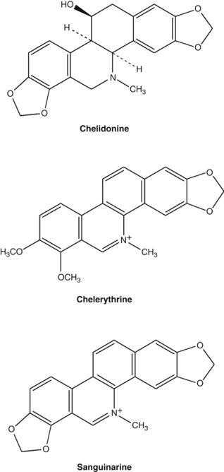
• Isoquinoline alkaloids (0.35 to 1.3%),6 including the major alkaloids chelidonine (>0.07%), chelerythrine, sanguinarine, berberine, coptisine and dl-stylopine

• Other alkaloids: sparteine (which is usually found in the Leguminosae (pea) family)6,7

• Flavonoids, phenolic acids.7

The isoquinoline group of alkaloids contains many structural types including the benzophenanthridines (chelidonine, chelerythrine, sanguinarine) and protoberberines (berberine, coptisine). An analysis of 20 Chelidonium samples from different parts of China found the total alkaloids varied from 0.89% to 1.70%, with coptisine the highest alkaloid present (average content of 0.5%) and berberine the lowest at 0.013%.8

The milky orange sap of Chelidonium contains defensive proteins, including an extracellular peroxidase with nuclease activity.9,10

image

Pharmacodynamics

Hepatoprotective and choleretic activity

Oral administration of an alcohol extract of dried Chelidonium reduced carbon tetrachloride-induced liver injury in rats.11 Significant reductions in elevated plasma levels of liver enzymes and bilirubin occurred in the treated group. A follow-up study was undertaken to clarify the underlying aspects of tissue recovery. There was an absence of fibrotic changes in the Chelidonium-treated rats (125 mg/kg/day for 3 weeks, oral), which was thought to be related to a reduced degree of cellular necrosis and a reduction in fibroblast-stimulating factors.12

Extracts of dried Chelidonium were tested for choleretic activity using the isolated perfused rat liver. The 70% ethanolic extract of the herb significantly induced choleresis (bile flow). However, it did this without increasing the total output of bile acids (that is, there was an increased flow of more dilute bile). In contrast, the phenolic and alkaloidal fractions of the total extract, tested individually and in combination, did not significantly increase bile flow, although small increases were observed. The authors concluded that the increased bile flow is due to an additive effect from all compounds in the total extract of Chelidonium, not one or two specific active constituents or fractions.13

Antimicrobial activity

Isolated chelerythrine and an alkaloid fraction from the dried roots of Chelidonium containing chelerythrine and sanguinarine were found to be ineffective against Gram-negative bacteria in vitro. However, a significant antimicrobial effect was observed against Gram-positive bacteria such as Staphylococcus aureus and two strains of Streptococcus, and also against the fungus Candida albicans.14 Chelerythrine inhibited the adherence of Streptococcus mutans and was thereby considered to possess significant anticariogenic activity.15 This alkaloid also inhibited the growth of this organism in vitro with an MIC (minimum inhibitory concentration) of 0.78 mg/mL, but chelidonine was inactive.16 Sanguinarine is also well described as an alkaloid with activity against dental plaque.1719

In an in vitro screening of the ethanolic extracts from 12 Siberian herbs for antimicrobial activity against five common pathogenic organisms, the extract of Chelidonium aerial parts was not active. In contrast, the root extract demonstrated marked activity against Bacillus cereus, Candida albicans and Salmonella enteritidis.20

Extracts of Chelidonium were found to exert antiviral effects in vitro against adenovirus types 12 and 5 and herpes simplex virus type 1 (HSV-1). The most promising antiviral alkaloid was found in greater concentrations in the fresh and aerial plant samples. This alkaloid, which belonged to the benzophenanthridine group, was not identified.21 A crude extract of Chelidonium was found to inhibit HIV-1 from infecting cells in vitro.22 A low sulphated polyglycosaminoglycan appeared to be responsible for this action and protected mice from the negative effects of murine retrovirus infection after iv administration. Such a large molecule is unlikely to have oral activity.

Alkaloids from Chelidonium and sanguinarine inhibited the growth of Trichomonas vaginalis in vitro. Sanguinarine also caused the protozoa to undergo deformation followed by disintegration within 2 h.23

An in vitro study demonstrated that the alkaloids extracted from Chelidonium, chelerythrine, and a mixture of chelerythrine and sanguinarine, exerted an antifungal effect on some Trichophyton strains, Microsporum canis, Epidermophyton floccosum and Aspergillus fumigatus.24 Another in vitro study investigated the effect of Chelidonium extracts on several Candida species and other dermatophytes. Liquid extracts of Chelidonium prepared from dried plant material collected in late July and early September (Europe) were compared. Both extracts showed greater than average antifungal activity against some organisms involved in skin infections. The July extract was active against Candida albicans, but the September extract showed no activity.25

When six species of clinically resistant yeast fungi were exposed to isolated Chelidonium alkaloids, 8-hydroxydihydrosanguinarine and 8-hydroxydihydrochelerythrine demonstrated potent activity, with MIC ranges of 2 to 80 and 4 to100 μg/mL, respectively.26 Other alkaloids also had some degree of antifungal activity. The two above compounds were also quite active against methicillin-resistant Staph. aureus.27

Fusarium species have the capacity to cause opportunistic human infections. Extracts of Chelidonium showed some activity against certain strains of this organism in vitro, with Chelidonium root extracts being more active than shoot extracts.28

Antitumour activity

An ethanolic extract of rhizomes and roots of Chelidonium exhibited cytotoxicity against a carcinoma of the nasopharynx in vitro. One of the cytotoxic principles was found to be the alkaloid coptisine.29 Chelidonium exerted an antimutagenic effect in vitro against several mutagens in the Ames test.30 The extract caused changes in the mitotic index of transplanted ascitic cells, showing marked antimitotic activity.3

Chelidonine and sanguinarine induce apoptosis in leukaemia cells in vitro, but only the former induced cell cycle arrest.31 Sanguinarine and chelerythrine induce DNA damage and cytotoxic effects in normal and cancer cells, whereas chelidonine does not.32 Chelidonine may inhibit tumour cell growth by reducing telomerase activity.33 A methanolic extract of Chelidonium also induced apoptosis in two leukaemia cell lines.34

The milky sap from fresh Chelidonium contains two nuclease enzymes with apoptotic activity against the HeLa tumour cell line, but not CHO cells.35 A lectin isolated from Chelidonium inhibited growth of two tumour cell lines, but not normal mouse fibroblasts.36

The activities of aqueous and alcoholic extracts of Chelidonium, the partially purified methanol extract and chelidonine and protopine were screened using transplanted tumours in mice. The water-soluble, purified methanol extract of dried Chelidonium demonstrated high tumour inhibition with relatively mild cytotoxic side effects. Intraperitoneal administration of 700 mg/kg for 7 days resulted in 55% inhibition of sarcoma 180 and Ehrlich carcinoma. The aqueous extract showed insignificant activity, and chelidonine and protopine (both of which are insoluble in water) showed negligible tumour inhibition and were associated with cytotoxic side effects. The crude methanol extract also showed more pronounced toxic side effects.37

The numbers of stomach tumours in rats treated with oral doses of a Chelidonium extract after initial exposure to a carcinogen were significantly lower, compared with untreated but exposed animals.38 An ethanolic whole plant extract of Chelidonium (0.1 mL/mouse of a 1:20 diluted extract) reduced the incidence of chemically induced liver cancer in mice after 60 and 120 days.39

Ukrain is described as a semi-synthetic eastern European anticancer drug derived from Chelidonium. It purportedly contains one molecule of thiophosphoric acid conjugated (bonded) to three molecules of chelidonine and is administered by intravenous injection.40 However, chemical analysis revealed that some commercial samples of Ukrain were just a mixture of Chelidonium alkaloids, with no trimeric structure evident.41 This was also the case 6 years later in 2006, when another research team found that the Ukrain sample they tested lacked the purported trimeric ‘Ukrain molecule’ and instead resembled an alkaloidal extract of Chelidonium, with chelidonine, sanguinarine and chelerythrine present as major components.42 Their in vitro studies suggested chelidonine was a particularly active inducer of tumour cell apoptosis.

A 2005 review of the anticancer research on Ukrain identified 36 in vitro studies and 46 in vivo experiments. These publications suggest that it has the capacity to exert selective cytotoxic and cytostatic effects on tumour cells, while favourably modifying the immune response.43 Specifically, diminished DNA, RNA and protein synthesis, inhibition of cellular oxygen consumption, inhibition of tubulin polymerisation44 and induction of apoptosis have all been described.45 Ukrain also modifies the host immune response via an increase in T cells and normalisation of the T-helper/T-suppressor lymphocyte ratio. Tumour mass reductions have also been demonstrated in vivo.45 More recent examples of in vivo studies include growth inhibitory effects against B16 melanoma cells46 and Ehrlich’s carcinoma,47 both in mice. It is uncertain how much, if any, of this research would have relevance to the oral use of Chelidonium extracts.

Anti-inflammatory activity

The alkaloids sanguinarine and chelerythrine are potent in vitro inhibitors of 5-lipoxygenase in polymorphonuclear leucocytes and 12-lipoxygenase in mouse epidermis. An extract of Chelidonium also inhibited 5-lipoxygenase. The inhibitory effects against lipoxygenase enzymes appear to be due to a specific enzyme interaction, rather than a non-specific redox mechanism.48

The Chelidonium alkaloids chelerythrine and sanguinarine (5 and 10 mg/kg, oral and sc) demonstrated anti-inflammatory activity in the carrageenan rat paw oedema test.14 A Chelidonium extract (40 and 400 mg/kg/day for 28 days, oral) suppressed the progression of collagen-induced arthritis in mice. Decreased levels of cytokines and activated immune cells were observed, together with increased numbers of regulatory T cells.49

Other effects

Chelidonium extract and isolated components (coptisine and caffeoylmalic acid) weakly antagonised experimentally induced contraction of isolated rat ileal smooth muscle.50 Two ethanolic dry extracts of Chelidonium and their three main alkaloids were studied in different antispasmodic test models on isolated guinea-pig ileum.51 Both extracts induced relaxation in barium chloride, carbachol and electric field stimulated ileum, as did the alkaloids.

Radioreceptor assays suggest that the alkaloids sanguinarine, chelerythrine, stylopine, allcryptopine and particularly protopine interact with the chloride channel of the GABA-A receptor.52 Chelidonium extract inhibited GABA-activated current via G proteins in vitro, suggesting an analgesic mechanism (see below).53 Studies in mice found that allocryptopine and protopine increased the brain concentration of the neurotransmitter GABA and the activity of its synthesising enzyme GAD.54

Chelidonium alkaloids acted as irreversible inhibitors of liver mitochondrial monoamine oxidase in vitro. Chelidonine was the strongest inhibitor.55 The same alkaloids also reversibly inhibited acetylcholinesterase in vitro, with sanguinarine and berberine exhibiting the strongest activity.56 Of the minor alkaloids, 8-hydroxydihydrochelerythrine and 8-hydroxydihydrosanguinarine had potent acetylcholinesterase inhibitory activity.57

Chelerythrine chloride exerted an in vitro antiplatelet effect that was believed to be due to inhibition of thromboxane formation and phosphoinositide breakdown.58

An extract of Chelidonium inhibited human keratinocyte proliferation, with sanguinarine being the most potent constituent. The mechanism of action appears to be inhibition of the inflammatory mediators leukotriene B4 and 12(S)-HETE, both of which have a known role in stimulating epidermal keratinocyte proliferation. Although the alkaloids have demonstrated cytotoxic activity in low concentrations, sanguinarine and chelerythrine did not cause more damage to cell membranes than the antipsoriatic drug anthralin, as observed by the release of lactate dehydrogenase activity (an indicator of plasma membrane damage).59

The antinociceptive action of aminophenazone in mice was potentiated by the Chelidonium alkaloids allocryptopine, chelidonine and sanguinarine.60 Chelidonium extract suppressed glycine-induced responses and elevated those induced by glutamate in isolated rat periaqueductal grey neurons.61 This might activate the descending pain control system leading to an analgesic effect.

A liquid extract of Chelidonium (2.5, 5 and 10 mL/kg, oral) dose-dependently protected against indomethacin-induced gastric ulceration in rats.62 However, it was not as active as other digestive herbs such as licorice and peppermint.

High doses of Ukrain caused slight osteopenic effects in rats, possibly due to inhibition of locomotor activity.63 However, it was anabolic on bone in ovariectomised female rats (doses 7, 14 and 28 mg/kg, ip).

Pharmacokinetics

No data available.

Clinical trials

Spasmolytic and cholagogue effects

In an early uncontrolled trial, a Chelidonium extract exerted good to very good results in two-thirds of patients treated for cholangitis, cholelithiasis and cholecystitis without stones. Forty patients received 3 mL/day (for 43 to 50 days) of a fresh plant tincture standardised to 20 mg alkaloids/100 mL.64 An early clinical trial investigated the effect of a suspension of Silybum marianum, Chelidonium and Curcuma on 28 patients. Compared to a control liquid, the herbal mixture demonstrated a greater increase in bile flow and pancreatic secretion.65

In 60 Berlin practices, 608 patients were treated in an uncontrolled study over a 3-month period with a standardised preparation of dried Chelidonium, which acted as a plant-based spasmolytic. The main presenting symptoms were cramp-like pains in the gastrointestinal tract (43%) or gall ducts (48.2%), but also included dyspeptic symptoms. Each Chelidonium tablet contained 125 mg of a 5:1 to 7:1 hydroethanolic extract with 2.85 mg of total alkaloids, including 0.79 mg of chelidonine. The dose was initially 5 tablets/day and this was reduced to 3 tablets/day in patients who responded to treatment. The average duration of treatment was 22 days and the longest treatment time was 2.5 months. A good or very good therapeutic effect on symptoms with a quick response was observed in 87.4% of cases. In most cases symptom relief occurred within 30 minutes of taking the herbal medication (62.3%). In 46.1% of patients, the average duration of efficacy of each tablet dose was more than 3 h. This study suggests value for Chelidonium in the treatment of cramp-like abdominal pains associated with irritable bowel syndrome and other causes.66

In a retrospective, open label study conducted over a 6-month period, 206 patients with epigastric complaints related to gallstones or gallbladder removal were evaluated.67 Patients received either a Chelidonium capsule (125 mg/day of a hydromethanolic extract containing 0.68 mg of chelidonine) or a liquid (three times 20 drops daily containing 0.15 mg chelidonine). There was a noted improvement in symptoms such as flatulence, diarrhoea, constipation and upper abdominal pain. Pain-free intervals were increased and inflammatory markers decreased.

The efficacy and tolerability of a standardised Chelidonium extract was investigated in a randomised, placebo-controlled, double blind trial involving 60 patients with functional epigastric symptoms.68 These included cramp-like pains, sensation of pressure or fullness, flatulence and nausea. Patients receiving active treatment took 6 tablets/day, each containing 66 to 167 mg of a Chelidonium dry extract (5.3:1 to 7.5:1) delivering 4 mg of total alkaloids calculated as chelidonine. Following 6 weeks of treatment, there was a clear difference in physician-rated response rates: 60% in the Chelidonium group versus 27% in placebo (p=0.0038). The treatment was without significant side effects compared with the placebo.

A double blind, placebo-controlled trial investigated the impact of a Chelidonium and turmeric root combination in 76 patients with upper abdominal pain attributed to functional disorders of the biliary system.69 Patients received either 3 capsules/day (each containing Chelidonium (104 to 131 mg of a 5:1 to 10:1 extract containing 4 mg of alkaloids calculated as chelidonine) and turmeric (45 mg of a 12.5:1 to 25:1 extract)) or a matching placebo for 3 weeks. In the first week there was a significant reduction in pain for the patients receiving the herbal treatment, compared with placebo. No other symptoms changed significantly relative to placebo.

A proprietary formula known as STW 5 contains liquid extracts of Chelidonium, Matricaria recutita (chamomile) flower, Iberis amarus (bitter candywort) herb, Carum carvi (caraway) fruit, Angelica archangelica (garden angelica) root, Glycyrrhiza glabra (licorice) root, Silybum marianum (milk thistle) fruit, Melissa officinalis (lemon balm) leaf and Mentha × piperita (peppermint) leaf and has been extensively researched for indigestion and functional dyspepsia. Two meta-analyses of the clinical studies on this formula have been published.70,71 They found that STW 5, at a dose of 1 mL 3 times per day, significantly reduced symptoms compared with placebo. It also demonstrated similar efficacy to cisapride and metoclopramide.

Warts, polyps

In a small, uncontrolled trial, an infusion of dried Chelidonium was administered as an enema for colonic polyposis. Administration of 10 or more enemas resulted in the complete disappearance of colonic polyps in several cases.72 In a later study, the fresh plant was made into a paste and administered 2 or 3 h after an evacuant enema. In most cases, two or three courses (consisting of 10 to 20 enemas each) were deemed to be necessary. This regime was ineffective for treating malignant regenerated or degenerated polyps. Over a 2-year period treating 149 patients with various forms of polyposis, 59% showed improvement with 27% making a complete recovery.73

An ethanolic extract of Chelidonium was used as a topical application to treat nursing mothers for warts, papillomas, condylomas and nodules in an uncontrolled trial. The extract was applied to the affected area approximately 200 times per day for 2 to 3 weeks or until improvement was observed. Complete resolution of the warts occurred after 15 to 20 days in 135 women.74

Respiratory conditions

Chelidonium was given as a syrup or extract (equivalent to 15 g of herb per day) to patients with chronic bronchitis in an uncontrolled study. The effective response rate was around 80%. It was more effective in the simple type than the asthmatic type.3 Chelidonium syrup or a decoction of the fresh herb was used to treat whooping cough in an uncontrolled study. Dosages were: infants under 6 months, 5 to 8 mL; 6 to 12 months, 8 to 10 mL; 1 to 3 years, 10 to 15 mL; 3 to 6 years, 5 to 20 mL; and above 6 years, 20 to 30 mL. Treatment was for a course of 8 to 10 days. Of 500 cases so treated, 355 were ‘cured’ and 116 improved.3

Chelidonium tincture improved tonsillar function and immunity and reduced recurrence of infection in an open comparative study in children with tonsillitis (article in Russian).75

Anticancer activity

Chelidonium was one of three herbs used to examine the efficacy of traditional Chinese herbs on squamous cell carcinoma of the oesophagus. A 30 mL dose of a decoction of Chelidonium (equivalent to 30 g of crude herb) was given orally to 30 patients twice daily for 2 weeks prior to surgery. Histological examination of the excised tissue demonstrated a greater degree of stromal lymphoid cell infiltration and cancer tissue degeneration in the patients given Chelidonium than in those given the herb plus endoxan or in the control group. The antitumour action of Chelidonium was thought to be due to the activation of an immunological rejection mechanism.76

A systematic review of seven clinical trials on Ukrain given intravenously to patients with colorectal, pancreatic, bladder and breast cancer found benefit in improving survival compared with various control groups.40 Trials were generally of low quality and clear interpretation of results was difficult due to various methodological and reporting flaws. As noted previously, the relevance of Ukrain to the clinical use of Chelidonium is uncertain.

HIV infection

An uncontrolled trial reported on the efficacy of a combination of freeze-dried Chelidonium, Ulmus rubra (slippery elm) bark and Sanguinaria canadensis (bloodroot) in 13 HIV positive patients.77 Each capsule contained 175 mg Chelidonium, 20 mg slippery elm and 5 mg bloodroot. The dose used was 9 capsules per day. The most dramatic response was a general improvement in lymphadenopathy in the patients affected by this. Minor improvements in CD4+ T cell counts and energy levels were also noted.

Toxicology and other safety data

Toxicology

No harmful or toxic effects from therapeutic doses have been established. The LD50 of the decoction in mice by intraperitoneal injection is 9.5 g/kg3 and the LD50 of the alkaloids in mice is 300 mg/kg (subcutaneous).4

In an antitumour experiment, intraperitoneal administration of a methanol extract of Chelidonium (350 mg/kg/day for 7 days) to mice resulted in a 20% mortality rate.37 After 4 weeks of feeding Chelidonium (1.5 and 3 g/kg/day) to rats, no toxic or hepatotoxic effects were observed.78 This suggests the herb is not inherently hepatotoxic and that the observed adverse hepatic reactions are rare, idiosyncratic responses.

Contraindications

Pre-existing serious liver disease or damage.

Special warnings and precautions

Given the nature of the alkaloid content of this herb and the rare hepatotoxic reactions, long-term use (except topical) is not preferred. Caution should be observed during pregnancy and lactation and in patients with gallstones. Use of Chelidonium should not be combined with heavy alcohol consumption.

Interactions

None documented.

Use in pregnancy and lactation

Category C-has caused or is associated with a substantial risk of causing harmful effects on the fetus or neonate without causing malformations.

Intramuscular injection of Ukrain on days 6 to 11 of gestation to hamsters and on days 6 to 15 of gestation to rats (0.1 to 28 mg/kg/day) did not produce teratogenic effects in either species compared with controls. Slight embryotoxic effects (increased post-implantation losses), and in consequence decreased number of average litter size, were noted in hamsters exposed to Ukrain at doses that were otherwise not embryotoxic to rats.79

Chelidonium use is strongly discouraged during breastfeeding.

Effects on ability to drive and use machines

No adverse effects expected.

Side effects

The potential association of Chelidonium with idiosyncratic hepatotoxicity was first reported in 1996. A 69-year-old woman developed symptoms of acute hepatitis after taking tablets containing several herbs including Chelidonium over a period of 6 weeks. Symptoms returned with rechallenge.80 Three additional cases were then reported (1997, 1998).8183 In one series of observations over 2 years (1997–1999) in an area of approximately 1 million inhabitants in Germany, preparations of Chelidonium apparently induced 10 cases of acute hepatitis. Investigations and tests excluded viral causes, alcohol intake and hereditary causes. Although immunological factors could not be safely excluded, the evidence, including liver biopsy, suggested a treatment-related pathology. Cholestasis was observed in half the cases, but there were no cases of liver failure and the condition improved quickly in all cases when the Chelidonium was stopped. In one case a rechallenge led to a second attack of hepatitis.84 Three cases of acute hepatitis associated with Chelidonium were then reported in the literature in 2002 and May 2003,85,86 and another in 2006.87 Again, patients returned to normal when the herbal treatment was ceased. Another case report of cholestatic hepatitis (with complete recovery) including a review of 16 cases documented in the literature, was published in 2009.88 Assessment of causality for this case suggested a probable relationship with Chelidonium consumption. Generally these hepatotoxic reactions have been observed after using higher-dose German products.

A case of contact dermatitis has been linked to exposure to the plant.89

A case of haemolytic anaemia was reported after the oral ingestion of Chelidonium extract. The patient was treated with corticosteroids, blood transfusions and haemodialysis and recovered after about 12 days.90

When Chelidonium was used in traditional Chinese medicine studies, various degrees of dry mouth, dizziness, gastric discomfort, diarrhoea, abdominal distension, nausea and mild leucopenia were reported in a minority of patients. Symptoms generally disappeared within 3 to 5 days without the discontinuation of treatment.3

Overdosage

Critical consideration of the often-cited fatal case of poisoning in a 4-year-old boy recorded in 1936 suggests that it is by no means certain that Chelidonium was involved. More than 500 g of Chelidonium is said to be required to cause toxic effects in horses and cattle.91

Safety in children

No information available, but prolonged use is probably unsuitable in children, although it has been used to treat chronic tonsillitis.

Regulatory status in selected countries

Chelidonium is covered by a positive Commission E Monograph and can be used for cramp-like disorders of the biliary and gastrointestinal tracts.

Chelidonium is not on the UK General Sale List. Under the terms of the British Medicines Act 1968 and the Statutory Instrument SI 2130 (Retail Sale or Supply of Herbal Remedies) Order 1977 (Schedule Part III), the sale of Chelidonium is restricted to herbal practitioners. It may be prescribed at a maximum dosage of 2 g three times per day.

Chelidonium does not have GRAS status. However, it is freely available as a ‘dietary supplement’ in the USA under DSHEA legislation (1994 Dietary Supplement Health and Education Act).

Chelidonium is not included in Part 4 of Schedule 4 of the Therapeutic Goods Act Regulations of Australia and is freely available for sale. However, Chelidonium-containing products must contain the following warning: ‘Greater Celandine may harm the liver in some people. Use only under the supervision of a healthcare practitioner.’

References

1. Felter HW, Lloyd JU. King’s American Dispensatory, Vol 1. 18th ed., rev 3. Portland: 1905. Reprinted by Eclectic Medical Publications; 1983. pp. 491–493.

2. Grieve M, A Modern Herbal, New York, Dover Publications, 1971;Vol 1. pp. 178–179

3. Chang HM, But PP, Pharmacology and Applications of Chinese Materia Medica, Singapore, World Scientific, 1987;vol 1. pp. 390–394

4. Huang KC. The Pharmacology of Chinese Herbs. Boca Raton: CRC Press, 1993. pp. 144–145

5. Launert EL. The Hamlyn Guide to Edible and Medicinal Plants of Britain and Northern Europe. London: Hamlyn, 1981. p. 26

6. Wagner H, Bladt S. Plant Drug Analysis: A Thin Layer Chromatography Atlas, 2nd ed. Berlin: Springer-Verlag, 1996. p. 10

7. Colombo ML, Bosisio E. Pharmacol Res. 1996;33(2):127–134.

8. Gu Y, Qian D, Duan JA, et al. J Sep Sci. 2010;33(8):1004–1009.

9. Nawrot R, Kalinowski A, Gozdzicka-Jozefiak A. Phytochemistry. 2007;68(12):1612–1622.

10. Nawrot R, Lesniewicz K, Pienkowska J, et al. Fitoterapia. 2007;78(7-8):496. 501

11. Mitra S, Gole M, Samajdar K, et al. Int J Pharmacog. 1992;30(2):125–128.

12. Mitra S, Sur RK, Roy A, et al. Phytother Res. 1996;10(4):354–356.

13. Vahlensieck U, Hahn R, Winterhoff H, et al. Planta Med. 1995;61(3):267–271.

14. Lenfeld J, Kroutil M, Marsalek E, et al. Planta Med. 1981;43(10):161–165.

15. Cheng RB, Chen X, Liu SJ, et al. Shanghai Kou Qiang Yi Xue. 2007;16(1):68–72.

16. Cheng RB, Chen X, Liu SJ, et al. Shanghai Kou Qiang Yi Xue. 2006;15(3):318–320.

17. Kuftinec MM, Mueller-Joseph LJ, Kopczyk RA. J Can Dent Assoc. 1990;15(suppl 7):31–33.

18. Laster LL, Lobene RR. J Can Dent Assoc. 1990;56(suppl 7):19–30.

19. Hannah JJ, Johnson JD, Kuftinec MM. Am J Orthod Dentofacial Orthop. 1989;96(3):199–207.

20. Kokoska L, Polesny Z, Rada V, et al. J Ethnopharmacol. 2002;82(1):51–53.

21. Kery A, Horvath J, Nasz I, et al. Acta Pharm Hung. 1987;57(1–2):19–25.

22. Gerencer M, Turecek PL, Kistner O, et al. Antiviral Res. 2006;72(2):153–156.

23. Bodalski T, Pelozarska H, Ujec M. Arch Immunol Terapii Doswiadcjalny. 1958;6(4):705–711.

24. Hejtmánková N, Walterova D, Preininger V. Fitoterapia. 1984;55(5):291–294.

25. Vukusic I, Pepeljnjak S, Kustrak D, et al. Planta Med. 1991;57(suppl 2):A46.

26. Meng F, Zuo G, Hao X, et al. J Ethnopharmacol. 2009;125(3):494–496.

27. Zuo GY, Meng FY, Hao XY, et al. J Pharm Pharm Sci. 2008;11(4):90–94.

28. Matos OC, Baeta J, Silva MJ, et al. J Ethnopharmacol. 1999;66(2):151–158.

29. Kim HK, Farnsworth NR, Blomster RN, et al. J Pharm Sci. 1969;58(3):372–374.

30. Shi GZ. Chung-Hua Yu Fang I Hsueh Tsa Chih. 1992;26(3):165–167.

31. Pilchenkov A, Kaminskyy V, Zavelevich M, et al. Toxicol In Vitro. 2008;22(2):287–295.

32. Kaminskyy V, Lin KW, Filyak Y, et al. Cell Biol Int. 2008;32(2):271–277.

33. Noureini SK, Wink M. World J Gastroenterol. 2009;15(29):3603–3610.

34. Nadova S, Miadokova E, Alfoldiova L, et al. Neuro Endocrinol Lett. 2008;29(5):649–652.

35. Nawrot R, Wolun-Cholewa M, Gozdzicka-Jozefiak A. Folia Histochem Cytobiol. 2008;46(1):79–83.

36. Fik E, Wolun-Cholewa M, Kistowska M, et al. Folia Histochem Cytobiol. 2001;39(2):215–216.

37. Sokoloff B, Saelhof CC, Yoshichi MD, et al. Growth. 1964;28:225–231.

38. Kim DJ, Ahn B, Han BS, et al. Cancer Lett. 1997;112(2):203–208.

39. Biswas SJ, Bhattacharjee N, Khuda- Bukhsh AR. Food Chem Toxicol. 2008;46(5):1474–1487.

40. Ernst E, Schmidt K. BMC Cancer. 2005;5:69.

41. Panzer A, Joubert AM, Eloff JN, et al. Cancer Lett. 2000;160(2):237–241.

42. Habermehl D, Kammerer B, Handrick R, et al. BMC Cancer. 2006;6:14.

43. Uglyanitsa KN, Nefyodov LI, Doroshenko YM, et al. Drugs Exp Clin Res. 2000;26(5–6):341–356.

44. Panzer A, Hamel E, Joubert AM, et al. Cancer Lett. 2000;160(2):149–157.

45. Jagiello-Wojtowicz E, Kleinrok Z, Urbanska EM. Drugs Exp Clin Res. 1998;24(5–6):213–219.

46. Skivka L, Susak Y, Trompak O, et al. J Oncol Pharm Pract, 2010. [Epub ahead of print]

47. Susak YM, Skivka LM, Rudik MP, et al. Exp Oncol. 2010;32(2):107–110.

48. Vavreckova C, Gawlik I, Müller K. Planta Med. 1996;62(5):397–401.

49. Lee YC, Kim SH, Roh SS, et al. J Ethnopharmacol. 2007;112(1):40–48.

50. Boegge SC, Kesper S, Verspohl EJ, et al. Planta Med. 1996;62(2):173–174.

51. Hiller KO, Ghorbani M, Schilcher H. Planta Med. 1998;64(8):758–760.

52. Häberlein H, Tschiersch KP, Boonen G, et al. Planta Med. 1996;62(3):227–231.

53. Kim Y, Shin M, Chung J, et al. Am J Chin Med. 2001;29(2):265–279.

54. Jagiello-Wójtowicz EWA, Feldo M, Kleinrok Z. Polish J Pharmacol Pharm. 1992;44(suppl):144.

55. Iagodina OV, Mikol’skaia EB, Faddeeva MD. Tsitologiia. 2003;45(10):1032–1037.

56. Kuznetsova LP, Sochilina EE, Faddeeva MD, et al. Ukr Biokhim Zh. 2005;77(2):147–153.

57. Cho KM, Yoo ID, Kim WG. Biol Pharm Bull. 2006;29(11):2317–2320.

58. Ko FN, Chen IS, Wu SJ, et al. Biochim Biophys Acta. 1990;1052:360–365.

59. Vavreckova C, Gawlik I, Müller K. Planta Med. 1996;62(6):491–494.

60. Jagiello-Wójtowicz EWA, Feldo M, Chodkowska A, et al. Polish J Pharmacol Pharm. 1992;44(Suppl):143.

61. Shin MC, Jang MH, Chang HK, et al. Clin Chim Acta. 2003;337(1–2):93–101.

62. Khayyal MT, el-Ghazaly MA, Kenawy SA, et al. Arzneimittelforschung. 2001;51(7):545–553.

63. Jablonski M. Drugs Exp Clin Res. 2000;26(5–6):317–320.

64. Neumann-Mangoldt P. Med Welt. 1977;28(4):181–185.

65. Baumann JC, Heintze K, Muth HW. Arzneimittelforschung. 1971;21(1):98–101.

66. Kniebel R, Urlacher W. Zeit Allg Med. 1993;69(25):680–684.

67. Ardjah H. Fortschr Med Suppl. 1991;115:2–8.

68. Ritter R, Schatton WFH, Gessner B, et al. Comp Ther Med. 1993;1:189–193.

69. Niederau C, Gopfert E. Med Klin (Munich). 1999;94(8):425–430.

70. Melzer J, Rösch W, Reichling J, et al. Aliment Pharmacol Ther. 2004;20(11–12):1279–1287.

71. Rösch W, Liebregts T, Gundermann KJ, et al. Phytomedicine. 2006;13(suppl 1):114–121.

72. Aminev AM, Stoliarenko AI. Vop Onkol. 1960;6(8):81–82.

73. Aminev AM. Am J Proctol. 1963;14(1):25–27.

74. Demchenko PF. Vrachebn Delo. 1957;12:1335–1338.

75. Khmel’nitsakaia NM, Vorob’ev KV, Kliachko LL, et al. Vestn Otorinolaringol. 1998;4:39–42.

76. Xian MS, Hayashi K, Lu JP, et al. Acta Med Okayama. 1989;43(6):345–351.

77. D’Adamo P. J Naturopathic Med. 1992;3(1):31–34.

78. Mazzanti G, Di Sotto A, Franchitto A, et al. J Ethnopharmacol. 2009;126(3):518–524.

79. Juszkiewicz T, Minta M, Wlodarczyk B, et al. Drugs Exp Clin Res. 1992;18(suppl):23–29.

80. De Smet PA, Van den Eertwegh AJ, Lesterhuis W, et al. BMJ. 1996;313(7049):92.

81. Greving I, Niedereichholz U, Meister V, et al. Poster No. PO19, Europäischer Pharmakovigilanz Kongress, Berlin: 1997.

82. Greving I, Meister V, Monnerjahn C, et al. Pharmacoepidemiol Drug Saf. 1998;7:S66–S69.

83. Strahl S, Ehret V, Dahm HH, et al. Dtsch Med Wochenschr. 1998;123(47):1410–1414.

84. Benninger J, Schneider HT, Schuppan D, et al. Gastroenterology. 1999;117(5):1234–1237.

85. Crijns AP, de Smet PA, van den Heuvel M, et al. Ned Tijdschr Geneeskd. 2002;146(3):124–128.

86. Stickel F, Poschl G, Seitz HK, et al. Scand J Gastroenterol. 2003;38(5):565–568.

87. Rifai K, Flemming P, Manns MP, et al. Internist (Berl). 2006;47(7):749–751.

88. Moro PA, Cassetti F, Giugliano G, et al. J Ethnopharmacol. 2009;124(2):328–332.

89. Etxenagusia MA, Anda M, Gonzales-Mahave I, et al. Contact Dermatitis. 2000;43(1):47.

90. Pinto Garcia V, Vicente PR, Barez A, et al. Sangre (Barc). 1990;35(5):401–403.

91. Frohne D, Pfander HJ. A Colour Atlas of Poisonous Plants: A Handbook for Pharmacists, Doctors, Toxicologists, and Biologists. London: Wolfe Publishing, 1984. translated from the 2nd German edition by NG Bisset. pp. 160–162

Devil’s claw

(Harpagophytum procumbens DC ex Meissner)

Synonyms

Grapple plant (Engl), Harpagophyti radix (Lat), Teufelskralle, Trampelklette, Sudafrikanische (Ger), tubercule de griffe du diable (Fr), venustorn (Dan), duiwelsklou (Afrik).

What is it?

Harpagophytum procumbens, a native to the savannah of the Kalahari of South Africa, Namibia and Botswana, has been wildcrafted and imported into Europe and subsequently elsewhere since 1953. The fruit is a capsule protected by numerous curved spines which, after the splitting of the fruit, take on a claw-like appearance. The names Harpagophytum (from the Greek harpagos, a grappling hook) and devil’s claw are derived from this. However, the secondary root tuber is the part used medicinally. Devil’s claw is also known as wood spider, grapple plant, burdock and Windhoeks’ root. Recognition of the medicinal value of the plant by Europeans is apparently traced to German soldiers, and later to GA Menhert during the Hottentot rebellion in 1904. Menhert observed the recovery of a Hottentot (who had been given up as lost by doctors) when treated by a local witch doctor. He then followed the witch doctor and discovered what plant was used, subsequently promoting the use of the root under the name ‘harpagophytum tea’.

Effects

Reduces inflammation and pain; acts as a bitter tonic.

Traditional view

Not much is known about the use of the herb in early traditional African medicine. Devil’s claw has been used in recent times in South Africa by Europeans, Euro-Africans, Bushmen, Hottentots and the Bantu for its purgative action, as a bitter tonic for digestive disturbances, and for febrile illnesses, allergic reactions and migraine. Externally it has been used in the form of an ointment for ulcers, wounds, cutaneous lesions and boils. Amongst the Bushmen, Hottentots and Bantu, women ingest the pulverised root and apply an ointment to the abdomen during labour to alleviate pain.1,2

Summary actions

Anti-inflammatory, analgesic, antirheumatic, bitter.

Can be used for

Indications supported by clinical trials

Rheumatic and arthritic conditions, including muscle pain. Possible value for pain in endometriosis.

Traditional therapeutic uses

Digestive disturbances, febrile illnesses, allergic reactions and to relieve pain. Externally for wounds, ulcers, boils and the relief of pain.

May also be used for

Extrapolations from pharmacological studies

Cardiac arrhythmias.

Preparations

Decoction of dried root, tablets, capsules or liquid extract for internal use; dried root or liquid extract for external use as an ingredient in ointments and creams.

Dosage

In tablet form, devil’s claw has been used in doses up to 6 g/day without side effects.3 In view of this and other recent clinical trials, the doses given in the British Herbal Pharmacopoeia 1983 are inadequate for antirheumatic and analgesic activity.4 For these applications the equivalent of 3 to 6 g/day of the dried herb should be prescribed.5,6 This corresponds to 6 to 12 mL/day of a 1:2 extract or 15 to 30 mL/day of 1:5 tincture. Even higher doses have been used in some clinical trials. Tablets containing a 3:1 powdered extract should be taken at the rate of 1000 to 2000 mg/day of extract. For gastrointestinal complaints, much lower doses can be used.

Some studies have indicated that the analgesic and anti-inflammatory effects of devil’s claw are decreased by the acidity of the stomach. While these findings do not necessarily discount the use of galenical preparations (such as teas and liquid extracts), they do suggest that enteric-coated extracts of devil’s claw may be more beneficial clinically. At the very least, devil’s claw preparations should be administered between meals when gastric activity is at its lowest.

Duration of use

No restriction on long-term use.

Summary assessment of safety

No adverse effects are expected if used as recommended, but, given its bitter properties, devil’s claw should be prescribed with caution in patients with peptic ulcers.

Technical data

Botany

Harpagophytum procumbens, a member of the Pedaliaceae family, is a weedy, perennial plant with creeping stems spreading from a tuberous rootstock. The greyish-green leaves are placed either alternately or directly opposite each other. Red-violet, yellow-violet or violet flowers are found at the juncture between leaf and stem. The characteristic fruits have long branching arms with anchor-like hooks (which assist their dissemination by animals). The primary root descends up to 2 m with secondary roots spreading out for up to 1.5 m on all sides, which allows it to conserve water.2,7 Two subspecies are defined: Harpagophytum procumbens subsp. procumbens and Harpagophytum procumbens subsp. transvaalensis.8

Adulteration

Occasionally the harpagoside-poor primary roots of devil’s claw are found as an adulterant, as are the roots of other intensely bitter tasting African plants, such as Elephantorrhiza spp. and Acanthosicyos naudinianus and Harpagophytum zeyheri.9 Even though Harpagophytum procumbens and H. zeyheri can easily be distinguished in the field, it is impossible to tell them apart visually when in the form of dried and sliced tubers. Both species are harvested and traded as devil’s claw in Namibia. Between 1985 and 1986 it was estimated that about 50% of the harvested wild material was mixed H. procumbens and H. zeyheri.10 Devil’s claw was proposed for inclusion in Appendix II of the Convention on International Trade in Endangered Species (CITES) in April 2000, but the proposal was not accepted,11 and it remains off the list as of August 2011.

Key constituents

• Iridoid glycosides (0.5% to 3.0%), primarily harpagoside (which has a bitter taste), isoharpagoside, harpagide (which has a slightly sweet taste), procumbide12

• Sugars (over 50%), triterpenes, phytosterols, phenolic acids and glycosides, flavonoids.6,13,14 The sugars lead to an unusually high water-soluble fraction of 50% to 70%.6,14

image

The European Pharmacopoeia recommends that devil’s claw contain not less than 1.2% harpagoside, calculated with reference to the dried herb.15H. zeyheri is physically similar to H. procumbens and is considered an inferior substitution species. Chemical testing indicates that harpagoside can be completely absent from H. zeyheri8 and the ratio of harpagoside to 8-p-coumaroylharpagide can be used to readily distinguish between H. procumbens and H. zeyheri.16

Pharmacodynamics

Anti-inflammatory and antirheumatic activity

Many of the studies undertaken to examine the anti-inflammatory effects of devil’s claw have demonstrated limited activity in standard inflammatory models. The anti-inflammatory effect varies with the route of administration and nature of the condition (acute or subacute).

Most non-steroidal anti-inflammatory drugs (NSAIDs) act by inhibiting prostaglandin biosynthesis by cyclo-oxygenase (COX). Despite earlier studies suggesting no activity in such models,17 more recent in vitro assays indicate that harpagoside and/or devil’s claw extracts inhibit the expression of COX-2 and inducible nitric oxide,1822 the production of inflammatory cytokines (for example, interleukin-1beta, interleukin-6, tumour necrosis factor-alpha),23,24 and leukotriene,25 the activity of COX-1,21,22 and the production of matrix metalloproteinases (cartilage-degrading enzymes)26 and elastase.27 However, a study that examined the effect of devil’s claw (2 g/day of powder for 21 days) on prostaglandin production during blood clotting in healthy humans found no significant differences in prostaglandin levels between the before and after measurements.28 Three known triterpenoids from devil’s claw root showed significant inhibitory activity against experimentally stimulated neutrophil respiratory burst.29 Several iridoids from devil’s claw also showed inhibitory activity against macrophage respiratory burst in vitro.30

Anti-inflammatory effects have been more convincingly demonstrated in subacute animal models rather than acute, but overall results have been mixed.5 In subacute animal models utilising formaldehyde-, Freund’s adjuvant- and granuloma-induced experimental arthritis, aqueous and methanolic extracts of devil’s claw appear to be efficient in reducing inflammation. In one study using the croton oil-induced granuloma pouch test in rats, the reduction in inflammation produced by the 12-day oral administration of aqueous and methanolic extracts of devil’s claw (200 mg/kg/day) was similar to phenylbutazone.31 In the formaldehyde-induced arthritis test, an effect comparable to 40 mg/kg/day of phenylbutazone was demonstrated for an aqueous extract of devil’s claw (20 mg/kg/day for 10 days, ip).32 Another study using different but similar models found that oral administration of devil’s claw did not produce significant effects on primary or secondary inflammatory reactions in rats.33 More recently, devil’s claw extract reduced the acute inflammation of fresh egg albumin-induced paw oedema (50 to 800 mg/kg, ip),34 and reduced oedema in Freund’s adjuvant-induced arthritis (assessing acute and chronic inflammation; extract administered orally).35

Devil’s claw extract administered by intraperitoneal injection reduced the intensity of carrageenan-induced inflammatory response in hindpaws of normal and adrenalectomised rats. The extract was ineffective when administered orally. Two and four hours after ip injection of the extract there was a significant reduction in the number of circulating leukocytes in normal rats, indicative of a hyperactive response by the adrenal cortex. No change was observed after oral administration. Overall, these results suggest that the anti-inflammatory response observed for devil’s claw after injection does not depend on the release of adrenal corticosteroids. For both routes the devil’s claw extract was administered at doses of 100 to 800 mg/kg (extract strength undefined).36

Oral administration of standardised devil’s claw extract exerted a potential chondroprotective effect in an experimental model (joint destabilisation). There was a trend toward chondroid regeneration and increased elastic and collagen fibres in the hip cartilage of the surgically altered limb of devil’s claw treated rabbits. Investigation of RNA activity suggests the effect may be due to the inhibition of matrix metalloproteinase-2.37

Little or no activity of oral doses of aqueous extracts of devil’s claw or harpagoside was demonstrated in studies using acute models, such as carrageenan-induced oedema.5,17,31,32 However, intraperitoneal pretreatment with an aqueous extract of devil’s claw produced significant, dose-dependent anti-inflammatory effects in this model.38 The highest dose tested (equivalent to 400 mg/kg of root) was more effective than pretreatment with 10 mg/kg of indomethacin.38 Harpagoside does not appear to be involved in this anti-inflammatory effect, since it did not protect against carrageenan inflammatory effects at levels corresponding to 400 mg of dried root. When devil’s claw root was treated with acid at similar levels to the stomach, it lost all activity via intraperitoneal injection.38 A later study confirmed the loss of anti-inflammatory activity following passage through the stomach.39

Topical anti-inflammatory activity has been demonstrated using an ex vivo porcine skin model. An ethanolic extract of devil’s claw and some constituents (including harpagoside) decreased COX-2 expression. Another constituent, harpagide, caused an increase in the levels of COX-2 expression after 6 hours. The overall anti-inflammatory activity of a topically applied extract may depend upon the ratio of these compounds.4042 Topical application of methanol extract of devil’s claw inhibited 12-O-tetradecanoylphorbol-13-acetate-induced COX-2 expression in mouse skin. (12-O-Tetradecanoylphorbol-13-acetate has tumour-promoting activity.)43

Analgesic activity

Injection of devil’s claw extract and harpagoside exhibited dose-dependent peripheral analgesic effects comparable to aspirin.38 This activity was abolished by acid pretreatment of the herbal extract. In earlier work, intraperitoneal administration of harpagoside (20 mg/kg) produced an analgesic effect comparable to phenylbutazone (50 mg/kg).32 However, harpagoside hydrolysed by emulsion (which would produce harpagogenin) was inactive.32 No consistent analgesic effects were found in mice after oral doses of devil’s claw extracts.31 In a later study, oral pretreatment with devil’s claw extract (30 to 300 mg/kg, standardised to contain 1.9% harpagoside) did produce significant analgesic effects in the formalin test of mice. Naloxone (5 mg/kg, sc) significantly attenuated the analgesic effect of devil’s claw, suggesting that the opioidergic system may be involved.44 Devil’s claw extract has also demonstrated analgesic activity against heat- and chemical-induced pain in mice (50 to 800 mg/kg, 64:1 extract, ip injection)34 and against heat-induced pain in rats (extract administered orally).35

Cardiovascular activity

In conscious normotensive rats, the dried methanolic extract of devil’s claw caused a significant dose-dependent reduction of arterial blood pressure, and a concomitant decrease of heart rate. The reduction of blood pressure was only significant at the higher oral dosage of 400 mg/kg. Under the same experimental conditions, an equivalent quantity of harpagoside was not as effective as the full plant extract. The methanolic extract of devil’s claw caused a mild decrease in heart rate with a concomitant mild positive inotropic effect at lower concentrations in vitro, and marked negative inotropic effect at higher concentrations.45 Devil’s claw extract also demonstrated a dose-dependent protective effect against arrhythmias induced by aconitine and particularly those provoked by calcium and epinephrine-chloroform, both in vivo (100 to 400 mg/kg, oral) and in vitro.45

In another study on isolated rat hearts, methanolic extracts of devil’s claw showed a significant, dose-dependent protection against hyperkinetic ventricular arrhythmias induced by reperfusion following ischaemia. Additionally, it was demonstrated that both the methanolic extract and harpagoside inhibited hyperkinetic arrhythmias triggered by digitoxin. The mechanism of action of devil’s claw was thought to be analogous to that of verapamil. Thus devil’s claw may modify the cellular metabolism that causes transmembrane ionic fluxes, thereby producing a calcium antagonistic effect.46

Another in vivo study demonstrated that the iridoids, triterpenes and the flavonoids (luteolin and kaempferol) were all responsible for the electrophysiological and haemodynamic effects of devil’s claw. The oral doses used in this study were 20 and 40 mg/kg of a methanolic extract.47

Other activity

Devil’s claw extract and tincture inhibited free radical generation in vitro.48 Water-soluble constituents of devil’s claw extract were antioxidant, but harpagoside did not contribute significantly to the antioxidant activity.49

The effect of an aqueous extract of devil’s claw root on longitudinal, tubular uterine horn muscle strips taken from both non-pregnant and pregnant young adult, female rats was investigated. Devil’s claw extract (10 to 800 μg/mL, 28:1 extract) increased the tone and induced contraction of oestrogen-dominated rat longitudinal uterine horn muscle strips taken from stilboestrol-pretreated, non-pregnant female rats. The same effect was observed for longitudinal, tubular uterine horn muscle strips taken from female rats in the early, middle and late stages of pregnancy. Moderate to high concentrations (200 to 1000 μg/mL, 28:1 extract) provoked powerful contractions of isolated longitudinal, tubular uterine horn muscle preparations of both non-pregnant and pregnant rats.50

The potential anticonvulsant activity of a devil’s claw aqueous extract (28:1) administered by intraperitoneal injection was investigated against pentylenetetrazole-, picrotoxin- and bicuculline-induced seizures in mice. At the dosage of 100 to 800 mg/kg, the average onset of convulsions was delayed and the average duration of convulsions reduced. The activity was greater in the pentylenetetrazole and picrotoxin models. Two reference drugs were also tested. By comparison, animals were 90% protected after administration of the reference drug diazepam (0.5 mg/kg, ip) against convulsions caused by picrotoxin, while the highest dose of devil’s claw (800 mg/kg) resulted in 70% protection.51

Intraperitoneal injection of devil’s claw aqueous extract (50 to 800 mg/kg, 64:1 extract) produced dose-dependent, significant reductions in blood glucose concentrations of both fasted normal and fasted diabetic rats.34

The antimicrobial activity of plant extracts against 29 species of aerobic and anaerobic bacteria and yeasts was tested in vitro. An aqueous standardised extract of devil’s claw was effective against all microbes tested, but generally only at higher concentrations (100 μg/mL of a stock solution comprising 200 mg/mL of the 1.5:1 to 2.5:1 extract). Harpagoside was not active.52

Pharmacokinetics

There has been controversy over the effects of stomach and acid hydrolysis on devil’s claw extract and its active ingredients, as suggested by some of the above studies. Some authors have proposed that the compounds obtained after acid hydrolysis, especially harpagogenin, are the true active compounds producing the anti-inflammatory and antiarthritic properties. In contrast, other studies suggest that the extract, and harpagoside in particular, may be partially inactivated by the acid milieu of the stomach.7,39 In other words, the basic issue is whether harpagoside and other iridoid glycosides (and perhaps other compounds in the root) are more active after oral doses than their respective hydrolysis products, such as harpagogenin.38,39

An in vitro experiment investigating the simulated gastric disintegration of devil’s claw extract tablets was published in 2000. The tablets (which were not enterically coated) disintegrated after around 18 minutes in artificial gastric fluid. The harpagoside content was decreased by about 10% in this medium after periods of 1 and 3 h. Harpagoside remained stable in artificial intestinal fluid for a period of 6 h.53

Another consideration complicates this picture. The main iridoids of devil’s claw, namely harpagide, harpagoside and 8-O-p-coumaroylharpagide, were transformed into the pyridine monoterpene alkaloid aucubinine B by human faecal flora and by specific bacterial species isolated from the flora. Aucubinine B was also generated from harpagide, harpagoside and 8-O-p-coumaroylharpagide by beta-glucosidase in the presence of the ammonium ion.54 Hence this alkaloid could be an active agent after oral doses of devil’s claw.

The hepatobiliary excretion of harpagoside was investigated in rats. The bile-to-blood distribution ratio (AUCbile/AUCblood) was 986 for an intravenous dose of 3 mg/kg. The ratio dropped significantly to 6.41 or 221.2 after co-administration of cyclosporin A (10 mg/kg) or verapamil (1.2 mg/kg), respectively. In other words, both drugs increased the concentration of harpagoside in the blood. Elimination of harpagoside via the bile is probably regulated by P-glycoprotein (P-gp), since cyclosporin A and verapamil are P-gp inhibitors.55

The pharmacokinetics of devil’s claw was investigated in three pilot, single-dose studies with small numbers of healthy volunteers. Three different standardised extracts were orally administered at varying doses. Maximum concentrations of harpagoside in plasma (Cmax) were reached after 1.3 to 2.5 h and ranged from around 8 to 50 ng/mL. The half-life was short, ranging between 3.7 and 6.4 h, and clearance was about 15 L/min. A linear relationship between the dose of harpagoside and the first Cmax and area under the concentration (AUC) curve was observed.25

Clinical trials

Antirheumatic activity

There are several reports of uncontrolled and controlled trials in the late 1970s to 1990 investigating the oral administration of devil’s claw for the treatment of rheumatic conditions.7 Examples of two early uncontrolled trials follow. In one large, open study involving 630 patients with various rheumatic illnesses, 42% to 85% of the patients showed a significant improvement after 6 months of devil’s claw (3 to 9 g/day). Efficacy varied with the site of the arthritis; 80% of patients with arthritis in the large joints or spinal column experienced a significant improvement in symptoms, whilst the remaining 20% experienced no therapeutic effect even at maximum dosage.56 However, one small open study involving 13 patients with rheumatoid arthritis and psoriatic arthropathy found no benefit from devil’s claw treatment.57

Several reviews have assessed the quality of the clinical trial data. The most comprehensive systematic review was published in 2003 and examined 20 trials of devil’s claw in the treatment of chronic musculoskeletal pain, including low back pain and arthritis.58 Of the 20 trials included in the review, eight were uncontrolled or observational studies, two were open comparisons with conventional treatments (phenylbutazone or various conventional treatments) and 10 were randomised, double-blinded comparisons: eight against placebo and two against non-steroidal anti-inflammatory drugs (NSAIDs: diacerhein and rofecoxib).

In the opinion of the reviewers, the uncontrolled studies provided mainly preliminary information. The two open label trials were subject to performance, detection and/or selection bias. Of the eight randomised, double blind comparisons against placebo, six were affected by lack of transparency; one trial3 could not provide definitive evidence in terms of its pre-selected principal outcome measure (consumption of the rescue analgesic drug tramadol), and the remaining trial59 provided good-quality evidence of a dose-dependent superiority over placebo. One of the randomised controlled comparison trials (devil’s claw versus rofecoxib) was intended only as a pilot study.60 The other comparative trial61 provided evidence that devil’s claw powder is as effective as the weak NSAID diacerhein.58

More details are provided below for the latter four trials:

• For the secondary outcome measure – the number of patients with low back pain who were pain free – treatment with devil’s claw extract (containing 60 mg/day of harpagoside for 4 weeks) was significantly better than placebo (p=0.008) in a randomised trial involving 118 patients.3

• More patients with an exacerbation of chronic low back pain receiving devil’s claw extract (equivalent to 4.5 g/day or 9 g/day of root and containing 50 mg/day or 100 mg/day of harpagoside, respectively) for 4 weeks were pain-free compared with placebo. Treatment with extract containing 100 mg/day of harpagoside was more effective than 50 mg/day in terms of the number of pain-free patients, but not in terms of improvement in the pain component of the Arhus low back pain index. This was a randomised, double blind study involving 197 patients.59

• Devil’s claw extract (containing 60 mg/day of harpagoside) over a 6-week period reduced pain in patients with low back pain to about the same extent as rofecoxib (12.5 mg/day).60 This was a randomised, double-dummy, double blind trial involving 88 patients.

• Devil’s claw powder (2.6 g/day, containing 57 mg/day of harpagoside) taken over a period of 4 months significantly reduced both spontaneous pain and functional disability, and exhibited similar efficacy to diacerhein (100 mg/day) in a trial involving 122 patients with osteoarthritis of the knee or hip. The number of patients using other NSAIDs and analgesic drugs (diclofenac or paracetamol-caffeine) at the completion of the study was significantly lower in the devil’s claw group.61

A 2006 Cochrane review of herbal medicine for low back pain62 included three of these devil’s claw trials,3,59,60 considering them to be of high quality.

Three postmarketing surveillance studies6365 and a retrospective analysis of existing trial data66 have been undertaken subsequent to the 2003 systematic review. Some of these studies were a continuation of prior work, or involved subgroups of patients from previous surveillance studies. Patients had low back pain or osteoarthritis of the knee or hip. Results were obtained in some cases for up to 54 weeks of treatment. In general, treatment with devil’s claw extract (50 to 60 mg/day harpagoside) was clinically beneficial. For example, in one trial maximum pain relief occurred after 3 to 4 months66 and in another trial 34% of patients had responded to treatment by month 1, with 51% responding by month 2.64

An uncontrolled trial involving 259 patients was conducted in the UK, with results published in 2007. Patients with mild to moderate rheumatic disorders were treated for 8 weeks with devil’s claw extract (about 2.2 g/day dried herb equivalent). There were statistically significant (p<0.0001) improvements in patients’ assessment of global pain, stiffness and function, with significant reductions in mean pain scores for the hand, wrist, elbow, shoulder, hip, knee and back.67

In one interesting study, patients with slight to moderate muscular tension or pain in the back, shoulder and neck were enrolled in a randomised, double blind, placebo-controlled trial: 63 patients received either a placebo or devil’s claw (960 mg/day of a 5:1 60% ethanolic extract) for 4 weeks.68 By the end of the trial, devil’s claw treatment was significantly better than placebo in terms of muscle pain (visual analogue scale), muscle tenderness (pressure algometer test) and muscle stiffness (all p<0.001).

Other activity

A preliminary observational study found that devil’s claw extract (400 mg capsules, four per day for 12 weeks) was beneficial in 12 patients with histologically proven endometriosis. A reduction of symptoms such as dysmenorrhoea and dyspareunia was observed after 4 weeks of treatment in six patients. All patients reported improvement in symptoms after 8 weeks and quality of life was also enhanced.69 A patent lodged for this application70 indicates the capsule contained a 30% aqueous ethanolic concentrate (2.6:1 to 3.1:1), suggesting a daily dose of about 4.5 g dried herb equivalent.

Regression of follicular lymphoma occurred in two patients who self-treated with devil’s claw (dosage undefined). One patient was also taking the herbal formulation known as Essiac. CT scan images at baseline and follow-up (10 and 11 months later) provided objective evidence of regression of the tumours. Spontaneous regression of low-grade lymphoma has been previously reported in seven of 44 patients taking neither herbal medicines nor COX-2 inhibitors. However, the timing of the response in these two patients suggests a possible therapeutic effect. The chance of observing spontaneous regression in two consecutive lymphoma patients is approximately 2%.71

Devil’s claw has also generated interest for its veterinary applications. The effect of devil’s claw on degeneration of the proximal intertarsal, distal interdistal and tarsometatarsal joints and of muscular disorders was investigated in ten race horses by comparison with a control group of ten horses treated with phenylbutazone.72 The horses were given 0.5 mg/kg of an approximately 3:1 extract of devil’s claw for 60 days. Six horses receiving the devil’s claw showed a marked improvement of symptoms, even compared with the control group receiving the drug. The dose used in this study appears to be relatively low.

Toxicology and other safety data

Toxicology

Devil’s claw demonstrated low toxicity in acute and subacute toxicity studies.17,31 No significant haematological or gross pathological findings were evident in rats after oral administration of 7.5 g/kg of devil’s claw for 21 days. Hepatotoxic effects could not be demonstrated in terms of liver weight, levels of microsomal protein and liver enzymes after 7 days of oral treatment with 2 g/kg.31Table 1 lists the LD50 data recorded for devil’s claw extract and its constituents.

Table 1 LD50 data recorded for devil’s claw extract and its constituents

image

Contraindications

None advised, but see below.

Special warnings and precautions

The Commission E advises that the use of devil’s claw is contraindicated in patients with gastric or duodenal ulcers and should only be used with professional supervision in patients with gallstones.74 However, any health risks are theoretical in nature and are projected from the bitter tonic activity of the herb. Bitters should be used with caution in oesophageal reflux and in states of hyperacidity.

Interactions

The in vitro inhibitory activity of a devil’s claw extract (undefined) towards the cytochrome P450 enzymes 1A2 and 2D6 was relatively low (IC50>900 μg/mL). CYP 2C8, CYP 2C9, CYP 2C19 and CYP 3A4 were moderately inhibited, with IC50 values in the range of 100 to 350 μg/mL. (IC50 is the concentration that is required to produce 50% inhibition.)75 Three standardised extracts of devil’s claw inhibited P-gp activity in vitro (using two tests: calcein-AM and esterase). Harpagoside was almost inactive. Expression of P-gp was also tested. In cells cultured for 3 days in the presence of devil’s claw extracts or pure harpagoside, a dose-dependent increase in P-gp expression was observed.76

Devil’s claw may potentiate the effect of warfarin. A case of purpura was reported in a patient taking warfarin and devil’s claw.77 The patient’s medical condition, other medications, and the doses and duration of the warfarin and devil’s claw ingestion were not reported.

Devil’s claw has demonstrated a protective effect against arrhythmia in vitro and in vivo45,46 and it has been proposed that it may interact with antiarrhythmic drugs.5 However, this is a theoretical concern of unknown clinical relevance.

Use in pregnancy and lactation

Category B2 – no increase in frequency of malformation or other harmful effects on the foetus from limited use in women. Animal studies are lacking.

In South African traditional medicine, low doses of the dried tuber (such as 0.25 g three times daily) are administered to pregnant women to relieve pain, and this is continued postpartum at a reduced dose. The fresh tuber is also made into an ointment and applied to the abdomen of women who anticipate a difficult birth.78 While devil’s claw has increased the contraction of isolated uterine strips,50 the clinical relevance of this is uncertain.

Devil’s claw is probably compatible with breastfeeding, since it has been used in low doses in the postpartum period in traditional South African medicine.78

Effects on ability to drive and use machines

No adverse effects expected.

Side effects

A systematic review published in 2008 investigated the safety of devil’s claw preparations. Twenty-eight clinical studies (uncontrolled, observational and controlled) were identified from 1985 up to early 2007 involving a total of 6892 patients. Twenty-one of the studies were of short duration (up to 8 weeks) and two postmarketing surveillance studies were carried out over 54 weeks. Double blind trials assessed 615 patients, uncontrolled and observational studies included 6277 patients. For the double blind trials, the incidence of adverse events during treatment with devil’s claw was not higher than during placebo treatment. Minor adverse events were described in 20 studies (n=4274) in a total of 138 patients, which corresponds to an overall adverse event rate of around 3%. Some of the adverse events, such as gastrointestinal complaints and allergies, were probably related to intake of devil’s claw.79

One case of conjunctivitis, rhinitis and asthma has been reported after occupational exposure to devil’s claw. Allergy was confirmed by a provocation test.80

Overdosage

No incidents found in published literature.

Safety in children

No information available, but adverse effects are not expected.

Regulatory status in selected countries

Devil’s claw is official in the European Pharmacopoeia 2001 and the British Pharmacopoeia 2011.

Devil’s claw is covered by a positive Commission E Monograph and has the following applications:

• Lack of appetite, dyspeptic complaints

• In supportive therapy for degenerative disorders of the musculoskeletal system.

Devil’s claw is on the UK General Sale List. Products have achieved Traditional Herbal Registration in the UK with the traditional indication of relief of symptoms (aches and pains) associated with the muscles and joints.

Devil’s claw does not have GRAS status. However, it is freely available as a ‘dietary supplement’ in the USA under DSHEA legislation (1994 Dietary Supplement Health and Education Act).

Devil’s claw is not included in Part 4 of Schedule 4 of the Therapeutic Goods Act Regulations of Australia and is freely available for sale.

References

1. Ragusa S, Circosta C, Galati EM, et al. J Ethnopharmacol. 1984;11(3):245–257.

2. Van Wyk B-E, Van Oudtshoorn B, Gericke N. Medicinal Plants of South Africa. Arcadia: Briza Publications, 1997. pp. 144–145

3. Chrubasik S, Zimpfer CH, Schutt U, et al. Phytomedicine. 1996;3(1):1–10.

4. British Herbal Medicine Association’s Scientific Committee. British Herbal Pharmacopoeia. Cowling: BHMA; 1983. p. 111.

5. Scientific Committee of ESCOP (European Scientific Cooperative on Phytotherapy). ESCOP Monographs: Harpagophytum radix. UK: European Scientific Cooperative on Phytotherapy Secretariat; 1996.

6. British Herbal Medicine Association. British Herbal Compendium, Vol 1. Bournemouth: BHMA; 1992. pp. 78–80.

7. Wenzel P, Wegener T. Dtsch Apoth Ztg. 1995;135(13):1131–1144.

8. van Wyk BE. J Ethnopharmacol. 2008;119(3):342–355.

9. Bisset NG, ed. Herbal Drugs and Phytopharmaceuticals: A Handbook for Practice on a Scientific Basis. Stuttgart: Medpharm Scientific Publishers, 1994. p. 250

10. Hachfeld B, Schippmann U, Medicinal Plant Conservation, Bonn, Bundesamt fur Naturschutz, 2000;vol 6. pp. 3–9

11. Convention of International Trade in Endangered Species of Wild Fauna and Flora, Eleventh meeting of the Conference of the Parties, Gigiri (Kenya): 2000. pp. 10–20.

12. Wagner H, Bladt S. Plant Drug Analysis: A Thin Layer Chromatography Atlas, 2nd ed. Berlin: Springer-Verlag, 1996. p. 76

13. Burger JFW, Brandt EV, Ferreira D. Phytochemistry. 1987;26(5):1453–1457.

14. Ziller KH, Franz G. Planta Med. 1979;37(12):340–348.

15. European Pharmacopoeia, 3rd ed. Strasbourg: European Department for the Quality of Medicines within the Council of Europe; 1996. pp. 716–717.

16. Baghdikian B, Lanhers MC, Fleurentin J, et al. Planta Med. 1997;63(2):171–176.

17. Whitehouse LW, Znamirowska M, Paul CJ. Can Med Assoc J. 1983;129(3):249–251.

18. Huang TH, Tran VH, Duke RK, et al. J Ethnopharmacol. 2006;104(1–2):149–155.

19. Kaszkin M, Beck KF, Koch E, et al. Phytomedicine. 2004;11(7–8):585–595.

20. Jang MH, Lim S, Han SM, et al. J Pharmacol Sci. 2003;93(3):367–371.

21. Anauate MC, Torres LM, de Mello SB. Phytother Res. 2010;24(9):1365–1369.

22. Bermejo Benito P, Díaz Lanza AM, Silván Sen AM, et al. Planta Med. 2000;66(4):324–328.

23. Inaba K, Murata K, Naruto S, et al. J Nat Med. 2010;64(2):219–222.

24. Fiebich BL, Heinrich M, Hiller KO, et al. Phytomedicine. 2001;8(1):28–30.

25. Loew D, Möllerfeld J, Schrödter A, et al. Clin Pharmacol Ther. 2001;69(5):356–364.

26. Schulze-Tanzil G, Hansen C, Shakibaei M. Arzneimittelforschung. 2004;54(4):213–220.

27. Boje K, Lechtenberg M, Nahrstedt A. Planta Med. 2003;69(9):820–825.

28. Moussard C, Alber D, Toubin MM, et al. Prostagland Leuk Essent Fatty Acids. 1992;46(4):283–286.

29. Qi J, Li N, Zhou JH, et al. Planta Med. 2010;76(16):1892–1896.

30. Qi J, Chen JJ, Cheng ZH, et al. Phytochemistry. 2006;67(13):1372–1377.

31. Erdos A, Fontaine R, Friehe H, et al. Planta Med. 1978;34(1):97–108.

32. Eichler O, Koch C. Arzneimittelforschung. 1970;20:107–109.

33. McLeod DW, Revell P, Robinson BV. Br J Pharmacol. 1979;66(1):140P–141P.

34. Mahomed IM, Ojewole JA. Phytother Res. 2004;18(12):982–989.

35. Andersen ML, Santos EH, Seabra Mde L, et al. J Ethnopharmacol. 2004;91(2–3):325–330.

36. Catelan SC, Belentani RM, Marques LC, et al. Phytomedicine. 2006;13(6):446–451.

37. Chrubasik JE, Lindhorst E, Neumann E, et al. Phytomedicine. 2006;13(8):598–600.

38. Lanhers MC, Fleurentin J, Mortier F, et al. Planta Med. 1992;58(2):117–123.

39. Soulimani R, Younos C, Mortier F, et al. Can J Physiol Pharmacol. 1994;72(12):1532–1536.

40. Abdelouahab N, Heard C. J Nat Prod. 2008;71(5):746–749.

41. Ouitas NA, Heard CM. Int J Pharm. 2009;376(1–2):63–68.

42. Ouitas NA, Heard C. Phytother Res. 2010;24(3):333–338.

43. Kundu JK, Mossanda KS, Na HK, et al. Cancer Lett. 2005;218(1):21–31.

44. Uchida S, Hirai K, Hatanaka J, et al. Biol Pharm Bull. 2008;31(2):240–245.

45. Circosta C, Occhiuto F, Ragusa A, et al. J Ethnopharmacol. 1984;11(3):259–274.

46. Costa de Pasquale R, Busa G, Circosta C, et al. J Ethnopharmacol. 1985;13(2):193–199.

47. Occhiuto F, de Pasquale A. Pharmacol Res. 1990;22(suppl 3):72–73.

48. Grant L, McBean DE, Fyfe L, et al. Phytother Res. 2009;23(1):104–110.

49. Betancor-Fernández A, Pérez-Gálvez A, Sies H, et al. J Pharm Pharmacol. 2003;55(7):981–986.

50. Mahomed IM, Ojewole JA. J Smooth Muscle Res. 2009;45(5):231–239.

51. Mahomed IM, Ojewole JA. Brain Res Bull. 2006;69(1):57–62.

52. Weckesser S, Engel K, Simon-Haarhaus B, et al. Phytomedicine. 2007;14(7–8):508–516.

53. Chrubasik S, Sporer F, Dillmann-Marschner R, et al. Phytomedicine. 2000;6(6):469–473.

54. Baghdikian B, Guiraud-Dauriac H, Ollivier E, et al. Planta Med. 1999;65(2):164–166.

55. Wu Q, Wen XD, Qi LW, et al. J Chromatogr B Analyt Technol Biomed Life Sci. 2009;877(8–9):751–756.

56. Belaiche P. Phytotherapy. 1982;1:22–28.

57. Grahame R, Robinson BV. Ann Rheum Dis. 1981;40(6):632.

58. Chrubasik S, Conradt C, Black A. Phytomedicine. 2003;10(6–7):613–623.

59. Chrubasik S, Junck H, Breitschwerdt H, et al. Eur J Anaesthesiol. 1999;16(2):118–129.

60. Chrubasik S, Model A, Black A, et al. Rheumatology. 2003;42(1):141–148.

61. Chantre P, Cappelaere A, Leblan D, et al. Phytomedicine. 2000;7(3):177–183.

62. Gagnier JJ, van Tulder M, Berman B, et al. Cochrane Database Syst Rev. 2006;2:CD004504.

63. Chrubasik S, Künzel O, Thanner J, et al. Phytomedicine. 2005;12(1–2):1–9.

64. Chrubasik S, Chrubasik C, Künzel O, et al. Phytomedicine. 2007;14(6):371–376.

65. Wegener T, Lüpke NP. Phytother Res. 2003;17(10):1165–1172.

66. Thanner J, Kohlmann T, Künzel O, et al. Phytother Res. 2009;23(5):742–744.

67. Warnock M, McBean D, Suter A, et al. Phytother Res. 2007;21(12):1228–1233.

68. Göbel H, Heinze A, Ingwersen M, et al. Schmerz. 2001;15(1):10–18.

69. Arndt D, Bobermien K, Heyer H, et al. Geburtsh Frauenheilk. 2006;S1(67): PO-E-03–10

70. WO/2006/114422. Use of Devil’s Claw (Harpagophytum procumbens) Root Extracts for Endometriosis Treatment. 2006.

71. Wilson KS. Curr Oncol. 2009;16(4):67–70.

72. Montesano D, Ferrara L. Rev Fitoterapia. 2002;2(S1):105. Abstract A045

73. Van Haelen M, Van Haelen-Fastre R, Samaey-Fontaine J, et al. Phytotherapy. 1983;5:7–13.

74. Blumenthal M, ed. The Complete German Commission E Monographs: Therapeutic Guide to Herbal Medicines. Austin: American Botanical Council, 1998. p. 120

75. Unger M, Frank A. Rapid Commun Mass Spectrom. 2004;18(19):2273–2281.

76. Romiti N, Tramonti G, Corti A, et al. Phytomedicine. 2009;16(12):1095–1100.

77. Shaw D, Leon C, Kolev S, et al. Drug Saf. 1997;17(5):342–356.

78. Watt JM, Breyer-Brandwijk MG. The Medicinal And Poisonous Plants of Southern and Eastern Africa: Being an Account of Their Medicinal and Other Uses, Chemical Composition, Pharmacological Effects and Toxicology in Man and Animal. Edinburgh: Churchill Livingstone, 1962. p. 830

79. Vlachojannis J, Roufogalis BD, Chrubasik S. Phytother Res. 2008;22(2):149–152.

80. Altmeyer N, Garnier R, Rosenberg N, et al. Arch Mal Prof. 1992;53:289–291.

Dong quai

(Angelica sinensis (Oliv.) Diels)

Synonyms

Angelica polymorpha var. sinensis (botanical synonym), dong quai (Engl), Radix Angelica sinensis (Lat), dang gui (Chin), toki (Jap), tanggwi (Kor), kinesisk kvan (Dan).

What is it?

The root of dong quai is an extremely popular herb that has been used by the Chinese for thousands of years as a tonic and a spice. Its reputation in the West as a Chinese herb is second only to ginseng. Women have especially used dong quai to maintain their health and it is sometimes regarded as a ‘women’s ginseng’. Angelica acutiloba, which is indigenous to Japan, became a substitute in that country for genuine dong quai, but probably has some different phytochemistry (see Adulteration below). The popular use of dong quai in the West as a herb to treat menopausal symptoms such as hot flushes appears to be ill advised (although there are some positive trials for its use in combination). Nonetheless, it may have value in menopause as a tonic.

Effects

Regulates menstruation, alleviates dysmenorrhoea; treats blood deficiency; relieves constipation by lubricating the bowel; treats and prevents cardiovascular disorders; protects the liver; has blood-building and tonic effects (in combination with Astragalus).

Traditional view

Dong quai is sweet, acrid, bitter and warm. It strengthens the heart, lung and liver meridians and lubricates the bowel. Dong quai tonifies the Blood, regulates menstruation, invigorates and harmonises the Blood and is used to treat congealed blood conditions, blood deficiency and Deficient Blood patterns. It is an important herb in the treatment of gynaecological problems.1

Summary actions

Antianaemic, antiplatelet, female tonic, mild laxative, antiarrhythmic, anti-inflammatory.

Can be used for

Indications supported by clinical trials

To relieve dysmenorrhoea and treat infertility (uncontrolled trials); chronic hepatitis and cirrhosis (uncontrolled trial), chronic obstructive pulmonary disease and cor pulmonale (uncontrolled trials). No benefits were found in the treatment of menopausal symptoms with the use of dong quai alone; trials where it was used in combination have yielded mixed results.

Traditional therapeutic uses

Irregular menstruation, amenorrhoea, dysmenorrhoea; constipation; congealed blood (abdominal pain, traumatic injuries, swellings, contusions, bruising); blood deficiency (tinnitus, blurred vision, palpitations); as a tonic, especially for women.

May also be used for

Extrapolations from pharmacological studies

For the prevention of atherosclerosis; as an antiplatelet agent.

Preparations

Dried root as a decoction; liquid extract, tablets or capsules for internal use.

Dosage

• 3 to 15 g/day of the dried root by decoction

• 4 to 8 mL/day of a 1:2 liquid extract; 10 to 20 mL/day of 1:5 tincture; or equivalent doses as tablets or capsules.

Duration of use

May be taken long term.

Summary assessment of safety

No adverse effects from ingestion of dong quai are expected, providing the suggested contraindications are observed.

Technical data

Botany

Dong quai, a member of the Umbelliferae (Apiaceae, carrot) family, is a fragrant, perennial herb native to China, Korea and Japan that grows to a height of 0.5 to 1 m. The inferior leaves are tripinnate, superior leaves are pinnate, on long, sheathed petioles. The umbels number 10 to 14, with irregular rays, each umbel containing 12 to 36 white flowers. The root’s exterior is grey-dark brown in colour and its surface is covered in wrinkles. The root consists of a head, body and tail.2 Different properties are ascribed to these parts: the head is most tonic and the tail moves the blood most strongly. Such preparations are very expensive and the entire root is usually prescribed.

Adulteration

Ligusticum glaucescens and Levisticum officinale are regarded as substitutes of lower quality for dong quai.2Angelica acutiloba is a therapeutically interchangeable species for A. sinensis in Japan (but in China would be regarded as a substitute of lower quality).2,3A. dahurica, A. pubescens and Ligusticum chuanxiong are also used in traditional Chinese medicine, Ligusticum porteri is traditionally used in Mexico and is sold in the USA. These plants are morphologically similar to Angelica sinensis and may occur as substitutes.3

A. acutiloba contains phthalides as main constituents and can be difficult to distinguish from A. sinensis by thin layer chromatography,4 although ferulic acid was detected in A. sinensis and not in A. acutiloba.5 Senkyunolide A, analysed using a high performance liquid chromatography method, is a useful standard compound for quality evaluation and chemical differentiation between Ligusticum chuanxiong rhizome and Angelica sinensis root.6

Key constituents

• Essential oil (0.4% to 0.7%), mainly consisting of the phthalides ligustilide and n-butylidenephthalide2 (n-butylidenephthalide has a penetrating characteristic fragrance,7 like all plant phthalides)

• Phytosterols, ferulic acid, coumarins (angelol, angelicone).2

image

Pharmacodynamics

Effects on sexual function

The essential oil relaxed the isolated uterus, but other components increased uterine contraction.8 Butylidenephthalide demonstrated spasmolytic activity by inhibiting rat uterine contractions. Its effect was non-specific, similar to papaverine, but with a different mechanism of action.9 In vitro tests with isolated rat uterus suggest that ligustilide has a non-specific antispasmodic activity.10 Some experiments on the whole root of dong quai have shown a stimulant activity in vivo, while others have shown that it can relax or coordinate (make more rhythmic) uterine contractions, depending on uterine tone. Different parts of the root exhibited a similar spasmolytic action on isolated rabbit uterus.11

A review suggested that the in vitro experiments provide conflicting evidence concerning the oestrogenicity of dong quai.11 In contrast, oral administration of a standardised ethanolic dong quai extract at ‘doses based on typical clinical human doses’ produced oestrogenic activity in ovariectomised rats. The uterine cellular structure was stimulated, vaginal cornification provoked and serum luteinising hormone decreased.12 However, dong quai had no effect on uterine weight when administered orally to mice for a period of 4 days.13

One study showed increased sexual activity in female animals and a reduction in signs of vitamin E deficiency in male mice.2 Intraperitoneal injection of dong quai protected mice ovaries from the damaging effects of gamma radiation.14

An aqueous extract of dong quai stimulated the growth of breast cancer cells in vitro.15 In an earlier study this activity was also demonstrated for the ethanolic extract.13 Clinical implications (such as adverse effects from the use of dong quai during breast cancer) cannot yet be drawn from this research.

Cardiovascular activity

Dong quai has a quinidine-like action on the isolated heart. It can prolong the refractory period and correct experimental atrial fibrillation induced by atropine, pituitrin, strophanthin, acetylcholine or electrical stimulation.8 Extracts of dong quai relaxed isolated aorta16 and promoted the growth of vascular endothelial cells in vitro and in vivo.17,18 Dong quai increased the production of vascular endothelial growth factor (VEGF) in a rat model of myocardial infarction.19 It had an antioxidant activity in vitro and reversed the morphological changes induced in vascular endothelial cells by hyperlipidaemic serum.20,21 Ferulic acid and ligustilide exhibit antiplatelet activity in vitro or in vivo, as does the aqueous extract.8 Both the aqueous extract of dong quai and ferulic acid inhibited platelet aggregation and serotonin release.22

Dong quai lowered blood pressure, dilated coronary vessels, increased coronary flow, reduced serum cholesterol and reduced respiratory rate. Mixed in feed at 5%, it reduced atherosclerosis formation in animals.23 The herb exerted a stimulating action on haematopoiesis in vitro in bone marrow,23 and a recent in vitro and in vivo (oral doses) publication observed that polysaccharide components might be responsible for this effect.24 A decoction of dong quai (15 g/kg orally to mice) for 4 weeks reduced the mortality rate and cardiotoxicity following subsequent administration of the chemotherapeutic drug doxorubicin.25

Renal effects

Danggui Buxue Tang (DBT) is a combination of dong quai and Astragalus in the ratio of 1:5, usually prepared by decoction. This decoction given orally alleviated hyperlipidaemia in nephrotic rats. The combination also retarded the progression of renal fibrosis and deterioration of renal function.26,27 Renoprotective effects were demonstrated in both an in vitro28 and an in vivo model,29 including positive microvasculature changes with an increase of VEGF production in vivo.30 DBT is a traditional antifibrotic agent in China. Combined treatment with the ACE (angiotensin-converting enzyme) inhibitor enalapril was significantly more effective than the drug alone in decreasing tubulointerstitial fibrosis in a rat model of obstructive uropathy.31 The antifibrotic activity might be associated with the enhanced production of nitric oxide that was observed in a similar model following oral doses of DBT (14 g/kg/day).32

DBT induced the mRNA expression of erythropoietin (EPO) in vitro (albeit in cultured human hepatocellular carcinoma cells rather than renal cells) in a dose-dependent manner.33 Use of a myelosuppression mouse model found that DBT (10 mg/kg/day) significantly increased the recovery of megakaryocytes.34 DBT also enhanced EPO gene expression in cyclophosphamide-induced anaemia in rats and somewhat countered the reduced levels of red and white blood cells, platelets and vitamin B12.35

Cytotoxic effects

An antitumour action for n-butylidenephthalide was demonstrated in vitro and in vivo for the brain tumour cell glioblastoma multiforme. The survival rate of inoculated rats was significantly prolonged.36 Dong quai extracts (20 or 60 mg/kg of different extracts, ip) also showed activity in vivo against the same cancer.37 Phthalides from dong quai exhibited in vitro cytotoxic activity against human colon cancer HT-29 cells, but their effect was greater when combined with other ingredients in the herb extract.38 Cytotoxic bioassay-guided fractionation of a methanolic extract of dong quai demonstrated that the phthalides in particular, and also two polyacetylenes, possessed significant in vitro activity.39 A recent study found only weak in vitro activity for an aqueous extract of dong quai against a range of tumour cell lines, perhaps as might be expected from the above focus on relatively non-polar components of the herb.40

An in vitro and in vivo study found that dong quai reduced the metastasis of BL16-BL6 mouse melanoma cells, possibly by inhibiting their adherence and migration characteristics.41 In contrast, dong quai promoted proliferation of normal melanocytes, melanin synthesis and tryosinase activity in vitro.42

Immune function

Some studies have shown a pronounced inhibition of antibody production, while others have shown a sometimes weak stimulation of phagocytosis and lymphocyte proliferation.8 Dong quai can somewhat counter the immunosuppressive effects of hydrocortisone in vivo, but is not as effective as Astragalus.43 Combined with Astragalus as DBT, it improved thrombocytopenic purpura in rabbits43 and markedly induced cell proliferation, secretion of interleukin-2 and macrophage phagocytosis in vitro.44 The immunomodulating impact of DBT was revealed to be strongest at the traditionally used 1:5 ratio.44

Other activity

Ligustilide demonstrated an antiproliferative effect on smooth muscle cells in vitro45 and a muscle relaxing activity in rats, which was believed to be of central origin.46 The compound given to guinea pigs (0.14 mL/kg, ip) inhibited the asthmatic reaction induced by acetylcholine and histamine.47

Feeding rats 5% dong quai for 4 weeks increased metabolism and oxygen utilisation in the liver. Glutamic acid and cysteine oxidation were also enhanced.8

Sodium ferulate pretreatment (via intragastric administration) demonstrated hepatoprotective activity in mice.48 A water extraction of dong quai protected the liver from carbon tetrachloride toxic hepatitis and prevented loss of liver glycogen.2 Dong quai protected against experimentally induced injury in rat lungs by decreasing alveolitis and the release of inflammatory factors.49

Injection of dong quai extract inhibited the progress of radiation-induced pulmonary fibrosis in mice, possibly by downregulating the expression of the pro-inflammatory cytokine transforming growth factor beta-1.50,51

Aqueous extract of dong quai stimulated the proliferation, alkaline phosphatase activity, protein secretion and type I collagen synthesis of bone cells in vitro.52 An unidentified multicomponent factor from an aqueous extract of dong quai enhanced the deposition of hyaluronic acid and proliferation of osteoblasts in vitro, as well as bone regeneration in the rat calvarial defect model.53 Together these results suggest a beneficial effect in periodontal regeneration.

Dong quai essential oil (30 mg/kg, oral) had a modest anxiolytic activity in mice.54 Butylidenephthalide (100 mg/kg by injection) reduced the impairment of inhibitory avoidance performance induced by drugs in rats. This finding reflected on activation of the central cholinergic neuronal system via muscarinic and nicotinic receptors.55 Phthalide dimers from the herb were found to be active in an in vitro GABA-A receptor-binding assay.56 Ligustilide (5 and 20 mg/kg by injection) protected against neuronal damage induced by transient cerebral ischaemia in mice.57 An ethanolic extract of dong quai also protected against beta-amyloid peptide-induced neurotoxicity in vitro.58

Pharmacokinetics

When an ethanol extract of dong quai was orally administered to a rabbit, 32 compounds (including ligustilide and butylidenephthalide) present in the extract were also found in the blood. At least 10 other components not in the original extract were found in plasma, indicating that these compounds were metabolites.59 The bioavailability of ferulic acid from dong quai in mice was substantially increased by combination with cinnamon bark.60

Clinical trials

Female reproductive tract conditions

Ligustilide at 450 mg/day was used to treat 112 cases of dysmenorrhoea in an uncontrolled trial. The effective rate was 77% compared to 38% for aqueous extract of dong quai.61 In combination with Corydalis, Paeonia lactiflora and Ligusticum, dong quai showed a 93% improvement rate for the treatment of dysmenorrhoea in an uncontrolled trial. The decoction was given daily, starting 5 days before and until cessation of menstruation. (After treatment for about four cycles, 72% were ‘cured’.62)

Infertility due to tubal occlusion was treated for up to 9 months with uterine irrigation of dong quai extract in an uncontrolled trial; 79% of patients regained tubal patency and 53% became pregnant.63

In November 2007 a case of a woman with atypical polypoid adenomyoma (APA) and infertility was reported. Oral use of dong quai (dose not specified) for 4 months corrected her endometrium to that of a normal secretory endometrium. (APA is an unusual form of precancerous endometrial proliferation.) Pregnancy soon followed. Her doctors suggested that dong quai may have acted as an ovulation inducer.64

In a randomised, double blind, placebo-controlled clinical trial, 71 postmenopausal women (FSH levels of >30 mIU/mL with hot flushes) received either dong quai (4.5 g dried root per day) or placebo for 24 weeks. Dong quai did not produce oestrogen-like responses in endometrial thickness or in vaginal maturation. The incidence of symptoms dropped in both groups but there was no significant difference between dong quai and placebo.65

In a controlled clinical trial, 55 postmenopausal women were randomly assigned to receive either a herbal tablet containing dong quai and chamomile (Matricaria recutita) or a placebo over 12 weeks. The average number per week of daytime and night hot flushes declined significantly in the herbal group compared with the placebo patients (p<0.001). There was also a significant decline in the intensity of hot flushes for the herbal group versus placebo (p<0.001), and they were almost completely eliminated by the third month of the herbal treatment. There were no differences in serum levels of oestrogen, FSH or LH recorded, nor was there any morphological change noted on vaginal ultrasound scans. The authors concluded that the herbal combination appeared to be an effective and safe treatment for the vasomotor symptoms of menopause.66 The details provided on the dong quai and chamomile product used in this successful trial are vague. Daily doses were 375 mg for dong quai and 150 mg for chamomile in tablet form, but it is not clear if these were doses of dried extract or dried herb. If it is the latter then the doses used in the trial were quite low.

Treatment with a preparation containing extracts of dong quai, soy and black cohosh reduced the frequency and severity of menstrual migraine in a randomised, double blind trial. The average number of migraines (weeks 9 to 24) compared to baseline for the treatment group was 4.7%, and for the placebo group was 10.3%. The difference in the results between the two groups was significant (p<0.01). The differences in the results (favouring herbal treatment) were also significant for headache severity score and doses of triptan and analgesics (weeks 20 to 24). Although the extract strengths were not defined, the daily dose of actives corresponded to 1 mg ligustilide (dong quai), 60 mg soy isoflavones and 4 mg triterpenes (black cohosh).67

A complex formulation containing extracts of black cohosh, dong quai, milk (St Mary’s) thistle, red clover, American ginseng and chaste tree was tested in a randomised, placebo-controlled, double blind pilot study in 50 healthy but symptomatic peri- and postmenopausal women.68 There was a high dropout rate and only 35 women completed the 3-month study. Statistical analysis (not using intention-to-treat) revealed a significant 73% reduction in hot flushes for the herbal group versus 38% for placebo (p=0.026), with similar results for night sweats. There were no changes in vaginal ultrasonography, or levels of oestradiol and FSH.

In contrast, DBT failed to demonstrate any superiority over placebo in a 6-month randomised, double blind study involving 103 women suffering hot flushes.69 However, the formulation was statistically superior to placebo in the treatment of mild flushes.

The HALT study (Herbal Alternatives for Menopause Trial) used a 1-year randomised, double blind, placebo-controlled design to assess the impact of various herbal treatments in 351 peri- and postmenopausal women.70,71 The trial consisted of five treatment arms: black cohosh 160 mg/day, a complex herbal formulation that included black cohosh (200 mg/day) and dong quai (400 mg/day), the formulation plus dietary soya intake, conjugated equine oestrogen (with or without medroxyprogesterone acetate) and placebo. At 3, 6 and 12 months patients receiving the herbal interventions had the same change in vasomotor symptoms (hot flushes, night sweats) as those receiving placebo (except for more severe symptoms in the soya plus herbal formulation group). In contrast oestrogen substantially decreased vasomotor symptoms.71 None of the herbal treatments exhibited any effects on vaginal epithelium, endometrium or reproductive hormones.72

A randomised, double blind, placebo-controlled trial was conducted in 22 men receiving luteinising hormone-releasing hormone agonist therapy for prostate cancer and experiencing hot flushes.73 After 3 months there was no significant influence on the severity, frequency or duration of hot flushes from dong quai treatment compared with placebo.

Other conditions

Dong quai has been successfully used to treat Buerger’s disease and constrictive aortitis8 and is often combined with dan shen in the treatment of angina, peripheral vascular disorders and stroke. This information is based on case studies and uncontrolled trials.

Dong quai improved abnormal protein metabolism, improved abnormal thymol turbidity test and increased plasma protein level in 60% of patients with chronic hepatitis or hepatic cirrhosis after 1 to 3 weeks of treatment in an uncontrolled trial.74

Dong quai extract, given orally to patients with chronic obstructive pulmonary disease for 50 to 60 days in an uncontrolled trial, significantly increased the forced expiratory volume. In cases of chronic cor pulmonale (pulmonary heart disease) dong quai lowered the mortality rate, improved blood gas measurements and improved ECG (electrocardiogram).75

A case report described a 56-year-old man with end-stage renal disease and chronic anaemia due to resistance to EPO.76 He experienced marked improvement in his anaemia and well-being after self-initiating once-weekly consumption of a herbal decoction prepared from about 12 g dong quai and 52 g Paeonia lactiflora root.

Toxicology and other safety data

Toxicology

The following LD50 data have been recorded for dong quai and its constituents:

image

The minimum lethal dose of dong quai root was 30 to 90 g/kg in mice (route unknown). Respiration was inhibited and blood pressure fell in anaesthetised rabbits, cats and dogs intravenously administered the essential oil (1 mg/kg).8

Ferulic acid, which also occurs naturally in many fruits and vegetables, demonstrated chromosome-damaging activity in vitro at a concentration of 25 mg/mL.78,79 However, it has also demonstrated antigenotoxic and anticarcinogenic activity in vivo after oral and intraperitoneal administration and topical application.79 In any case, it occurs at quite low levels in dong quai.

Safrole, a carcinogenic compound, has been detected in dong quai root,2 although not confirmed. If present it is likely to occur at very low levels.

Subcutaneous administration of dong quai aqueous extract (0.1 to 0.4 mL/day) for 5 days did not affect fertility or exhibit teratogenic effects in mice.80

At concentrations higher than 2500 µg/mL, aqueous extracts of dong quai root exerted a general cytotoxicity to melanocytes in culture. Prior treatment of the dong quai extract to reduce its coumarin content resulted in reduced cytotoxicity.81

Contraindications

Contraindications according to traditional Chinese medicine are as follows: diarrhoea caused by weak digestion, haemorrhagic disease, bleeding tendency or very heavy periods, first trimester of pregnancy, tendency to spontaneous abortion and acute viral infections such as colds and influenza.2 At the doses used in the West, the main concern would be in the first trimester of pregnancy.

Special warnings and precautions

Caution is advised for patients receiving chronic treatment with warfarin.

Interactions

The effects of dong quai on the pharmacodynamics and pharmacokinetics of warfarin were studied in rabbits. Single subcutaneous doses of warfarin (2 mg/kg) were administered with or without 3 days’ treatment with oral dong quai extract (2 g dried herb/kg, twice daily). The dong quai treatment did not affect prothrombin time on its own, but significantly lowered the value 3 days after co-administration with warfarin. No significant variation in the pharmacokinetic parameters of warfarin was observed after dong quai treatment for both single-dose administration or steady-state concentrations of warfarin.82 Caution is therefore advised for patients receiving chronic treatment with warfarin.

One female patient stabilised on warfarin presented with widespread bruising and an increased international normalised ratio (INR),83 and another developed an increased INR and prothrombin time after taking dong quai concurrently for 4 weeks.84

A survey completed by 28 professional members of the American Herbalists Guild in 2004 found that of the 25 who regularly used dong quai, only one respondent reported having seen potentiation of an anticoagulant drug when combined with the herb. Nineteen reported no evidence of potentiation and five had not combined the two agents.85

Use in pregnancy and lactation

Category C – has caused or is associated with a substantial risk of causing harmful effects on the fetus or neonate without causing malformations. (Apparently, based on traditional considerations, dong quai is contraindicated in the first trimester of pregnancy and in women with a tendency to spontaneous abortion.2)

The essential oil relaxed the isolated uterus, but other components of dong quai increased uterine contraction. Some experiments on the whole root have shown a stimulant action in vivo, while others have shown that it can relax or coordinate uterine contractions, depending on uterine tone.8,75 Subcutaneous administration of dong quai aqueous extract (0.1 to 0.4 mL/day) for 5 days did not affect fertility or exhibit teratogenic effects in mice.80

Dong quai is considered compatible with breastfeeding.

Effects on ability to drive and use machines

No adverse effects expected.

Side effects

A 35-year-old man developed gynaecomastia (mammary glandular hyperplasia) after ingestion of dong quai capsules for 1 month. The label indicated ‘100% dong quai (Angelica sinensis) root powder. No fillers or additives’. The patient discontinued the pills and the gynaecomastia had regressed completely when he was reviewed 3 months later.86

‘Angelica-Paeonia Powder’ has been reported to cause mild lassitude, drowsiness and urticaria after oral administration.8 The species of Angelica and the formula was not defined.

A case of liver toxicity in a female patient was reported after the ingestion of a Chinese herbal preparation for 6 months. Dong quai was one of nine herbs in the formula. The patient’s liver function normalised within several months of ceasing the herbal mixture.87 It is not known if the dong quai was implicated.

A case was reported in 2001 of a woman diagnosed with grade 1 endometrioid adenocarcinoma of the endometrium ‘whose history was notable for extensive use of supplemental phytoestrogens’. Herbs used included chaste tree, dong quai, black cohosh and licorice.88 No causality was demonstrated.

A survey completed by 28 professional members of the American Herbalists Guild in 2004 found that of the 25 who regularly used dong quai, 11 respondents reported observing side effects with the use of dong quai. The most notable side effects included bleeding gums, increased menstrual flow, headaches, digestive disturbance and increased hot flushes rash.85

Overdosage

No effects known.

Safety in children

No information available, but adverse effects are not expected.

Regulatory status in selected countries

Dong quai is official in the Pharmacopoeia of the People’s Republic of China (English edition, 1997). It is not covered by a Commission E Monograph and is not on the UK General Sale List.

Dong quai does not have GRAS status. However, it is freely available as a ‘dietary supplement’ in the USA under DSHEA legislation (1994 Dietary Supplement Health and Education Act). Dong quai has been used as an ingredient in products offered over the counter for use as an aphrodisiac. The FDA, however, advises that: ‘based on evidence currently available, any OTC drug product containing ingredients for use as an aphrodisiac cannot be generally recognised as safe and effective’.

Dong quai is not included in Part 4 of Schedule 4 of the Therapeutic Goods Act Regulations of Australia and is freely available for sale.

References

1. Bensky D, Gamble A. Chinese Herbal Medicine Materia Medica. Seattle: Eastland Press, 1986. pp. 474–476

2. Zhu DP. Am J Chin Med. 1987;15(3–4):117–125.

3. Zschocke S, Liu JH, Stuppner H, et al. Phytochem Anal. 1998;9:283–290.

4. Wagner H., Bauer R, Peigen X, et al, eds., Chinese Drug Monographs and Analysis: Radix Angelicae sinensis – Danggui, Kotzting/Bayer, Verlag fur Ganzheitliche Medizin, 2001;vol. 3, no.14..

5. Sheu SJ, Ho YS, Chen YP, et al. Planta Med. 1986;53:377–378.

6. Yi T, Leung KS, Lu GH, et al. Chem Pharm Bull. 2005;53(11):1480–1483.

7. Lin M, Zhu GD, Sun QM, et al. Yao Hsueh Hsueh Pao. 1979;14(9):529–534.

8. Chang HM, But PP, Pharmacology and Applications of Chinese Materia Medica, Singapore, World Scientific, 1987;vol 1. pp. 489–505

9. Ko WC. Jpn J Pharmacol. 1980;30(1):85–91.

10. Du J, Bai B, Kuang X, et al. J Ethnopharmacol. 2006;108(1):54–58.

11. Piersen CE. Integr Cancer Ther. 2003;2(2):120–138.

12. Circosta C, Pasquale RD, Palumbo DR, et al. Phytother Res. 2006;20(8):665–669.

13. Amato P, Christophe S, Mellon PL. Menopause. 2002;9(2):145–150.

14. Zhang D, Shan S, Zhang L. Acta Acad Med Hubei. 1996;17(3):216–218.

15. Lau CB, Ho TC, Chan TW, et al. Menopause. 2005;12(6):734–740.

16. Rhyu MR, Kim JH, Kim EY. J Cardiovasc Pharmacol. 2005;46(1):99–104.

17. Lei Y, Gao Q, Li YS. Zhongguo Zhong Xi Yi Jie He Za Zhi. 2003;23(10):753–756.

18. Lam HW, Lin HC, Lao SC, et al. J Cell Biochem. 2008;103(1):195–211.

19. Meng H, Guo J, Sun JY, et al. Am J Chin Med. 2008;36(3):541–554.

20. Wang BH, Ouyang JP, Liu YM, et al. Sheng Li Xue Bao. 2001;53(3):240–243.

21. Xiaohong Y, Jing-Ping OY, Shuzheng T. Clin Hemorheol Microcirc. 2000;22(4):317–323.

22. Yin ZZ, Zhang LY, Xu LN. Yao Hsueh Hsueh Pao. 1980;15(6):321–326.

23. Huang KC. The Pharmacology of Chinese Herbs. Boca Raton: CRC Press, 1993. pp. 247–248

24. Liu PJ, Hsieh WT, Huang SH, et al. Exp Hematol. 2010;38(6):437–445.

25. Xin YF, Zhou GL, Shen M, et al. Basic Clin Pharmacol Toxicol. 2007;101(6):421–426.

26. Li J, Yu L, Li N, et al. Chin Med J. 2000;113(4):310–314.

27. Wang H, Li J, Yu L, et al. Life Sci. 2004;74(13):1645–1658.

28. Li B, Tang JW, Cai SQ, et al. Beijing Da Xue Xue Bao. 2006;38(4):381–384.

29. Song JY, Li S, Meng LQ, et al. Beijing Da Xue Xue Bao. 2009;41(2):196–202.

30. Song J, Meng L, Li S, et al. Vascul Pharmacol. 2009;50(5–6):185–193.

31. Wojcikowski K, Wohlmuth H, Johnson DW, et al. Phytother Res. 2010;24(6):875–884.

32. Meng L, Qu L, Tang J, et al. Vascul Pharmacol. 2007;47(2–3):174–183.

33. Gao QT, Cheung JK, Choi RC, et al. Planta Med. 2008;74(4):392–395.

34. Yang M, Chan GC, Deng R, et al. J Ethnopharmacol. 2009;124(1):87–97.

35. Chang MS, Kim do R, Ko EB, et al. J Med Food. 2009;12(3):637–642.

36. Tsai NM, Chen YL, Lee CC, et al. J Neurochem. 2006;99(4):1251–1262.

37. Lee WH, Jin JS, Tsai WC, et al. Pathobiology. 2006;73(3):141–148.

38. Kan WL, Cho CH, Rudd JA, et al. J Ethnopharmacol. 2008;120(1):36–43.

39. Chen QC, Lee J, Jin W, et al. Arch Pharm Res. 2007;30(5):565–569.

40. Chu Q, Satoh K, Kanamoto T, et al. Anticancer Res. 2009;29(8):3211–3219.

41. Gu Q, Xu JY, Cheng LG, et al. Zhong Yao Cai. 2007;30(3):302–305.

42. Deng Y, Yang L. Di Yi Jun Yi Da Xue Xue Bao. 2003;23(3):239–241.

43. Luo B, Li SC, Cui WY, et al. J Beijing Med Univ. 1987;19(6):419–422.

44. Gao QT, Cheung JK, Li J, et al. Planta Med. 2006;72(13):1227–1231.

45. Kobayashi S, Mimura Y, Notoya K, et al. Jpn J Pharmacol. 1992;60(4):397–401.

46. Ozaki Y, Sekita S, Harada M. Yakugaku Zasshi. 1989;109(6):402–406.

47. Tao JY, Ruan YP, Mei QB, et al. Yao Xue Xue Bao. 1984;19(8):561–565.

48. Wang H, Peng RX. Chung Kuo Yao Li Hsueh Pao. 1994;15(1):81–83.

49. Xu Q, Liu W, Lin Y. Acta Acad Med Hubei. 1997;18(1):20–23.

50. Han G, Zhou YF, Zhang MS, et al. Radiat Res. 2006;165(5):546–552.

51. Zhong YH, Han G, Zhou YF, et al. Zhonghua Yu Fang Yi Xue Za Zhi. 2007;41(2):105–109.

52. Yang Q, Populo SM, Zhang J, et al. Clin Chim Acta. 2002;324(1–2):89–97.

53. Zhao H, Alexeev A, Sharma V, et al. Phytother Res. 2008;22(7):923–928.

54. Chen SW, Min L, Li WJ, et al. Pharmacol Biochem Behav. 2004;79(2):377–382.

55. Hsieh MT, Wu CR, Lin LW, et al. Planta Med. 2001;67(1):38–42.

56. Deng S, Chen SN, Lu J, et al. Phytochem Anal. 2006;17(6):398–405.

57. Kuang X, Yao Y, Du JR, et al. Brain Res. 2006;1102(1):145–153.

58. Huang SH, Lin CM, Chiang BH. Phytomedicine. 2008;15(9):710–721.

59. Wang YL, Liang YZ, Chen BM, et al. Anal Bioanal Chem. 2005;383(2):247–254.

60. Yang ZY, Pei J, Liu RM, et al. Zhongguo Zhong Yao Za Zhi. 2006;31(12):1012–1015.

61. Gao YM, Zhang H, Duan ZX. J Lanzhou Med Coll. 1988;1:36–38.

62. Liu MA, Qi CH, Yang JC. Beijing J Trad Chin Med. 1988;5:30–31.

63. Fu YF, Xia Y, Shi YP, et al. Jiangsu J Trad Chin Med. 1988;9(1):15–16.

64. Wong AY, Chan KS, Lau WL, et al. Fertil Steril. 2007;88(5):1438. e7–1438,e9

65. Hirata JD, Swiersz LM, Zell B, et al. Fertil Steril. 1997;68(6):981–986.

66. Kupfersztain C, Rotem C, Fagot R, et al. Clin Exp Obstet Gyn. 2003;30(4):203–206.

67. Burke BE, Olson RD, Cusack BJ. Biomed Pharmacother. 2002;56(6):283–288.

68. Rotem C, Kaplan B. Gynecol Endocrinol. 2007;23(2):117–122.

69. Haines CJ, Lam PM, Chung TK, et al. Climacteric. 2008;11(3):244–251.

70. Newton KM, Reed SD, Grothaus L, et al. Maturitas. 2005;52(2):134–146.

71. Newton KM, Reed SD, LaCroix AZ, et al. Ann Intern Med. 2006;145(12):869–879.

72. Reed SD, Newton KM, LaCroix AZ, et al. Menopause. 2008;15(1):51–58.

73. Al-Bareeq RJ, Ray AA, Nott L, et al. Can Urol Assoc J. 2010;4(1):49–53.

74. Zhou QJ, Chang HM, ed. Advances in Chinese Medicinal Materials Research, Singapore, World Scientific, 1985:217.

75. Mei QB, Tao JY, Cui B. Chin Med J. 1991;104(9):776–781.

76. Bradley RR, Cunniff PJ, Pereira BJ, et al. Am J Kidney Dis. 1999;34(2):349–354.

77. Opdyke DL. Food Cosmet Toxicol. 1979;17(3):251.

78. Stich HF, Rosin MP, Wu CH, et al. Cancer Lett. 1981;14(3):251–260.

79. Stich HF. Mutat Res. 1991;259(3–4):307–324.

80. Matsui AS, Rogers J, Woo YK, et al. Med Pharmacol Exp. 1967;16:414–424.

81. Raman A, Lin ZX, Sviderskaya E, et al. J Ethnopharmacol. 1996;54(2–3):165–170.

82. Lo AC, Chan K, Yeung JH, et al. Eur J Drug Metab Pharmacokinet. 1995;20(1):55–60.

83. Ellis GR, Stephens MR. BMJ. 1999;319:650.

84. Page RL, Lawrence JD. Pharmacotherapy. 1999;19(7):870–876.

85. Romm A, Upton R. J Am Herbalists Guild. 2004;5(2):40–45.

86. Goh SY, Loh KC. Singapore Med J. 2001;42(3):115–116.

87. Kane JA, Kane SP, Jain S. Gut. 1995;36(1):146–147.

88. Johnson EB, Muto MC, Yanushpolsky EH, et al. Obstet Gynecol. 2001;98(5 Pt 2):947–950.