Duration of use

No restriction on long term use.

Summary assessment of safety

Gotu kola is a safe herb. Other than infrequent gastrointestinal upset or contact dermatitis, almost no adverse effects are seen with this herb.

Technical data

Botany

Centella asiatica, a member of the Apiaceae (Umbelliferae, celery) family, is a tropical, evergreen, perennial creeping herb with reddish stems. The 2 to 3 cm long and 3 to 4 cm wide fleshy leaves are reniform with crenated margins and long, thin petioles with sheating bases. Leaves cluster at each stem node. The plant produces tiny pale pink flowers in small white umbels from June to September (northern hemisphere). Its non-branching rhizomes have many root hairs. Gotu kola produces an intricate network of stolons. Fruit is a disk-shaped, reticulate, diachene. The whole plant is aromatic and tastes spicy and sweet.11

Adulteration

There is no common adulterant, although theoretically Hydrocotyle spp. adulteration could occur. Usually these plants have single leaves at nodes and separate stipules, not sheathing leaf bases. Unintentional adulteration may occur due to confusion over the identity of ‘brahmi’, a common name used for both gotu kola and Bacopa monnieri in India. Trade samples of gotu kola herb often contain unacceptably low levels of triterpenes.

Key constituents

• Triterpene saponins (centellosides), mainly madecassoside and asiaticoside, together with their respective aglycones madecassic and asiatic acids12

• Monoterpenoids and sesquiterpenoids, including myrcene, farnesene, germacrene, caryophyllene and pinene;12 a range of polyacetylenes12

• Flavonoids, including quercetin and kaempferol glucosides.12

image

Most work has focused on the three major triterpenoids in gotu kola, which comprise 1% to 8% of the plant.13 One study found that leaves had the highest concentration of triterpenoids compared to roots and petioles.14

Pharmacodynamics

Wound-healing activity

Several in vitro studies have examined the impact of gotu kola or its components on cells involved in wound healing. Early research found that ‘asiaticoside’ (probably TECA) activated the dermal layer and stimulated keratinisation in the epidermis of cultured pig skin.15 Fifty-four genes with known functions for cell proliferation and synthesis of extracellular matrix (ECM) were significantly upregulated when skin fibroblasts were exposed to pure asiaticoside in much later work.16,17 Exposure of human foreskin fibroblasts to TECA and human skin fibroblasts to TTF led to increased collagen synthesis in both cases.18,19 Both type I and type III collagen production was stimulated in human skin fibroblasts by gotu kola saponins.20 The aglycones were also able to stimulate type I collagen synthesis in vitro.21 Ageing of the skin is primarily related to reductions in the level of type I collagen.22 Asiaticoside was found to induce human type I collagen synthesis via activation of the transforming growth factor beta (TGF-beta) receptor I kinase-independent Smad pathway. The Smad signalling cascade is known to perform an important function in human collagen production and this might explain how asiaticoside inhibits hypertrophic scar formation.23

Systemic doses of gotu kola extract or its phytochemical constituents have been shown to improve healing or strengthen connective tissue in various animal models. In an early study TECA (100 mg/kg, oral) shortened the healing time of iterative wounds in rats.24 A crude ethanol extract of gotu kola (1 mL/day) was applied topically or given orally to rats for 24 days after wounding.25 Increased cellular proliferation and collagen synthesis at the wound site was observed, as evidenced by increases in the DNA, protein and collagen content of granulation tissue. Quicker and better maturation and cross-linking of collagen were found and the wounds healed faster. Compared with topical application, the oral use of gotu kola was generally more active for the above parameters.

Topical and oral asiaticoside were significantly more effective at promoting wound healing in guinea pigs than vehicle controls.26 Applications of 0.2% asiaticoside solution increased the proline and collagen content of the healing tissue, increased its tensile strength and enhanced epithelialisation. Diabetic animals also showed improved wound healing after the application of a 0.4% asiaticoside solution. In the chick chorioallantoic membrane assay, 40 μg/disk of asiaticoside increased angiogenesis.

Gotu kola extract (800 mg/kg/day, oral) significantly improved wound strength, connective tissue content and healing rates in rats.27 It also overcame the suppressive action of dexamethasone on wound healing, something that was not observed for asiaticoside alone at oral doses of 2 mg/kg.28 However, oral doses of asiaticoside alone did dose-dependently increase the tensile strength of wounds after 4 and 6 days.28

Osteoarthritis is a degenerative joint disease in which focal cartilage destruction is a primary feature. A triterpene isolate of gotu kola containing 42% asiaticoside and 55% aglycones (0.3 mg/mouse/day, oral for 11 days) inhibited zymosan-induced cartilage degeneration without affecting inflammatory cell filtration and joint swelling.29 This suggests a chondroprotective activity, which was supported by additional in vitro experiments on bovine cartilage explants and chondrocytes.

The impact of oral asiaticoside (10 to 20 mg/day for 10 to 15 days) on parameters of dermal wound healing was studied in 15 patients in an early human pharmacological study.30 The higher dose for longer periods tended to increase leucine aminopeptidase activity and decrease thiol groups in the skin, which were considered to be favourable for accelerated healing.

As already touched on above, gotu kola or its components have also enhanced wound healing when applied topically. Three formulations (ointment, cream and gel) of an aqueous extract of gotu kola increased wound cellular proliferation, collagen synthesis and rates of healing in rats.31 The gel formulation appeared to confer greatest activity. Local application of TECA improved rates of connective tissue regeneration and remodelling in a rat model.32 Glycosaminoglycan synthesis was also increased. Asiaticoside exerted a preferential stimulation of collagen synthesis and was active at low doses only.32 A study in rats found that asiaticoside application (0.2%, topical) twice daily for 7 days led to increased enzymatic (such as catalase and superoxide dismutase) and non-enzymatic (ascorbic acid and vitamin E) antioxidants in the wounds of rats.33 However, these differences were not apparent after 14 days of continuous application, leading the authors to suggest that enhancement of antioxidant levels by asiaticoside early in healing may be an important factor in its wound-healing ability.

Topical application of asiaticoside at 0.5 or 1.0% three times daily for 1 to 3 months remarkably alleviated scar formation in the rabbit ear model.34 TGF-beta1 expression was decreased and inhibitory Smad7 expression was remarkably enhanced (see earlier).

One early study of burns failed to find any benefit of intraperitoneal injection of TECA on wound healing or mortality in mice compared with the control group.35 However, the topical application of a 70% ethanolic extract of gotu kola or isolated asiaticoside (at quite low concentrations) significantly accelerated burn wound repair in mice, possibly via enhanced angiogenesis.36 Oral doses of madecassoside (6, 12 and 24 mg/kg) facilitated burn wound closure in a dose-dependent manner in mice.37 Investigations suggested that several mechanisms may be involved, including antioxidant activity, regulation of collagen synthesis and promotion of angiogenesis.

Oral doses of gotu kola may also improve healing of gastric ulcers. An aqueous extract of gotu kola (0.1 to 0.25 g/kg, oral) and asiaticoside (5 to 10 mg/kg, oral) accelerated healing in rats with acetic-acid-induced gastric ulcers.38 This was associated with increased expression of basic fibroblast growth factor, faster epithelialisation and increased angiogenesis in the healing ulcer crater. The same research group found that the same doses of gotu kola given to rats before ethanol administration significantly inhibited gastric lesion formation and decreased mucosal myeloperoxidase activity (an indicator of neutrophil infiltration).39 These results suggested that the herb strengthened the gastric mucosal barrier and indirectly reduced free radical damage. Later in vivo research by this group suggested that an anti-inflammatory activity might also be involved.27 Increased mucosal defence was supported by another study that used fresh gotu kola juice.40

Antifibrotic effects

Interestingly, given the data cited above clearly showing that gotu kola and its constituents can increase collagen synthesis in wounds, other research suggests it can decrease collagen in situations of fibrosis. Using an in vitro model of overactive hepatic stellate cells, asiatic acid and asiaticoside both inhibited excessive collagen formation.41 Antifibrotic effects were also seen after applying gotu kola extract in vitro to mammalian renal cells.42 One in vitro study found that gotu kola and its saponins inhibited the growth of keratinocytes, suggesting antipsoriatic activity.43 There is also the suggestion from one model that asiaticoside might decrease post-burn hypertrophic scars.44 Rats given TTF by gastric lavage also exhibited significantly less liver fibrosis following administration of dimethylnitrosamine.45 Given these amphoteric effects, gotu kola could perhaps act as a collagen modulator (see also the Clinical trials section regarding scleroderma and keloid formation).

Anti-inflammatory and analgesic activities

Asiatic acid has been shown in vitro to moderately inhibit multiple inflammatory pathways, including inhibiting NF-kappaB and the expressions of cyclo-oxygenase-2 and inducible nitric oxide synthase, and by blocking production of pro-inflammatory cytokines in stimulated macrophages.46 Asiaticoside was less active than asiatic acid in this test system. Madecassic acid exerted similar activity in vitro using stimulated macrophages.47

Centellosides given orally (50 to 200 mg/kg) or subcutaneously (50 to 100 mg/kg) to rats after implantation of glass rods dose-dependently reduced granuloma after 3 weeks.48 All four individual compounds tested in this model were more or less equally effective.

Aqueous extract of gotu kola (4 and 10 mg/kg, ip) was effective at inhibiting PGE2-induced inflammation in rats (including more effectively than mefenamic acid in the case of the higher dose).49 Pain was inhibited in mice by an aqueous extract of gotu kola (assessed by the acetic acid and hot plate tests) in this same study.

Madecassoside (3, 10 and 30 mg/kg/day, oral from days 21 to 42) attenuated the inflammatory response in collagen-induced arthritis in mice.50 A similar study by a different research group confirmed this finding.51 The latter authors suggested that gotu kola might have clinical value in rheumatoid arthritis.

Asiatic acid blocks the binding of angiotensin II to its receptor in vitro.52 This may also have implications in terms of gotu kola reducing or preventing hypertension.

Central and peripheral nervous system activity

Various extracts of gotu kola and pure asiaticoside have demonstrated anxiolytic activity in rats and mice.53,54 In mice, asiaticoside 10 mg/kg (oral) was as effective as diazepam 0.3 mg/kg.54 Anxiolytic activity was not linked to sedation in rats.53 Anxiolytic effects were seen in two older studies in rodents, as well as anticonvulsant activity (in one case as potent as that of diazepam).55,56 In both of these studies, gotu kola extracts also potentiated the sedative effects of barbiturates. Mice given 10 to 20 mg/kg asiaticoside (oral) exhibited similar antidepressant effects to clomipramine at 50 mg/kg.57 Some of these results suggested gotu kola acts as a GABAergic agent, although in other research aqueous gotu kola extracts appeared to acts by decreasing the turnover of catecholamines.58

In vitro, an aqueous extract of gotu kola increased phosphorylation of cAMP-response element binding protein (CREB) in neuroblastoma cells expressing beta-amyloid.59 CREB is a crucial regulator of genes involved in memory formation. Various synthetic derivates of asiaticoside directly protected neurons against the toxic effects of beta-amyloid in vitro.60 This potential activity in Alzheimer-type dementia has been further explored in animal models. An aqueous extract of gotu kola (100, 200 and 300 mg/kg/day, oral for 21 days) prevented streptozotocin-induced cognitive deficit and oxidative stress in rats.61 In mice spontaneously expressing beta-amyloid plaque formation, an undefined extract of gotu kola (2.5 and 5.0 g/kg, oral) decreased beta-amyloid formation after 8 months, partially by modulating oxidative stress and protecting against DNA damage.62

The potential neuroprotective activity of gotu kola has also been explored in experimental models. Asiatic acid exerted a significant neuroprotective effect in cultured cortical cells challenged with glutamate-induced exitotoxicity.63 In vivo, a chloroform-methanolic extract of gotu kola (100 and 200 mg/kg, oral) protected against the free radical generation and excitotoxicity induced by feeding monosodium glutamate to rats.64 Asiatic acid (37, 75 and 165 mg/kg, oral) demonstrated neuroprotective activity in a mouse model of cerebral ischaemia65 and a 50% ethanolic gotu kola extract (300 mg/kg, oral) protected against MPTP-induced neurotoxicity in aged rats (a model of parkinsonism).66

As alluded to above, much of the neuroprotective activity of gotu kola might result from its ability to enhance endogenous antioxidant protective mechanisms. This hypothesis was further examined in several experimental models. An aqueous extract of gotu kola (200 mg and 300 mg/kg, oral) increased brain levels of glutathione and catalase and decreased measures of oxidative damage in rats.67 This was coupled with improved cognition in a series of tests, which the authors suggested was linked to the observed antioxidant effects. Age-related decline in cerebral antioxidant defences and commensurable increases in measures of brain oxidation were considerably countered in aged rats by a gotu kola extract (300 mg/kg/day, oral for 60 days).68 Similar antioxidant findings were observed in the brains of young mice fed a gotu kola extract (0.5% and 1.0% of diet) for 4 weeks.69 Short-term oral intake of an aqueous extract of gotu kola (5 mg/kg/day for 10 days) conferred marked resistance against 3-nitropropionic acid-induced oxidative stress and mitochondrial dysfunction in the brains of young mice.70,71

There is even a suggestion from experimental models that gotu kola might possess neuroregenerative and neurodevelopmental activities. A gotu kola ethanolic extract (300 to 330 mg/kg/day, oral for 18 days) accelerated peripheral nerve regeneration following damage in rats.72 More rapid functional recovery and increased axonal regeneration (larger calibre axons and greater numbers of myelinated axons) were observed compared with controls, indicating that axons grew at a faster rate. The same investigation found that an ethanolic extract and asiatic acid increased neurite outgrowth in vitro, but an aqueous extract was inactive.

Fresh leaf extract of gotu kola (4 and 6 mL/kg/day) fed to rat pups for 4 to 6 weeks increased dendritic length and branching in hippocampal neurons, a region of the brain involved in learning and memory.73,74 Similar findings were also observed by the same research group in adult rats.75 This might explain observations of enhanced learning and memory in young rodents fed gotu kola juice or aqueous extract76,77 and suggests neuroplastic effects from the herb.

Anticonvulsant activity for gotu kola has also been observed in a few studies. An aqueous extract of gotu kola (300 mg/kg, oral) decreased pentylenetetrazole-induced seizures in rats and showed improvement in the associated learning deficit.78 A different research group found this activity was associated with a recovery of brain levels of acetylcholine and acetylcholinesterase. A study in mice found the ethyl acetate extract of gotu kola exerted an anticonvulsant effect that was additive to some antiepileptic drugs.79

Circulatory activity

A review of pharmacological investigations, including human pharmacological studies, discussed the desirable properties of gotu kola (specifically TTF) in the context of the management of CVI, a more severe clinical manifestation of varicose veins.80 These included an action on fibroblasts in the vein wall (improving collagen synthesis and remodelling), enhanced microcirculation with decreased oedema, improved lymphatic drainage, and a possible decrease in endothelial cell damage.

More details of some of these studies in human volunteers follow. An early open study found that 60 mg/day of an unspecified gotu kola extract (probably an isolate) for 30 days increased venous return in patients with CVI.81 In 20 patients with varicose veins, TTF (60 mg/day for 90 days) significantly lowered the elevated serum levels of uronic acids and lysosomal enzymes.82 These results were interpreted as an indirect confirmation of improved connective tissue metabolism in the vein wall.

The vacuum suction chamber produces a wheal on the skin, with the disappearance time (DT) determined by capillary filtration and permeability. After 2 weeks of 180 mg/day of TTF there was a significant decrease in DT in the limbs of patients with either superficial (n=22) or deep (n=12) venous incompetence. This was confirmed by laser Doppler flowmetry and clinical signs and symptoms.83 TTF (90 mg/day for 3 weeks) also reduced the number of circulating endothelial cells in the veins of 15 patients with a history of deep vein thrombosis in an open, controlled study.84 This finding was interpreted as indicating a reduction of endothelial cell injury and an improvement in vascular integrity in these patients with diseased veins.

Some cardioprotective activity has been observed for gotu kola and madecassoside. Pretreatment with a hydroethanolic extract of gotu kola whole plant (100, 500 and 1000 mg/kg/day, oral for 7 days) dose-dependently reduced left ventricular necrosis and measures of oxidative stress in a rat model of myocardial ischaemia-reperfusion injury.85 Madecassoside demonstrated similar protective activity in analogous models in rats86 and rabbits.87 Pretreatment with an aqueous extract of gotu kola (200 mg/kg, oral) protected against the cardiotoxicity of doxorubicin in rats by improving mitochondrial function88 and improving antioxidant responses.89

Antimicrobial and antiparasitic activities

Aqueous but not ethanolic extracts (and pure asiaticoside) of gotu kola were active against herpes simplex virus I and II in vitro.90,91 Two in vitro studies failed to find any activity for gotu kola extracts against Mycobacterium leprae or M. tuberculosis.92,93 One study found that liposomal asiaticoside was significantly more active against M. leprae in vitro.94 A tincture of gotu kola administered orally to dogs (30 mg/kg/day) dramatically reduced blood levels of infection with the microfilarial parasite Dirofilaria immitis without causing any adverse effects.95

Antitumour activity

Gotu kola extracts and triterpenoids have demonstrated activity against a range of cancer cells in vitro, including melanoma, uterine cancer and gastric cancer.96,97 Asiaticoside in vitro induced apoptosis in a human colon cancer cell line.98 A methanolic extract of gotu kola exerted the same effect on MCF-7 human breast cancer cells.99 Asiaticoside has also been shown to enhance the lethality of vincristine against a range of multidrug resistant cancer cell lines.100 Life-span was increased in mice with solid and ascites tumours when they were fed gotu kola crude (1 g/kg) and purified (40 mg/kg) extracts.101 Only simultaneous or prophylactic dosing was protective. Aqueous gotu kola extract (10 and 100 mg/kg, oral) decreased the incidence of colon cancers formed in rats exposed to the carcinogen azoxymethane.102

Other activity

The aqueous extract103 (10, 100 and 300 mg/kg/day, orally for 7 days in mice) or suspension (100 mg/kg/day, orally for 7 days in rats)104 of gotu kola enhanced both cellular and humoral immune responses. In mice, injections of a methanolic extract of gotu kola enhanced phagocytic activity without affecting B lymphocytes or antibody responses.105

Traditionally, gotu kola has been regarded as somewhat of an adaptogen and there is experimental support for this. Cold and restraint stress effects were countered in rats106 and an ethanolic extract (100 mg/kg, oral) exhibited significant antistress activity in rats in a range of experimental models.107,108

Gotu kola may exert antitoxic activity against arsenic. Its aqueous extract (100, 200 or 300 mg/kg, oral) protected against arsenic toxicity in rats, especially in terms of measures of oxidative stress.109 Arsenic concentrations in tissues were not changed.110

Protection against radiation-induced taste aversion was noted in a study in rats (100 mg/kg gotu kola extract, ip).111 Gotu kola extract (100 mg/kg, oral) improved survival against radiation in mice112 and protected against radiation-induced liver damage.113

Pretreatment with asiaticoside (5 to 20 mg/kg/day, oral for 3 days) demonstrated ‘remarkable’ hepatoprotective activity following lipopolysaccharide and galactosamine administration to mice, possibly related to inhibition of TNF (tumour necrosis factor)-alpha and cell kinases.114

Pharmacokinetics

An early study in rats using radioactively labelled compounds found that asiatic and madecassic acids were well absorbed, up to 50% of a single oral dose.115 Once absorbed, the acids were subject to phase II conjugation to form glucuronides and sulphates and largely excreted via the bile. Oral asiaticoside is first converted to asiatic acid by the bowel microflora, leading to the slow and prolonged appearance of asiatic acid in rat plasma.

Oral administration of a gotu kola extract to beagles (containing 540 mg saponins of which asiaticoside was about 72%) revealed the following pharmacokinetic parameters for asiatic acid: half-life 4.29 h, Tmax 2.7 h and Cmax 0.74 µg/mL.116

In a double blind, randomised, crossover, multiple-dose study, six healthy male and six healthy female volunteers each received asiatic acid (6 mg) or asiaticoside (12 mg) twice daily for 9 days, with a single dose on the tenth day.117 Pharmacokinetic parameters (for asiatic acid in plasma) were assessed only after the single dose on the tenth day and were as follows: Cmax 0.98 μg/mL and 0.65 mg/mL, Tmax 4.0 and 5.4 h and pre-dose level 0.39 and 0.50 μg/mL for administration of asiatic acid and asiaticoside, respectively. Despite these differences, the steady state area-under-the-curve values (reflecting total absorption) were similar for the two compounds. (Note that although the mg doses of the two compounds differed, their molar quantities were similar, owing to the higher molecular weight of asiaticoside compared to its aglycone asiatic acid.)

After oral administration of TTF at 30 or 60 mg twice daily for 7 days to 12 healthy men, the time to maximum plasma concentrations of asiatic acid was stable at just over 4 h, while the half-life and area-under-the-curve increased significantly from baseline to the end of study, reaching 6.3 or 10.3 h and 10.5 or 20.8 μg/mL/h for the 30 or 60 mg doses, respectively.118 There were significant intra-individual differences in all parameters studied. Steady state Cmax values for asiatic acid were 1.03 and 1.69 μg/mL, respectively. Asiaticoside was determined to be converted to asiatic acid.

Clinical trials

Vascular diseases

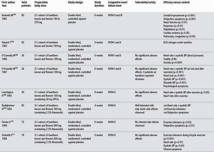
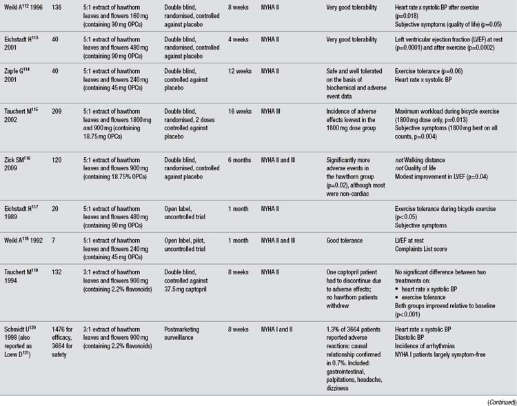
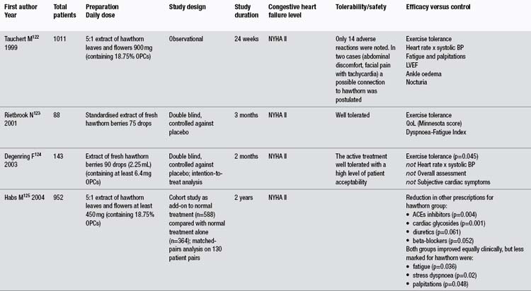
In more than any other patient group, gotu kola has demonstrated a clinical ability to help people with chronic problems with varicose veins in the lower limbs accompanied by oedema, itch, skin atrophy and ulceration, and discomfort or pain (known as chronic venous insufficiency or CVI). The larger clinical trials have been summarised in Table 1. Unfortunately, all key studies to date have been conducted by the same research group and have not been well designed. As noted in the table, apparently one of those studies has been published twice and another essentially three times, with considerable time having elapsed between the first and last publications. Several less rigorous trials have also been reported using TTF for CVI patients, but only those including 40 or more patients were included in this review and Table 1.

Table 1 Summary of trials of gotu kola extracts in patients with chronic venous insufficiency

image

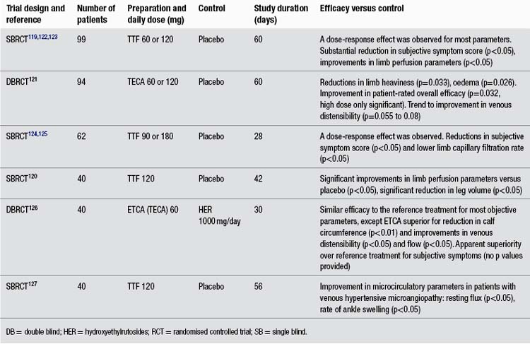
The reasonably methodologically sound randomised clinical trials compared TTF to placebo in 99 and 40 patients (both used 60 mg twice per day, and one study also included a group receiving only 60 mg/day).119,120 However, both were only single blind trials. A more rigorous double blind, randomised trial assessed TECA, 60 or 120 mg/day against placebo.121 This trial found a modest benefit for both doses of the extract over placebo at improving symptomatology. However, this benefit was not as clinically significant as reported in the less rigorous trials.

It is useful to reflect here on why a herb with a well-established reputation for promoting healing has developed into a treatment for CVI. Varicose veins and CVI have long been regarded as disorders of valvular incompetence. However, recent evidence suggests that changes in the vein wall could well precede such incompetence. For example, varicosities are often observed below competent valves and often can occur before valvular incompetence.128 Defects in ECM and collagen composition in the vein wall are thought to be part of this process.129 Endothelial damage from abnormally sustained venous pressure could contribute as well. Given the above, the noted pharmacological properties of gotu kola in terms of effects on fibroblasts, collagen and ECM in the vein wall, together with its potential for reducing endothelial damage and improving microcirculation, would suggest that it is indeed a plausible (and perhaps disease-modifying) treatment for CVI.

Two controlled trials have investigated the activity of oral TTF in patients with microvascular damage due to diabetes. The largest trial involved 100 patients with or without neuropathy and compared TTF (120 mg/day) with placebo over 12 months and also in 40 healthy controls.130 TTF was significantly more effective at improving microcirculatory parameters and oedema (p<0.05). A smaller trial with 50 patients compared 60 mg TTF twice daily with placebo or no treatment for 6 months.131 TTF resulted in significant improvement in parameters linked to microscopic vascular damage, including capillary permeability (p<0.01 to p<0.05).

In a randomised trial, the impact of TTF (180 mg/day) for 2 days before, the day of, and 1 day after, a long airline flight (up to 14 h) against no treatment was compared in patients with mild-to-moderate chronic venous insufficiency.132 Ankle oedema was significantly less in the TTF group (p<0.05), as were microcirculatory parameters such as rate of ankle swelling and venoarteriolar reflex (p<0.05). All parameters deteriorated with the length of flight, a phenomenon that was only partially compensated by the herbal treatment.

An open trial of TTF (60 mg/day) combined with bulk-forming laxatives (as appropriate) was rated as effective in relieving symptoms in over 90% of 210 patients with first or second degree haemorrhoids.133

Two interesting trials suggest that gotu kola may be able to stabilise vulnerable arterial plaque, indicating a possible role for the herb in modifying cardiovascular outcomes. Most atherosclerotic plaques do not trigger life-threatening events. A vulnerable plaque is one that is at high short-term risk of rupture. Plaque rupture is by far the most frequent cause of arterial thrombosis. It is deemed responsible for about 75% of coronary thrombi leading to heart attacks or death and about 90% of thrombosed carotid plaques causing ischaemic stroke. Only plaque with very thin fibrous caps is at risk of rupture and even just a small lesion is life-threatening. These plaques are essentially unstable because of a deficiency of connective tissue in their matrix. Even in the presence of widespread arterial disease, rarely more than a few plaques appear to be at risk of rupture at any given moment.134 One group of researchers observed: ‘It is not clear why some plaques lead to clinical manifestations, whereas many others remain asymptomatic and heal with subsequent fibrosis…’.135 In one sense arterial plaque is a type of wound on the blood vessel wall and vulnerable plaque can be viewed as a wound that is either not healing appropriately or in the early stages of healing (fibrosis).

In two placebo-controlled clinical trials, TTF stabilised low-density carotid136 and femoral artery plaques.137 The dose used in both trials was 180 mg/day for 12 months. These clinical outcomes were assessed by significant and marked increases in the ultrasound echogenicity of plaques compared with placebo (p<0.05). For the patients with carotid lesions, symptomatic cerebral events were observed in 6.5% of patients taking TTF compared with 11% of controls (p<0.05) and positive magnetic resonance images (indicating cerebral ischaemic lesions) were 7% in the herbal group, versus 17% for the control group (p<0.05). Arterial plaque which is echolucent (low echogenicity by ultrasound) has a limited connective tissue component and the plaque is weaker and prone to ulceration, rupture and embolisation. This is not quite the same as vulnerable coronary artery plaque, but shares many similarities.

On the basis of these findings it would be interesting to examine the impact of the long-term intake of gotu kola on health outcomes in patients with coronary artery disease.

Wounds, ulcers, and other skin and epithelial disorders

Early publications comprise around 30 mostly small, uncontrolled, open label clinical trials or case observation studies, dating from the late 1950s. These papers variously refer to the use of ‘asiaticoside’ or a proprietary triterpenoid isolate product, which were probably identical and represent an early version of TECA. The TECA was typically administered by intramuscular (and sometimes intralesional) injection and was often applied topically as well. Although injection was used, the good oral bioavailability of the gotu kola triterpenoids, and the fact that typical oral doses tend to be much higher than the injected doses used, suggest that these early data still have clinical relevance to the oral use of the herb. Conditions with favourable outcomes included bladder ulcers,138 corneal lesions,139 episiotomies140 and other postsurgical lesions,141 leg ulcers,142 skin grafts,143 skin ulcers144 and traumatic injury.145 Not all studies yielded positive outcomes.146,147 A few of the larger, less dated and/or interesting studies are featured below.

Following a positive series of published case reports148 and their experience with a burn patient who demonstrated a substantial reduction in hypertrophic scars, a group of Canadian clinicians conducted a clinical investigation of TECA as an antikeloid agent.149 Their first clinical evaluations involved the injectable form, but later they were able to report on 227 patients with keloids or hypertrophic scars who were treated with the oral form of TECA (60 to 90 mg/day, but up to 150 mg/day in stubborn cases). In an open label trial, 116 of 139 patients were found to respond after 2 to 18 months, either by relief of symptoms or by a gradual disappearance of inflammation. These positive results remained after the cessation of therapy. In most cases the treatment was started on well-established, non-responsive lesions, some of many years’ duration. A small, concurrent double blind study demonstrated a significant benefit over placebo (p<0.05). The authors also studied the preventative action of TECA in 88 patients undergoing surgical scar revision, with treatment started a few weeks prior to surgery and maintained for 3 months after. Improvement was noted in 79% of patients, but there was no comparative control group to account for the benefit of surgery alone.

In a recent clinical trial conducted in Thailand, 200 patients with diabetic foot ulcer were randomised to receive gotu kola extract or placebo.150 General symptoms, wound characteristics, wound size and wound depths were examined at days 7, 14 and 21. Wound contraction was determined by the decrease in the volume of the wound. Twenty cases dropped out from the study and 10 cases were terminated before the end of treatment because termination criteria were met. (Termination criteria included patient refusal, wound infection, delayed primary sutured wound and secondary healing wound.) Results for the 170 patients who completed the trial were analysed.

By the end of each week, wound contraction was significantly better in the group treated with gotu kola compared with those treated with placebo (p≤0.001). The trend to good wound contraction occurred earlier for the herb group than in the placebo group: for example, by day 7, 28.5% of patients receiving gotu kola had good wound contraction, compared with 12.8% in the placebo group; by day 14, the results for good contraction were 38.1% and 18.6% for herbal and placebo treatment, respectively. More granulation tissue formed in the placebo group than the herb group, and the difference between the groups was significant at days 7, 14, and 21 (p<0.001). There were no systemic side effects reported and there was no significant difference in wound infection between the two groups. The dose used was unclear, with the capsules variously described as containing ‘50 mg of extracted asiaticoside’ and ‘50 mg freeze dry lyophilized extract’. The trial author disclosed the dose in a personal communication (March 2011) as 300 mg/day of extract, corresponding to 11.2 g of fresh leaf.

There is some suggestion from case reports in the 1970s that gotu kola might assist liver pathologies. In one study, 30 patients with clinical hepatic cirrhosis (mainly due to bilharzia) and previous splenectomy were given TECA (20 mg/day, im) for 55 days.151 The nodularity and firm consistency of the liver decreased markedly in all the cases and biopsy revealed a substantial reduction in collagen fibres in both the fine and medium portal tracts. Serum alkaline phosphatase and glycoproteins were also reduced. In a second study, ongoing administration of TECA (90 to 150 mg/day, presumably oral) resulted in improvement in 5 of 12 patients with recently diagnosed alcoholic cirrhosis or chronic liver disease.152 Benefits were mainly observed in the patients with alcoholic cirrhosis, where biopsy revealed reduced sclerosis or steatosis.

Gotu kola might have value in obstetrics according to studies from the 1960s. Results for 131 cases of episiotomies treated with TECA (25 mg/day, im on the second, fourth and sixth days after surgery) from an open label trial demonstrated improved healing and reduced symptoms in 106 cases compared with other (unspecified) cases treated by different means.153 Clinical observation of 114 patients with perineal lesions (especially following episiotomy) found injected TECA (with concurrent topical use in some cases) accelerated healing, resulting in reduced hospital stays.140 Accelerated healing of perineal tears was found in another study.154

Some novel uses of gotu kola preparations have also been investigated. Open trials have demonstrated the benefit of topical madecassoside and either intramuscular madecassoside or asiaticoside in patients with previously non-healing wounds or skin ulcers.155,156 In a prospective, open clinical trial, 30 patients with active leprosy were given 500 mg powdered gotu kola (for 12 months), 60 mg asiaticoside (for 6 months) or dapsone three times per day.157 Improvement in leprosy ulcers was similar in all groups. Intramuscular (20 mg/day) and/or topical application of TECA in 90 patients with perforating leg ulcers due to leprosy were compared against placebo, and both treatments were found to be significantly superior at healing the ulcers (p<0.01).158 A combination of dapsone, hydnocarpic acid (from Hydnocarpus wightiana) and isolated asiaticoside (50 mg/day) for 6 to 11 months in 10 patients with active leprosy was just as effective, but less toxic, than standard multidrug treatments.159

The value of gotu kola compounds in the connective tissue disorder scleroderma has been assessed. An open trial involving 13 women with scleroderma and using either 20 or 60 mg/day TECA (im) observed improvement of symptoms in 11.160 TECA 30 mg/day (oral), sometimes with topical TECA ointment, improved local and systemic scleroderma in the majority of 54 patients investigated in another open trial.161 Benefits were also seen in another seven patients with scleroderma (TECA, 30 to 60 mg/day, oral),162 but a review of 15 cases in children found little benefit from current treatments, including asiaticoside (probably TECA).163

Some of the early studies on skin disorders also focused on topical application. Topical application of a cream of gotu kola cleared psoriasis lesions in five and improved the remaining two of seven patients in one case series.164 A cream containing 1% of an extract of gotu kola completely healed previously non-healing, chronic, infected skin ulcers in 17 out of 22 patients after 3 weeks in an open trial.165 One double blind trial in 18 patients failed to find that a 2% TECA powder had any better effect than placebo on healing after skin graft removal.143

In recently published studies there has been a further focus on the topical use of gotu kola compounds, probably thereby underestimating the unique value of the internal use of the herb. In a preliminary open trial, 12 healthy volunteers had a small wound induced on their forearms and then 0.3% madecassoside combined with copper, zinc and manganese was applied daily for 22 days.166 The researchers concluded that inflammation was resolved more quickly, epidermal regrowth was more rapid, and there was less scarring, apparently compared with no treatment. An ointment containing extracts of gotu kola, Viola tricolor and Mahonia aquifolium was not superior to a base cream in a randomised double blind, vehicle-controlled, half-side comparison in 88 patients with mild-to-moderate atopic dermatitis.167

Some of the topical trials are more cosmetic in nature. One early double blind trial involving 80 pregnant women investigated the effects of a topical cream containing gotu kola, vitamin E and collagen-elastin hydrolysate compared with placebo at preventing striae gravidarum (stretch marks).168 The cream was applied once per day. After 12 weeks, the group applying the active cream developed significantly fewer striae than the placebo group.

In a double blind, randomised clinical trial, a gel containing gotu kola extract, Rosmarinus officinalis extract, tetrahydrocurcumin, and dimethylaminoethanol was compared with placebo in 28 women.169 Each woman treated half their face with the gel or placebo for 4 weeks. At the end of this time, skin was significantly softer on the gel-treated skin compared with placebo, as rated both objectively and subjectively. Three women left the trial due to mild skin irritation. A cream with 0.1% asiaticoside applied around the eyes of 27 women for 12 weeks in an open trial was associated with improvement or elimination of wrinkles in 18.170

A 6-month randomised, double blind study was conducted on photo-aged (sun-damaged) skin in 20 female volunteers to assess the impact of a topical treatment containing 5% vitamin C and 0.1% madecassoside.171 There was a significant improvement in the clinical score for deep and superficial wrinkles, suppleness, firmness, roughness and skin hydration. These results were corroborated by objective tests. The reappearance of a normally structured, ‘young’ elastic fibre network was observed.

Nervous system and adaptogenic effects

A single dose of 12 g of crude gotu kola was more effective than placebo at reducing the acoustic startle response in 40 healthy adults in one double blind, placebo-controlled, randomised trial.172 An uncontrolled trial observed that a 70% ethanolic extract of gotu kola (500 mg/day corresponding to about 4.5 g of starting herb) over 2 months significantly improved measures of stress, anxiety, adjustment, depression and attention (all p<0.01 compared with baseline).173 Results after 2 months were substantially better than after 1 month.

An uncontrolled trial used gotu kola to treat 33 volunteers with generalised anxiety disorder.173 They received gotu kola extract (about 9 g/day dried herb equivalent) for 60 days. Participants were initially assessed for mental status using the Brief Psychiatric Rating Scale. Results using self-assessment questionnaires revealed significant improvements (p<0.01) from baseline in anxiety, stress, depression, adjustment and attention at day 30 and day 60.

Early studies focusing on cognitive function yielded conflicting results. A daily 500 mg tablet of gotu kola herb for 12 weeks significantly improved mental function and behaviour in 30 intellectually impaired children in a placebo-controlled trial.174 However, a double blind, placebo-controlled trial using the same tablet daily for 1 year observed no effect on the mental ability of children with normal baseline intelligence.175

A later trial was more positive. Twenty-eight healthy volunteers with a mean age of 65 years were randomly divided into four groups.176 One group received a placebo and the others were given respectively 250, 500 and 750 mg/day of a gotu kola extract for 2 months. The gotu kola extract was standardised for total phenolic content and also contained around 5% asiaticoside and asiatic acid. Assessment using a battery of cognitive performance tests was undertaken 1 h after the first dose (for acute effects) and after 1 and 2 months (for chronic effects).

For the acute effect (single dose) assessment, only the highest dose of gotu kola demonstrated a significant effect above placebo for just two of the nine tests used. These were the reaction time of spatial memory and the percentage accuracy of numeric working memory. Based on what is known about these tests, the authors suggested that these acute effects of gotu kola might partly occur via the modulation of dopamine and norepinephrine in the prefrontal cortex, together with the modulation of acetylcholine and serotonin in the hippocampus.

The repeated administration of gotu kola for a further 2 months also showed the same significant increases in spatial memory reaction time and percentage accuracy of numeric working memory. In addition, significant improvements were seen above placebo for choice reaction time, numeric working memory reaction time, accuracy of word recognition and accuracy of picture recognition. These effects were most consistently demonstrated at the highest dose of gotu kola. There was no significant effect on simple reaction time, the digit vigilance test, the accuracy of spatial memory, word recognition reaction time and picture recognition reaction time. The above effects were interpreted by the authors as gotu kola enhancing working memory. The extract not only improved cognitive performance, but also anxiety: the highest dose increased calmness and alertness after both 1 and 2 months of treatment.

When gotu kola powder (dosage not specified) was administered to 43 healthy but impoverished East Indian adults for 6 months, it elevated erythrocytes and haemoglobin and decreased blood urea nitrogen in a low-quality placebo-controlled trial.177,178 Gotu kola extract at 250, 500 and 750 mg/day significantly improved physical strength and fitness measured by the 30-second chair stand test in 80 healthy older volunteers (average age around 65 years). The two higher doses also improved the physical subscale of a quality of life scale (SF-36). This was a placebo-controlled, randomised, double blind trial and implies an adaptogenic activity for the herb.179

Other conditions

An uncontrolled study examined the effect of 60 mg/day oral TECA and 20 mg three times per week (im) in 64 patients with gastric and duodenal ulcers.180 After 10 weeks, all but two patients had healed completely (and the majority had healed after 6 weeks). In an open randomised trial, patients who had extracts of gotu kola and Punica granatum (pomegranate) applied to periodontal pockets experienced significantly better healing than those who received only standard medical care.181 This confirmed the preliminary results of an earlier study using the same extracts.182

Toxicology and other safety data

Toxicology

Gotu kola has been consumed as a leafy vegetable, particularly in Bangladesh, Thailand, Indonesia (West Java), Sri Lanka and South Africa,183185 and appears to have no harmful effect when used as a food.186 The leaf and stolon are eaten raw and cooked.187

Acute toxicity testing indicated a low toxicity following oral administration to rats (LD50 >675 mg/kg of gotu kola extract, equivalent to >4 g/kg dried leaf). Chronic administration of 150 mg/kg/day of extract (equivalent to 0.9 g/kg dried leaf) for a period of 30 days did not produce any adverse effects.188 Mice receiving up to 1 g/kg of gotu kola extract (2.5 g/kg dried plant) by mouth did not exhibit adverse effects.55 Aqueous ethanol extract of gotu kola entire plant demonstrated a maximum tolerated dose value of 250 mg/kg after ip injection in mice.189 Subcutaneous injection of 40 to 50 mg/kg of asiaticoside was toxic to mice and rabbits, while 20 to 250 mg/kg resulted in increased bleeding time. An oral dose of 1 g/kg was well tolerated.190

The local toxicity of asiaticoside was investigated by the measurement of skin respiration and histological analysis. (The death of a cell is accompanied by loss of respiratory activity.) Compared with other therapeutic agents, the toxicity of asiaticoside was not excessive and was comparable to that of many common antibiotics. Histological effects on guinea-pig skin indicated that moderate concentrations of asiaticoside produced swollen, abnormally staining cells. Higher concentrations resulted in necrotic cultures, showing signs of ‘thickening’ of the epidermis, even though the cells had mostly disintegrated. This may have been due to the cells becoming rapidly keratinised. Although fairly high concentrations of asiaticoside were required to produce this effect, it occurred in vivo (5 mg, sc) as well as in vitro.35

An ethanol extract of gotu kola exhibited mutagenic activity to strain TA98 (Salmonella/microsome test) only in the presence of S9 mix.191 A water extract of gotu kola was not toxic towards TA98 and TA100 with or without addition of S9 mix at the tested concentration (5 mg/plate). Gotu kola weakly inhibited the mutagenicity of the indirect mutagen IQ (2-amino-3-methylimidazo[4,5-f]quinoline).192 In another experiment, gotu kola water extract (1 mL of a 1:5 decoction) showed mutagenic activity in strain TA 98 with metabolic activation only.193 Gotu kola methanol extract induced abnormal metaphases and increased chromosome aberrations in the Vicia faba root meristem assay.194

Asiaticoside was found to be a weak tumour promoter in the hairless mouse epidermis model and was very weakly carcinogenic to the dermis after topical application.195

Contraindications

Gotu kola is contraindicated in cases of known allergy.

Special warnings and precautions

None suggested.

Interactions

No clinically relevant interactions have been found. In an in vivo study investigating wound healing with drugs, the anti-inflammatory drugs dexamethasone and phenylbutazone individually combined with asiaticoside caused a reduction in the tensile strength (and hence therapeutic effect) produced by asiaticoside alone.28 In this study the test substances were administered by intramuscular injection (asiaticoside is a saponin and has surfactant activity), so it is not known if the observed results extrapolate to the topical use of asiaticoside or oral use of gotu kola in humans.

In open trials, asiaticoside has been used topically in combination with an antibiotic and corticosteroid196 and was well tolerated.197

Use in pregnancy and lactation

Category B1 – no increase in frequency of malformation or other harmful effects on the fetus from limited use in women. No evidence of increased fetal damage in animal studies.

Gotu kola has been traditionally used in Bengal as a contraceptive agent. A reduction in conception rate was observed in female mice fed gotu kola (juice of whole plant, equivalent to 20 to 80 g fresh whole plant per kg) by gavage. Two sets of animals received the herb for 14 days (7 days before and 7 days during cohabitation) and 21 days (7 days before and 14 days during cohabitation). In the first set, sterile mating occurred for 50% to 60% of animals versus 15% in the controls, and for the second set 50% to 55% versus 20%. An isolated triterpenoid glycoside (40 to 120 mg/kg) and a compound derived from it also demonstrated antifertility activity. In all treatment groups there was no significant decrease in the number of young per litter and birth weight of the young were normal. The authors noted that the isolated glycoside and the compound derived from it caused a consistent reduction of fertility.198 These were very high doses, well above those normally used in clinical practice. Moreover, an antifertility effect does not imply harm during pregnancy.

Antifertility activity was also demonstrated in vivo in an early study for gotu kola (part undefined). Teratological effects have been studied in the rabbit and found to be negative for that animal.149

Gotu kola is compatible with breastfeeding.

Effects on ability to drive and use machines

No adverse effects expected.

Side effects

As with all herbs rich in saponins, oral use may cause irritation of the gastric mucous membranes and reflux.

Cases of allergic contact dermatitis have been reported from the use of gotu kola, TTF and asiaticoside, but they are considered to be low-risk treatments.199206 Both the extract and the triterpene constituents are weak sensitisers,149 although asiaticoside has been classified as a contact allergen.207 Patch tests in many cases confirmed that gotu kola or its constituents were responsible,199,200,203,204 although in some cases other constituents in the preparations were also responsible (such as propylene glycol,202 geraniol, lavender essence and neomycin201).

Traditional sources indicate that gotu kola may produce photosensitisation when used in tropical areas, although whether from use by oral or topical application is not indicated.5,208 Occasional gastrointestinal intolerance has been observed.149 A review of the use of TTF for the treatment of chronic venous insufficiency indicated that it was safe and well tolerated. Most trials used dosage of TTF in the range of 60 to 120 mg/day.80

Three cases of idiosyncratic hepatotoxicity have been reported from Argentina in association with extracts of gotu kola, including at least one that was clearly triggered by rechallenge.209 However, no other such cases are on record and the identity of the herb was not confirmed in these cases. One case of night-eating syndrome was attributed to use of a 70% ethanolic tincture of gotu kola, possibly with other herbs.210

Overdosage

There are no reliable reports of overdose with gotu kola.

An early case (prior to 1896) is recorded concerning a ‘Dr Boiteau who, in treating himself, progressively increased the dose and found that after two months the drug had produced all the effects of a violent, cumulative poison. … the plant, properly prepared and administered, is a powerful stimulant of the circulatory system, its action chiefly affecting the vessels of the skin and mucous membrane’.211 Traditional sources writing in the early to mid-20th century indicate that the plant can be a stupefying narcotic in large doses and in some cases produces headache or vertigo with a tendency to coma.207

Safety in children

Adverse effects are generally not expected. Gotu kola dried herb has been assessed in a clinical trial in India as a mental tonic for mentally disabled children.173 Gotu kola is used in leaf concentrate meals, which are prepared as porridge for preschool children in Sri Lanka to combat nutritional deficiencies.212 (Although the leaf composition varies with location, fresh gotu kola leaves typically contain 2% protein, 7 mg/100 g of vitamin C, 0.09 mg/100 g of vitamin B1 and 5.6 mg/100 g of iron.)213

However, one case of gotu kola-induced hepatotoxicity in a child has been recently reported.214

Regulatory status in selected countries

Gotu kola is official in the Pharmacopoeia of the People’s Republic of China 1997. The usual adult dosage, typically administered in the form of a decoction, is listed as 15 to 30 g, or 30 to 60 g of the fresh herb.

Gotu kola for external use is included on the General Sale List in the UK. It was not included in the Commission E assessment. Gotu kola is official in the British Pharmacopoeia 2011 and the European Pharmacopoeia 2011.

Gotu kola does not have GRAS status in the USA. However, it is freely available as a ‘dietary supplement’ in the USA under DSHEA legislation (Dietary Supplement Health and Education Act of 1994).

Gotu kola is not included in Part 4 of Schedule 4 of the Therapeutic Goods Regulations and is freely available for sale in Australia.

References

1. Nadkarni AK, Nadkarni KM. Indian Materia Medica. Bombay: Popular Prakashan, 1976. pp. 662–666

2. Grieve M, A Modern Herbal, New York, Dover Publications, 1971;vol 1.

3. Sharma PV. Dravyaguna-vijnana. Varanasi: Chaukambha Bharati Academy, 2003. pp. 3–6

4. Chen JK, Chen TT. Chinese Medical Herbology and Pharmacology. CA: Art of Medicine Press, 2004. pp. 147–148

5. British Herbal Medicine Association Scientific Committee. British Herbal Pharmacopoeia. Bournemouth: BHMA, 1983.

6. Felter HW, Lloyd JU. King’s American Dispensatory. ed 18, rev 3, 1905. Portland: reprinted Eclectic Medical Publications; 1983.

7. Farnsworth NR, Bunyapraphatsara N, eds. Thai Medicinal Plants. Bangkok: Medicinal Plant Information Center, 1992.

8. Dharma AP. Indonesian Medicinal Plants. Jakarta: Balai Pustaka, 1987.

9. Cambie RC, Ash J. Fijian Medicinal Plants. Australia: CSIRO, 1994.

10. van Wyk B-E, van Oudtshoorn B, Gericke N. Medicinal Plants of South Africa. Arcadia: Briza Publications, 1997.

11. Applequst W. The Identification of Medicinal Plants: A Handbook of the Morphology of Botanicals in Commerce. St Louis: Missouri Botanical Garden Press, 2006. pp. 45–46

12. Tang W, Eisenbrand G. Chinese Drugs of Plant Origin. Berlin: Springer-Verlag, 1992. pp. 273–275

13. Brinkhaus B, Linder M, Schuppan D, et al. Phytomedicine. 2000;7(5):427–448.

14. Zainol NA, Voo SC, Sarmidi MR, et al. Malaysian J Anal Sci. 2008;12(2):322–327.

15. May A. Eur J Pharmacol. 1968;4(3):331–339.

16. Lu L, Ying K, Wei S, et al. Br J Dermatol. 2004;151(3):571–578.

17. Lu L, Ying K, Wei S, et al. Int J Dermatol. 2004;43(11):801–807.

18. Marquart FX, Bellon G, Gillery P, et al. Connect Tissue Res. 1990;24:107–120.

19. Tenni R, Zanaboni G, De Agostini MP, et al. Ital J Biochem. 1988;37:69–77.

20. Bonte F, Dumas M, Chaudagne C, et al. Ann Pharm Fr. 1995;53(1):38–42.

21. Bonte F, Dumas M, Chaudagne C, et al. Planta Med. 1994;60(2):133–135.

22. Lee J, Jung E, Kim Y, et al. Planta Med. 2006;72(4):324–328.

23. Qi SH, Xie JL, Pan S, et al. Clin Exp Dermatol. 2008;33(2):171–175.

24. Poizot A, Dumez D. C R Acad Sci. 1978;286:789–792.

25. Suguna L, Sivakumar P, Chandrakasan G. Indian J Exp Biol. 1996;34(12):1208–1211.

26. Shukla A, Rasik AM, Jain GK, et al. J Ethnopharmacol. 1999;65:1–11.

27. Shetty BS, Udupa SL, Udupa AL, Somayaji SN. Int J Low Extrem Wounds. 2006;5(3):137–143.

28. Velasco M, Romero E. Curr Ther Res Clin Exp. 1976;19(1):121–125.

29. Hartog A, Smith HF, van der Kraan PM, et al. Exp Biol Med (Maywood). 2009;234(6):617–623.

30. Tincani GP, Riva PC, Baldini E. G Ital Dermatol. 1963;104:429–440.

31. Sunilkumar, Parameshwaraiah S, Shivakumar HG. Indian J Exp Biol. 1998;36(6):569–572.

32. Maquart FX, Chastang F, Simeon A, et al. Eur J Dermatol. 1996;9(4):289–296.

33. Shukla A, Rasik AM, Dhawan BN. Phytother Res. 1999;13(1):50–54.

34. Ju-Lin X, Shao-Hai Q, Tian-Zeng L, et al. J Cutan Pathol. 2009;36(2):234–239.

35. Lawrence JC. Eur J Pharmacol. 1967;1:414–424.

36. Kimura Y, Sumiyoshi M, Samukawa K, et al. Eur J Pharmacol. 2008;584(2–3):415–423.

37. Liu M, Dai Y, Li Y, et al. Planta Med. 2008;74(8):809–815.

38. Cheng CL, Guo JS, Luk J, Koo MWL. Life Sci. 2004;74:2237–2249.

39. Cheng CL, Koo MW. Life Sci. 2000;67(21):2647–2653.

40. Sairam K, Rao CV, Goel RK. Indian J Exp Biol. 2001;39(2):137–142.

41. Dong MS, Jung SH, Kim HJ, et al. Arch Pharm Res. 2004;27:512–517.

42. Wojcikowski K, Wohlmuth H, Johnson DW, et al. Nephrology (Carlton). 2009;14(1):70–79.

43. Qi S, Xie J, Li T. Zhonghua Shao Shang Za Zhi. 2000;16(1):53–56.

44. Zhang T, Rong XZ, Yang RH, et al. Nan Fang Yi Ke Da Xue Xue Bao. 2006;26(1):67–70. [In Chinese]

45. Ming ZJ, Liu SZ, Cao L. Zhongguo Zhong Xi Yi Jie He Za Zhi. 2004;24(8):731–734. [In Chinese]

46. Yun KJ, Kim JY, Kim JB, et al. Int Immunopharmacol. 2008;8:431–441.

47. Won JH, Shin JS, Park HJ, et al. Planta Med. 2010;76(3):251–257.

48. Vogel HG, De Souza N, D’Sa A. Acta Therapeutica. 1990;16:285–298.

49. Somchit MN, Sulaiman MR, Zuraini A, et al. Indian J Pharmacol. 2004;36(6):377–380.

50. Li H, Gong X, Zhang L, et al. Phytomedicine. 2009;16(6–7):538–546.

51. Liu M, Dai Y, Yao X, et al. Int Immunopharmacol. 2008;8(11):1561–1566.

52. Caballero-George C, Vanderheyde PM, Okamoto Y, et al. Phytother Res. 2004;18:729–736.

53. Wijeweera P, Arnason JT, Koszycki D, et al. Phytomedicine. 2006;13:668–676.

54. Chen SW, Wang WJ, Li WJ, et al. Pharmacol Biochem Behav. 2006;85:339–344.

55. Sakina MR, Dandiya PC. Fitoterapia. 1990;61:291–296.

56. Diwan PV, Karwande I, Singh AK. Fitoterapia. 1991;3:253–257.

57. Liang X, Huang YN, Chen SW, et al. Pharmacol Biochem Behav. 2008;89:444–449.

58. Nalini K, Aroor AR, Karanth KS, et al. Fitoterapia. 1992;63(3):232–237.

59. Xu Y, Cao Z, Khan I, et al. J Alzheimers Dis. 2008;13:341–349.

60. Mook-Jung I, Shin JE, Yun SH, et al. J Neurosci Res. 1999;58(3):417–425.

61. Veerendra Kumar MH, Cupta YK. Clin Exp Pharmacol Physiol. 2003;30(5–6):336–342.

62. Dhanasekaran M, Holcomb LA, Hitt AR, et al. Phytother Res. 2009;23(1):14–19.

63. Lee MK, Kim SR, Sung SH, et al. Res Commun Mol Pathol Pharmacol. 2000;108(1–2):75–86.

64. Ramanathan M, Sivakumar S, Anandvijayakumar PR, et al. Indian J Exp Biol. 2007;45(5):425–431.

65. Krishnamurthy RG, Senut MC, Zemke D, et al. J Neurosci Res. 2009;87(11):2541–2550.

66. Haleagrahara N, Ponnusamy K. J Toxicol Sci. 2010;35(1):41–47.

67. Veerendra Kumar MH, Gupta YK. J Ethnopharmacol. 2002;79(2):253–260.

68. Subathra M, Shila S, Devi MA, et al. Exp Gerontol. 2005;40(8–9):707–715.

69. Shinomol GK, Muralidhara. Phytomedicine. 2008;15(11):971–984.

70. Shinomol GK, Muralidhara. Neurotoxicology. 2008;29(6):948–957.

71. Shinomol GK, Ravikumar H, Muralidhara. Phytother Res. 2010;24(6):885–892.

72. Soumyanath A, Zhong YP, Gold SA, et al. J Pharm Pharmacol. 2005;57(9):1221–1229.

73. Mohandas Rao KG, Muddanna Rao S, Gurumadhva Rao S. Evid Based Complement Altern Med. 2006;3(3):349–357.

74. Mohandas Rao KG, Muddanna Rao S, Gurumadhva Rao S. Evid Based Complement Altern Med. 2009;6(2):203–210.

75. Gadahad MR, Rao M, Rao G. J Chin Med Assoc. 2008;71(1):6–13.

76. Rao MKG, Rao MS, Rao GS. Neuroanatomy. 2005;4:18–23.

77. Rao SB, Chetana M, Uma Devi P. Physiol Behav. 2005;86:449–457.

78. Gupta YK, Veerendra Kumar MH, Srivastava AK. Pharmacol Biochem Behav. 2003;74(3):579–585.

79. Vattanajun A, Watanabe H, Tantisira MH, et al. J Med Assoc Thai. 2005;88(suppl 3):S131–S140.

80. Incandela L, Cesarone MR, Cacchio M, et al. Angiology. 2001;52(suppl 2):S9–S13.

81. Cospite M, Ferrara F, Milio G, et al. Giorn Ital Angiol. 1984;4(3):200–205.

82. Arpaia MR, Ferrone R, Amitrano M, et al. Int J Clin Pharmacol Res. 1990;10(4):229–233.

83. Belcaro GV, Grimaldi R, Guidi G. Angiology. 1990;41(7):533–540.

84. Montecchio GP, Samaden A, Carbone S. Haematologica. 1991;76(3):256–259.

85. Pragada RR, Veeravalli KK, Chowdary KP, et al. J Ethnopharmacol. 2004;93(1):105–108.

86. Bian GX, Li GG, Yang Y, et al. Biol Pharm bull. 2008;31(3):458–463.

87. Li GG, Bian GX, Ren JP, et al. Yao Xue Xue Bao. 2007;42(5):475–480.

88. Gnanapragasam A, Yogeeta S, Subhashini R, et al. Mol Cell Biochem. 2007;294(1–2):55–63.

89. Gnanapragasam A, Ebenezar KK, Sathish V, et al. Life Sci. 2004;76(5):585–597.

90. Zheng MS. J Trad Chin Med. 1989;9:113–116.

91. Yoosook C, Bunyapraphatsara N, Boonyakiat Y, et al. Phytomedicine. 2000;6(6):411–419.

92. Herbert D, Paramsivan CN, Prabhakar R, et al. Indian J Leprosy. 1994;66:65–68.

93. Newton SM, Lau C, Gurcha SS, et al. J Ethnopharmacol. 2002;79:57–67.

94. Medda S, Das N, Mahato SB, et al. Indian J Biochem Biophys. 1995;32(3):147–151.

95. Chakraborty T, Sinha Babu SP, Sukul NC. Fitoterapia. 1995;57(2):110–112.

96. Park BC, Bosire KO, Lee ES, et al. Cancer Lett. 2005;218(1):81–90.

97. Yoshida M, Fuchigami M, Nagao T, et al. Biol Pharm Bull. 2005;28(1):173–175.

98. Tang XL, Yang XY, Jung HJ, et al. Biol Pharm Bull. 2009;32(8):1399–1405.

99. Babykutty S, Padikkala J, Sathiadevan PP, et al. Afr J Tradit Complement Altern Med. 2008;6(1):9–16.

100. Huang YH, Zhang SH, Zhen RX, et al. Ai Zheng. 2004;23(12):1599–1604. [In Chinese]

101. Babu TD, Kuttan G, Padikkala J. J Ethnopharmacol. 1995;48(1):53–57.

102. Bunpo P, Kataoka K, Arimochi H, et al. Food Chem Toxicol. 2004;42(12):1987–1997.

103. Punturee K, Wild CP, Kasinrerk W, et al. Asian Pac J Cancer Prev. 2005;6(3):396–400.

104. Patil JS, Nagavi BG, Ramesh M, et al. Ind Drugs. 1998;38:711–714.

105. Jayathirtha MG, Mishra SH. Phytomedicine. 2004;11(4):361–365.

106. Chatterjee TK, Chakraborty A, Pathak M, et al. Indian J Exp Biol. 1992;30(10):889–891.

107. Sarma DNK, Khosa RL. Phytother Res. 1996;10(2):181–183.

108. Sarma DNK, Khosa RL, Chansauria JPN, et al. Phytother Res. 1995;9(8):589–590.

109. Gupta R, Flora SJ. J Appl Toxicol. 2006;26(3):213–222.

110. Flora SJ, Gupta R. Phytother Res. 2007;21(10):980–988.

111. Shobi V, Goel HC. Physiol Behav. 2001;73(1–2):19–23.

112. Sharma J, Sharma R. Phytother Res. 2002;16(8):785–786.

113. Sharma J, Sharma R. Phytother Res. 2005;19(7):605–611.

114. Zhang L, Li HZ, Gong X, et al. Phytomedicine. 2010;17(10):811–819.

115. Chasseaud LF, Fry BJ, Hawkins DR, et al. Arzneimittelforschung. 1971;21(9):1379–1384.

116. Zheng XC, Wang SH. J Chromatogr B Analyt Technol Biomed Life Sci. 2009;877(5–6):477–481.

117. Rush WR, Murray GR, Graham DJ. Eur J Drug Metab Pharmacokinet. 1993;18(4):323–326.

118. Grimaldi R, De Ponti F, D’Angelo L, et al. J Ethnopharmacol. 1990;8:235–241.

119. Incandela L, Cesarone MR, Cachio M, et al. Angiology. 2001;52(10 suppl 2):S61–S67.

120. Cesarone MR, Belcaro G, Rulo A, et al. Angiology. 2001;52(suppl 1–2):S45–S48.

121. Pointel JP, Boccalon H, Cloarec M, et al. Angiology. 1987;38(1 pt 1):46–50.

122. Belcaro G, Laurora G, Cesarone MR, et al. Curr Ther Res. 1989;46(6):1015–1026.

123. Cesarone MR, Laurora G, De Sanctis MT, et al. Minerva Cardioangiol. 1994;42(6):299–304.

124. De Sanctis MT, Belcaro G, Incandela L, et al. Angiology. 2001;52(10 suppl 2):S55–S59.

125. Belcaro GV, Rulo A, Grimaldi R. Angiology. 1990;41(1):12–18.

126. Monteverde A, Occhipinti P, Rossi F, et al. Acta Ther. 1987;13:629–636.

127. Cesarone MR, Belcaro G, De Sanctis MT, et al. Angiology. 2001;52(suppl 2):S15–S18.

128. Lim CS, Davies AH. Br J Surg. 2009;96(11):1231–1242.

129. Naoum JJ, Hunter GC. Vascular. 2007;15(5):242–249.

130. Incandela L, Belcaro G, Cesarone MR, et al. Angiology. 2001;52(suppl 2):S27–S31.

131. Cesarone MR, Incandela L, De Sanctis MT, et al. Angiology. 2001;52(suppl 1–2):S49–S54.

132. Cesarone MR, Incandela L, De Sanctis MT, et al. Angiology. 2001;52(suppl 1–2):S33–S37.

133. Guarerio F, Sansonetti G, Donzelli R, et al. Giorn Ital Angiol. 1986;6(1):46–52.

134. Thim T, Hagensen MK, Bentzon JF, et al. J Intern Med. 2008;263(5):506–516.

135. Schoenhagen P, Tuzcu EM, Ellis SG. Circulation. 2002;106(7):760–762.

136. Cesarone MR, Belcaro G, Nicolaides AN, et al. Angiology. 2001;52(suppl 2):S19–S25.

137. Incandela L, Belcaro G, Nicolaides AN, et al. Angiology. 2001;52(suppl 2):S69–S73.

138. Etrebi A, Ibrahim A, Zaki K. J Egypt Med Assoc. 1975;58(5–6):324–327.

139. Peyresblanques J. Bull Soc Ophtalmol Fr. 1959;8:771–781.

140. Baudon-Glanddier B. Gaz Med Fr. 1963;70:2463–2464.

141. Sevin P. Progr Med (Paris). 1962;90:23–24.

142. Huriez CL. Lille Med. 1972;3(suppl. 17):574–579.

143. O’Keeffe P. Br J Plast Surg. 1974;27(2):194–195.

144. Balina LM, Cardama JE, Gatti JC, et al. Dia Med. 1961;33:1693–1696.

145. Stassen P. Rev Med Liege. 1964;19:305–308.

146. Mayall RC, Mayall AC, Bertolotti JG, et al. Rev Bras Med. 1975;32:26–29.

147. Mekkawi MF. Bull Ophthalmol Soc Egypt. 1975;68:77–79.

148. El-Hefnawi H. Dermatologica. 1962;125:387–392.

149. Bosse JP, Papillon J, Frenette G, et al. Ann Plast Surg. 1979;3(1):13–21.

150. Paocharoen V. J Med Assoc Thai. 2010;93(7):S166–S170.

151. El-Zawahry MD, Khalil AM, El-Banna MH. Bull Soc Int Chir. 1975;34(4):296–297.

152. Darnis F, Orcel L, de Saint-Maur PP, et al. Sem Hop. 1979;55(37–38):1749–1750.

153. Castellani L, Gillet JY, Lavernhe G, et al. Bull Fed Soc Gynecol Obstet Lang Fr. 1966;18(2):184–186.

154. Torre MP, Donnadieu JM, Braditch JL. Clinique (Paris). 1963;58:203–206.

155. Kiesewetter H. Wien Med Wochenschr. 1964;114:124–126. [In German]

156. Wolfram ST. Wien Med Wochenschr. 1965;115:439–442. [In German]

157. Chakrabarty T, Deshmukh S. Sci Culture. 1976;11:573.

158. Nebout M. Bull Soc Pathol Exot Filiales. 1974;67:471–478. [In French]

159. Chaudhury S, Hazra S, Podder GC, et al. Indian J Dermatol. 1987;32(3):63–67.

160. Sasaki S, Shinkai H, Akashi Y, et al. Jap J Clin Derm. 1971;25(6):585–593.

161. Guseva NG, Starovoitova MN, Mach ES. Ter Arkh. 1998;70(5):58–61. [In Russian]

162. Szczepanski A, Dabrowska H, Blaszczyk M. Przegl Dermatol. 1974;61(5):701–703.

163. Frati Munari AC, Culebro Nieves G, Velazquez E, et al. Bol Med Hosp Infant Mex. 1979;36(2):201–214.

164. Natarajan S, Paily PP. Indian J Dermatol. 1973;18(4):82–85.

165. Kosalwatna S, Shaipanich C, Bhanganada K. Siriraj Hosp Gaz. 1988;40(6):455–461.

166. Rougier A, Humbert F. J Am Acad Dermatol. 2008;2 suppl 2(58): AB144. Abstract P3109

167. Klövekorn W, Tepe A, Danesch U. Int J Clin Pharmacol Ther. 2007;45(11):583–591.

168. Mallol J, Belda MA, Costa D, et al. Int J Cos Sci. 1991;13:51–57.

169. Sommerfeld B. Phytomedicine. 2007;14:711–715.

170. Lee J, Jung E, Lee H, et al. Int J Cos Sci. 2008;30(3):167–173.

171. Haftek M, Mac-Mary S, Le Bitoux MA, et al. Exp Dermatol. 2008;17(11):946–952.

172. Bradwejn J, Zhou Y, Koszychki D, et al. J Clin Psychopharmacol. 2000;20:680–684.

173. Jana U, Sur TK, Maity LN, et al. Nepal Med Coll J. 2010;12(1):8–11.

174. Appa Rao MVR, Srinivasan K, Koteswara Rao T. J Res Indian Med. 1973;8(4):9–15.

175. Kuppurajan K, Srinivasan K, Janaki K. J Res Indian Med. 1978;13(1):37–41.

176. Wattanathorn J, Mator L, Muchimapura S, et al. J Ethnopharmacol. 2008;116(2):325–332.

177. Appa Rao MVR, Usha SP, Rajagopalan SS, et al. J Res Indian Med. 1967;2:79–85.

178. Appa Rao MVR, Rajapopalan SS, Srinivasan VR, et al. Nagarjun. 1969;12:33–41.

179. Mato L, Wattanathorn J, Muchimapura S, et al. Evid Based Complement Altern Med, 2009. At <http://ecam.oxfordjournals.org/cgi/reprint/nep177v1>. Accessed 6.7.10.

180. Rhee J, Choi KW. Korean J Gastroenterol. 1981;13(1):35–40.

181. Sastravaha G, Gassmann G, Sangtherapitikul P, et al. J Int Acad Periodontol. 2005;7(3):70–79.

182. Sastravaha G, Yotnuengnit P, Booncong P, et al. J Int Acad Periodontol. 2003;5(4):106–115.

183. Bagchi CD, Puri HS. Herba Hung. 1988;27(2–3):137–140.

184. Dharma AP. Indonesian Medicinal Plants. Jakarta: Balai Pustaka, 1987. pp. 24–25

185. van Wyk B-E, Gericke N. People’s Plants: A Guide to Useful Plants of Southern Africa. Arcadia: Briza Publications, 2000. pp. 68, 142

186. Rattanapanone V, Sanpitak N, Phornphibul B. Chiang Mai Med Bull. 1971;10:17–23.

187. Kays SJ, Silva Dias JC. Econ Bot. 1995;49(2):115–152.

188. dede Lucia R, Sertie JAA. Fitoterapia. 1997;68(5):413–416.

189. Dhar ML, Dhar MM, Dhawan BN, et al. Indian J Exp Biol. 1968;6(4):232–247.

190. Boiteau P, Ratsimamanga AR. Therapie. 1956;11:125–149.

191. Ieamworapong C, Kangsadalumpai K, Rojanapo W. Environ Mol Mutagen. 1989;14(suppl 15):93.

192. Yen GC, Chen HY, Peng HH. Food Chem Toxicol. 2001;39:1045–1053.

193. Rivera IG, Martins MT, Sanchez PS, et al. Environ Toxicol Water Qual. 1996;9(2):87–93.

194. Gopalan HNB, Wairimu AN. Environ Mol Mutagen. 1989;14(suppl 15):73.

195. Laerum OD, Iversen OH. Cancer Res. 1972;32(7):1463–1468.

196. Kartnig T. Herb Spice Med Plant. 1988;3:145–173.

197. Hadida E, Sayag J, Bonerandi JJ, et al. Bull Soc Fr Dermatol Syphiligr. 1970;77(4):522–525.

198. Dutta T, Basu UP. Indian J Exp Biol. 1968;6:181–182.

199. Hausen BM. Contact Dermatitis. 1993;29(4):175–179.

200. Danese P, Carnevali C, Bertazzoni MG. Contact Dermatitis. 1994;31(3):201.

201. Santucci B, Picardo M, Cristaudo A. Contact Dermatitis. 1985;13(1):39.

202. Izu R, Aguirre A, Gil N, et al. Contact Dermatitis. 1992;26(3):192–193.

203. Eun HC, Lee AY. Contact Dermatitis. 1985;13(5):310–313.

204. Huriez C, Martin P. G Ital Dermatol Minerva Dermatol. 1969;44(9):463–464.

205. Gonzalo Garijo MA, Revenga Arranz F, Bobadilla Gonzalez P. Allergol Immunopathol. 1996;24(3):132–134.

206. Vena GA, Angelini GA. Contact Dermatitis. 1986;15(2):108–109.

207. Goossens A, Beck MH, Haneke E, et al. Contact Dermatitis. 1999;40:112–113.

208. Chopra RN, Chopra IC, Handa KL, et al. Chopra’s Indigenous Drugs of India, 2nd ed., 1958. Calcutta: reprinted Academic Publishers; 1982. pp. 351–353.

209. Jorge OA, Jorge AD. Rev Esp Enferm Dig. 2005;97(2):115–124.

210. O’Brien B. Ir Med J. 2005;98(10):250–251.

211. Chopra RN, Badhwar RL, Ghosh S, Poisonous Plants of India, New Delhi, Indian Council of Agricultural Research, 1965;vol I. pp. 433–434

212. Cox DN, Rajasuriya S, Soysa PE, et al. Int J Food Sci Nutr. 1993;44:123–132.

213. Peiris KHS, Kays SJ. Hort Tech. 1996;6(1):13–18.

214. Dantuluri S, North-Lewis P, Karthik SV. Dig Liver Dis. 2011;43(6):500.

Hawthorn

(Crataegus spp.)

Synonyms

Crataegi (Lat), Weiβdorn (Ger), aubépine (Fr), biancospino (Ital), alm. hvidtjørn (Dan).

What is it?

The leaf, flower and berry of several species of hawthorn are used medicinally, most often: Crataegus laevigata (Poiret) DC (synonyms: C. oxyacantha auct. non L, C. oxyacanthoides Thuill.) and C. monogyna Jacq. The Greek meaning of Crataegus oxyacantha refers to hard (wood) and sharp thorns. References to hawthorn are extensive throughout history and the shrub has been utilised in many ways including for wood, cultivation as a hedge and for flavouring of liquor by the berries. Although the berries were traditionally used as medicine, modern research has tended to focus on preparations from the leaves or leaves and flowers.

The first use of hawthorn in cardiac therapy is attributed to Dr Green, an Irish doctor who used a tincture of the fresh berries. With the increasing incidence of heart disease in the Western world at the time, its use rapidly spread to other countries, notably France, the United States, England and Germany. In Western herbal medicine hawthorn is now considered to be the most significant herb for ischaemic and congestive heart disease and there is considerable objective evidence to support its status.

Effects

Increases force of myocardial contraction, increases coronary blood flow, reduces myocardial oxygen demand, protects against myocardial damage, hypotensive, improves heart rate variability, antiarrhythmic.

Traditional view

Hawthorn berries have been traditionally used to treat cardiovascular problems (including hypertension with myocardial weakness, angina pectoris and tachycardia) and other circulatory disorders (atherosclerosis, Buerger’s disease). The flowers and berries were also used as an astringent for sore throats and as a diuretic for kidney problems and dropsy.1,2 The fruit and bark were used by the Eclectics as a heart remedy for indications such as pain, praecordial oppression, dyspnoea, cardiac hypertrophy, valvular insufficiency and anaemia.3 The fruit of other Crataegus species (C. pinnatifida, C. cuneata) have been used in traditional Chinese medicine to improve digestion, stimulate circulation and remove blood stasis.4

Summary of actions

Cardiotonic (mild), cardioprotective, antioxidant, collagen stabilising, mild astringent, hypotensive (mild), antiarrhythmic.

Can be used for

Indications established by clinical trials

Congestive heart disease due to ischaemia, hypertension or other causes (particularly corresponding to NYHA stages I and II, good evidence), mild hypertension (moderate evidence for a mild effect); topically for acne (uncontrolled trial); anxiety (in combination).

Traditional therapeutic uses

As a treatment for mild heart conditions (angina pectoris, coronary artery disease, cardiac arrhythmias, myocardial weakness) and to assist prevention of arterial degeneration caused by atherosclerosis.

May also be used for

Extrapolations from pharmacological studies

Antioxidant activity; co-factor for vitamin C intake; stabilisation of connective tissue tone; for hyperlipidaemia (especially the berries).

Other applications

Cosmetic and hair care products for antiseborrhoeic and anti-inflammatory activities and to increase hydration and elasticity of the skin.5

Preparations

Dried or fresh leaf, flower or fruit for infusion or decoction, liquid extract and tablets or capsules for internal use. Decoction or extract for topical use.

Dosage

• 1.5 to 3.5 g/day of dried flower, leaf or berry, as infusion or decoction

• Hawthorn tablets or capsules (for example containing 1 g leaves and flowers, standardised to 15 to 20 mg oligomeric procyanidins and 6 to 7 mg flavonoids) three to four per day or more

• 3 to 6 mL/day of 1:2 liquid extract of hawthorn leaf, 3 to 7 mL/day of 1:2 liquid extract of hawthorn berry, 7.5 to 15 mL/day of 1:5 tincture of hawthorn leaf, 7.5 to 17.5 mL/day of 1:5 tincture of hawthorn berry. Higher doses than these may be necessary for hypertension

• Concentrated extracts (3:1 or 5:1), typically from the leaves and flowers and standardised to various levels of flavonoid and/or oligomeric procyanidin (OPC) content are also available in solid dosage form. These have been used in clinical trials, with doses (of extract) ranging from 160 to 1800 mg/day (see Table 1).

Table 1 Clinical trials of hawthorn in congestive heart failure

image image image

Duration of use

There is no restriction on the long-term use of hawthorn and it should be prescribed over a period of at least 2 months if used to treat heart conditions.

Summary assessment of safety

No adverse effects from ingestion of hawthorn are expected. Hawthorn may act in synergy with digitalis glycosides and beta-blockers. However, no specific interactions with the pharmacokinetics of digoxin were identified in human subjects and adverse effects are not generally anticipated. Modification of drug dosage may be required, but is unlikely.

Technical data

Botany

Hawthorn, a member of the Rosaceae (rose) family, is a deciduous, thorny shrub or small tree up to 10 m tall. The leaves are broader than long and have three to five lobes. The white flowers, with their red anthers, are arranged in groups of five to ten at the apex of small branches. The dark red, false fruits are oval and contain a small kernel which is the true fruit.6,7

Note: There has been confusion and debate over the nomenclature of Crataegus. species It was suggested in 1975 that the name C. oxyacantha L. should be rejected since it is a source of confusion8 and C. laevigata used in preference.9 In addition, extensive hybridisation has occurred between C. monogyna and C. laevigata.1012 Where a research publication refers to the use of ‘C. oxyacantha’, this has generally been described in this monograph as pertaining to C. laevigata.

Adulteration

No adulterants known.

Key constituents

• Oligomeric procyanidins (OPCs), mainly procyanidin B-2.13 The monomers epicatechin and catechin are also present and are generally included in chemical tests for OPC levels.

• Flavonoids, including quercetin glycosides (hyperoside, rutin) and particularly flavone C-glycosides (vitexin and related compounds).13 The minor flavonoids in C. laevigata and C. monogyna show differences that enable their differentiation.14

• Amines, catechols, carboxylic and triterpene acids (crataegus acids, especially in the berries).15

The flowers contain the highest levels of flavonoids and the leaves contain the highest levels of OPCs. The key constituents of Crataegus monogyna, with variations with plant part and time of harvest, are outlined below.16

  OPC % Major flavonoids %
Leaves with flowers (average of seven samples) 2.50 0.92
Flowers (average of four samples) 2.67 1.31
Leaves (spring, flowers open) 3.02 0.53
Leaves (summer, berries green) 2.71 0.74
Leaves (autumn, berries ripe) 2.06 0.76
Berries (summer, green) 3.18 0.15
Berries (autumn, ripe) 1.74 0.13

The relative astringency of an OPC increases with its degree of polymerisation. OPCs from Crataegus oxyacantha (laevigata) berries, with an average number of monomers of four, have an astringency relative to tannic acid of 0.73.17 While the stability of flavonoids in C. oxyacantha (laevigata) tinctures was good, OPCs demonstrated relatively poor stability in this dosage form.18

image

Pharmacodynamics

The therapeutic value of hawthorn may extend beyond its cardiovascular applications, largely due to the significant OPC content. Although most of the research is not specifically on the OPCs found in hawthorn, it is likely that the hawthorn OPCs will share many of the described pharmacological properties for this class of compounds. In 1966 the French scientist Masquelier and his co-workers analysed the bark of several conifers from Quebec. Some species, especially Tsuga canadensis (previously known as Pinus canadensis), were rich in OPCs. However, Masquelier’s interest in this tree was based on the accounts of the French explorer Jacques Cartier.19 During the winter of 1534 Cartier’s group in Canada was afflicted by scurvy, which was cured on the advice of a native who directed that the sailors use a decoction of the bark and leaves of the ‘Anneda’ tree. The liquid was consumed and the solid residue applied as a poultice to the swollen joints. On the basis of Cartier’s description, Masquelier postulated that ‘Anneda’ was Tsuga canadensis. He further speculated that the leaves provided a source of ascorbic acid and the bark, by providing OPCs, acted as a vitamin C synergist (perhaps in a similar manner to the chemically related flavonoids).

Masquelier’s original interest in OPCs was for their ‘vitamin P-like’ activity.20 He later postulated that they may be important in the prevention of atheroma.21 Since then, pharmacological and clinical studies of OPCs, as either pine bark (especially Pinus pinaster) or grape seed (Vitis vinifera) extracts, have revealed an impressive variety of therapeutic properties.

Effect on the cardiovascular system

Effect on cardiac contractility and frequency and coronary blood flow

In early research the crataegus acids increased coronary blood flow in vitro and decreased blood pressure in vivo.2224 Crataegolic and ursolic acids increased coronary flow in vitro and crataegolic acid was also positively inotropic.25

Hawthorn extract exhibited a positive inotropic effect (increasing the contraction amplitude) of cardiac myocytes in vitro, with a relatively small increase in their energy requirements.26 Hawthorn also prolonged the apparent refractory period in the presence and absence of isoprenaline (a beta-adrenergic agonist), indicating an antiarrhythmic potential.27 A standardised leaf and flower extract exerted a positive inotropic effect on isolated left ventricular muscle strips from explants from patients with congestive heart failure.28 Hawthorn reinforced the positive inotropic action of cardiac glycosides without an increase in glycoside toxicity and increased coronary blood flow, even after experimental damage, in vitro.29

Two flavonoid fractions of hawthorn were positively inotropic in isolated heart. One fraction had an effect on frequency, being negatively chronotropic (decreasing the heart rate). In isolated hearts, vitexin-2″-O-rhamnoside was positively inotropic and negatively chronotropic at higher doses, while the flavonoid aglycones were inactive.25

The main flavonoids of hawthorn (O-glycosides: luteolin-7-glucoside, hyperoside, rutin; and C-glycosides: vitexin, vitexin-2″-O-rhamnoside and monoacetyl vitexin-2″-O-rhamnoside) increased coronary flow, demonstrated slight positive inotropic effects and raised heart rate in isolated hearts. An inhibition of cAMP phosphodiesterase was also observed.30

OPC subfractions were more positively inotropic in isolated heart than flavonoid fractions, but had no effect on frequency.31 In another study, hawthorn OPCs had a mild negative chronotropic effect in isolated heart, but caused a marked increase in contraction and blood flow. Procyanidin dimers were more active than oligomers with 3 to 4 units, and the OPC fraction from hawthorn leaves was more active than that from the berries. In general, the effects of the OPCs are similar to those of flavonoids, but are elicited at lower doses.25

Crataegus OPCs increased coronary blood flow in dogs after oral administration.32 Vitexin-2″-O-rhamnoside increased coronary blood flow by up to 64% after intracoronary administration.33 Intravenous or intracoronary administration of hawthorn flavonoids increased coronary blood flow, decreased blood pressure and increased heart contractility. No change was observed in heart rate.3436

Hawthorn (C. laevigata) has also been studied in a model of congestive heart failure. A standardised hawthorn leaf and flower extract had no significant effect on the characteristic immunomodulatory response induced by 6 months of pressure overload in a rat model of heart failure.37 However, the same research group found that the extract at the same doses (1.3, 13 and 130 mg/kg, oral) modified left ventricular remodelling and counteracted myocardial dysfunction in early (4 weeks) pressure overload-induced cardiac hypertrophy in rats,38 but only exhibited a modest beneficial effect in the long-term (5 months) model.39

Cardioprotective activity and effects under hypoxic conditions

The cardioprotective effect of several fractions of standardised hawthorn leaf and flower extract (containing 18.75% OPCs) was investigated in vitro and in vivo. The lipophilic fraction containing flavonoids was as active as the whole extract in inhibiting human neutrophil elastase, but only half as active as a radical scavenger. The water-soluble fraction was weak in both in vitro systems. In contrast, the inhibiting potencies of the OPC-rich (and flavonoid-free) fraction were significantly higher than the extract. Oral administration of the OPC-rich fraction (20 mg/kg/day) to rats afforded similar protection against ischaemia-reperfusion-induced pathologies to treatment with a higher dose of the standardised extract (100 mg/kg/day).40

Oral pretreatment with hawthorn extract (2% of diet) to rats for 3 months resulted in myocardial protection, as evidenced by attenuation of levels of lactase dehydrogenase (LDH) release after induced ischaemia and reperfusion in the isolated hearts. This attenuation of LDH by hawthorn pretreatment suggests a preservation of the cell membrane and a protection from myocardial damage.41 Pretreatment with hawthorn or garlic extract, but particularly the combination of both, resulted in protective effects against isoprenaline-induced heart, liver and pancreas damage in rats.42 Oral administration of hawthorn reduced the deleterious metabolic influence of hypoxia on ventricular muscle fibres in rabbits. Hawthorn may protect the sodium pump against anoxia.43 In early research, administration of hawthorn to volunteers improved the anoxic symptoms produced by inhalation of an 8% oxygen gas mixture44 and decreased the signs of ischaemia as assessed by an exercise ECG test in patients with ischaemic heart disease.45

Administration of the purified flavonoids from hawthorn leaves to rats with myocardial infarction resulted in a smaller necrotic focus and improved revascularisation of finer vessels when compared to controls.46 Pretreatment with hawthorn (C. laevigata) fresh berry tincture (5 mL/kg/day for 30 days, oral) prevented the increase in lipid peroxidation and the decrease in antioxidant enzymes, increased the rate of ADP-stimulated oxygen intake and protected against pathological changes induced by isoproterenol in the hearts of rats administered this agent.47 The beneficial effects of hawthorn were observed to extend to cardiac mitochondria in a second similar study (maintenance of mitochondrial antioxidant status, prevention of mitochondrial lipid peroxidation and increased Krebs cycle enzymes).48

In one earlier study the water-soluble fraction of hawthorn extract demonstrated a cardioprotective effect on the ischaemic-reperfused heart in vitro (which mimics myocardial infarction). The cardioprotective effect was not accompanied by an increase in coronary flow.49 In contrast, no cardioprotective effects were seen during ischaemia and reperfusion, and there was even aggravation of arrhythmias, after oral pretreatment of Wistar rats with C. oxyacantha (laevigata) extract (0.5 g/kg/day for 8 weeks, standardised to 2.2% flavonoids).50 However, pretreatment with a different leaf and flower extract (10 or 100 mg/kg/day, oral for 7 days) improved cardiac function and reduced infarct size in a similar rat model of prolonged ischaemia and reperfusion.51

In vitro and in vivo (rats, 100 mg/kg, oral) studies suggested that hawthorn extract protected against ischaemia- reperfusion injury by inhibiting apoptotic pathways, possibly by regulating Akt (an anti-apoptotic kinase) and HIF-1 (hypoxia-inducible factor 1) signalling pathways.52,53

Microcirculatory and vasorelaxant activity

Compared with a digoxin preparation, hawthorn extract increased the erythrocyte flow rate in all the vascular networks examined and reduced both leucocyte endothelial adhesion and diapedesis in the venular network in rat mesenteric vessels.54 Even after removal of tannins, extracts of Crataegus monogyna still potently inhibited ADP-induced human platelet serotonin release and platelet aggregation in vitro.55 Hawthorn flower extract inhibited thromboxane A2 biosynthesis in vitro.56

In early research intravenous administration of a hawthorn extract decreased blood pressure and increased peripheral blood flow to skeletal muscle in vivo.57,58 In a placebo- controlled, crossover study, the effect of a single dose of 900 mg of a hawthorn leaf and flower extract (3:1, standardised to 2.2% flavonoids) on the cutaneous microcirculation was compared to 0.3 mg medigoxin. Six hours after taking hawthorn, the haematocrit had dropped by a mean of 3.2%, whereas erythrocyte aggregation increased significantly by 19% 3 h after taking the digoxin. No significant changes were recorded for the other measured parameters (plasma viscosity, flow rate in nail bed capillaries, heart rate, blood pressure).59

OPCs in hawthorn extract may be responsible for the observed endothelium-dependent nitric oxide-mediated relaxation in isolated rat aorta, possibly via activation of tetraethylammonium-sensitive K+ channels.60 A standardised leaf and flower extract induced a concentration-dependent vasodilation of rat aorta and human mammarian artery in vitro, with activity again residing in the OPC fraction.61 The mechanism involved appeared to be an endothelium-dependent, NO-mediated vasorelaxation, a finding supported yet again by another in vitro study from a different laboratory.62 Flavonoids and OPCs from hawthorn demonstrated inhibitory activity on angiotensin converting enzyme (ACE) in vitro.63

Mechanistic studies

Hawthorn may exert its activity in vitro in a similar way to phosphodiesterase inhibitors, raising cAMP levels in cardiac muscle cells. The total flavonoids from hawthorn were shown to inhibit phosphodiesterase activity in vitro.26 However, an in vitro study of hawthorn leaf and flower extract suggested it exerted direct positive inotropic effects, implying a pharmacologic mechanism similar to the cAMP-independent positive inotropic action of cardiac glycosides.28 Hawthorn extract prolonged the effective refractory period in isolated heart compared to four other inotropic drugs (including adrenaline and digoxin), which all shortened it. Thus, hawthorn has a relatively lower risk of causing arrhythmias and may in fact have antiarrhythmic activity.64 Multi-electrode techniques have demonstrated that hawthorn extract prolonged the action potential duration and delayed recovery of the maximum upstroke velocity of guinea pig papillary muscle.65 A hawthorn extract demonstrated negative chronotropic effects in a cultured murine cardiomyocyte assay, independent of beta-adrenergic receptor blockade, possibly involving muscarinic receptor activation.66

An interesting in vitro study compared the effects of a hawthorn flower, leaf and berry extract against a berry extract (species not specified) using rat cardiomyocytes.67 Markedly different effects were observed; for example, the former extract resulted in the initiation of robust calcium transits with eventual calcium overload, whereas the latter initiated calcium transits but with no calcium overload. In other words the flower, leaf and berry extract was more potent. Mechanistic explorations suggested that cardiac activity is exerted via the sodium-potassium pump (Na+/K+-ATPase). Another in vitro study in cultured cardiomyocytes comparing a berry and a leaf extract of C. laevigata found similar negative chronotropic effects, unrelated to beta-adrenergic receptor blockade.68

A hawthorn leaf and flower extract inhibited induced intimal hyperplasia in vitro and in vivo (rats, oral doses), probably by directly inhibiting platelet-derived growth factor-beta.69 This might have clinical implications in angioplasty-related restenosis. Reduced endothelium-dependent vasodilator responses with increased synthesis of endothelin-1 (ET-1) are characteristics of endothelial dysfunction in heart failure and are predictive of mortality. Hawthorn and grape seed OPCs equipotently inhibited ET-1 synthesis in cultured endothelial cells.70

Hypocholesterolaemic activity

Hawthorn berry tincture administered simultaneously to rats fed an atherogenic diet significantly increased the in vitro binding of LDL (low density lipoprotein) to liver plasma membranes, increased bile acid excretion and depressed hepatic cholesterol synthesis.71 The results of adding hawthorn (2% of diet) to a rabbit diet high in cholesterol suggested that the mechanism by which Chinese hawthorn berry (Crataegus pinnatifida) decreases serum cholesterol involves, at least in part, the inhibition of cholesterol absorption via downregulation of intestinal acyl-CoA-cholesterol acyltransferase (ACAT) activity.72 Chinese hawthorn berry also inhibited ACAT in Caco-2 (intestinal) cells, largely due to the activity of the triterpenic acids (oleanolic and ursolic acid).73 Further to this, a berry dichloromethane extract (0.37% of diet) demonstrated a synergistic effect with plant sterols in lowering plasma cholesterol in hamsters fed a high cholesterol diet.73 The triterpene acids were also quite active in this model at 0.01% of diet.

In contrast, three flavonoids (quercetin, hyperoside and rutin) and chlorogenic acid from Chinese hawthorn berry weakly inhibited HMG-CoA reductase activity in vitro and demonstrated synergy in combination, thereby perhaps suggesting mild statin-like activity.74 A synergistic activity on lowering cholesterol and triglycerides was also demonstrated in a mouse model after oral doses of 2.85 mg/kg/day of each compound or the combination (see also Chapter 2).

Antioxidant activity

Hawthorn extracts obtained using acetone, methanol and water demonstrated antioxidant activity on hepatic microsomal preparations in vitro. A correlation was demonstrated between the total phenolic content (mainly flavonoids and OPCs) and antioxidant activity for leaf, flower and berry at all stages of herb growth. The most active individual components were (−)-epicatechin and procyanidin B-2.75 One recent study found that the ethanolic extract of C. monogyna berries possesses more antioxidant activity than the aqueous extract (with catechin and epicatechin mainly responsible for activity),76 while another on the berry tincture of the same species attributed the greatest antioxidant contribution to epicatechin and hyperoside.77

Most of the polyphenolic compounds in hawthorn demonstrated a dose-dependent protection of human LDL from copper-mediated oxidation, and prevented the peroxyl free radical-induced oxidation of alpha-tocopherol. In addition, supplementation of a hawthorn fruit powder (C. pinnatifida) at 2% of diet significantly elevated serum alpha-tocopherol by 18% to 20% in rats fed a 30% polyunsaturated canola oil diet, as compared to the control.78 In another study, the in vitro inhibition of LDL oxidation was also linked to hawthorn’s polyphenols, in particular the dimeric procyanidin B2 and the flavonol glycoside hyperoside. OPCs were more active than the majority of the flavonoids tested.79

Chinese research found that the Chinese hawthorn berry (C. pinnatifida) was a potent inducer of superoxide dismutase (SOD) activity in mice.80 Oral doses of aqueous extracts were used and the SOD activity was measured in red blood cells. (SOD is the enzyme which combats the harmful effects of the superoxide radical.) Further to this, a combination of tinctures of mango bark (Mangifera indica) and C. laevigata berries (5 mg/kg of each, oral) in rats fed a high cholesterol diet not only significantly lowered LDL-cholesterol and raised HDL-cholesterol, but also restored the observed reductions in SOD, catalase, glutathione peroxidase and glutathione induced by the diet.81

Other activity

Oral administration of hawthorn tincture to rats for 30 days afforded good protection against abnormal changes in liver function tests after myocardial infarction (alanine aminotransferase, aspartate aminotransferase, LDH and alkaline phosphatase) and in protein-bound carbohydrate alterations (hexose, hexosamine, fucose and sialic acid). These hepatoprotective effects included a reversal of histological changes in the liver.82

Induction of epidermal ornithine decarboxylase (ODC) activity, stimulation of hydroperoxide production and increased DNA synthesis are three biochemical effects linked to skin tumour promotion by the tumour promoter 12-O-tetradecanoylphorbol 13-acetate (TPA). Topical application of OPCs to mouse epidermis prior to administration of TPA resulted in inhibition of ODC activity. The inhibition of ODC activity increased with the degree of polymerisation of the OPCs: trimer (procyanidin C-1) > dimer (procyanidin B-2) > monomer (epicatechin).83 Further to this, an extract of C. pinnatifida berries inhibited skin tumour promotion and demonstrated topical anti-inflammatory activity in various models.84

An extract from a mixture of C. monogyna and C. laevigata berries demonstrated moderate activity against Gram-positive bacteria in vitro, with no effect on Candida albicans,85 and flavonoids and OPCs from C. sinaica were inhibitory against herpes simplex virus type 1 in vitro.86

High doses (100 to 1000 mg/kg, ip) of an extract of C. monogyna fruit pulp exhibited CNS depressant and analgesic activities in mice.87 Pretreatment with hawthorn (C. laevigata) extract (100 mg/kg/day for 15 days, oral in rats) reduced brain damage and improved neurological score by reducing oxidative stress in a model of cerebral ischaemia-reperfusion injury.88

Flavonoids in a hawthorn leaf extract inhibited alpha-glucosidase in vitro.89 An aqueous extract of hawthorn leaf (C. laevigata) demonstrated a significant dose-dependent decrease of blood glucose levels in streptozotocin-induced diabetic rats (doses were 150 or 300 mg/kg/day, oral for either 1 or 9 days), but had no such effect in normal rats.90

A leaf and flower extract of C. laevigata demonstrated a range of effects on isolated human neutrophil function that could be interpreted as reflecting anti-inflammatory activity.91 These included inhibition of release of chemoattractants and cytokines and inhibition of neutrophil oxidative burst. The mixed species berry combination mentioned above at 50, 100 and 200 mg/kg/day (oral) produced a dose-dependent anti-inflammatory effect in the rat paw oedema model.85 At similar doses it also exhibited a dose-dependent gastroprotective action in a gastric ulcer model in rats.

Hawthorn fruit (as extract equivalent to 0.5, 1.0 and 2.0 g/kg/day of berries, oral) dose-dependently exerted a protective activity in two experimental models of murine colitis.92 Anti-inflammatory effects were demonstrated in the colon and the authors reflected on the potential value of this herb in patients with inflammatory bowel disease (the equivalent human dose for 2.0 g/day in a mouse is approximately 12 g/day of berries, which is achievable).

An isolate of hawthorn leaf flavonoids (4, 8 and 16 mg/kg/day, oral for 8 days) significantly increased the activity of muscle lipoprotein lipase (LPL) in mice, with a commensurate decrease of activity in adipose tissue.93 This indicates that hawthorn flavonoids may reduce lipid accumulation in adipose tissue by regulating LPL expression.

A C. monogyna fruit extract significantly protected human lymphocytes against gamma irradiation damage in vitro.94 Radioprotective activity was also demonstrated in vivo for an extract of an Iranian hawthorn species (C. microphylla fruit) given to mice (25, 50, 100 and 200 mg/kg, single dose, ip), as assessed by genotoxic changes in bone marrow cells.95 Following on from this research, the same investigative group gave a single oral dose of 500 mg of this extract to five human volunteers and then subjected their blood samples to a defined level of gamma irradiation.96 For samples collected 1 h after the hawthorn dose there was a 44% decrease in genotoxic damage in lymphocytes, compared to samples collected beforehand.

Pharmacokinetics

To date there have been no human pharmacokinetic studies of hawthorn. The pharmacokinetics of oral doses of OPCs given to mice have been studied in early research using an isotopic labelling technique.97 OPCs showed rapid absorption and preferential localisation in tissues rich in glycosaminoglycans. The plasma half-life was about 5 h, indicating a prolonged presence in the bloodstream. In contrast, the flavonoid glycoside rutin showed poor absorption, the bulk of the radioactivity residing with the contents of the digestive tract. This work and that of others reviewed by Middleton98 suggests that OPCs, or OPC fragments resulting from bacterial activity in the gut, are more bioavailable than some flavonoids.

More recent research, all from China, has mainly focused on the pharmacokinetics of the typical hawthorn flavonoids using a variety of models. Oral administration of a hawthorn fruit extract (C. pinnatifida) to rats revealed only significant bioavailability for catechin.99 Chlorogenic acid, hyperoside and isoquercitrin present in the extract could not be detected in the rat plasma. The same research group found that epicatechin, isoquercitrin and hyperoside exhibited limited permeability using in vitro models of intestinal absorption and confirmed that only epicatechin was detectable as the parent compound after oral administration to rats.100

The oral bioavailability of vitexin-2″-O-rhamnoside (VOR) was determined to be only 3.57% in rats.101 In vitro modelling of intestinal absorption suggested that this molecule exhibits high permeability via passive diffusion, but its absorption is limited by P-glycoprotein acting as an efflux pump.102

The pharmacokinetics of VOR and vitexin-4″-O-glucoside (VOG) after a single oral dose of a hawthorn leaf flavonoid isolate (from C. pinnatifida) was determined in rats.103 After a dose of extract containing 83.3 mg/kg of VOG and 342 mg/kg of VOR, the same Tmax of 0.75 h was observed, with Cmax recorded at 4.1 μg/mL and 16.5 μg/mL, respectively. The mean elimination half-lives of VOG and VOR were 2.53 h and 2.32 h, respectively. High levels of tissue distribution of VOG and VOR were observed in liver and kidney, but none was detected in brain tissue. There was no long-term accumulation of VOG and VOR in the rat tissues examined. The total recovery of the dose in 24 hours was 64.91% (0.70% in urine, 64.21% in faeces) for VOG and 89.01% (0.72% in urine, 88.29% in faeces) for VOR. The cumulative VOG and VOR excreted in bile represented 0.58% and 13.38% of the doses, respectively. The authors concluded that VOG and VOR were not efficiently absorbed. (For further details of the pharmacokinetics of flavonoids and OPCs see Chapter 2.)

Clinical trials

Heart disease

The New York Heart Association (NYHA) classifies loss of cardiac output and heart failure: for stage I the patient is symptom-free when at rest and on treatment; stage II patients have loss of capacity with medium effort; for stage III even minor effort results in dyspnoea, with no symptoms at rest; in stage IV symptoms are present when at rest. There have been a number of clinical studies of the effect of hawthorn on heart failure which are summarised in Table 1. Many of these have been subjected to systematic review and meta-analysis, as discussed below.

A 2008 Cochrane review evaluated the information reported in good quality double blind clinical trials comparing hawthorn leaf and flower extract against placebo in patients with chronic heart failure (NYHA Classes I to III).126 Fourteen clinical trials were found to be of sufficiently high quality to be included in the evidence review.126 In most of these studies the hawthorn extract was used in conjunction with conventional treatments, including cardiac drugs. Pooling of the results of 10 trials using meta-analysis found that the average maximum cardiac work capacity was significantly higher in the patients receiving hawthorn compared to placebo (p<0.02). The review also found that a measure of cardiac oxygen need (systolic blood pressure × heart rate) was significantly lower. These findings suggest that hawthorn treatment helped the heart muscle tissue to work more efficiently. The capacity of patients to tolerate exercise was increased and symptoms such as shortness of breath and fatigue were significantly improved. Reported side effects were mild and infrequent and there was no suggestion of any adverse interaction with conventional drugs. The authors concluded that there is significant benefit in both symptom control and physiological outcomes from hawthorn extract as an adjunctive treatment for chronic heart failure.

The 13 published trials included in the 2008 Cochrane review are summarised in date order as the first 13 entries in Table 1. Following these, the table includes another eight published clinical studies of hawthorn in chronic heart failure, also in date order. These trials were not included in the Cochrane review because they did not meet the selection criteria. Also it should be noted that the Cochrane review used an earlier interim conference report of the trial by Zick and co-workers. The version of this study included in Table 1 is the final published analysis from 2009.

Other clinical trials in heart failure patients are as follows. The large SPICE (Survival and Prognosis Investigation of Crataegus Extract) study was a randomised, double blind, placebo-controlled study conducted at 156 centres in 13 European countries.127 In the 2-year clinical trial, 2681 patients with NYHA Class II or III chronic heart failure were given either hawthorn (900 mg/day of a 5:1 leaf and flower extract containing 18.75% OPCs) or a placebo. All of their conventional medications were continued. The trial was designed to assess whether the regular use of hawthorn by cardiac patients could delay or reduce their likelihood of a ‘cardiac event’, defined in the study as cardiac death (due either to sudden cardiac arrest, death from progressive heart failure or a fatal heart attack), a non-fatal heart attack or hospitalisation due to progressive heart failure.

While the study did show some minor positive findings, it was probably not large enough or long enough to demonstrate any definitive benefit from hawthorn. The rate of heart attacks (fatal and non-fatal) was about the same in the two groups (hawthorn or placebo). There was a difference in sudden cardiac deaths (20 fewer in the hawthorn group) that failed to reach statistical significance, except in the subgroup of patients with milder heart failure (p=0.025), suggesting a reduced risk of dangerous arrhythmias.

One strong conclusion from the SPICE trial is the excellent safety and low side-effect profile of the hawthorn extract. This was despite the fact that the patients were also taking a wide variety of cardiac drugs including beta-blockers, ACE inhibitors, diuretics and digoxin.

In a retrospective analysis of their data (see the entry in Table 1), Zick and co-workers examined the clinical progression of the chronic heart failure patients participating in their placebo-controlled trial.128 Progression of heart failure occurred in 46.6% of the hawthorn group and 43.3% of the placebo group. However, hawthorn did appear to increase the risk of progression earlier in the trial, and overall for the more severe patients. By their own admission the authors conceded that their study was limited due to the small sample size and the fact that it was a secondary data analysis from a trial designed to measure different outcomes. Such a negative finding was not supported by the larger and longer SPICE study (see above), which in contrast was designed to assess clinical progression as a primary outcome.

The sinus activity of the heart undergoes a natural fluctuation. Short-term fluctuations occur with inhalation and exhalation (sinus arrhythmia) but there are also second-grade variations associated with blood pressure rhythm. With age, diabetes and damage to the heart, this variability of heart rate decreases. Low heart rate variability (HRV) is a risk factor in coronary heart disease and there is a positive correlation between HRV and life-expectancy.129 The effect of an extract of hawthorn leaves, flowers and berries (containing 45 mg/day flavonoids) was studied in 20 geriatric patients over 6 weeks in a randomised, double blind, placebo-controlled trial.130 Patients with a coefficient of variation (CV) in heart rate of less than 5% were chosen for the study. Those with frequent ectopic beats and diabetes were excluded and the average age of patients was about 80 years. A small but statistically significant positive effect was seen for the hawthorn group (p<0.01): the CV of heart rate rose steadily over the 6-week treatment period from 1.9% to 2.5% while there was no change in the placebo group. The authors postulated that this effect might become more marked over longer treatment periods. The improvement in HRV was dramatic in some patients given hawthorn.

The same extract was given at the same dose to nine healthy volunteers for 4 weeks in a placebo-controlled trial.131 At the end of the trial, the heart rate × systolic blood pressure products were significantly lower in the hawthorn group after exercise (p=0.04). Responsiveness to exogenous catecholamines was not altered.

A case study was described of a 64-year-old male patient scheduled for internal defibrillator placement.132 After the patient was prescribed a combination of oral coenzyme Q10 (100 mg/day), hawthorn extract (450 mg/day) and chelated magnesium (350 mg/day) his symptoms and ventricular function improved and 11 months later he was not yet needing the surgical procedure.

Hawthorn was also beneficial in combination with passionflower. In a randomised, double blind, placebo-controlled study, 40 patients with dyspnoea commensurate with NYHA stage II received either a hawthorn and passionflower extract combination or placebo over 6 weeks. Exercise capacity, measured in terms of a walking test, increased significantly in those patients receiving the herbal preparation (p<0.05). A slight but significant decrease in heart rate at rest and mean diastolic blood pressure during exercise, and a decrease in total plasma cholesterol, were observed in the group receiving the extract.21 The two groups did not differ significantly in the other tests: maximum exercise capacity measured during a bicycle ergometer test, subjective assessment of breathlessness or blood lactate levels, although the trend favoured the herbal group.

In early studies, patients previously unsuccessfully treated with digoxin alone were compensated for rest and slight stress with relatively low oral doses of the glycoside in combination with hawthorn.133,134

Blood pressure

In an early uncontrolled trial, mean systolic pressure fell from 205 mmHg to 148 mmHg and mean diastolic pressure fell from 112 mmHg to 83 mmHg in hypertensive patients receiving hawthorn berry tincture. When treatment was stopped, blood pressures returned to their initial values over a 2-week period. There was only a slight effect on subjects with normal blood pressure.135 Other clinical studies (see Table 1) sometimes demonstrate that hawthorn extract resulted in a slight reduction in blood pressure in patients with chronic heart failure.

A pilot study investigated the hypotensive potential of hawthorn extract (500 mg/day of a 3:1 extract of leaves and flowers containing 1.8% flavonoids) and magnesium (600 mg/day), individually and in combination, compared to a placebo in a randomised, double blind trial.136 In all, 36 mildly hypertensive patients completed the 10-week study. Due to the small patient numbers in each group, only the hawthorn group demonstrated any clinical effects, with trends to lowered diastolic blood pressure (p=0.081) and reduced anxiety (p=0.094).

A follow-up randomised, controlled trial was undertaken among outpatients in general practices to investigate the effect of hawthorn on hypertension in patients with type 2 diabetes taking prescribed drugs. Patients (n=79) were randomised 1200 mg/day of the same hawthorn extract as above (n=39) or placebo (n=40) for 16 weeks. Hypotensive drugs were already being used by 71% of the study population, with a mean intake of 4.4 hypoglycaemic and/or hypotensive drugs per patient. After 16 weeks, the hawthorn group showed greater reductions in diastolic blood pressure (baseline: 85.6 mmHg, 95% CI 83.3 to 87.8; outcome: 83.0 mmHg, 95% CI 80.5 to 85.7) than the placebo group (baseline: 84.5 mmHg, 95% CI 82 to 87; outcome: 85.0 mmHg, 95% CI 82.2 to 87.8) (p=0.035). There was no group difference in systolic blood pressure reduction from baseline. No herb-drug interactions were found and minor health complaints were reduced from baseline in both groups.137

In a double blind, placebo-controlled clinical trial involving 92 men and women with primary mild hypertension, a hydroalcoholic extract of the leaves and flowers of Crataegus curvisepala showed a significant decrease in both systolic and diastolic blood pressures after 3 months (p<0.05).138

Interestingly a combination of hawthorn berry extract and camphor has successfully treated orthostatic and chronic hypotension in a number of clinical trials.139141 The typical daily doses from the combination are 25 mg of camphor and around 1000 mg of fresh hawthorn berry extract. The same combination also improved low blood pressure and cognitive function in the elderly in a randomised, placebo-controlled, double blind trial.142

Other conditions

In an uncontrolled, multicentre trial, 50 patients with and without acne in various stages of development uniformly applied one to two ampoules of liposome-containing hawthorn extract to the most affected areas of the skin each day for at least 30 days. The hawthorn extract demonstrated a general capillary-protective activity which resulted in the reduction or disappearance of capillary congestion. A mild anti-inflammatory activity was demonstrated by a significant reduction in acne and erythema. (The improvement in stratum corneum hydration and roughness of skin and the antiseborrhoeic effect were attributed to the phosphatidylcholine liposomes.)5

A combination of hawthorn leaves and flowers extract (300 mg/day), Californian poppy (Eschscholtzia californica) extract (80 mg/day) and magnesium (300 mg/day) was assessed against placebo in a 3-month randomised, double blind clinical trial involving 264 patients with mild-to-moderate generalised anxiety (DSM-III-R).143 Total and somatic Hamilton anxiety scale scores and subjective patient-rated anxiety fell during treatment and were significantly different to placebo (p=0.005, p=0.054 and p=0.005, respectively). Adverse reactions were similar in the treatment and placebo groups.

Toxicology and other safety data

Toxicology

Studies involving excessive dosing of hawthorn flower extract (600 mg/kg/day, undefined extract strength, 4.4% flavonoids) over 30 days in rats showed unremarkable adverse effects.144

The acute oral toxicity in undefined animals of hawthorn was 6 g/kg. No target organ toxicity was defined at 100 times the human dose (2.7 mg/kg) of concentrated hawthorn extract. Standard mutagenic and clastogenic tests were also negative.145 A study in mice demonstrated a high LD50 of 13.5 g/kg for an aqueous extract of C. laevigata.90

Schimmer and co-workers found that an ethanolic extract of Crataegus was weakly mutagenic in the Salmonella test, a finding which they attributed to the quercetin content of the extract.146 Popp and co-workers found a DNA-damaging potency of commercial Crataegus preparations in human lymphocyte cultures.147 The active principles were not identified but were probably flavonoids. Several procyanidins with different degrees of polymerisation (dimers, a trimer and a polymer) were found to be non-mutagenic in the Salmonella mutagenesis assay system.148

Contraindications

None known.

Special warnings and precautions

Not to be used concomitantly with heart and blood pressure medication unless supervised by a suitably qualified herbal practitioner or physician.

Interactions

Speculation on a harmful interaction with digoxin has not been borne out in any study.149 A large number of patients, previously unsuccessfully treated with digoxin alone, were compensated for rest and slight stress with relatively low oral doses of the glycoside in combination with hawthorn and without evidence of adverse effects.133,134 A randomised, crossover trial published in 2003 involving eight healthy volunteers confirmed that standardised hawthorn extract (leaf and flower extract, 900 mg/day) and digoxin may be safely co-administered.150

Hawthorn may act in synergy with digitalis, glycosides, beta-blockers and other hypotensive drugs. Modification of drug dosage may be required. There is also a suggestion from one in vitro study that hawthorn may interfere with one specific method of serum digoxin assay (the Digoxin III immunoassay).151

Use in pregnancy and lactation

Category B1 – no increase in frequency of malformation or other harmful effects on the fetus from limited use in women. No evidence of increased fetal damage in animal studies.

At a high oral dose of 2.8 g/kg administered from days 1 to 15 of gestation, hawthorn (part undefined) did not have an adverse effect on reproductive outcome in rats. Fetal weights were slightly increased when the herb was administered from days 8 to 15 of gestation. There were no differences in placental weight, the number of resorptions and litter size and no externally visible malformations. The herb was administered as an ethanol extract and the dose administered was the highest possible for which ethanol remained below the teratogenic threshold.152 Pregnant rats given 56 times the human dose of hawthorn orally did not exhibit any adverse effects.153 Hawthorn also did not influence embryonic development in vitro.

Hawthorn is compatible with breastfeeding.

Effects on ability to drive and use machines

No adverse effects expected.

Side effects

An assessment has been made of the safety data from all available human studies on hawthorn products, including library and on-line searches, and data from the WHO Centre for International Drug Monitoring (18 case reports) and manufacturers. A total of 7311 patients had been enrolled in 24 clinical trials, and data from 5577 patients were available for analysis. Overall, 166 adverse events were identified. Most of these adverse events were mild to moderate. The most frequent adverse events were dizziness/vertigo (n=15), gastrointestinal complaints (n=24), headache (n=9), migraine (n=8) and palpitation (n=11). More severe reactions were falls (n=2), gastrointestinal haemorrhage (n=2), circulation failure (n=2) and erythematous rash (n=2). There were no reports of drug interactions.154 (See also Table 1 and the discussion of the SPICE study for further information regarding side effects during clinical trials.)

One case of a type I hypersensitivity reaction to hawthorn has been reported.155

Overdosage

Not known.

Safety in children

No information available but adverse effects are not expected.

Regulatory status in selected countries

Hawthorn berries, leaf and flower are official in the British Pharmacopoeia 2011 and the European Pharmacopoeia 2011.

Hawthorn leaf with flower is official in the United States Pharmacopeia–National Formulary USP31 NF26 2008.

Hawthorn berry, hawthorn leaf and hawthorn flower are covered by null Commission E monographs. The Commission E suggests that, as the efficacy of hawthorn berry, leaf or flower has not been documented, no therapeutic application can be recommended. In contrast, hawthorn leaf with flower is covered by a positive Commission E monograph and can be used to treat decreased cardiac output as described in functional stage II of NYHA.

Hawthorn is now on the UK General Sale List.

Hawthorn does not have GRAS status. However, it is freely available as a ‘dietary supplement’ in the USA under DSHEA legislation (1994 Dietary Supplement Health and Education Act).

Hawthorn is not included in Part 4 of Schedule 4 of the Therapeutic Goods Act Regulations which lists herbal substances restricted from free sale within Australia.

References

1. Grieve M, A Modern Herbal, New York, Dover Publications, 1971;vol. 1. p. 385

2. British Herbal Medicine Association Scientific Committee. British Herbal Pharmacopoeia. Cowling: BHMA, 1983. pp. 74–75

3. Felter HW, Lloyd JU. King’s American Dispensatory. ed 18, rev 3, vol 2, 1905. Portland: Reprinted by Eclectic Medical Publications; 1983. p. 1613.

4. Chang HM, But PP, Pharmacology and Applications of Chinese Materia Medica, Singapore, World Scientific, 1987;vol. 1. pp. 100–107

5. Longhi MG, Rocchi P, Gezzi A, et al. Fitoterapia. 1984;55(2):87–99.

6. Launert EL. The Hamlyn Guide to Edible and Medicinal Plants of Britain and Northern Europe. London: Hamlyn, 1981. p. 76

7. Chiej R. The Macdonald Encyclopedia of Medicinal Plants. London: Macdonald, 1984. Entry no. 99

8. Byatt JI. Bot J Linn Soc. 1974;69:15–21.

9. Mabberley DJ. The Plant Book, 2nd ed. Cambridge: Cambridge University Press, 1997. p. 190

10. Byatt JI. Watsonia. 1975;10:253–264.

11. Bevan J. Watsonia. 1980;13(2):119–121.

12. Christensen KI. Acta Univ Upsaliensis Symb Bot Ups. 1996;31(3):211–220.

13. Wagner H, Bladt S. Plant Drug Analysis: A Thin Layer Chromatography Atlas, 2nd ed. Berlin: Springer-Verlag, 1996. p. 198

14. Prinz S, Ringl A, Heufner A, et al. Chem Biodivers. 2007;4(12):2920–2931.

15. Bisset NG, ed. Herbal Drugs and Phytopharmaceuticals. Stuttgart: Medpharm Scientific Publishers, 1994. p. 162

16. Kartnig T, Hiermann A, Azzam S. Sci Pharm. 1987;55(2):95–100.

17. Porter LJ, Woodruffe J. Phytochemistry. 1984;23(6):1255–1256.

18. Bilia AR, Eterno F, Bergonzi MC, et al. J Pharm Biomed Anal. 2007;44(1):70–78.

19. Masquellier J, Beal JL, Reinhard E, eds. Natural Products as Medicinal Agents, Stuttgart, Hippokrates-Verlag, 1981:243–256.

20. Michaud J, Masquellier J. Prod Probl Pharm. 1973;28(7):499–520.

21. Von Eiff M, Brunner H, Haegeli A, et al. Acta Therapeut. 1994;20(1–2):47–66.

22. Ammon HPT, Handel M. Planta Med. 1981;43(2):105–120.

23. Ammon HPT, Handel M. Planta Med. 1981;43(3):209–239.

24. Ammon HPT, Handel M. Planta Med. 1981;43(4):313–322.

25. Occhiuto F, Circosta C, Costa R, et al. Plantes Med Phytother. 1986;20(1):52–63.

26. Petkov E, Nikdov N, Uzunov P. Planta Med. 1981;43(2):183–186.

27. Popping S, Rose H, Ionescu I, et al. Arzneimittelforschung. 1995;45(11):1157–1161.

28. Schwinger RH, Pietsch M, Frank K, et al. J Cardiovasc Pharmacol. 2000;35(5):700–707.

29. Trunzler G, Schuler E. Arzneimittelforschung. 1962;12:198–202.

30. Schussler M, Holzl J, Fricke U. Arzneimittelforschung. 1995;5(8):842–845.

31. Leukel A, Fricke U, Holzl J. Planta Med. 1986;52:545–546.

32. Roddewig C, Hensel H. Arzneimittelforschung. 1977;27(7):1407–1410.

33. Manalov P, Daleva L. Farmatsiya (Sofia). 1969;19(3):38–44.

34. Manalov P. Suvrem Med. 1971;22:20–23.

35. Petrov L, Gagov S, Popova A. Acta Physiol Pharmacol Bulg. 1974;2:82–89.

36. Liévre M, Andrieu JL, Baconin A. Ann Pharm Franc. 1985;43(5):471–477.

37. Bleske BE, Zineh I, Hwang HS, et al. Med Sci Monit. 2007;13(12):BR255–BR258.

38. Hwang HS, Bleske BE, Ghannam MM, et al. Cardiovasc Drugs Ther. 2008;22(1):19–28.

39. Hwang HS, Boluyt MO, Converso K, et al. Pharmacotherapy. 2009;29(6):639–648.

40. Chatterjee SS, Koch E, Jaggy H, et al. Arzneimittelforschung. 1997;47(7):821–825.

41. Al Makdessi S, Sweidan H, Mullner S, et al. Arzneimittelforschung. 1996;46(1):25–27.

42. Ciplea AG, Richter KD. Arzneimittelforschung. 1988;38(11):1583–1592.

43. Kanno T, Toshihiro S, Yamamoto M. Jpn Heart J. 1976;17:512–520.

44. Frank E, Heymanns E. Arztl Forsch. 1956;10:248–254.

45. Kandziora J. MMW. 1969;111:295–298.

46. Guendjev Z. Arzneimittelforschung. 1977;27(8):1576–1579.

47. Jayalakshmi R, Niranjali Devaraj S. J Pharm Pharmacol. 2004;56(7):921–926.

48. Jayalakshmi R, Thirupurasundari CJ, Devaraj SN. Mol Cell Biochem. 2006;292(1–2):59–67.

49. Nasa Y, Hashizume H, Hoque AN, et al. Arzneimittelforschung. 1993;43(9):945–949.

50. Rothfuss MA, Pascht U, Kissling G. Arzneimittelforschung. 2001;51(1):24–28.

51. Veveris M, Koch E, Chatterjee SS. Life Sci. 2004;74(15):1945–1955.

52. Swaminathan JK, Khan M, Mohan IK, et al. Phytomedicine. 2010;17(10):744–752.

53. Jayachandran KS, Khan M, Selvendiran K, et al. J Cardiovasc Pharmacol. 2010;56(5):526–531.

54. Ernst F-D, Reuther G, Walper A. Munch Med Wochenschr. 1994;136(suppl 1):S57–S59.

55. Rogers KL, Grice ID, Griffiths LR. Eur J Pharm Sci. 2000;9(4):355–363.

56. Vibes J, Lasserre B, Gleye J, et al. Prostagland Leukot Essent Fatty Acids. 1994;50(4):173–175.

57. Stepka W, Winters AD. Lloydia. 1973;36:436.

58. Braasch W, Bienroth W. Arzneimittelforschung. 1960;10:127–129.

59. Fischer K, Jung F, Koscielny J, et al. Munch Med Wochenschr. 1994;136(suppl 1):S35–S38.

60. Kim SH, Kang KW, Kim KW, Kim ND. Life Sci. 2000;67(2):121–131.

61. Brixius K, Willms S, Napp A, et al. Cardiovasc Drugs Ther. 2006;20(3):177–184.

62. Anselm E, Socorro VF, Dal-Ros S, et al. J Cardiovasc Pharmacol. 2009;53(3):253–260.

63. Lacaille-Dubois MA, Franck U, Wagner H. Phytomedicine. 2001;8(1):47–52.

64. Joseph G, Zhao Y, Klaus W. Arzneimittelforschung. 1995;45(12):1261–1265.

65. Muller A, Linke W, Zhao Y, et al. Phytomedicine. 1996;3(3):257–261.

66. Salehi S, Long SR, Proteau PJ, et al. Nat Med (Tokyo). 2009;63(1):1–8.

67. Rodriguez ME, Poindexter BJ, Bick RJ, et al. J Med Food. 2008;11(4):680–686.

68. Long SR, Carey RA, Crofoot KM, et al. Phytomedicine. 2006;13(9–10):643–650.

69. Furst R, Zirrgiebel U, Totzke F, et al. Atherosclerosis. 2010;211(2):409–417.

70. Corder R, Warburton RC, Khan NQ, et al. Clin Sci (Lond). 2004;107(5):513–517.

71. Rajendran S, Deepalakshmi PD, Parasakthy K, et al. Atherosclerosis. 1996;123(1–2):235–241.

72. Zhang Z, Ho WK, Huang Y, et al. J Nutr. 2002;132(1):5–10.

73. Lin Y, Vermeer MA, Trautwein EA. Evid Based Complement Altern Med., 2009. [Published online 19 February 2009]

74. Ye XL, Huang WW, Chen Z, et al. J Agric Food Chem. 2010;58(5):3132–3138.

75. Bahorun T, Trotin F, Pommery J, et al. Planta Med. 1994;60(4):323–328.

76. Bernatoniene J, Masteikova R, Majiene D, et al. Medicina (Kaunas). 2008;44(9):706–712.

77. Masteikova R, Muselik J, Bernatoniene J, et al. Ceska Slov Farm. 2008;57(1):35–38.

78. Zhang Z, Chang Q, Zhu M, et al. J Nutr Biochem. 2001;12(3):144–152.

79. Quettier–Deleu C, Voiselle G, Fruchart JC, et al. Pharmazie. 2003;58(8):577–581.

80. Dai Y, Gao CM, Tian QL, et al. Planta Med. 1987;53(3):309–310.

81. Akila M, Devaraj H. Vascul Pharmacol. 2008;49(4–6):173–177.

82. Thirupurasundari CJ, Jayalakshmi R, Niranjali, et al. J Med Food. 2005;8(3):400–404.

83. Gali HU, Perchellet EM, Gao XM, et al. Planta Med. 1994;60(3):235–239.

84. Kao ES, Wang CJ, Lin WL, et al. Food Chem Toxicol. 2007;45(10):1795–1804.

85. Tadic VM, Dobric S, Markovic GM, et al. J Agric Food Chem. 2008;56(17):7700–7709.

86. Shahat AA, Cos P, De Bruyne T, et al. Planta Med. 2002;68(6):539–541.

87. Can OD, Ozkay UD, Ozturk N, et al. Pharm Biol. 2010;48(8):924–931.

88. Elango C, Jayachandaran KS, Devaraj NS. Int J Dev Neurosci. 2009;27(8):799–803.

89. Li H, Song F, Xing J, et al. J Am Soc Mass Spectrom. 2009;20(8):1496–1503.

90. Jouad H, Lemhadri A, Maghrani M, et al. J Herb Pharmacother. 2003;3(2):19–29.

91. Dalli E, Milara J, Cortijo J, et al. Pharmacol Res. 2008;57(6):445–450.

92. Fujisawa M, Oguchi K, Yamaura T, et al. Am J Chin Med. 2005;33(2):167–180.

93. Fan C, Yan J, Qian Y, et al. J Pharmacol Sci. 2006;100(1):51–58.

94. Leskovac A, Joksic G, Jankovic T, et al. Planta Med. 2007;73(11):1169–1175.

95. Hosseinimehr SJ, Azadbakht M, Mousavi SM, et al. J Radiat Res (Tokyo). 2007;48(1):63–68.

96. Hosseinimehr SJ, Mahmoudzadeh A, Azadbakht M, et al. Radiat Environ Biophys. 2009;48(1):95–98.

97. Laparra J, Michaud J, Masquelier J. Plantes Med Phytother. 1977;11:133.

98. Middleton E, Jr. Trends Pharmacol Sci. 1984;5(8):335–338.

99. Chang Q, Zuo Z, Ho WK, et al. J Clin Pharmacol. 2005;45(1):106–112.

100. Zuo Z, Zhang L, Zhou L, et al. Life Sci. 2006;79(26):2455–2462.

101. Liang M, Xu W, Zhang W, et al. Biomed Chromatogr. 2007;21(4):422–429.

102. Xu YA, Fan G, Gao S, et al. Drug Dev Ind Pharm. 2008;34(2):164–170.

103. Ma LY, Liu RH, Xu XD, et al. Phytomedicine. 2010;17(8–9):640–645.

104. Iwamoto M, Ishizaki T, Sato T. Planta Med. 1981;42(1):1–16.

105. Hanack T, Bruckel MH. Therapiewoche. 1983;33:4331–4333.

106. O’Connolly M, Jansen W, Bernhoft G, et al. Fortschr Med. 1986;104(42):805–808.

107. O’Connolly M, Bernhoft G, Bartsch G. Therapiewoche. 1987;37(38):3587–3600.

108. Leuchtgens H. Fortschr Med. 1993;111(20–21):36–38.

109. Bodigheimer K, Chase D. Munch Med Wochenschr. 1994;136(suppl 1):S7–S11.

110. Forster A, Forster K, Buhring M, et al. Munch Med Wochenschr. 1994;136(suppl 1):S21–S26.

111. Schmidt U, Kuhn M, Ploch M, et al. Phytomedicine. 1994;1:17–24.

112. Weikl A, Assmus KD, Neukum-Schmidt A, et al. Fortschr Med. 1996;114(24):291–296.

113. Eichstadt H, Stork T, Mockel M, et al. Perfusion. 2001;14(6):212–217.

114. Zapfe G. Phytomedicine. 2001;8:262–266.

115. Tauchert M. Am Heart J. 2002;143(5):910–915.

116. Zick SM, Vautaw BM, Gillespie B, et al. Eur J Heart Fail. 2009;11(10):990–999.

117. Eichstadt H, Bader M, Danne O, et al. Therapiewoche. 1989;39:3288–3296.

118. Weikl A, Noh H. Herz Gefasse. 1992;12:516–524.

119. Tauchert M, Ploch M, Hubner WD, et al. Munch Med Wochenschr. 1994;136(suppl 1):S27–S33.

120. Schmidt U, Albrecht M, Podzuweit H, et al. Z Phytother. 1998;19:22–30.

121. Loew D, Albrecht M, Podzuweit. Phytomedicine. 1996;3(suppl 1):92. Abstract SL–70

122. Tauchert M, Gildor A, Lipinski J. Herz. 1999;24:465–474.

123. Rietbrock N, Hamel M, Hempel B, et al. Arzneimittelforschung. 2001;51(10):793–798.

124. Degenring FH, Suter A, Weber M, et al. Phytomedicine. 2003;10(5):363–369.

125. Habs M. Forsch Komplementarmed Klass Naturheilkd. 2004:36–39.

126. Pittler MH, Guo R, Ernst E. Cochrane Database Syst Rev. 2008;1:CD005312.

127. Holubarsch CJ, Colucci WS, Meinertz T, et al. Eur J Heart Fail. 2008;10(12):1255–1263.

128. Zick SM, Gillespie B, Aaronson KD. Eur J Heart Fail. 2008;10(6):587–593.

129. Kleiger RE, Miller JP, Bigger JT, Jr., et al. Am J Cardiol. 1987;59(4):256–262.

130. Rudolph HT, Erben C, Buhring M. International Congress on Phytotherapy. Munich, Sept 10–13, 1992.

131. Hellenbrecht D, Saller R, Ruckbeil C, et al. Eur J Pharmacol. 1990;183(2):525–526.

132. Islam J, Uretsky BF, Sierpina VS. Explore (NY). 2006;2(4):339–341.

133. Wolkerstorfer H. MMW. 1966;108:438–441.

134. Jaursch U, Landers E, Schmidt R, et al. Med Welt. 1969;27:1547–1552.

135. Graham JDP. Br Med J. 1939;2(4114):951–953.

136. Walker AF, Marakis G, Morris AP, et al. Phytother Res. 2002;16(1):48–54.

137. Walker AF, Marakis G, Simpson E, et al. Br J Gen Pract. 2006;56(527):437–443.

138. Asgary S, Naderi GH, Sadeghi M, et al. Drugs Exp Clin Res. 2004;30(38843):221–225.

139. Kroll M, Ring C, Gaus W, et al. Phytomedicine. 2005;12(6–7):395–402.

140. Belz GG, Butzer R, Gaus W, et al. Phytomedicine. 2002;9(7):581–588.

141. Schandry R, Duschek S. Phytomedicine. 2008;15(11):914–922.

142. Werner NS, Duschek S, Schandry R. Phytomedicine. 2009;16(12):1077–1082.

143. Hanus M, Lafon J, Mathieu M. Curr Med Res Opin. 2004;20(1):63–71.

144. Fehri B, Aiache JM, Boukef K, et al. J Pharm Belg. 1991;46(3):165–176.

145. Schlegelmilch R, Heywood R. J Am Coll Toxicol. 1994;13(2):103–111.

146. Schimmer O, Hafele F, Kruger A. Mutat Res. 1988;206(2):201–208.

147. Popp R, Paulini H, Volkl S, et al. Planta Med. 1989;55(7):644–645.

148. Yu CL, Swaminathan B. Food Chem Toxicol. 1987;25(2):135–140.

149. Miller LG. Arch Intern Med. 1998;158(20):2200–2211.

150. Tankanow R, Tamer HR, Streetman DS, et al. J Clin Pharmacol. 2003;43(6):637–642.

151. Dasgupta A, Kidd L, Poindexter BJ, et al. Arch Pathol Lab Med. 2010;134(8):1188–1192.

152. Yao M, Brown-Woodman PDC, Ritchie H. Teratology. 2001;64:320–325.

153. Yao M, Ritchie HE, Brown-Woodman PD. J Ethnopharmacol. 2008;118(1):127–132.

154. Daniele C, Mazzanti G, Pittler MH, Ernst E. Drug Saf. 2006;29(6):523–535.

155. Steinman HK, Lovell CR, Cronin E. Contact Dermatitis. 1984;11(5):321.

Horsechestnut seed

(Aesculus hippocastanum L.)

Synonyms

Hippocastani semen (Lat), Robkastaniensamen (Ger), graine de marronier d’Inde, aescule (Fr), eschilo (Ital), hestekastanje (Dan).

What is it?

The horsechestnut tree (Aesculus hippocastanum L.) is mainly grown as an ornamental in parks and gardens in Europe, although it is in fact a native of Asia Minor. Horsechestnut seeds and bark have been extensively used in European herbal medicine since the 16th century and a wine based on the flowers was imbibed for neuralgia and arthritis. The flowers and flower buds are now used to make two of the Bach Flower Remedies. However, this monograph will only describe the herbal use of the seed, principally for the improvement of vein health. Unlike true chestnuts, the seeds of the horsechestnut are not edible, although a specially prepared seed meal has been used as fodder. While it is sometimes regarded as a toxic herb, there is no suggestion from published trials that the normal use of the seed causes toxic effects.

Effects

Increases venous tone, improves capillary resistance, decreases capillary permeability, improves circulation by toning veins; decreases oedema resulting from lymphatic congestion or inflammation.

Traditional view

Horsechestnut seed (hereafter referred to as horsechestnut) was traditionally used in the treatment of rheumatism and neuralgia and conditions of venous congestion, particularly with dull, aching pain and fullness. Other major uses include rectal complaints (particularly haemorrhoids, rectal neuralgia and proctitis) and reflex conditions attributed to rectal involvement (including headache, spasmodic asthma, dizziness and disturbed digestion). It was regarded as a remedy for congestion and engorgement. Uneasy and throbbing sensations, with dull aching pain in any part of the body, but especially in the hepatic region, was one specific indication.1,2

Summary actions

Venotonic, anti-oedematous, anti-inflammatory.

Can be used for

Indications supported by clinical trials

Chronic venous insufficiency (high level evidence), varicose veins, varicose ulcer, oedema of the lower limbs. Prophylactic use to decrease the incidence of deep venous thrombosis following surgery (low level evidence). Topically for haematoma, contusions, non-penetrating wounds and sports injuries involving oedema (typically in combination).

Traditional therapeutic uses

Venous problems (especially varicose veins, haemorrhoids); rheumatism; neuralgia; rectal complaints; disease states associated with inflammatory congestion.

May also be used for

Extrapolations from pharmacological studies

To improve circulation by improving venous tone (peripheral vascular disorders, slow-healing leg ulcers); disorders where local tissue oedema may be involved (such as carpal tunnel syndrome, Bell’s palsy, congestive dysmenorrhoea, trigeminal neuralgia, intervertebral disc lesions, compression neuropathies); conditions requiring treatment in the early phase of inflammation, such as soft tissue injuries, swelling, minor surgery.

Other applications

Skin care products: for normal skin, baby skin, sensitive skin; to tone the skin; as an anti-inflammatory; to treat fragile capillaries, pimples, sunburn or cellulite.3 Topically for antiageing effects on skin.

Preparations

Decoction of dried or fresh seeds, tincture, liquid extract, capsules and tablets for internal use. Decoction, extract, cream, gel or ointment for topical use.

Dosage

• 1 to 2 g/day of dried seed

• Horsechestnut tablets or capsules (200 mg of 5:1 concentrated extract, standardised to contain 40 to 50 mg beta-escin): two to three tablets/day

• 2 to 6 mL/day of 1:2 liquid extract, 5 to 15 mL/day of 1:5 tincture

• Preparations containing at least 100 mg/day of beta-escin.

Duration of use

There is no suggestion that the long-term use of horsechestnut should be restricted.

Summary assessment of safety

Despite its inclusion in texts on poisonous plants, there is a very low risk associated with the oral or topical administration of horsechestnut seed.4

Technical data

Botany

The horsechestnut, a member of the Hippocastanaceae (buckeye family), is a deciduous tree with grey bark that grows to 25 m. The leaves are opposite and palmate with five to seven strongly ribbed leaflets. The flowers are white with yellow to pink spots, contain five petals and are arranged in noticeable panicles up to 30 cm long. The fruit has a leathery, prickly capsule (the conker) and contains one to two brown seeds with large whitish scar.5,6

Adulteration

No known adulterants.

Key constituents

• Saponins (3% to 6%), referred to as escin (which is a complex mixture of over 30 individual pentacyclic triterpene diester glycosides).7 Beta-escin is a subfraction of escin containing only 22-O-acetyl compounds

• Flavonoids, lipids, sterols.8,9

Although from a phytochemical perspective escin and beta-escin are not equivalent, in most products and studies the ‘escin’ being referred to is actually beta-escin.

image

Pharmacodynamics

Escin (also spelt ‘aescin’) was a registered drug in Germany and is the active ingredient in a number of preparations used either topically or orally for the treatment of peripheral vascular disease, in particular that related to altered capillary permeability and resistance. (For conditions associated with oedema it is mainly administered by injection see under Clinical trials.)

Venotonic, vascular protective and anti-oedema activity

Most of the pharmacological research on horsechestnut and escin was conducted prior to 2000; hence a 2001 review still has relevance. This review noted research supporting three key pharmacological actions, namely anti-oedematous, anti-inflammatory and venotonic.10 The review suggested that all of these appear to be due to a basic molecular mechanism: selective vascular permeabilisation, allowing a higher sensitivity of calcium channels (for example) to molecular ions, resulting in increased venous and arterial tone. In a sense the anti-oedematous effect is a key feature of the anti-inflammatory activity of horsechestnut and underlies much of its value in conditions linked to local inflammation, with associated swelling and pressure on other structures.

The review goes on to state that these sensitising effects to ions and other molecules such as serotonin probably result in enhanced venous contractile activity, leading to the venotonic effect.10 In fact, escin can be used as a pharmacological tool to assess the sensitivity of vascular tissues to different agonists.

Escin reduced the localised oedema associated with inflammation11 probably by reducing capillary permeability to water, thereby decreasing exudation into intercellular spaces.12 It induced contraction of isolated portal vein and stimulated the generation and release of prostaglandin F2-alpha in vitro. Hence this antiexudative activity of escin may be mediated by prostaglandin F2-alpha.13 Escin administered by injection inhibited oedema induced by several agents in rat paw, but was not effective in models representing the late reparative (proliferative) phase of inflammation.14,15 This suggests it acts more specifically on the initial stages of inflammation. In contrast, a study using carrageenan-induced paw oedema and induced capillary permeability in mice found that escin (2 mg/kg, by injection) was a potent and long-lasting anti-inflammatory agent without immunosuppressive activity.16 Parenteral administration of escin to rats indicated the antiexudative activity resulted from an influence on the small pores of the capillary wall (through which fluid is exchanged).17 Tests conducted on adrenalectomised and hypophysectomised animals indicated the normal production of corticosteroids is necessary for the anti-oedema activity. Escin thus mimics and relies upon the activity of corticosteroids1820 and it exerts a synergistic anti-inflammatory effect with low doses of glucocorticoids in vivo and in vitro.21 Oral administration of escin demonstrated antiexudative and anti-inflammatory activity in another in vivo study. The activity occurred in both prophylaxis and treatment and was due to a beneficial effect on permeability and diuresis.22 Topical application of escin also significantly inhibited exudation in vivo.23

Additional studies have examined the endothelial cell protective activity of escin. Human vascular endothelial cells were exposed to cobalt chloride to mimic hypoxia and to Escherichia coli lipopolysaccharide to mimic inflammation.24 Pretreatment with escin prevented both the induced hypoxia and inflammation, as measured by factors such as the expression of vascular cell adhesion molecule 1 and the reduction of platelet endothelial cell adhesion molecule 1. Escin enhanced endothelium-dependent relaxation in rat aortic rings induced by acetylcholine when such relaxation had been reduced by pyrogallol, a generator of the superoxide radical.25

The whole extract of the horsechestnut also shares these properties of escin. In fact, some writers suggest that the combination of escin with flavonoids, as found in the natural plant extract, is a superior treatment to escin alone. Horsechestnut extract demonstrated venotonic activity in vitro by inducing the contraction of isolated vein preparations. Perfusion with horsechestnut extract increased the venous pressure of normal veins and, with prior administration, pathological veins.26 During perfusion in the opposite direction to blood flow, a clear contractile effect on the valves was obtained. Horsechestnut extract (2.5, 5.0 mg/kg, iv) increased femoral venous pressure and flow, as well as thoracic lymphatic flow, with no change in arterial parameters. A more recent in vitro study found that horsechestnut extract dose-dependently contracted both veins and arteries, with veins being the most sensitive.27 ADP-induced platelet aggregation was also significantly reduced. Escin (as the sodium salt) in vitro also contracts blood vessels at lower concentrations (rat aorta).28

Oral administration of a horsechestnut standardised extract (HCSE, 50 to 400 mg/kg, containing 70% escin) reduced cutaneous capillary hyperpermeability in rodents.26 It also increased skin capillary resistance in guinea pigs fed a vitamin C-deficient diet, as measured by the petechiae method. The extract (200 to 400 mg/kg) decreased the formation of oedema of lymphatic or inflammatory origin induced in rat hind paw, suppressed plasmatic extravasation and leucocyte migration into the pleural cavity in experimental pleurisy (200 to 400 mg/kg, oral, and 1 to 10 mg/kg, iv), and decreased connective tissue formation in subchronic inflammatory granuloma (400 mg/kg, oral, and 5 to 10 mg/kg, sc).26

In a randomised, double blind, placebo-controlled crossover study, the influence of oral doses of horsechestnut on capillary resistance was tested in 12 healthy volunteers. After 7 days of treatment with a high dose of a HSCE (1500 mg/day, corresponding to 300 mg/day escin), capillary resistance was significantly improved (as measured by the petechiae test). There was no effect from the placebo.29

Pharmacological and clinical studies indicate that oral administration of horsechestnut extract can improve connective tissue and circulation by toning the veins. In a double blind, placebo-controlled study, a decrease in the vascular capacity (as measured by increased flow) and filtration coefficients was observed in volunteers with healthy circulation treated with HCSE (600 mg/day, containing 100 mg escin).30,31 The anti-oedematous activity demonstrated by HCSE in chronic venous insufficiency was mainly dependent on the inhibition of proteoglycan degradation and lysosomal enzyme activity, as determined in a human study after administration of 900 mg/day HCSE for 12 days.32

The effect of oral HCSE (360 mg/day, containing 90 mg escin) in 14 healthy volunteers on the venous tone of a segment of the lower leg was compared with placebo controls. Horsechestnut resulted in significant reduction of the pressure-dependent vein capacity (p<0.02), which is an indication of reduced deformation of the veins and an increase in venous tone. An intravenous infusion of escin did not result in a noticeable change, suggesting other components of the extract had this activity.33 However, in a double blind, placebo-controlled trial involving 20 healthy volunteers, 100 mg of HCSE (containing 16% or 70% escin) demonstrated similar venotonic activity on peripheral venous pressure–volume curves to the placebo.34 This lack of a positive effect may reflect inadequate dosage.

In an uncontrolled trial, the velocity of blood in varicose veins was assessed after patients received HCSE for 12 days. Blood viscosity was significantly lowered and correlated to subjective improvement in 73% of cases.35 A single dose of HCSE (600 mg, containing 100 mg escin) prevented or significantly reduced the increase in ankle and foot oedema (p<0.05) in healthy humans during a 15 h air flight. The study was of randomised, double blind design and the oedema was compared with preflight levels.36

Gastrointestinal activity

The inhibitory effects of oral doses of the saponin fraction of horsechestnut extract and its principal constituents escins Ia, Ib, IIa and IIb on gastric emptying were investigated in mice. Gastric emptying of a 1.5% carboxymethyl cellulose sodium salt meal was inhibited by 11.1% to 54.2%. Escins Ia to IIb (50 mg/kg) also inhibited gastric emptying of a 40% glucose meal by 21.1% to 23.5% (except for escin Ia), a milk meal by 18.4% to 33.1%, and a 30% ethanol meal by 13.5% to 15.9%.37 Further studies were conducted to assess the likely mechanism involved. Results suggested a possible involvement of capsaicin-sensitive sensory nerves (CPSN) stimulating the synthesis and/or release of dopamine to release prostaglandins (PGs) via central dopamine-2 receptors.38,39 Escins Ib and IIb also demonstrated enhanced absorption of magnesium at 12.5 and 25 mg/kg orally in mice, respectively. The mechanism was suggested to involve constitutive nitric oxide synthase (NOS), but not endogenous PGs or the sympathetic nervous system (SNS).40,41

Further to these investigations, the effect of oral pretreatment with escins Ia, IIa and IIb on ethanol-induced gastric mucosal lesions, and the roles of CPSN, endogenous NO, sulphydryls, PGs, gastric secretion and the SNS, were studied in rats.42 Escins Ia to IIb (10 to 50 mg/kg) potently inhibited ethanol-induced gastric mucosal lesions, whereas their hydrolysed products desacylescins I and II showed no effect. Endogenous PGs, NO, capsaicin-sensitive afferent neurons and the SNS all participated in this activity.

Postoperative adhesions form after trauma through complex processes involving injured tissues and the peritoneum. Escin (0.45 to 3.6 mg/kg, iv, as the sodium salt) was administered in different rodent models to investigate its effect on inflammation, gastrointestinal transit and postoperative adhesion formation.43 It was shown that escin could inhibit acute inflammation and granuloma formation, accelerate gastrointestinal transit, help recover intestinal mobility and attenuate the formation of postoperative adhesions. The authors suggest that escin attenuated adhesion formation by inhibiting inflammation and promoting gastrointestinal transit. However, an invited commentary noted that the design of the study, for example the different animal species used, made the results difficult to interpret.44 Gastrointestinal transit acceleration from escin was postulated to involve constitutive NOS and the SNS.33,45

Antitumour activity

A few studies have examined the antitumour activity of escin, mainly in vitro. For example, beta-escin inhibited proliferation and induced apoptosis in human hepatocellular carcinoma cells by inhibiting STAT-3 (signal transducer and activator of transcription 3).46 At 1.4 and 2.8 mg/day, for 7 days, ip administration of escin inhibited hepatocellular carcinoma growth in mice.47 Escin was observed to chemosensitise human cancer cells in vitro through inhibition of NF-kappaB48 and beta-escin acted synergistically with 5-fluorouracil in human hepatocellular carcinoma cells.49

However, some retractions of published work in this area,5052 and the fact that escin does not appear to be as active as other similar saponins,53 together with its limited oral bioavailability as such, all suggest that more attractive antitumour prospects exist elsewhere.

Oral doses of beta-escin do appear to have chemopreventative activity. The chemopreventative activity of dietary beta-escin on azoxymethane-induced colonic aberrant crypt foci (ACF) was evaluated in vivo.54 Rats were fed diets containing 0%, 0.025% or 0.05% beta-escin for 1 week. Treatment was then continued for 8 weeks after the addition of azoxymethane (15 mg/kg once weekly for 2 weeks). Both the 0.025% and 0.05% diets significantly suppressed total colonic ACF formation, up to around 40% (p<0.001) and 50% (p<0.0001), respectively, compared with the saline control. The same researchers observed that beta-escin induced cancer growth arrest in HT-29 human colon cancer cells at the G1-S phase, which was associated with induction of the cyclin-dependent kinase inhibitor p21.54

Dermatological activity

Contraction forces generated by non-muscle cells such as fibroblasts play important roles in determining cell morphology, vasoconstriction and/or wound healing. They can influence the morphology and mechanical properties of the skin, but few agents are known that can help generate such contraction forces. A screen of around 100 plant extracts found that horsechestnut extract induced the strongest contraction force in cultured human fibroblasts.55,56 A postulated mechanism was the formation of stress fibres accompanied by actin polymerisation.

Further to this, the effect of horsechestnut extract on various kinases involved in contraction force generation in fibroblasts was examined in vitro.57 Contraction forces induced in fibroblasts by stimuli such as lysophosphatidic acid and thrombin are accompanied by stress fibre formation, regulated by myosin light chain kinase and Rho kinase. Results suggested that horsechestnut extract produced force generation in fibroblasts via direct activation of Rho kinase through Rho protein, preceded by the formation of stress fibres.

Other activity

Horsechestnut extract demonstrated strong active oxygen-scavenging activity and protective activity in vitro against cell damage induced by active oxygen.58 HCSE (containing 70% escin) inhibited enzymatic and non-enzymatic lipid peroxidation in vitro and counteracted the deleterious effects of free radical oxidative stress in mice and rats (200 to 400 mg/kg, oral, and 25 mg/kg, iv, respectively).26

The inhibitory action of plant constituents on the activity of the connective tissue enzymes elastase and hyaluronidase were investigated in vitro. Saponin constituents from horsechestnut showed inhibitory effects on hyaluronidase. The activity was mainly linked to escin and, to a lesser extent, its genin (aglycone).59

Triterpene saponins from horsechestnut (escin Ia, Ib, IIa and IIb) exhibited an inhibitory effect on ethanol absorption and a hypoglycaemic activity in the oral glucose tolerance test in rats.60 Saponins can inhibit absorption of small molecules that rely on transporter systems (see Chapter 2).

Beta-escin (15, 30 and 60 mg/kg/day for 7 days, oral) was given to rats before induced ischaemia/reperfusion.61 Higher doses significantly decreased neurological deficit (p<0.05). Beta-escin potently inhibited caspase-3 activation and the release of cytochrome c, increasing the expression of Bcl-2 after cerebral ischaemia/reperfusion, supporting an inhibitory effect on apoptosis. Additional research by the same Chinese research group using a similar model of cerebral ischaemia-reperfusion injury identified that beta-escin (sodium salt) downregulated expression of adhesion molecules and subsequent migration of neutrophils62 and boosted antioxidant activity, while reducing infarct size and neurological deficit.63 Some years later, different Chinese investigators observed that escin (0.45, 0.9 and 1.8 mg/kg/day for 3 days, iv) given post-ischaemia to mice improved learning and memory responses and reduced hippocampal injury relative to controls.64

Beta-escin (1.0 to 6.0 mg/kg/day, ip) exhibited potent anti-allergic activity and reduced airway inflammation in two mouse models.65 Its activity was comparable or superior to dexamethasone, a standard reference compound.

An isolate from horsechestnut seeds rich in escin (100 mg/kg/day for 5 weeks, oral) decreased leptin by 31.6% (p<0.05) in mice fed a high fat diet.66

Pharmacokinetics

High concentrations of escin were measured in skin and muscle tissue underlying the site of topical application of radio-labelled sodium escinate, but low values were measured in internal organs, blood, urine, skin and musculature from other parts of the body. Between 0.5% and 1% of the applied dose was excreted in urine within 24 h of administration. Total elimination (bile and urine) was calculated at 1% to 2.5% of the administered dose. Less than one half of this was excreted as escin, the remainder as metabolites.67 However, the true availability of escin to skin and muscle tissue may not be as high as reported in this study, since the radioactivity detected may have been carried by metabolites of escin, as well as by escin itself.

Studies indicate that escin is eliminated quickly following intravenous injection, with two-thirds excreted via the bile and one-third by renal elimination.68 Two studies of the bioavailability of beta-escin following oral doses of various horsechestnut preparations were conducted using healthy volunteers. Validated radioimmunosorbent assay (RIA) was used to determine levels of beta-escin in plasma. One study on two solid-dose preparations in 18 volunteers found a large variation in absorption parameters for beta-escin. Cmax after a dose containing 50 mg escin varied from 0.19 to 45.1 ng/mL, Tmax varied from 0.73 to 8.5 h and the area under the curve (AUC, an assessment of concentration over time) varied from 24.6 to 389 ng/h/mL.69 The second study, also of two solid-dose preparations (one sustained-release) and using 24 volunteers found more consistent results. This may have been because a horsechestnut extract containing a defined dose of escin was used, rather than just escin alone. Parameters for the sustained-release tablet were superior. For example, after a dose containing 50 mg escin, Cmax for the sustained-release tablet was 9.81 ± 8.9 ng/mL, Tmax was 2.23 ± 0.9 h and AUC averaged 187.1 ng/h/mL.70 The half-life for both preparations was about 20 h.

In a steady-state crossover study over 7 days in 18 healthy volunteers, the relative bioavailability of 100 mg beta-escin after oral administration of an immediate release, enteric-coated test formulation of HCSE was evaluated against a sustained-release product.71 RIA was used for analysis. The two tested products were bioequivalent with Cmax around 16 to 18 ng/mL for the first dose of the day (containing 50 mg beta-escin) and 10 to 11 ng/mL for the second, the difference apparently being due to food intake.

A review of the pharmacokinetic data published for HCSE up to 2000 identified five single- and four multiple-dose bioequivalence studies, including those reviewed above.72 Considerable variation was observed for the key pharmacokinetic parameters, leading the authors to suggest that these differences might be due to variations in the relative saponin concentrations from batch to batch (since escin is a complex mixture of many individual saponins). Hence the need was expressed for either specific validation of the RIA technique, or the use of an alternative analytical technology, to better understand the pharmacokinetics of this herb.

Since the review, the comparative bioavailability of beta-escin (from HCSE) was evaluated for two test products in two randomised, open label crossover trials using a multiple-dose treatment schedule in 18 healthy volunteers each.73 A normal and a sustained-release product were compared and shown to have similar bioavailability, as assessed using RIA. Peak serum concentrations were reached approximately 2 to 4 h (Tmax) after dosing of 100 mg of beta-escin and concentration/time profiles and steady-state concentrations were similar for the two formulations in both trials. Average Cmax concentrations ranged from 12 to 18 ng/mL.

Saponins are large molecules containing highly polar groups and their intact bioavailability can be expected to be low after oral doses. This has been confirmed in all the above studies, since the pharmacokinetic parameters indicate absorptions of less than 1% of the administered oral dose of beta-escin. However, saponins can be hydrolysed by intestinal flora, leaving the less polar aglycone or sapogenin available for absorption. These sapogenins, or their hepatic metabolites, may in fact be the main active form of escin following oral doses. More studies are needed to clarify this issue.

Pursuing this line of reasoning, the effect of human intestinal bacterial enzymes on the biotransformation of escin Ia was examined, and structures of biotransformation products were determined in vitro. Escin Ia was incubated with crude enzymes or Lactobacillus brevis. Biotransformation products were isolated and structures determined by spectroscopic techniques. Results suggested that escin Ia is indeed a prodrug and was converted by both the enzymes and Lactobacillus. Biotransformation products included isoescin Ia, desacylescin I, 21beta-O-tigloylprotoaescigenin and protoaescigenin. Of these, desacylescin I showed inhibitory action on mouse sarcoma-180 tumour cell growth, hepatic carcinoma H(22) and lung carcinoma in vivo, thereby indicating biological activity.74

Clinical trials with escin

There are several early clinical studies where escin was mainly given by injection, for example to treat road accident victims with severe head injury. Here it reduced the dangerous rise in intracranial pressure, leading to a more favourable prognosis at iv doses of 10 to 20 mg/day.75 Escin has also been effective in the treatment of cerebral oedema following cranial fracture and cranial trauma (with or without retrograde amnesia), cerebral tumours, intracranial aneurysms, cerebral sclerosis, subdural haematoma, encephalitis, meningitis and cerebral abscess.76 Depending on the severity of the condition, disappearance of cephalgia, vertigo and general discomfort were observed within 3 to 16 days. Cerebral oedema due to acute vasomotor insufficiency was resolved quickly, while in chronic disease remission occurred slowly over a long period of administration.76 Other trials examined the value of intravenous escin during routine surgery. For example, in a placebo-controlled trial in patients undergoing surgery of the hand, iv administration of escin (20 mg/day) produced a fast reduction in postoperative inflammation and oedema.77 Oral escin was also used in some trials. For example a dose of 120 mg/day for up to 2 months markedly and significantly (p<0.01) improved symptoms, bleeding and swelling in an early, double blind, placebo-controlled trial involving 80 patients suffering from acute haemorrhoids.78

Recent studies on the clinical use of escin are as follows. The impact of escin was examined in patients experiencing cutaneous pruritus to test the traditional Chinese medicine theory that ‘wind should be treated by regulating blood disorder, and wind disappears after activating blood’.79 A total of 51 patients were randomly divided into either an escin-treated group (n=30) or a loratadine-treated group (n=21) in an open label trial. The dose of escin was 300 mg twice daily for 4 weeks and that of loratadine 10 mg four times daily, both higher than average doses. After 4 weeks the effective treatment rate for escin and loratadine were 86.7% and 80%, respectively. No statistically significant difference was noted in total symptom scores, or specific scores of pruritus and lesion shape between the two groups (p>0.05). However, the score for lesion range was lower for the escin group compared with the loratadine group (p<0.05). The conclusion was that escin has a satisfactory effect in treating pruritus caused by ‘blood stasis and wind-dryness’. Note that, while this study refers to ‘escin’ as the treatment, the active agent might well have been HCSE (based on the incorrect attribution in other clinical studies from China of ‘escin’).

Following on from positive pharmacological studies (see earlier), an open label, controlled pilot trial was conducted in 64 abdominal surgery patients to assess the impact of escin (0.3 mg/kg, iv, immediately after surgery) on intestinal ileus.43 Times to first bowel sounds, passage of gas and defecation were all significantly less in the escin group, compared with a saline control treatment (p<0.01). Another pilot trial (of similar design, but undertaken by a different research team) in 72 postoperative colorectal cancer patients demonstrated a dose-response effect for escin (5, 15 and 25 mg, iv) on the above parameters, with the higher doses achieving statistical significance.80

A combination of oral escin (1250 mg/day) and the flavonoid derivative troxerutin (2250 mg/day) was assessed against the drug pentoxyphylline (600 mg/day) in 68 patients with inner ear perfusion disturbances.81 This 6-week, open label, controlled trial found that 23 of the 34 patients receiving the escin-troxerutin combination demonstrated a hearing increase of at least 10 dB, compared with only six of 34 in the drug control group (p<0.05).

The pathological mechanism involved in Bell’s palsy, the most common acute facial paralysis, is believed to involve inflammatory oedema and entrapment neuropathy. It has been postulated that the impact of beta-escin on local oedema and effusion suggests it could be a valuable treatment for Bell’s palsy.82

Topical use

Topical preparations containing escin have been successfully used for a variety of applications: treatment of oedema and haematoma in surgical practice,83 the prevention and treatment of sports injuries, including acute injuries, blunt injuries (non-penetrating wounds) and oedema.8489 It has been used alone, or more typically in combination with heparin, buphenin, diethylamine salicylate (DEAS) or polyunsaturated phosphatidylcholine in venous disorders (inflammation of veins, venous insufficiency, varicose veins);9093 in combination with l-thyroxine for the treatment of hypertrophic scars, keloid scars, stretch marks and lymphoedema after mastectomy,9496 and in combination with heparin and phospholipids for the treatment of joint and venous diseases,97,98 anorectal varicose pathologies (particularly in gynaecology and obstetrics),99101 postoperative treatment of episiotomies102 and during oral and periodontal surgery.103 Details of some more recent, larger and interesting studies follow.

In a randomised, double blind trial, 81 patients with contused traumas following limb injuries received treatment with a 2% escin and 5% salicylate gel or placebo gel for 9 days. Compared with placebo, the mobility of the injured extremity increased significantly in comparison to the uninjured extremity in those treated with the active gel (p<0.02). The circumferences of the lower extremities returned to almost normal (compared with the uninjured leg) in the treatment group, but remained unchanged in the placebo group. The active gel was also superior for reduction in lower leg swelling, subjective complaints and remission frequencies (p<0.05).104

A topically applied 2% escin gel was compared with a placebo in experimentally induced haematoma in a randomised, double blind trial. Efficacy was measured over 9 h after a single application of gel. The escin gel significantly reduced tenderness to pressure within 1 h and then at all other time points during the trial.105

The effect of topical escin on pain from blunt injuries caused by sports and leisure activities was examined in a double blind, placebo-controlled clinical trial.106 In all, 126 patients were randomly assigned to one of four groups: three active preparations (containing various amounts of escin, DEAS and sulphated escin) or a placebo. The gel was applied topically at 0, 4 and 8 h after injury and the variable measured was the pressure required at the centre of the lesion to elicit a pain reaction at different time points up to 24 h after the injury. There was a significant difference observed for tenderness at 6 h (p=0.0001) for all treatment groups compared with placebo, with similar findings after 24 h.

In a similar trial, the clinical efficacy and safety of an escin-containing gel was investigated on blunt impact injuries.107 Participants in various sports competitions were enrolled within 2 h of sustaining a strain, sprain or contusion and randomised to one of either two active treatment groups or a placebo group. Topical treatment occurred three times within a period of 8 h. The gels contained either 1% or 2% escin, together with 5% DEAS and heparin. A total of 156 patients were evaluated and results demonstrated that the active gels were significantly more effective than placebo at reducing tenderness (p=0.0001 and p=0.0002, respectively). Both active gel preparations produced more rapid pain relief than the placebo, as well as showing good safety and tolerability.

A proprietary escin-containing gel with 1% escin, 1% essential phospholipids and 10 000 IU sodium heparin has been developed for local treatment of venous and microcirculatory problems, sports injuries and varices in pregnancy. A review suggested that the gel is effective and safe, without contraindications or side effects.108 A series of small, placebo-controlled and somewhat repetitive clinical trials, all from the same research group, have observed benefits on microcirculation and other related parameters and symptoms in patients with venous hypertensive microangiopathy (with ulcers), diabetic microangiopathy and superficial vein thrombosis.109115 Patient numbers ranged from 10 to 35 and treatment times varied from a single application to up to 8 weeks.

A few years later the same research team evaluated a similar topical escin plus phospholipids product, but without the heparin, in a series of four open label clinical trials involving patients with chronic venous insufficiency (hypertension). A 2-week trial in 15 patients compared with 15 normal controls found that the gel significantly increased transcutaneous oxygen levels (PO2) compared with baseline (p<0.05).116 The three other 2-week trials tested the gel in similar patient numbers. Significant changes (p<0.05) observed relative to baseline included improvements in skin flux (as measured by laser-Doppler flowmetry),117 plasma free radicals118 and transcutaneous carbon dioxide (pCO2).119

Clinical trials with horsechestnut

Chronic venous insufficiency

Chronic venous insufficiency (CVI) is an imprecise term frequently referred to and not easily defined. It describes the impairment of venous return, usually from the legs, often with oedema and sometimes with stasis ulcers at the ankle. Other terms used are chronic deep vein incompetence, peripheral venous incompetence and chronic venous hypertension. According to more recent clinical, aetiological, anatomical and pathological elements, chronic venous disease has been classified into seven clinical classes, designated C0–6.120 These are defined as follows: C0: no visible sign of venous disease; C1: telangiectasia or reticular veins; C2: varicose veins; C3: oedema; C4a: pigmentation or eczema; C4b: lipodermatosclerosis; C5: healed ulcer; C6: active ulcer. Classes C4 to C6 have been designated as CVI. There have been a substantial number of clinical trials using various versions of HCSE in the management of CVI published over a time span of around 40 years. Most of these are reviewed below.

A meta-analysis of 16 trials was published in 2002.121 In all, 13 randomised, controlled trials (1051 patients) and three observational studies (10 725 patients) were identified as meeting the inclusion criteria (out of 75 studies located). Inclusion criteria included treatment of CVI with HCSE versus control (placebo or other therapies), duration of at least 20 days, and trials that permitted adequate data extraction. Examined objective outcomes were leg volume, ankle and calf circumference and oedema. Subjective outcomes were pain, sensation of tension, swelling, leg fatigue/heaviness, calf cramp and itching. Random and fixed effect models were used to pool outcomes and adverse events. Such models were applied separately for the randomised trials and the observational studies. Overall, results from the randomised trials indicated that HCSE improved signs and symptoms in patients with CVI. Leg volume was reduced by 46.4 mL compared with placebo (95% confidence interval (CI) 11.3 to 81.4) and likelihood of an improvement in leg pain was increased 4.1-fold (95% CI 0.98 to 16.8). Similarly, improvement probabilities were increased 1.5-fold (95% CI 1.2 to 1.9) for oedema and 1.7-fold (95% CI 0.01 to 3.0) for itching. Subjective improvement scores were transformed into a standardised scale before quantifying pooled effects (such as leg heaviness/fatigue and calf cramps). Treatment effects of HCSE were not as evident for these as the objective outcomes. Based on these results, the authors concluded that there is substantial evidence to support the efficacy, routine effectiveness and safety of HCSE in the treatment of CVI.

Subsequent to this analysis came the review from the Cochrane collaboration. This 2006 review was updated in 2010 with no changes to conclusions.122 Randomised, controlled clinical trials were included if they compared oral HCSE mono-preparations with placebo or a reference therapy in CVI. In all, 31 trials assessing HCSE in CVI were identified, including two unpublished trials, of which 17 met the inclusion criteria. Fourteen trials were excluded: two used topical application, eight assessed HCSE in combination with other active components, and four did not have appropriate clinical endpoints or were in healthy volunteers. Of the 17 trials included in the systematic review, 10 were placebo-controlled, two compared horsechestnut against treatment with compression stockings and placebo, four were controlled against a flavonoid derivative (beta-O-hydroxyethylrutoside) and one was controlled against pine bark extract. In all of the trials the extract was standardised to a defined level of escin and all the included trials bar one used a double blind design. Trials were scored for concealment of treatment allocation, where A=clearly concealed, B=unclear if concealed and C=clearly not concealed. Three trials scored A and the remaining 14 trials scored B. Methodological quality was evaluated using the scoring system developed by Jadad that measures the likelihood of bias inherent in a trial.123 The scale is from 1 to 5, where 5 denotes high quality with a low risk of bias. Nine of the 17 trials scored 4 or 5 and the average Jadad score for all the trials was 3.4. The majority of the included studies assessed clinical outcomes in terms of leg pain, oedema and pruritus. Other endpoints assessed included leg volume and circumference. For example, leg pain was assessed in seven placebo-controlled trials and six of these (543 patients) reported a statistically significant reduction (p<0.05) of leg pain using various measurement scales. Three other comparative studies reported no significant difference for horsechestnut extract relative to the reference treatments in terms of leg pain. Leg volume was assessed in seven placebo-controlled trials. Meta-analysis of six of these (502 patients) suggested a significant reduction in leg volume from HCSE versus placebo. One trial indicated that HCSE may be as effective as treatment with compression stockings. Adverse events were mild and infrequent. The evidence presented suggested that HCSE is an efficacious and safe short-term treatment for CVI. However, the authors noted that more and larger long-term trials are needed.

A brief summary follows of most of the clinical trials included in the above two studies.

In a double blind, placebo-controlled trial, 40 patients with leg oedema caused by chronic deep venous incompetence received either HCSE (738 to 824 mg/day, containing 150 mg escin) or placebo over 7 weeks. A significant reduction in average leg volume was observed for the treated group compared with placebo, both before and after an oedema provocation test (p<0.01). Leg pressure at rest was decreased (indicating better venous tone) and a pronounced alleviation of symptoms occurred in the treated group.124

A randomised, double blind, placebo-controlled trial assessed treatment with HCSE (600 mg/day, containing 100 mg escin) in 20 patients over a 4-week period. There was a significant improvement in volume changes of the foot and ankle (p<0.001) compared with the 20 patients treated with placebo. Symptoms such as oedema, pain, fatigue, feeling of tension and itching were also significantly improved (p<0.05). There were, however, no changes in venous capacity or calf muscle spasm.125

Seventy-four patients with CVI and lower leg oedema participated in a randomised, double blind, placebo-controlled trial. An anti-oedema effect was observed for those treated with HCSE (600 mg/day, containing 100 mg escin) over 8 weeks. Leg volume was reduced, while in the placebo group it increased. The progression of oedema was slowed in the treatment group, as were subjective symptoms.126 In a randomised, double blind, placebo-controlled, crossover trial involving 20 women with pregnancy-induced varicose veins or CVI, treatment with HCSE (containing 100 mg/day escin) for 4 weeks resulted in significant reduction in leg volume (p<0.01).127 The influence of HCSE (approximately 600 mg/day, containing 100 mg escin) for 4 weeks was tested in a randomised, placebo-controlled trial involving 30 patients with peripheral venous incompetence (CVI). Horsechestnut effected a reduction in leg circumference and improvement in subjective symptoms.128 In a double blind trial using the same dosage over 20 days involving 30 outpatients suffering from CVI, a significant reduction of leg circumference was demonstrated (p<0.05).129

One hundred and eighteen patients with varicose veins or CVI were treated for 40 days with 60 mg/day of HCSE (containing 70% escin) or placebo in a double blind trial. Significant improvements in symptoms (oedema, cramps, pain, fatigue, sensation of heaviness) were observed in the treated group (p<0.05).130 The dosage quoted for this trial is a low dose in comparison to the majority of trials conducted. Similar results were observed in a double blind, placebo-controlled, crossover trial (n=233) for patients treated with horsechestnut. Improvements were observed for oedema and pain (p<0.01), itchiness, fatigue and sensation of heaviness (p<0.05). Calf cramping, however, was not significantly improved.131

Treatment with HCSE (600 mg/day, containing 100 mg escin) for 2 weeks was superior to placebo in a trial in 20 pregnant women with oedema due to CVI. Significant reductions in oedema (p=0.009) and symptoms such as pain, fatigue and itching (p<0.05) were observed in the treatment group, and these patients also showed a greater resistance to oedema provocation. The trial was double blinded and crossover in design.132

In a randomised, partially blinded, placebo-controlled, parallel study published in The Lancet, 240 patients with CVI participated in a comparison of the efficacy of compression stockings class II with HCSE (600 mg/day, containing 100 mg escin) over 12 weeks. Lower leg volume decreased by a similar amount (43 to 47 mL) for both horsechestnut and compression therapy compared with placebo. A significant reduction in oedema was observed for horsechestnut (p=0.005) and compression (p=0.002) compared with placebo, and the two therapies were shown to be equivalent. Compression achieved high oedema reductions at the beginning of the study, while horsechestnut gradually decreased oedema volume, reaching a maximum by the end of the trial. (Patients allocated to compression treatment received a diuretic once daily during the first week of the trial to ensure the best possible stocking fit. Class II stockings provide a defined pressure.) Compliance was better for the herbal therapy.133

HCSE (720 to 824 mg/day, containing 150 mg escin) and beta-hydroxyethylrutosides (2000 mg/day) both demonstrated an oedema-protective effect in a randomised, double blind trial involving 40 patients with CVI and peripheral venous oedema.134 In a multicentre, randomised, double blind trial, the comparative efficacy of oxerutins (beta-hydroxyethylrutosides) and HCSE was investigated in 137 postmenopausal patients with grade II CVI. Patients received 600 mg/day of HCSE (containing 100 mg escin), 1000 mg/day of oxerutins for 12 weeks or 1000 mg/day of oxerutins for 4 weeks followed by 500 mg/day (of oxerutins) for 8 weeks. All treatments achieved a mean leg volume reduction of about 100 mL after 12 weeks of treatment, comparable to that achieved by compression therapy. A 6-week follow-up period without treatment indicated that both treatments also exhibited a substantial carry-over effect.135

HCSE (600 mg/day, containing 100 mg escin) was compared with a proprietary French maritime pine bark extract (360 mg/day) in an open, controlled, comparative study in 40 patients over 4 weeks. Outcomes assessed were the circumference of lower legs and the subjective symptoms of pain, cramps, night-time swelling, feeling of heaviness and reddening of the skin. HCSE moderately (but not statistically significantly) reduced the circumference of the lower limbs and marginally impacted subjective symptoms compared to baseline, but was inferior to pine bark. Both treatments were well tolerated.136

A previously unpublished study by Diehm and Schmidt from 2000 was reported by other authors in 2001.137 This was a 16-week, three-arm, randomised, double blind trial where HCSE (containing 100 mg/day escin) was compared with placebo or compression stockings in 355 patients with CVI. The drop-out rate was high at 69 patients. From intention-to-treat analysis, compression was significantly superior to placebo (p<0.001), whereas HCSE was not (p=0.115). Only in the per-protocol population (286 patients) did HCSE also demonstrate significance against placebo (p=0.018) for this parameter. Subjective symptoms favoured HCSE over compression, but the difference between the two treatments did not reach statistical significance.

Other published studies of interest that were not covered by, or were specifically excluded from, the Cochrane systematic review are discussed below.

HCSE (600 mg/day, containing 100 mg escin, for 3 weeks) significantly reduced subjective symptoms of patients with varicose veins (p<0.001) in a double blind, placebo-controlled trial.138 The impact of a single dose of HCSE was investigated in a randomised, double blind, placebo-controlled trial involvuing 22 patients with proven CVI. Three hours after taking 600 mg of horsechestnut extract (containing 100 mg escin), a significant decrease in the capillary filtration coefficient (22%) was observed in the treated group.139

In a case observation study involving more than 800 German general practitioners, more than 5000 patients with CVI were treated with HCSE and followed up at regular intervals. All the symptoms investigated (pain, tiredness, tension, swelling in the leg, itching and tendency towards oedema) improved markedly or completely disappeared. Horsechestnut extract was considered an economical, practice-relevant therapeutic tool which, in comparison with compression therapy, had the additional advantage of better compliance.140 In a postmarketing surveillance study, 1183 patients with CVI received the recommended dosage of HCSE over a 5-month period. A clear reduction in the objective and subjective symptoms was demonstrated.141

A proprietary fresh plant extract of horsechestnut seed, available as an oral tincture, tablets and a topical gel was reviewed for its efficacy in CVI and varicose veins.142 Five clinical trials were reviewed, of which only one was randomised and placebo-controlled. The trial details were as follows. A daily dose of fresh plant tincture containing 39 mg escin was given to 40 patients with CVI in a prospective, uncontrolled, multicentre trial over 4 weeks. In all, 77% of patients demonstrated a clinically relevant therapeutic result in terms of global efficacy, and more than 60% of patients rated the treatment as ‘good’ to ‘very good’ for subjective symptoms such as leg swelling and pruritus. Tablets delivering 120 mg/day escin were examined in 60 patients with CVI in a randomised, placebo-controlled, multicentre, double blind trial. The primary outcome was changes in the circumference of the leg measured just above the ankle, and the treatment group achieved a clinically relevant, statistically significant reduction compared with placebo (p<0.05). A tablet dose containing 100 mg/day escin was assessed in 87 patients with CVI in an open trial design. The primary assessment variable was safety. Fifty-seven of the 87 patients reported 91 adverse events; all were non-serious and only four were judged to be actually from the trial medication. A gel containing 2% escin was evaluated in 71 CVI patients in an open, uncontrolled, multicentre trial over 6 weeks. The primary trial outcome of ankle circumference decreased significantly (p<0.001). The fifth trial assessed tablets (60 mg/day escin) and the gel in 39 patients with varicose veins in an open, uncontrolled trial over 8 weeks. Trial outcomes were both subjective (by a visual analogue scale) and objective (reduction in ankle oedema). A significant improvement in heaviness and pain in the legs and blue discoloration was observed (p<0.0003) and a moderate rating was given by both therapists and patients for efficacy/satisfaction and tolerability.

Chronic venous ulceration

Fifty-four patients with venous leg ulcers were randomly assigned to treatment with HCSE tablets (containing 150 mg/day escin) or placebo in a parallel, triple blind, multicentre trial over 12 weeks.143 Assessment of ulceration was performed at 0, 4, 8 and 12 weeks using a wound assessment tool and the Alfred/Medseed Wound Imaging System. Primary outcomes measured were the number of healed leg ulcers, the change in wound surface area, depth, volume, pain and exudate. These variables were found not to be statistically significant between the treatment group and placebo. However, HCSE did have a significant effect on the percentage of wound slough over time (p=0.045) and the number of dressing changes at week 12 (p=0.009). Any assessable impact on the primary trial outcomes was limited by the small size of the trial.

The authors also conducted a 12-week cost–benefit analysis using the data from the above trial.144 The cost of HCSE, dressing materials, travel, staff salaries and infrastructure for each patient was taken into account. HCSE therapy combined with conventional therapy was found to be more cost-effective than conventional therapy alone, with an average saving of AUD 95 in organisational costs and AUD 10 in dressing materials per patient.

Deep vein thrombosis

A controlled trial involving 4 176 patients with thrombosis, pulmonary infarction or pulmonary embolism investigated horsechestnut as a prophylactic treatment for thrombosis and embolism arising from surgery over a 3-year period. Patients received an intravenous injection of horsechestnut extract (10 mL/day), strophanthin or digitalis, vitamin B complex and vitamin C or a similar injection without the horsechestnut extract for 4 days prior to surgery and continuing for up to 7 days after the operation. Horsechestnut significantly reduced the incidence of deep venous thrombosis following surgery compared with the control group (pulmonary embolism patients: p<0.01; other patients: p<0.001).145 It would be valuable to conduct a randomised, controlled trial of oral HCSE to assess its impact on this problem.

Topical use

A gel containing horsechestnut extract and heparin was found to be effective in the treatment of acute and chronic traumas and venopathies in an uncontrolled study. In particular, the gel quickly broke down haematomas.146 The tolerability and efficacy of a topical horsechestnut preparation were assessed in 15 patients with first- and second-degree CVI. The horsechestnut preparation contained 1.4% triterpene glycosides calculated as escin and was compared with a preparation containing heparin. Efficacy was assessed by the change in circumference of the lower, middle and upper leg and by changes in symptoms. Both treatments were well tolerated and the horsechestnut preparation showed a greater tendency to improvement than heparin.147

The effect of topical horsechestnut was investigated for its impact on skin ageing, based on pharmacodynamic studies suggesting the herb increased contraction forces in fibroblasts (see earlier).55 Clinical testing was carried out in 40 healthy women using a double blind, placebo-controlled design. The gel (containing 3% horsechestnut extract) was applied twice daily to the periphery of the eye for a total of 9 weeks. Outcomes were evaluated by visual scoring by a specialist, based on photo scales. The active gel showed significant wrinkle-smoothing efficacy at the corner of the eye and the lower eyelid compared with placebo (p<0.05 and p<0.001, depending on the site). Six weeks of treatment was deemed sufficient to have a significant wrinkle-smoothing effect.

Other uses

The impact of HCSE (containing 60 mg/day escin) on sperm quality was assessed against surgery or a control treatment (20 mg of vitamin E, together with 400 mg pentoxyphylline and 50 mg clomiphene) in an open label trial involving 219 patients with varicocele-associated infertility.148 Both surgery and HCSE were equally effective, and significantly superior to the control treatment, in terms of sperm density (p<0.05). However, only surgery was significantly better than the control treatment in terms of sperm motility (p<0.05). Patients with mild or moderate disease appeared to respond better to the HCSE treatment. Adverse effects were mild and infrequent.

The addition of HCSE to HIV treatment with indinavir was examined for its effect on delaying indinavir precipitation in urine, thereby preventing indinavir-associated nephrolithiasis. This pilot clinical trial was multicentre, randomised open label and controlled, with four crossover periods each of 4 weeks. One group of patients (n=22) received HCSE during the second and third treatment periods, the second (n=25) received HCSE during the first and fourth treatment periods. The dose used was 50 mg escin every 12 h (from 300 mg of HCSE) in combination with highly active antiretroviral activity (HAART). Thirty patients out of the 47 enrolled completed the study. Urine samples were collected at the end of each 4-week period and tested for indinavir crystallisation. The mean time to crystallisation averaged 14.7 min with HCSE and 9.9 min without (p=0.008). Urine and plasma concentrations of indinavir were unaffected by HCSE and no adverse effects were experienced.149

Toxicology and other safety data

Toxicology

The following LD50 data have been recorded for horsechestnut extract and its constituents:

image

These data demonstrate that horsechestnut seed extract has low oral toxicity. The substantially higher toxicity after ip or iv administration is probably a reflection of the low oral bioavailability of escin as such and its haemolytic activity.

No toxic effects were observed on the behaviour, growth, food consumption, haematological and biochemical tests or organ histology of rats fed horsechestnut seed extract at doses of 100 to 400 mg/kg/day for 34 weeks. The only toxic effect observed in dogs orally administered the extract (20 to 80 mg/kg/day, 5 days per week) for the same time period was vomiting in the highest dosage group at 8 weeks. This was eliminated by the use of enteric-coated tablets. No toxic effects were observed in rats after daily intravenous injection of 9 mg/kg of extract for 8 weeks. Lesions were primarily observed in the kidneys after administration of acutely toxic oral and intravenous doses.4

Oral administration of the sodium salt of escin (10 mg/kg, 70 mg/kg) to rats did not induce fatty degeneration of the liver.152 Intraperitoneal administration of escin (10 mg/kg) to juvenile male rats did not affect fertility or cause renal toxicity.153 Toxic effects in rodents following intravenous injection of high doses of escin were due to massive haemolysis. In contrast, continuous administration of escin (1.1 mg/kg/day) for 1 month was associated with minimal haemolysis in rabbits, only detectable by increased erythropoiesis.10,154 The route of administration was not clearly specified. However, as the dose was one-fifth of the LD50 of escin, it was probably administered by injection.

Horsechestnut seed extract demonstrated weak mutagenic activity in the Ames test in vitro. It was suggested that this effect might be due to the flavonoid quercetin.155 The potential genotoxicity of quercetin has been extensively studied and the results have been interpreted as being not relevant to human intake (see also Chapter 2).156

Contraindications

Because of the irritant effect of the saponins, horsechestnut should not be applied to broken or ulcerated skin. Do not use during pregnancy or lactation without professional advice.

Special warnings and precautions

Saponin-containing herbs are best kept to a minimum in patients with pre-existing cholestasis.

Interactions

A case of acute renal insufficiency after therapy with escin and the antibiotic gentamicin has been reported.157 (It is likely that the escin was administered by injection.) High doses of intravenous escin have been implicated in acute renal failure.158 (See the Overdosage section below.) Escin is a saponin that can cause haemolysis after injection. The liberated haemoglobin can deposit in the kidneys and cause renal failure. The risk of haemolysis after oral intake of horsechestnut is minimal because of the low absorption of saponins.

In vitro testing found that horsechestnut is quite a weak inhibitor of CYP3A4, which is unlikely to have clinical significance.159 It was also moderately active at inhibiting P-glycoprotein activity in vitro, a finding of uncertain clinical relevance.

Use in pregnancy and lactation

Category B3 – no increase in frequency of malformation or other harmful effects on the fetus from limited use in women. Evidence of increased fetal damage in animal studies exists, although the relevance to humans is unknown.

Standardised horsechestnut seed extracts have been successfully used in clinical studies127,132,138,160 to treat venous conditions in pregnant women at dosages of 600 mg/day (containing 100 mg escin) for 2 to 4 weeks. Some of these studies excluded women in the third trimester of pregnancy.127,132

Intravenous administration of standardised horsechestnut seed extract (9 and 30 mg/kg/day) to rats (days 6 to 15 gestation) and rabbits (days 6 to 18 gestation) did not result in teratogenicity or embryotoxicity. The same results were demonstrated in rats (100 and 300 mg/kg/day) and rabbits (100 mg/kg/day) after oral administration. Although no teratogenic effects were observed in rabbits orally administered very high doses of extract (300 mg/kg/day), fetal body weights were significantly reduced compared to controls.4

Horsechestnut is compatible with breastfeeding but caution should be exercised.

Effects on ability to drive and use machines

No adverse effects expected.

Side effects

A 2002 meta-analysis of adverse reactions found no significant difference between horsechestnut seed extract and placebo.121 Meta-analysis of three post-marketing surveillance studies, which included 10 725 patients, found an average of 1.51% of patients treated with horsechestnut seed extract reported mild adverse reactions.121 From 1968 until 1989 nearly 900 million individual doses of one brand of standardised horsechestnut seed extract were prescribed. In that time, only 15 patients reported significant side effects.161 Fourteen studies in the Cochrane review provided information on adverse events, which were usually mild and frequent. Gastrointestinal symptoms, dizziness, nausea, headache and pruritus were reported as adverse events in six studies. Four studies reported no adverse events and another four studies reported a good tolerability for the herbal treatment. The reviewers concluded that HCSE is a safe and effective treatment option for CVI and, according to available data, the risk/benefit ratio for treatment of CVI is positive.122

A case has been reported in Japan where pruritus, jaundice, elevated liver enzymes, liver cholestasis, centrilobular necrosis and mild eosinophilia developed 60 days after intramuscular injection of a product for pathological bone fracture containing horsechestnut extract. Drug-induced hepatic injury was suspected.162 The product has been in use in Japan since 1967 and only mild side effects such as nausea, vomiting, urticaria and, rarely, spasm and shock have otherwise been reported.162,163

A case of occupational asthma was reported where a 57-year-old man employed in the pharmaceutical industry developed bronchial asthma while working with products, including escin. Various tests were performed and other products eliminated, confirming escin as the causative factor. Characteristics of the asthma were suggestive of a non-IgE immunological mechanism, although an irritative mechanism secondary to long-term, low-level exposure could not be ruled out.164

Cases of pseudo-lupus (an autoimmune syndrome) after use of a product containing phenopyrazone, horsechestnut extract and cardiac glycosides have been reported.165 The ingredient or ingredients responsible for this reaction were not established. Urticaria and dyspnoea have been reported after the topical application of escin.10,166

As with all saponin-containing herbs, oral use may cause irritation of the gastric mucous membranes and reflux. However, the gastric irritation and reflux can be avoided by the use of enteric-coated preparations. Because of the irritant effect of the saponins, horsechestnut should not be applied to broken or ulcerated skin. Saponins and sapogenins in the bloodstream cause haemolysis but this effect is negligible at the oral doses used.

Overdosage

Very high doses will result in gastrointestinal irritation. If sufficient quantities of escin are absorbed through damaged or irritated gastrointestinal mucous membranes, haemolysis with associated kidney damage could possibly result.

Cases of acute renal failure have been reported which were suspected to have been caused by escin (510 to 540 μg/kg) administered intravenously for postoperative oedema.153,158 However, in trials designed to assess the effects of intravenous escin on renal function, no signs of impaired renal function developed in patients with normal renal function, and renal function did not worsen in patients with pre-existing renal impairment. Adults received intravenous escin (10 to 25 mg/day) for 3 to 10 days and two children with normal renal function were prescribed 0.2 mg/day for 6 days.10,153155

In the USA, an analysis of 3099 cases of human exposure to plant parts from eight different Aesculus spp. from 1985 to 1994 found that no effect or a non-toxic effect was recorded in 76.6% of cases. Most exposures (49.2%) occurred in children aged 0 to 5 years. Analysis of the 1993 to 1994 subset (571 cases) found that no cases of serious toxicity were reported and gastrointestinal symptoms occurred in only 5% of cases.167

Safety in children

Poisonings in children due to the ingestion of horsechestnut seeds or infusions made from the leaves and twigs have been reported, including fatalities.168 However, in an analysis of human exposures to Aesculus spp. which included 1527 children aged 0 to 5 years, serious toxicity was not reported and no effect or a non-toxic effect occurred in the majority of cases.167 Cases of toxicity in children attributed to horsechestnut seed might have actually resulted from ingestion of the seed capsule (pericarp).

Regulatory status in selected countries

A draft monograph of horsechestnut is being prepared for the European Pharmacopoeia.169,170

Horsechestnut seed is covered by a positive Commission E monograph and can be used to treat symptoms of venous disorders and chronic venous insufficiency, such as pain and a feeling of heaviness in the legs, night cramps, itching and swelling.

Horsechestnut is included in the UK General Sale List. Horsechestnut products have achieved Traditional Herbal Registration in the UK with the traditional indication of relief of symptoms associated with CVI and varicose veins such as tired heavy legs, pain, cramps and swelling.

Horsechestnut does not have GRAS status. However, it is freely available as a ‘dietary supplement’ in the USA under DSHEA legislation (1994 Dietary Supplement Health and Education Act).

Horsechestnut is not included in Part 4 of Schedule 4 of the Therapeutic Goods Act Regulations of Australia and is freely available for sale.

References

1. Grieve M, A Modern Herbal, New York, Dover Publications, 1971;vol. 1. p. 193

2. Felter HW. The Eclectic Materia Medica, Pharmacology and Therapeutics. 1922; Portland: Reprinted by Eclectic Medical Publications; 1983. p. 406.

3. Smeh NJ. Creating Your Own Cosmetics – Naturally. Garrisonville: Alliance Publishing, 1995. pp. 83, 134, 136, 139, 141, 142

4. Liehn HD, Franco PA, Hampel H, et al. Pan Med. 1972;14(3):84–91.

5. Launert EL. The Hamlyn Guide to Edible and Medicinal Plants of Britain and Northern Europe. London: Hamlyn, 1981. p. 57

6. Fitter R, Fitter A, Blamey M. The Wild Flowers of Britain and Northern Europe, 2nd ed. London: Collins, 1974. p. 36

7. Hostettmann K, Marston A. Chemistry and Pharmacology of Natural Products: Saponins. Cambridge: Cambridge University Press, 1995. p. 318

8. Wagner H, Bladt S. Plant Drug Analysis: A Thin Layer Chromatography Atlas, 2nd ed. Berlin: Springer-Verlag, 1996. p. 308

9. Leung AY, Foster S. Encyclopedia of Common Natural Ingredients Used in Food, Drugs and Cosmetics, 2nd ed. New York: John Wiley, 1996. pp. 304–306

10. Sirtori CR. Pharmacol Res. 2001;44(3):183–193.

11. Vogel G, Marek ML. Arzneimittelforschung. 1962;12:815–825.

12. Vogel G, Marek ML, Oertner R. Arzneimittelforschung. 1970;20(5):699–703.

13. Berti F, Omini C, Longiave D. Prostaglandins. 1977;14(2):241–249.

14. Lorenz D, Marek ML. Arzneimittelforschung. 1960;10:263–272.

15. Vogel G, et al. Arztl Forsch. 1965;19:98.

16. Wang T, Fu F, Zhang L, et al. Pharmacol Rep. 2009;61(4):697–704.

17. Vogel G, Strocker H. Arzneimittelforschung. 1966;16(12):1630–1634.

18. Preziosi P, Manca P. Folia Endocrinol. 1964;17:527–555.

19. Preziosi P, Manca P. Arzneimittelforschung. 1965;15(4):404–413.

20. Preziosi P, Manca P. Arzneimittelforschung. 1965;15(4):413–415.

21. Xin W, Zhang L, Sun F, et al. Phytomedicine. 2011;18(4):272–277.

22. Eisenburger R, Hofrichter G, Liehn HD, et al. Arzneimittelforschung. 1976;26(5):821–824.

23. Przerwa M, Arnold M. Arzneimittelforschung. 1975;25(7):1048–1053.

24. Montopoli M, Froldi G, Comelli M, et al. Planta Med. 2007;73(3):285–288.

25. Carrasco OF, Vidrio H. Vascul Pharmacol. 2007;47(1):68–73.

26. Guillaume M, Padioleau F. Arzneimittelforschung. 1994;44(1):25–35.

27. Felixsson E, Persson IA, Eriksson AC, et al. Phytother Res. 2010;24(9):1297–1301.

28. Li X, Chen GP, Li L, et al. Microvasc Res. 2010;79(1):63–69.

29. Wienert V. Int J Angiol. 1997;6(2):115–117.

30. Pauschinger P, Piechowiak H, Schnizer W, et al. Med Welt. 1974;25(14):603–607.

31. Pauschinger P. Ergebnisse Angiolog. 1984;30:129–137.

32. Enghofer E, Seibel K, Hammersen F. Therapiewoche. 1984;34(27):4130–4144.

33. Ehringer H. Arzneimittelforschung. 1968;18(4):432–434.

34. Lochs H, Baumgartner H, Honzatt H. Arzneimittelforschung. 1974;24(9):1347–1350.

35. Klemm J. Munch Med Wochenschr. 1982;124:579–582.

36. Marshall M, Dormandy JA. Phlebology. 1987;2:123–124.

37. Matsuda H, Li Y, Murakami T, et al. Eur J Pharmacol. 1999;368(2–3):237–243.

38. Matsuda H, Li Y, Yoskikawa M. Life Sci. 2000;67(24):2921–2927.

39. Matsuda H, Li Y, Yoshikawa M. Life Sci. 2000;66(3):PL41–PL46.

40. Li Y, Matsuda H, Wen S, et al. Eur J Pharmacol. 2000;387(3):337–342.

41. Li Y, Matsuda H, Wen S, et al. Bioorg Med Chem Lett. 1999;9(17):2473–2478.

42. Matsuda H, Li Y, Yoshikawa M. Eur J Pharmacol. 1999;373(1):63–70.

43. Fu F, Hou Y, Jiang W, et al. World J Surg. 2005;29(12):1614–1620.

44. Sayek I. World J Surg. 2005;29(12):1621–1622.

45. Matsuda H, Li Y, Yoshikawa M. Bioorg Med Chem. 1999;7(8):1737–1741.

46. Tan SM, Li F, Rajendran P, et al. J Pharmacol Exp Ther. 2010;334(1):285–293.

47. Zhou XY, Fu FH, Li Z, et al. Planta Med. 2009;75(15):1580–1585.

48. Harikumar KB, Sung B, Pandey MK, et al. Mol Pharmacol. 2010;77(5):818–827.

49. Ming ZJ, Hu Y, Qiu YH, et al. Phytomedicine. 2010;17(8–9):575–580.

50. [No authors listed] Leuk Lymphoma 2009;50(6):1061.

51. Niu YP, Wu LM, Jiang YL, et al. J Pharm Pharmacol. 2008;60(9):1213–1220.

52. Niu YP, Li LD, Wu LM. Leuk Lymphoma. 2008;49(7):1384–1391.

53. Chan PK. Biochem Pharmacol. 2007;73(3):341–350.

54. Patlolla JM, Raju J, Swamy MV, et al. Cancer Ther. 2006;5(6):1459–1466.

55. Fujimura T, Tsukahara K, Moriwaki S, et al. J Cosmet Sci. 2006;57(5):369–376.

56. Fujimura T, Tsukahara K, Moriwaki S, et al. Int J Cosmet Sci. 2007;29:140.

57. Fujimara T, Moriwaki S, Hotta M, et al. Biol Pharm Bull. 2006;29(6):1075–1081.

58. Masaki H, Sakaki S, Atsumi T, et al. Biol Pharm Bull. 1995;18(1):162–166.

59. Facino RM, Carini M, Stefani R, et al. Arch Pharm. 1995;328(10):720–724.

60. Yoshikawa M, Murakami T, Matsuda H, et al. Chem Pharm Bull. 1996;44(8):1454–1464.

61. Hu XM, Zhang Y, Zeng FD. Acta Pharmacol Sin. 2004;25(10):1267–1275.

62. Hu XM, Zhang Y, Zeng FD. Acta Pharmacol Sin. 2004;25(7):869–875.

63. Hu XM, Zeng FD. Yao Xue Xue Bao. 2004;39(6):419–423.

64. Zhang L, Fu F, Zhang X, et al. Neurochem Int. 2010;57(2):119–127.

65. Lindner I, Meier C, Url A, et al. BMC Immunol. 2010;11:24.

66. Avci G, Küçükkurt I, Küpeli AE, et al. Pharm Biol. 2010;48(3):247–252.

67. Lang W. Res Exp Med (Berl). 1977;169(3):175–187.

68. Markwardt F. Phlebology. 1996;11:10–15.

69. Schrader E, Schwankl W, Sieder C, et al. Pharmazie. 1995;50(9):623–627.

70. Oschmann R, Biber A, Lang F, et al. Pharmazie. 1996;51(8):577–581.

71. Kunz K, Lorkowski G, Petersen G, et al. Arzneimittelforschung. 1998;48(8):822–825.

72. Loew D, Schroedter A, Schwankl W, et al. Methods Find Exp Clin Pharmacol. 2000;22(7):537.

73. Bässler D, Okpanyi S, Schrödter A, et al. Adv Ther. 2003;20(5):295–304.

74. Yang XW, Zhao J, Cui JR, Guo W. Beijing Da Xue Xue Bao. 2004;36(1):31–35.

75. Put TR. Munch Med Wochenschr. 1979;121(31):1019–1022.

76. Heppner F, Ascher WP, Argyropoulos G. Wien Med Wochenschr. 1967;117(29):706–709.

77. Wilhelm K, Feldmeier C. Med Klin. 1977;72(4):128–134.

78. Pirard J, Gillet P, Guffens M, et al. Rev Med Liege. 1976;31(10):343–345.

79. Li FL, Xu R, Zhou R, et al. Zhong Xi Yi Jie He Xue Bao. 2004;2(6):426–428.

80. Xie Q, Zong X, Ge B, et al. World J Surg. 2009;33(2):348–354.

81. Siegers CP, Syed AS, Tegtmeier M. Phytomedicine. 2008;15(3):160–163.

82. Liu J, Li Y, Yuan X, et al. Med Hypotheses. 2008;71(5):762–764.

83. Isbary JW. Z Allgemeinmed. 1975;51(14):684–686.

84. Rothhaar J, Thiel W. Med Welt. 1982;33(27):1006–1010.

85. Crielaard JM, Franchimont P. Acta Bel Med Phys. 1986;9(4):287–298.

86. Pabst H, Kleine MW. Fortschr Med. 1986;104(3):44–46.

87. Arslanagic I, Brkic N. Med Arh. 1982;36(4):205–208.

88. Zuinen C. Rev Med Liege. 1976;31(5):169–174.

89. Anonymous. Munch Med Wochenschr. 1992;134(70):73.

90. Rocco P. Minerva Med. 1980;71(29):2071–2078.

91. Scremin S, Piccinni P, Potenza A. Eur Rev Med Pharmacol Sci. 1986;8:219–224.

92. Paciaroni E, Marini M. Policlin. 1982;89(3):255–264.

93. Pozza E, Menghi R, Pansini GC, et al. Acta Chir Ital. 1980;36:157–166.

94. Baruffaldi M, Turchi G. Gazzetta Med Ital. 1982;141(5):251–256.

95. Agostini F, Califano L. Clin Eur. 1979;18(6):1008–1012.

96. Dini D, Bianchini M, Massa T, et al. Minerva Med. 1981;72(35):2319–2322.

97. Tozzi E, Scatena M, Castellacci E. Clin Ter. 1981;98(5):517–524.

98. Wojcicki J, Samochowiec L, Lawczynski L, et al. Arch Immunol Ther Exp (Warsz). 1976;24(6):807–810.

99. Tolino A. Minerva Ginecol. 1979;31(3):169–174.

100. Malin L, Pollinzi V. G Clin Med. 1978;59(11):521–529.

101. Nappi R. Clin Ter. 1978;86(3):219–223.

102. Lapas K, Todorov I. Akush Ginekol (Sofiia). 1987;26(4):88–89.

103. Bertrand GL. Rev Odontostomatol Midi Fr. 1981;39(4):211–216.

104. Rothhaar J, Thiel W. Med Welt. 1982;33(27):1006–1010.

105. Calabrese C, Preston P. Planta Med. 1993;59(5):394–397.

106. Pabst H, Segesser B, Bulitta M, et al. Int J Sports Med. 2001;22:430–436.

107. Wetzel D, Menke W, Dieter R, et al. Br J Sports Med. 2002;36(3):183–188.

108. Belcaro G, Nicolaides AN, Geroulakos G, et al. Angiology. 2001;52(Suppl 3):S1–S4.

109. Incandela L, Belcaro G, Cesarone MR, et al. Angiology. 2001;52(Suppl 3):S17–S21.

110. De Sanctis MT, Incandela L, Belcaro G, et al. Angiology. 2001;52(Suppl 3):S29–S34.

111. Incandela L, Belcaro G, Cesarone MR, et al. Angiology. 2001;52(Suppl 3):S23–S27.

112. Incandela L, Belcaro G, Cesarone MR, et al. Angiology. 2001;52(Suppl 3):S35–S41.

113. Cesarone MR, Incandela L, Belcaro G, et al. Angiology. 2001;52(Suppl 3):S43–S48.

114. De Sanctis MT, Cesarone MR, Incandela L, et al. Angiology. 2001;52(Suppl 3):S57–S62.

115. Incandela L, De Sanctis MT, Cesarone MR, et al. Angiology. 2001;52(Suppl 3):S69–S72.

116. Cesarone MR, Belcaro G, Ippolito E, et al. Angiology. 2004;55(Suppl 1):S7–S10.

117. Belcaro G, Cesarone MR, Dugall M. Angiology. 2004;55(Suppl 1):S1–S5.

118. Ricci A, Ruffini I, Cesarone MR, et al. Angiology. 2004;55(Suppl 1):S11–S14.

119. Ruffini I, Belcaro G, Cesarone MR, et al. Angiology. 2004;55(Suppl 1):S19–S21.

120. Raffetto JD, Khalil RA. Phlebology. 2008;23(2):85–98.

121. Siebert U, Brach M, Sroczynski G, et al. Int Angiol. 2002;21(4):305–315.

122. Pittler MH, Ernst E. Cochrane Database Syst Rev. 2006;1:CD003230.

123. Jadad AR, Moore A, Carroll D, et al. Control Clin Trials. 1996;17:1–12.

124. Diehm C, Vollbrecht D, Amendt K, et al. Vasa. 1992;21(2):188–191.

125. Rudofsky G, Neiss A, Otto K, et al. Phlebol Proktol. 1986;15:47–54.

126. Lohr E, Garanin G, Jesau P, et al. Munch Med Wochenschr. 1986;128(31):579–581.

127. Steiner M, Hillemanns HG. Munch Med Wochenschr. 1986;128(31):551–552.

128. Erdlen F. Med Welt. 1989;40:994–996.

129. Pilz E. Med Welt. 1990;41:1143–1144.

130. Friedrich HC, Vogelsang H, Neiss A. Z Hautkr. 1978;53:369–374.

131. Neiss A, Bohm C. Munch Med Wochenschr. 1976;118(7):213–216.

132. Steiner M, Hillemanns HG. Phlebology. 1990;5:41–44.

133. Diehm C, Trampisch HJ, Lange S, et al. Lancet. 1996;347(8997):292–294.

134. Erler M. Med Welt. 1991;42:593–596.

135. Rehn R, Unkauf M, Klein P, et al. Arzneimittelforschung. 1996;46(5):483–487.

136. Koch R. Phytother Res. 2002;16(Suppl 1):S1–S5.

137. Ottillinger B, Greeske K. BMC Cardiovasc Disorders. 2001;1:5.

138. Alter H. Z Allge Med. 1973;49(27):1301–1304.

139. Bisler H, Pfeifer R, Kluken N, et al. Dtsch Med Wochenschr. 1986;111(35):1321–1329.

140. Greeske K, Pohlmann BK. Fortschr Med. 1996;114(15):196–200.

141. Leskow P. Therapiewoche. 1996;46:874–877.

142. Suter A, Bommer S, Rechner J. Adv Ther. 2006;23(1):179–190.

143. Leach MJ, Pincombe J, Foster G. J Wound Care. 2006;15(4):159–167.

144. Leach MJ, Pincombe J, Foster G. Ostomy Wound Manage. 2006;52(4):68–70. [72–4, 76–78]

145. Kronberger L, Gölles J. Med Klin. 1969;64:1207–1209.

146. Saffar H. Therapiewoche. 1981;31(36):5666–5667.

147. Buchherger, Metzner. 2nd International Congress on Phytomedicine, Munich, September 11–14, 1996.

148. Fang Y, Zhao L, Yan F, et al. Phytomedicine. 2010;17(3–4):192–196.

149. Grases F, Garc a–Gonz lez R, Redondo E, et al. Clin Ther. 2004;26(12):2045–2055.

150. Williams M, Olsen JD. Am J Vet Res. 1984;45(3):539–542.

151. Blaschek W, ebel S, Hackenthal E, et al. HagerROM 2002: Hagers Handbuch der Drogen und Arzneistoffe. Heidelberg: Springer, 2002.

152. Ulicna O, Volmut J, Kupcova V, et al. Bratisl Lek Listy. 1993;94(3):158–161.

153. Von Kreybig T, Prechtel K. Arzneimittelforschung. 1977;27:1465.

154. Pangiati D. Boll Chim Farm. 1992;131(8):320–321.

155. Schimmer O, Kruger A, Paulini H, et al. Pharmazie. 1994;49:448–451.

156. Ito N. Jpn J Cancer Res. 1992;83(3):312–313.

157. Voigt E, Junger H. Anaesthesist. 1978;27(2):81–83.

158. Hellberg K, Ruschewski W, de Vivie R. Thoraxchir Vask Chir. 1975;23(4):396–399.

159. Hellum BH, Nilsen OG. Basic Clin Pharmacol Toxicol. 2008;102(5):466–475.

160. Steiner M. Phebol Proktol. 1990;5:41–44.

161. Hitzenberger G. Wien Med Wochenschr. 1989;139(17):385–389.

162. Takegoshi K, Tohyama T, Okuda K, et al. Gastroenterol Jpn. 1986;21(1):62–65.

163. Mckenna DJ, Jones K, Hughes K, et al. Botanical Medicines: The Desk Reference for Major Herbal Supplements, 2nd ed. New York: The Haworth Herbal Press, 2002. p. 688

164. Munoz X, Culebras M, Cruz MJ, et al. Ann Allergy Asthma Immunol. 2006;96(3):494–496.

165. Grob PJ, Muller–Schoop JW, Hacki MA, et al. Lancet. 1975;2(7926):144–148.

166. Escribano MM, Munoz–Bellido FJ, Velazquez E, et al. Contact Dermatitis. 1997;37(5):233.

167. Maytunas N, Krenzelok E, Jacobson T, et al. J Toxicol Clin Toxicol. 1997;35:527–528.

168. Hardin JW, Arena JM. Human Poisoning From Native and Cultivated Plants. Durham, NC: Duke University Press, 1965. p. 80

169. [No authors listed]. Pharmeuropa 2008;20(3): 477–481.

170. [No authors listed]. Pharmeuropa 2008;20(3): 481–483.