Nutritional Secondary Hyperparathyroidism
Client Education Sheet Available on Website 
Basic Information 
Definition
• Nutritional secondary hyperparathyroidism (NSHP) is a clinical condition of reptiles that results from deficiencies of dietary calcium and/or vitamin D3.
• Most often, it occurs as the result of an imbalance in the calcium-to-phosphorus ratio in the diet and/or inadequate exposure to ultraviolet radiation.
• Persistent hypocalcemia increases the activity of the parathyroid gland (hyperparathyroidism) with a subsequent increase in the production of parathyroid hormone (PTH). Increased PTH results in the resorption of calcium from bones, eventually causing clinical metabolic bone disease. NSHP is the most common form of metabolic bone disease in reptiles.
Synonyms
Rubber jaw, nutrition-related metabolic bone disease, fibrous osteodystrophy, rickets, osteomalacia
Epidemiology
Species, Age, Sex
• Can potentially affect all reptile species but is most common in lizards and aquatic turtles
• NSHP has not been reported in snakes.
• Most commonly seen in neonatal and juvenile reptiles because of the high demand for calcium with bone development and growth
• More common in mature female reptiles as the demand for calcium increases with reproductive activity
Risk Factors
• Complexity of husbandry for some species of captive reptiles
• Increased incidence in herbivorous and insectivorous reptile species because their diets are difficult to balance for calcium and mineral
• Diurnal reptiles may be more susceptible because of the increased need for ultraviolet radiation.
Geography and Seasonality
In reptiles that breed seasonally, an increased incidence of NSHP may be seen in reproductively active females that are not nutritionally sound.
Clinical Presentation
History, Chief Complaint
• History of inappropriate husbandry, including:
 Feeding diets low in available calcium or high in phosphorus
No supplementation with calcium and vitamin D3
No exposure to unfiltered sunlight or ultraviolet light (UVB in the range of ≈285-320 nm)
Physical Exam Findings
Brachycephalic appearance to head
Deformity of mandibular and maxillary bones, soft and pliable
Exposure of mucous membranes along mandibular and maxillary surface, often covered with dried yellow/brown secretions/food material (fibrous osteodystrophy of mandibular/maxillary bones)
Firm swelling of long bones (fibrous osteodystrophy)
Horizontal rotation of the scapula
Kyphosis or scoliosis of spine with/without decreased neurologic function to the rear limbs
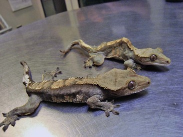
Nutritional Secondary Hyperparathyroidism Typical presentation of reptiles with nutritional secondary hyperparathyroidism (NSHP). Note the severe kyphosis in the crested gecko in the back, and that the animal in front is too weak to lift up its body to walk. (Photo courtesy Jörg Mayer, The University of Georgia, Athens.)
A large quantity of retained stool or urates may be palpated in the coelom.
Bloating and gastrointestinal motility issues are more common in some species of lizard such as juvenile bearded dragons.
Tremors and fasciculations may be evident, especially during handling
Seizure, tetany, and/or flaccid paresis of limbs/tail
Poor ability to elevate caudal body and proximal tail off the ground when ambulating
Prolapse of cloaca, colon, oviduct, hemipene
Gravid female lizards (higher demand for calcium) may present for NSHP with clinical signs, including:

Carapace and plastron may be abnormally soft and pliable, especially in young, growing chelonians.
Shape of the shell (especially the carapace) may be abnormal.
Often the marginal scutes on the carapace curl upward.
The turtle’s body may seem too large for its shell.
Mandibular and maxillary bones may be soft and pliable (less common than with lizards), and often the anterior maxilla may become elongated.
Fractures or swellings of long bones (less common than with lizards)
Inability to elevate the caudal plastron off the ground when ambulating
With abnormal shell growth of the carapace, conscious proprioceptive deficits or paresis/paralysis of the rear limbs may be noted.
Etiology and Pathophysiology
• NSHP in reptiles results from:
Dietary imbalance of calcium/phosphorus, typically low dietary calcium with excessive dietary phosphorus
Lack of exposure to natural sunlight or full-spectrum lighting or lack of dietary vitamin D3
Calcium deficiencies are common in herbivorous, insectivorous, and omnivorous reptiles.
Uncommon in snakes that eat warm-blooded prey.
Exacerbated in juvenile reptiles with the same (difficult to balance for calcium) diet but with an increased demand for calcium caused by rapid bone growth and formation
• Subsequent hypocalcemia results in partial depolarization of nerves and muscles (because of an increase in threshold potential), leading to tremors, twitching, and seizures in the reptile patient.
• Eventually, these dietary deficiencies (that result in low calcium uptake) will cause an increase in the production of parathyroid hormone from the parathyroid gland.
• Increased parathyroid hormone results in:
An increase in calcium resorption from bone to compensate for the low dietary uptake of calcium
Increased renal tubular reabsorption of calcium and increased excretion of phosphorus
Increased formation of 1,25-dihydroxycholecalciferol (DHCC), resulting in an increase in absorption of calcium in the intestinal tract
• Paresis and/or paralysis occurs with spinal fractures from weakened vertebral bones or compression of the spinal cord.
• Often innervation to the bowel and bladder is subsequently involved, resulting in elimination problems and constipation.
• Bloating, decreased gastrointestinal motility, and rectal/cloacal prolapse occur because of the effects of decreased calcium on the smooth muscle of the gastrointestinal tract.
• Paresis and paralysis related to spinal changes then result in prolapse of the colon/cloaca or reproductive organs, including the oviduct and hemipenes or phallus (in chelonians).
• NSHP is uncommon in adult reptiles:
No longer growing (especially long bones and shell) at the same rate as in a juvenile, so demand for calcium is lessened
Exception would be breeding female reptiles that are producing eggs/young, thus still with a demand for calcium.
Commonly misdiagnosed as NSHP when other metabolic diseases such as renal secondary hyperparathyroidism are more common in adult reptiles
Diagnosis 
Differential Diagnosis
Initial Database
• Obtain detailed historical information concerning diet and husbandry.
• In many cases, a diagnosis is made with historical dietary and husbandry information and physical exam findings.
• Serum/plasma biochemistry panel
Reptiles with secondary NSHP usually are normocalcemic; reptiles with renal disease generally are hypocalcemic and hyperphosphatemic (see Renal Disease).
Calcium and phosphorus values may provide baseline information needed to initiate calcitonin and monitor the therapeutic plan.
Uric acid, sodium, chloride, and total protein will be elevated with severe dehydration.
Treatment 
Therapeutic Goals
• Provide support for the presenting patient with supplemental heat and fluid therapy as needed.
• Address acute life-threatening presentations such as controlling seizures and stabilizing broken bones.
• Continue chronic treatment over weeks to months to correct deficiencies of calcium and/or vitamin D3.
• Correct specific husbandry issues related to NSHP such as diet (calcium and phosphorus intake) and exposure to ultraviolet lighting.
Acute General Treatment
• The patient should be warmed to appropriate physiologic temperatures (preferred optimum temperature zone [POTZ]), hydration status assessed, and fluid therapy (10-30 mL/kg q 24 h) initiated if necessary.
• For clinical signs of hypocalcemia, including fasciculations, tetany, and seizures, initiate immediate calcium supplementation with calcium gluconate (100 mg/kg IV or if given SQ give into a bolus of fluid or dilute before injection as it is caustic to tissues). Can be given every 6 h until clinical improvement:
Begin oral calcium supplementation with calcium glubionate at 23 mg/kg PO every 12 h.
Give 100 IU vitamin D3/kg IM or SC once weekly for two treatments.
If normocalcemic on serum/plasma biochemistry panel, or after treating with oral calcium glubionate for 3-7 days as described above, can give 50 IU/kg synthetic calcitonin intramuscularly weekly for two doses.
• A primary therapeutic goal is to stop bone loss and promote new bone production:
Calcitonin decreases circulating calcium and phosphorus.
With the use of calcitonin, bone repair may occur as early as 2-3 months as compared with 4-6 months without its use.
• Multiple pathologic fractures in small lizards and chelonians are best treated with strict cage rest:
• Larger lizards with one fractured limb may have the limb splinted to the body (lateral body wall for front limbs; tail for back limbs) with tape.
• All climbing accessories should be removed from the cage.
• Minimal handling is mandatory during treatment to avoid fracturing weakened bones.
• Reptile patients presented with associated obstipation/constipation may be given warm water soaks and gentle massage and manipulation to express fecal and urate material.
• Warm water enemas may be given using a gentle technique to guide the tube specifically into the colon (not just the cloaca); hydrostatic pressure should be minimized when the enema material is infused.
• During the treatment period (and beyond), optimal temperatures are necessary to allow adequate enzyme activity for gastrointestinal digestion and absorption of oral medications. Temperature gradients must be provided in the environment by focal basking or heat sources.
• If patients are not feeding voluntarily owing to soft mandibular and maxillary bones or other influences of NSHP gentle assist feeding, syringe feeding or tube feeding (chelonians) may be necessary. Start with small volumes, working up to more normal intakes over days to weeks as gastrointestinal motility may be affected by hypocalcemia.
Chronic Treatment
• Oral calcium glubionate treatment may be continued for a period of 1-3 months or longer.
• Follow-up treatments with parenteral vitamin D3 and calcitonin
• Correct nutrition and provide appropriate ultraviolet light exposure (natural sunlight or artificial lighting).
• Increase calcium in the diet, and limit phosphorus (calcium-to-phosphorus ratio, 2 : 1).
• Dietary recommendations are 2-3 mg/kcal of 0.6%-1.5% calcium and 0.5%-0.8% phosphorus on a dry matter basis (DM) for most reptiles.
• Calcium requirements can be higher for turtles and tortoises (1.4% calcium and 0.7% phosphorus DM).
• Excess phosphorus in the diet (bone meal or dicalcium phosphate) will exacerbate secondary hyperparathyroidism.
• Provide endogenous vitamin D3 through ultraviolet light exposure. Unfiltered sunlight is best, but if this is not feasible, full-spectrum lighting (285-320 nm) should be provided:
Positioning of the light is important for fluorescent tubes; they should be within 16-24 inches of the reptile to be useful (may be dangerous if too close to the reptile).
• The level of D3 as a supplement should range from 500-2000 IU D3 per kg.
• If a multivitamin is used to supplement oral vitamin D3, a vitamin A/vitamin D/vitamin E ratio of 100 : 10 : 1 is recommended.
• Use of a multivitamin alone typically will not provide enough calcium, and additional calcium must be given.
• Maximum tolerances suggested for many reptile species are 2.5% calcium, 1.6% phosphorus/DM, and 5000 IU/kg vitamin D3.
• General dietary recommendations for avoiding NSHP include:
For carnivores, feed whole animals, including bones and viscera (e.g., rodents, fish). Supplementation with additional calcium or multivitamins is not necessary unless feeding only neonatal prey items (e.g., newborn rodents).
For insectivores, feed invertebrate prey items a balanced diet (e.g., complete insect diet, complete mammalian chow). Dust invertebrates with a supplement (e.g., calcium carbonate, limestone) or a calcium/vitamin D3 combination.
Multivitamin supplements do not usually provide sufficient supplemental calcium and must be used in conjunction with a specific calcium source. Dusting is a very unscientific and “haphazard” method of supplementing the diet but has historically been successful. However, overzealous dusting may lead to vitamin toxicity (see Hypervitaminosis A).
For herbivores, feed vegetables with a good calcium-to-phosphorus ratio, such as collard greens, endive, parsley, and dandelion greens. For most herbivores, minimize fruit (should be no more than 10%-20% of total diet) in the diet because its high moisture content may dilute out necessary nutrients, and it tends to lack fiber. Salads must be supplemented with a calcium supplement such as calcium carbonate; 1 g (half teaspoon) of calcium carbonate per 100 g of food may be adequate.
Drug Interactions
Excessive levels of dietary vitamin D may be toxic, especially if combined with adequate exposure to unfiltered natural sunlight or full-spectrum lighting.
Possible Complications
Excessive calcium and phosphorus in the diet may interfere with the gastrointestinal absorption of other minerals such as zinc, copper, or iodine to deficiencies of these minerals.
Recommended Monitoring
• Regular rechecks in the first several weeks of treatment are critical to ensure that the patient is responding to therapy. These recheck visits also allow the clinician to review the husbandry to make sure the recommended changes have been made.
• Radiographic reevaluations can be useful at these follow-up visits to ensure bone healing progress.
• In lizards, malocclusion often occurs in association with fibrous osteodystrophy of the mandibular and maxillary bones. After treatment, the bones may heal, but lifelong malocclusion is a result. Severe underbites and overbites may occur, and management of chronic exposure gingivitis and ptyalism may be necessary. This is a chronic condition that cannot be resolved but can be managed with gentle cleansing and the application of a wax lip balm product to protect exposed tissue.
Prognosis and Outcome 
• Prognosis is good with aggressive therapy and appropriate husbandry changes.
• Spinal involvement (scoliosis and kyphosis, changes in the carapace in chelonians) has a more guarded to grave prognosis because neurologic damage may not resolve with treatment.
• If paresis and paralysis are associated with spinal involvement, and elimination complications result in obstipation/constipation, the prognosis is grave. Owners may be trained to help “assist” with elimination, but these patients may be difficult to manage. Often the spinal deformity worsens with growth, and an ascending pyelonephritis may result. Euthanasia may need to be considered in these cases.
• Females with spinal and pelvic changes may present with dystocia due to a narrowed pelvis. Prognosis for survival may be fair but a return to normal morphology poor, and lifelong management may be necessary.
• Prognosis is fair with signs of hypocalcemia such as tremors, ataxia, hyperreflexia, and even cloacal prolapse.
• Prognosis is worse with chronic disease often represented by fibrous osteodystrophy, pathologic fractures, paresis, and paralysis.
Pearls & Considerations 
Prevention
• Ensuring that clients with juvenile reptiles get proper husbandry information would prevent many cases.
• Encouraging clients to bring juvenile reptiles in for “new pet” exams provides a great opportunity to review husbandry and develop a relationship of trust, whereby the client will rely on your clinic for future care information.
Boyer, TH. Metabolic bone disease. In: Mader DR, ed. Reptile medicine and surgery. Philadelphia: WB Saunders; 1996:385–392.
Donoghue, S. Nutrition. In: Mader DR, ed. Reptile medicine and surgery. St Louis: Elsevier; 2006:251–298.
Frye, FL. Biomedical and surgical aspects of captive reptile husbandry, ed 2. Malabar, FL: Krieger Publishing; 1991.
Mader, DR. Metabolic bone disorders. In: Mader DR, ed. Reptile medicine and surgery. St Louis: Elsevier; 2006:841–851.