Arthroscopic Synovectomy of the Knee
Bryan D. Haughom, Brandon J. Erickson, Charles A. Bush-Joseph
The synovial lining is a specialized mesenchymal tissue that is integral to the normal functioning of a joint. Synovial disorders can involve varying amounts of the synovium. Rheumatoid arthritis shows total joint involvement, whereas on the other end of the spectrum, plica syndrome is caused by an isolated synovial lesion.
Volkman performed the first synovectomy in 1855 for tuberculous synovitis. Although the indications and technique have changed over time, the procedure is still performed, and the objective of removing the diseased synovium remains the same.1 Compared with open procedures, arthroscopic techniques have enabled surgeons to perform a synovectomy without a large arthrotomy, decreasing the risk of postoperative arthrofibrosis. Arthroscopy also serves as an effective technique to remove synovium in the posterior compartment and allows viewing of synovial lesions that may be missed with open procedures. Arthroscopic synovectomy can be used in the surgical treatment of rheumatoid arthritis, pigmented villonodular synovitis, hemophilic synovitis, plicae, synovial hemangioma, synovial osteochondromatosis, and degenerative synovitis.
As with all orthopaedic conditions, a complete workup including a thorough history and physical examination and complete imaging analysis is needed to evaluate these patients. Additionally, a trial of medical management should be performed before initiation of surgical treatment. Surgical treatment consists of arthroscopically removing varying amounts of synovium, the amount of which is based on the underlying disease process.
Clinical Evaluation
History
A complete history is important in the evaluation of patients with synovial disorders. The presence of other affected joints, the length of time symptoms have been experienced, exacerbating symptoms, and the amount of disability experienced by the patient on a daily basis are important pieces of information. Patients with rheumatoid arthritis may have more systemic complaints, including morning stiffness and other affected joints, particularly the small joints of the hands and feet. Pigmented villonodular synovitis (PVNS) is typically a monoarticular process that affects adults in the third or fourth decade of life. Symptoms are mechanical in nature and may be similar to those seen in patients with meniscal tears.2 Clinically patients have the insidious onset of localized warmth, swelling, and stiffness with occasional locking and a palpable mass. Plica syndrome is a finding in patients with anteromedial knee pain. Patients experience tightness, snapping, giving way, and pain with repetitive activities. Clinically it is difficult to distinguish plica syndrome from other causes of knee pain such as meniscal tears, patellar tendinitis, or patellofemoral pain syndrome.
Physical Examination and Laboratory Tests
A full rheumatologic workup should be completed for patients with systemic diseases, and appropriate laboratory tests should be up to date. Patients with hemophilia require a consultation with a hematologist. If surgical treatment is to be pursued, it is essential to have a well–thought-out plan for perioperative management of clotting factors.
Other joints may be affected in patients with rheumatic or autoimmune disorders, and these joints should be evaluated. Patients with rheumatoid arthritis often have a flexion contracture and quadriceps atrophy in the knee region.3 The skin should be examined and previous incisions and subcutaneous nodules should be evaluated. The knee should be examined to determine overall alignment, range of motion, the presence of an effusion, warmth, tenderness, crepitus, strength, meniscal integrity, and stability. Collateral ligament instability or bony malalignment suggests more severe articular loss, and patients with these conditions are poor candidates for a synovectomy.
The physical examination for PVNS is often nonspecific. An effusion is associated with diffuse involvement. Palpation of the joint may show warmth and tenderness. Aspiration of the joint fluid may show a dark-brown fluid that is a result of recurrent bleeding into the joint. Cytologic studies of the aspirate may show hemosiderin pigment and multinucleated foreign body giant cells, but often the findings of these studies are normal.4 Ligamentous instability is uncommon in persons with PVNS.
Plica syndrome begins insidiously. Tenderness over the medial parapatellar region is common. A plica may sometimes be directly palpated and rolled under the finger, recreating the patient's symptoms. If the medial border of the patella is palpated while pushing the patella medially with one hand and the other hand produces a valgus stress with external rotation of the tibia, pain may be elicited, suggesting plica syndrome.5 An effusion is not typically present in persons with plica syndrome.
Imaging
In patients with rheumatoid arthritis, cervical spine flexion and extension views should be obtained in preoperative patients to rule out cervical instability. Radiographs of the knee should be obtained. Patients with rheumatoid arthritis may have periarticular erosions and osteopenia. Radiographs in patients with PVNS can show erosive, cystic, and sclerotic lesions of the articular surface. If enough synovium that contains hemosiderin is present, soft tissue masses may be seen, but often the findings of the films are normal with well-maintained joint spaces. Magnetic resonance imaging is considered to be the most diagnostic study for PVNS. It may show nodular intraarticular masses of low signal intensity on T1- and T2-weighted images and also allows evaluation of the location and extent of disease.
Treatment
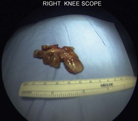
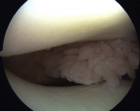
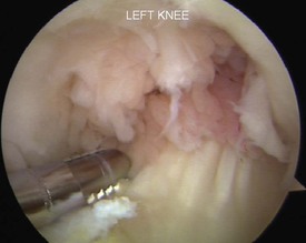
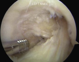
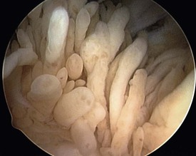
In disorders associated with a localized lesion, such as a localized PVNS (Figs. 95-1 and 95-2) or plica, arthroscopic intervention can remove the pathology in its entirety. Persons with diffuse conditions such as rheumatoid arthritis (Figs. 95-3 through 95-6) or hemophilia can undergo surgery to decrease the severity of disease symptoms once conservative measures have been exhausted. Recently, medical management of rheumatoid arthritis has improved significantly. The goals of medical treatment include reducing the number of painful and swollen joints, suppressing the acute phase response, decreasing the rheumatoid factor titer, and slowing radiographic progression of the disease. A patient with rheumatoid arthritis and minimal degenerative changes on radiographs would be a candidate for arthroscopic synovectomy after failure of approximately 6 months of medical management. Medical management should consist of a combination of disease-modifying antirheumatic drugs, nonsteroidal antiinflammatory drugs, an appropriate physical therapy regimen, activity modification, and intraarticular steroid injections (in general, no more than three steroid injections should be administered in one joint in a given year).6 Significant joint space narrowing or mechanical malalignment is a relative contraindication to synovectomy for inflammatory synovial knee disorders.

Hemophilic synovitis can also be associated with significant joint destruction and has shown favorable improvement in symptoms with synovectomy.7-9 Radiosynovectomy is indicated as the first procedure in persons with hemophilic synovitis, with satisfactory results in 80% of patients.10 No more than three radiosynovectomies can be performed per year. If the three radiosynovectomy procedures fail to relieve symptoms, an arthroscopic synovectomy is indicated.10 Although joint deterioration is not preventable, a synovectomy can reduce recurrent hemarthrosis and maintain range of motion. A hemophilic synovectomy requires an inpatient stay for coordinated management of clotting factors with the patient's hematologist.
Surgical Technique
Because arthroscopic synovectomy requires the use of multiple portals to access all spaces in the knee joint, good preoperative planning and patient setup is essential for a successful operation. Because a synovectomy can be a long procedure, induction of general anesthesia is recommended, and use of a Foley catheter should be considered. An epidural can be used if required for medical reasons and may also help with postoperative pain relief.
Examination After Inducement of Anesthesia
Both knees are examined for an assessment of the range of motion, ligamentous stability, patellar mobility, patellar tracking, and the presence of an effusion.
Positioning
The patient is placed supine on the operating room table. The well leg is appropriately padded and secured in a well leg holder after placement of a compressive stocking and sequential compression device. A leg holder is not used on the operative leg because it may interfere with use of the superomedial and superolateral portals. The foot of the operating table is dropped and the mid portion of the table is flexed to avoid hip hyperextension. A well-padded thigh tourniquet is placed high on the operative leg.
Surgical Steps
Anterior Compartments
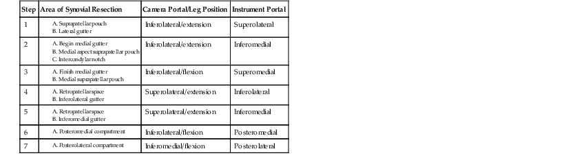
After standard prepping and draping is performed, the extremity is exsanguinated and the tourniquet is inflated to between 250 and 300 mm Hg. The tissue obtained from the synovectomy should be collected and sent for pathologic evaluation. The anterior aspect of the knee is addressed first. A superomedial outflow portal is created and the outflow cannula is placed here. Standard inferolateral and inferomedial portals are created. The arthroscope is placed in the inferolateral portal, and an initial diagnostic arthroscopy is performed. The synovectomy then proceeds with use of an arthroscopic shaver. While viewing from the inferolateral portal with the knee in extension, the shaver is used in the superolateral and inferomedial portals to remove all synovial tissue but avoiding injury to surrounding muscle, tendon, and fascia (Table 95-1).
TABLE 95-1
STEPS FOR PERFORMING AN ARTHROSCOPIC SYNOVECTOMY OF THE KNEE
| Step | Area of Synovial Resection | Camera Portal/Leg Position | Instrument Portal |
| 1 | Inferolateral/extension | Superolateral | |
| 2 | Inferolateral/extension | Inferomedial | |
| 3 | Inferolateral/flexion | Superomedial | |
| 4 | Superolateral/extension | Inferolateral | |
| 5 | Superolateral/extension | Inferomedial | |
| 6 | Inferolateral/flexion | Posteromedial | |
| 7 | Inferomedial/flexion | Posterolateral |
Posterior Compartments
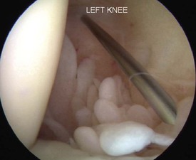
The anterior compartment should now be finished, and attention is turned to the posterior compartments, beginning with the posteromedial compartment. Typically the posterior compartments can be visualized with a 30-degree scope; however, a 70-degree scope can be used if difficulty is encountered. The arthroscope must first be placed in the posterior compartment. The blunt-tipped trocar is placed in the arthroscopic sheath and inserted into the inferolateral portal. The trocar is directed toward the medial femoral condyle, and when it is contacted, the trocar is carefully advanced posteriorly through the interval between the medial femoral condyle and the posterior cruciate ligament, raising the hand with insertion to match the slope of the tibia. If this maneuver proves difficult, a central and vertically oriented patellar tendon portal may provide easier access to the posterior compartment. The arthroscope is inserted, and the posterior portion of the medial femoral condyle and the posterior horn of the medial meniscus should be visible (Fig. 95-7). While looking medially, a spinal needle is inserted anterior to the medial head of the gastrocnemius into the posteromedial compartment. The needle is used to ensure that all areas in the posterior knee that are in need of synovectomy can easily be reached. Once the ideal portal location has been determined, a longitudinal incision is made in the skin. A hemostat is used to bluntly dissect and then penetrate the capsule, and a cannula is placed. The shaver is placed through the cannula, and the synovium in the posteromedial compartment is resected.
The posterolateral compartment is accessed in a manner similar to the posteromedial compartment. A blunt-tipped trocar is placed in the arthroscopic cannula in the inferomedial portal between the lateral femoral condyle and the anterior cruciate ligament. The hand is gently raised and advanced posteriorly, taking care not to violate the posterior capsule, which could put the neurovascular structures at risk. The arthroscope replaces the trocar, and the posterior lateral femoral condyle and the posterior horn of the lateral meniscus are viewed. Again, a spinal needle is used to make a posterolateral portal under direct visualization. Placing the needle in the soft spot, anterior to the biceps femoris muscle and posterior to the iliotibial band, helps protect the common peroneal nerve. The needle should be inserted posterior to the fibular collateral ligament and anterior to the lateral head of the gastrocnemius. Once it is determined that the spinal needle is placed so that all areas that require synovectomy can be reached, the skin is incised and a hemostat is used to dissect to the posterior capsule. The capsule is then punctured under direct visualization and a cannula is placed. The shaver is placed through the cannula, and the posterolateral compartment synovectomy is performed.
After the synovectomy is complete, the tourniquet is deflated and an electrocautery device is used to achieve hemostasis. It is common to use a suction drain for 24 hours to help minimize hemarthrosis. Ice, elevation, and a light compressive dressing are used to minimize swelling, and early motion is encouraged.
Postoperative Management
The patient can bear weight as tolerated after surgery. Physical therapy is begun on postoperative day 1 after drain removal and concentrates on closed chain exercises. The most immediate goals are regaining knee extension and quadriceps function. A continuous passive motion machine can be used for the first few days after surgery.
Complications
After synovectomy, a recurrent hemarthrosis may occur. The fluid can often be aspirated with a large-bore needle, but an arthroscopic washout may be needed. Joint stiffness and loss of extension may occur after a synovectomy. Performing aggressive and early range of motion exercises, using extension boards, and dynamic bracing may help with these symptoms. A septic joint or neurovascular injury can also occur. With use of careful technique, these complications can be minimized.
Results
Goetz et al.11 evaluated 32 knees at 14-year follow-up after combined arthroscopic and radiation synovectomy for rheumatoid arthritis. These investigators concluded that combined arthroscopic and radiation synovectomy led to a stable improvement of knee function for a minimum of 5 years, but repeat surgery was frequent, with 56% of patients having another operation by 10 years.11 If total knee arthroplasty was considered the end point, the joint survival rate was 88.5% at 5 years, 53.9% at 10 years, and 39.6% at 14 years.11 Carl et al.12 studied 11 patients with rheumatoid arthritis who were undergoing a synovectomy. These investigators found that a synovectomy led to an overall reduction of acute inflammatory infiltrates by 82.1% and of chronic inflammatory infiltrates by 62.5%.12
Although few follow-up data are available regarding synovectomy in patients with hemophilia in general, Verma et al.13 suggest that the primary predictor of outcome in hemophiliacs is the degree of intraarticular preexisting degenerative changes. In more severe cases, the results of arthroscopic synovectomy are less predictable, and total joint arthroplasty should be considered.
Rhee et al.14 performed a retrospective study to determine the long-term results of arthroscopic treatment of localized PVNS. Follow-up was 112 months for 11 patients. The authors concluded that excision of localized PVNS can improve symptoms and that patients can return to preoperative activity levels.14 Sharma and Cheng15 reported a recurrence-free survival of 62% at 2 years and 48% at 5 years in patients treated for diffuse and localized PVNS with arthroscopic synovectomy. Although synovectomy can prevent recurrence of PVNS, it appears that the joint may still progress to secondary osteoarthritis.
Summary
Arthroscopic synovectomy is a procedure that is the treatment of choice for a number of synovial disorders. The procedure requires thoughtful preparation and extensive preoperative workup. Results vary based on the underlying etiology, but the procedure does appear to help patients with the symptomatic management of synovial disorders.
For a complete list of references, go to expertconsult.com.