Excitation and Contraction of Smooth Muscle
Contraction of Smooth Muscle
The discussion in Chapters 6 and 7 was concerned with skeletal muscle. We now turn to smooth muscle, which is composed of far smaller fibers that are usually 1 to 5 micrometers in diameter and only 20 to 500 micrometers in length. In contrast, skeletal muscle fibers are as much as 30 times greater in diameter and hundreds of times as long. Many of the same principles of contraction apply to smooth muscle as to skeletal muscle. Most important, essentially the same attractive forces between myosin and actin filaments cause contraction in smooth muscle as in skeletal muscle, but the internal physical arrangement of smooth muscle fibers is different.
Types of Smooth Muscle
The smooth muscle of each organ is distinctive from that of most other organs in several ways: (1) physical dimensions, (2) organization into bundles or sheets, (3) response to different types of stimuli, (4) characteristics of innervation, and (5) function. Yet, for the sake of simplicity, smooth muscle can generally be divided into two major types, which are shown in Figure 8-1: multi-unit smooth muscle and unitary (or single-unit) smooth muscle.

Multi-Unit Smooth Muscle.
Multi-unit smooth muscle is composed of discrete, separate smooth muscle fibers. Each fiber operates independently of the others and often is innervated by a single nerve ending, as occurs for skeletal muscle fibers. Further, the outer surfaces of these fibers, like those of skeletal muscle fibers, are covered by a thin layer of basement membrane–like substance, a mixture of fine collagen and glycoprotein that helps insulate the separate fibers from one another.
Important characteristics of multi-unit smooth muscle fibers are that each fiber can contract independently of the others, and their control is exerted mainly by nerve signals. In contrast, a major share of control of unitary smooth muscle is exerted by non-nervous stimuli. Some examples of multi-unit smooth muscle are the ciliary muscle of the eye, the iris muscle of the eye, and the piloerector muscles that cause erection of the hairs when stimulated by the sympathetic nervous system.
Unitary Smooth Muscle.
Unitary smooth muscle is also called syncytial smooth muscle or visceral smooth muscle. The term “unitary” is confusing because it does not mean single muscle fibers. Instead, it means a mass of hundreds to thousands of smooth muscle fibers that contract together as a single unit. The fibers usually are arranged in sheets or bundles, and their cell membranes are adherent to one another at multiple points so that force generated in one muscle fiber can be transmitted to the next. In addition, the cell membranes are joined by many gap junctions through which ions can flow freely from one muscle cell to the next so that action potentials, or simple ion flow without action potentials, can travel from one fiber to the next and cause the muscle fibers to contract together. This type of smooth muscle is also known as syncytial smooth muscle because of its syncytial interconnections among fibers. It is also called visceral smooth muscle because it is found in the walls of most viscera of the body, including the gastrointestinal tract, bile ducts, ureters, uterus, and many blood vessels.
Contractile Mechanism in Smooth Muscle
Chemical Basis for Smooth Muscle Contraction
Smooth muscle contains both actin and myosin filaments, having chemical characteristics similar to those of the actin and myosin filaments in skeletal muscle. It does not contain the troponin complex that is required in the control of skeletal muscle contraction, and thus the mechanism for control of contraction is different. This topic is discussed in more detail later in this chapter.
Chemical studies have shown that actin and myosin filaments derived from smooth muscle interact with each other in much the same way that they do in skeletal muscle. Further, the contractile process is activated by calcium ions, and adenosine triphosphate (ATP) is degraded to adenosine diphosphate (ADP) to provide the energy for contraction.
There are, however, major differences between the physical organization of smooth muscle and that of skeletal muscle, as well as differences in excitation-contraction coupling, control of the contractile process by calcium ions, duration of contraction, and the amount of energy required for contraction.
Physical Basis for Smooth Muscle Contraction
Smooth muscle does not have the same striated arrangement of actin and myosin filaments as is found in skeletal muscle. Instead, electron micrographic techniques suggest the physical organization shown in Figure 8-2, which illustrates large numbers of actin filaments attached to dense bodies. Some of these bodies are attached to the cell membrane, and others are dispersed inside the cell. Some of the membrane-dense bodies of adjacent cells are bonded together by intercellular protein bridges. It is mainly through these bonds that the force of contraction is transmitted from one cell to the next.
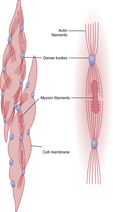
Interspersed among the actin filaments in the muscle fiber are myosin filaments. These filaments have a diameter more than twice that of the actin filaments. In electron micrographs, one usually finds 5 to 10 times as many actin filaments as myosin filaments.
To the right in Figure 8-2 is a postulated structure of an individual contractile unit within a smooth muscle cell, showing large numbers of actin filaments radiating from two dense bodies; the ends of these filaments overlap a myosin filament located midway between the dense bodies. This contractile unit is similar to the contractile unit of skeletal muscle, but without the regularity of the skeletal muscle structure; in fact, the dense bodies of smooth muscle serve the same role as the Z disks in skeletal muscle.
Another difference is that most of the myosin filaments have “sidepolar” cross-bridges arranged so that the bridges on one side hinge in one direction and those on the other side hinge in the opposite direction. This configuration allows the myosin to pull an actin filament in one direction on one side while simultaneously pulling another actin filament in the opposite direction on the other side. The value of this organization is that it allows smooth muscle cells to contract as much as 80 percent of their length instead of being limited to less than 30 percent, as occurs in skeletal muscle.
Comparison of Smooth Muscle Contraction and Skeletal Muscle Contraction
Although most skeletal muscles contract and relax rapidly, most smooth muscle contraction is prolonged tonic contraction, sometimes lasting hours or even days. Therefore, it is to be expected that both the physical and the chemical characteristics of smooth muscle versus skeletal muscle contraction would differ. Some of the differences are noted in the following sections.
Slow Cycling of the Myosin Cross-Bridges.
The rapidity of cycling of the myosin cross-bridges in smooth muscle—that is, their attachment to actin, then release from the actin, and reattachment for the next cycle—is much slower than in skeletal muscle; in fact, the frequency is as little as 1/10 to 1/300 that in skeletal muscle. Yet, the fraction of time that the cross-bridges remain attached to the actin filaments, which is a major factor that determines the force of contraction, is believed to be greatly increased in smooth muscle. A possible reason for the slow cycling is that the cross-bridge heads have far less ATPase activity than in skeletal muscle, and thus degradation of the ATP that energizes the movements of the cross-bridge heads is greatly reduced, with corresponding slowing of the rate of cycling.
Low Energy Requirement to Sustain Smooth Muscle Contraction.
Only 1/10 to 1/300 as much energy is required to sustain the same tension of contraction in smooth muscle as in skeletal muscle. This, too, is believed to result from the slow attachment and detachment cycling of the cross-bridges and because only one molecule of ATP is required for each cycle, regardless of its duration.
This low energy utilization by smooth muscle is important to the overall energy economy of the body because organs such as the intestines, urinary bladder, gallbladder, and other viscera often maintain tonic muscle contraction almost indefinitely.
Slowness of Onset of Contraction and Relaxation of the Total Smooth Muscle Tissue.
A typical smooth muscle tissue begins to contract 50 to 100 milliseconds after it is excited, reaches full contraction about 0.5 second later, and then declines in contractile force in another 1 to 2 seconds, giving a total contraction time of 1 to 3 seconds. This is about 30 times as long as a single contraction of an average skeletal muscle fiber. However, because there are so many types of smooth muscle, contraction of some types can be as short as 0.2 second or as long as 30 seconds.
The slow onset of contraction of smooth muscle, as well as its prolonged contraction, is caused by the slowness of attachment and detachment of the cross-bridges with the actin filaments. In addition, the initiation of contraction in response to calcium ions is much slower than in skeletal muscle, as will be discussed later.
The Maximum Force of Contraction Is Often Greater in Smooth Muscle Than in Skeletal Muscle.
Despite the relatively few myosin filaments in smooth muscle, and despite the slow cycling time of the cross-bridges, the maximum force of contraction of smooth muscle is often greater than that of skeletal muscle—as great as 4 to 6 kg/cm2 cross-sectional area for smooth muscle, in comparison with 3 to 4 kilograms for skeletal muscle. This great force of smooth muscle contraction results from the prolonged period of attachment of the myosin cross-bridges to the actin filaments.
The “Latch” Mechanism Facilitates Prolonged Holding of Contractions of Smooth Muscle.
Once smooth muscle has developed full contraction, the amount of continuing excitation can usually be reduced to far less than the initial level even though the muscle maintains its full force of contraction. Further, the energy consumed to maintain contraction is often minuscule, sometimes as little as 1/300 the energy required for comparable sustained skeletal muscle contraction. This mechanism is called the “latch” mechanism.
The importance of the latch mechanism is that it can maintain prolonged tonic contraction in smooth muscle for hours with little use of energy. Little continued excitatory signal is required from nerve fibers or hormonal sources.
Stress-Relaxation of Smooth Muscle.
Another important characteristic of smooth muscle, especially the visceral unitary type of smooth muscle of many hollow organs, is its ability to return to nearly its original force of contraction seconds or minutes after it has been elongated or shortened. For example, a sudden increase in fluid volume in the urinary bladder, thus stretching the smooth muscle in the bladder wall, causes an immediate large increase in pressure in the bladder. However, during the next 15 seconds to a minute or so, despite continued stretch of the bladder wall, the pressure returns almost exactly back to the original level. Then, when the volume is increased by another step, the same effect occurs again.
Conversely, when the volume is suddenly decreased, the pressure falls drastically at first but then rises in another few seconds or minutes to or near to the original level. These phenomena are called stress-relaxation and reverse stress-relaxation. Their importance is that, except for short periods, they allow a hollow organ to maintain about the same amount of pressure inside its lumen despite sustained, large changes in volume.
Regulation of Contraction by Calcium Ions
As is true for skeletal muscle, the initiating stimulus for most smooth muscle contraction is an increase in intracellular calcium ions. This increase can be caused in different types of smooth muscle by nerve stimulation of the smooth muscle fiber, hormonal stimulation, stretch of the fiber, or even change in the chemical environment of the fiber.
Smooth muscle does not contain troponin, the regulatory protein that is activated by calcium ions to cause skeletal muscle contraction. Instead, smooth muscle contraction is activated by an entirely different mechanism, as described in the next section.
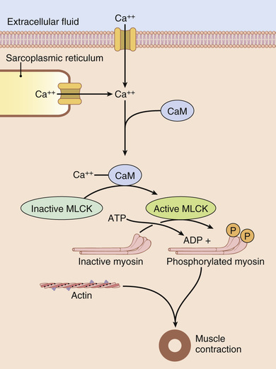
Calcium Ions Combine with Calmodulin to Cause Activation of Myosin Kinase and Phosphorylation of the Myosin Head.
In place of troponin, smooth muscle cells contain a large amount of another regulatory protein called calmodulin (Figure 8-3). Although this protein is similar to troponin, it is different in the manner in which it initiates contraction. Calmodulin initiates contraction by activating the myosin cross-bridges. This activation and subsequent contraction occur in the following sequence:
1. Calcium concentration in the cytosolic fluid of the smooth muscle increases as a result of the influx of calcium from the extracellular fluid through calcium channels and/or release of calcium from the sarcoplasmic reticulum.
2. The calcium ions bind reversibly with calmodulin.
3. The calmodulin-calcium complex then joins with and activates myosin light chain kinase, a phosphorylating enzyme.
4. One of the light chains of each myosin head, called the regulatory chain, becomes phosphorylated in response to this myosin kinase. When this chain is not phosphorylated, the attachment-detachment cycling of the myosin head with the actin filament does not occur. However, when the regulatory chain is phosphorylated, the head has the capability of binding repetitively with the actin filament and proceeding through the entire cycling process of intermittent “pulls,” the same as occurs for skeletal muscle, thus causing muscle contraction.
Source of Calcium Ions That Cause Contraction
Although the contractile process in smooth muscle, as in skeletal muscle, is activated by calcium ions, the source of the calcium ions differs. An important difference is that the sarcoplasmic reticulum, which provides virtually all the calcium ions for skeletal muscle contraction, is only slightly developed in most smooth muscle. Instead, most of the calcium ions that cause contraction enter the muscle cell from the extracellular fluid at the time of the action potential or other stimulus. That is, the concentration of calcium ions in the extracellular fluid is greater than 10−3 molar, in comparison with less than 10−7 molar inside the smooth muscle cell; this situation causes rapid diffusion of the calcium ions into the cell from the extracellular fluid when the calcium channels open. The time required for this diffusion to occur averages 200 to 300 milliseconds and is called the latent period before contraction begins. This latent period is about 50 times as great for smooth muscle as for skeletal muscle contraction.
Role of the Smooth Muscle Sarcoplasmic Reticulum.
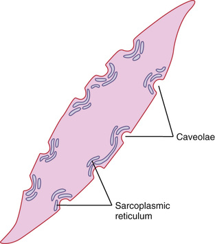
Figure 8-4 shows a few slightly developed sarcoplasmic tubules that lie near the cell membrane in some larger smooth muscle cells. Small invaginations of the cell membrane, called caveolae, abut the surfaces of these tubules. The caveolae suggest a rudimentary analog of the transverse tubule system of skeletal muscle. When an action potential is transmitted into the caveolae, this is believed to excite calcium ion release from the abutting sarcoplasmic tubules in the same way that action potentials in skeletal muscle transverse tubules cause release of calcium ions from the skeletal muscle longitudinal sarcoplasmic tubules. In general, the more extensive the sarcoplasmic reticulum in the smooth muscle fiber, the more rapidly it contracts.
Smooth Muscle Contraction Is Dependent on Extracellular Calcium Ion Concentration.
Although changing the extracellular fluid calcium ion concentration from normal has little effect on the force of contraction of skeletal muscle, this is not true for most smooth muscle. When the extracellular fluid calcium ion concentration decreases to about 1/3 to 1/10 normal, smooth muscle contraction usually ceases. Therefore, the force of contraction of smooth muscle is usually highly dependent on the extracellular fluid calcium ion concentration.
A Calcium Pump Is Required to Cause Smooth Muscle Relaxation.
To cause relaxation of smooth muscle after it has contracted, the calcium ions must be removed from the intracellular fluids. This removal is achieved by a calcium pump that pumps calcium ions out of the smooth muscle fiber back into the extracellular fluid, or into a sarcoplasmic reticulum, if it is present (Figure 8-5). This pump requires ATP and is slow acting in comparison with the fast-acting sarcoplasmic reticulum pump in skeletal muscle. Therefore, a single smooth muscle contraction often lasts for seconds rather than hundredths to tenths of a second, as occurs for skeletal muscle.
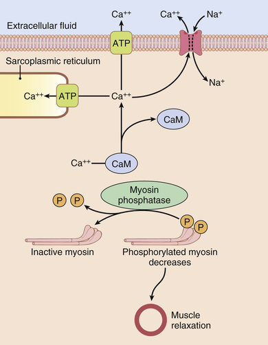
Myosin Phosphatase Is Important in Cessation of Contraction.
Relaxation of the smooth muscle occurs when the calcium channels close and the calcium pump transports calcium ions out of the cytosolic fluid of the cell. When the calcium ion concentration falls below a critical level, the aforementioned processes automatically reverse, except for the phosphorylation of the myosin head. Reversal of this situation requires another enzyme, myosin phosphatase (Figure 8-5), located in the cytosol of the smooth muscle cell, which splits the phosphate from the regulatory light chain. Then the cycling stops and contraction ceases. The time required for relaxation of muscle contraction, therefore, is determined to a great extent by the amount of active myosin phosphatase in the cell.
Possible Mechanism for Regulation of the Latch Phenomenon.
Because of the importance of the latch phenomenon in smooth muscle, and because this phenomenon allows long-term maintenance of tone in many smooth muscle organs without much expenditure of energy, many attempts have been made to explain it. Among the many mechanisms that have been postulated, one of the simplest is the following.
When the myosin kinase and myosin phosphatase enzymes are both strongly activated, the cycling frequency of the myosin heads and the velocity of contraction are great. Then, as the activation of the enzymes decreases, the cycling frequency decreases, but at the same time, the deactivation of these enzymes allows the myosin heads to remain attached to the actin filament for a longer and longer proportion of the cycling period. Therefore, the number of heads attached to the actin filament at any given time remains large. Because the number of heads attached to the actin determines the static force of contraction, tension is maintained, or “latched,” yet little energy is used by the muscle because ATP is not degraded to ADP except on the rare occasion when a head detaches.
Nervous and Hormonal Control of Smooth Muscle Contraction
Although skeletal muscle fibers are stimulated exclusively by the nervous system, smooth muscle can be stimulated to contract by nervous signals, hormonal stimulation, stretch of the muscle, and in several other ways. The principal reason for the difference is that the smooth muscle membrane contains many types of receptor proteins that can initiate the contractile process. Still other receptor proteins inhibit smooth muscle contraction, which is another difference from skeletal muscle. Therefore, in this section, we discuss nervous control of smooth muscle contraction, followed by hormonal control and other means of control.
Neuromuscular Junctions of Smooth Muscle
Physiologic Anatomy of Smooth Muscle Neuromuscular Junctions.
Neuromuscular junctions of the highly structured type found on skeletal muscle fibers do not occur in smooth muscle. Instead, the autonomic nerve fibers that innervate smooth muscle generally branch diffusely on top of a sheet of muscle fibers, as shown in Figure 8-6. In most instances, these fibers do not make direct contact with the smooth muscle fiber cell membranes but instead form diffuse junctions that secrete their transmitter substance into the matrix coating of the smooth muscle often a few nanometers to a few micrometers away from the muscle cells; the transmitter substance then diffuses to the cells. Furthermore, where there are many layers of muscle cells, the nerve fibers often innervate only the outer layer. Muscle excitation travels from this outer layer to the inner layers by action potential conduction in the muscle mass or by additional diffusion of the transmitter substance.
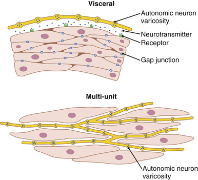
The axons that innervate smooth muscle fibers do not have typical branching end feet of the type found in the motor end plate on skeletal muscle fibers. Instead, most of the fine terminal axons have multiple varicosities distributed along their axes. At these points the Schwann cells that envelop the axons are interrupted so that transmitter substance can be secreted through the walls of the varicosities. In the varicosities are vesicles similar to those in the skeletal muscle end plate that contain transmitter substance. But in contrast to the vesicles of skeletal muscle junctions, which always contain acetylcholine, the vesicles of the autonomic nerve fiber endings contain acetylcholine in some fibers and norepinephrine in others, and occasionally other substances as well.
In a few instances, particularly in the multi-unit type of smooth muscle, the varicosities are separated from the muscle cell membrane by as little as 20 to 30 nanometers—the same width as the synaptic cleft that occurs in the skeletal muscle junction. These are called contact junctions, and they function in much the same way as the skeletal muscle neuromuscular junction; the rapidity of contraction of these smooth muscle fibers is considerably faster than that of fibers stimulated by the diffuse junctions.
Excitatory and Inhibitory Transmitter Substances Secreted at the Smooth Muscle Neuromuscular Junction.
The most important transmitter substances secreted by the autonomic nerves innervating smooth muscle are acetylcholine and norepinephrine, but they are never secreted by the same nerve fibers. Acetylcholine is an excitatory transmitter substance for smooth muscle fibers in some organs but an inhibitory transmitter for smooth muscle in other organs. When acetylcholine excites a muscle fiber, norepinephrine ordinarily inhibits it. Conversely, when acetylcholine inhibits a fiber, norepinephrine usually excites it.
Why are these responses different? The answer is that both acetylcholine and norepinephrine excite or inhibit smooth muscle by first binding with a receptor protein on the surface of the muscle cell membrane. Some of the receptor proteins are excitatory receptors, whereas others are inhibitory receptors. Thus, the type of receptor determines whether the smooth muscle is inhibited or excited and also determines which of the two transmitters, acetylcholine or norepinephrine, is effective in causing the excitation or inhibition. These receptors are discussed in more detail in Chapter 61 in relation to function of the autonomic nervous system.
Membrane Potentials and Action Potentials in Smooth Muscle
Membrane Potentials in Smooth Muscle.
The quantitative voltage of the membrane potential of smooth muscle depends on the momentary condition of the muscle. In the normal resting state, the intracellular potential is usually about −50 to −60 millivolts, which is about 30 millivolts less negative than in skeletal muscle.
Action Potentials in Unitary Smooth Muscle.
Action potentials occur in unitary smooth muscle (such as visceral muscle) in the same way that they occur in skeletal muscle. They do not normally occur in most multi-unit types of smooth muscle, as discussed in a subsequent section.
The action potentials of visceral smooth muscle occur in one of two forms: (1) spike potentials or (2) action potentials with plateaus.
Spike Potentials.
Typical spike action potentials, such as those seen in skeletal muscle, occur in most types of unitary smooth muscle. The duration of this type of action potential is 10 to 50 milliseconds, as shown in Figure 8-7A. Such action potentials can be elicited in many ways—for example, by electrical stimulation, by the action of hormones on the smooth muscle, by the action of transmitter substances from nerve fibers, by stretch, or as a result of spontaneous generation in the muscle fiber itself, as discussed subsequently.
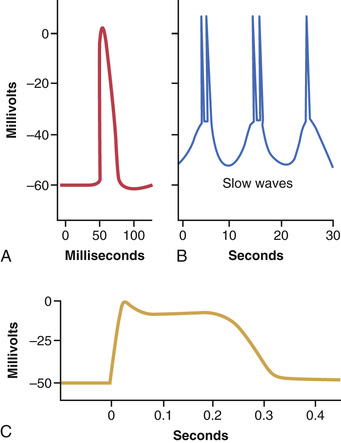
Action Potentials with Plateaus.
Figure 8-7C shows a smooth muscle action potential with a plateau. The onset of this action potential is similar to that of the typical spike potential. However, instead of rapid repolarization of the muscle fiber membrane, the repolarization is delayed for several hundred to as much as 1000 milliseconds (1 second). The importance of the plateau is that it can account for the prolonged contraction that occurs in some types of smooth muscle, such as the ureter, the uterus under some conditions, and certain types of vascular smooth muscle. (Also, this is the type of action potential seen in cardiac muscle fibers that have a prolonged period of contraction, as discussed in Chapters 9 and 10.)
Calcium Channels Are Important in Generating the Smooth Muscle Action Potential.
The smooth muscle cell membrane has far more voltage-gated calcium channels than does skeletal muscle but few voltage-gated sodium channels. Therefore, sodium participates little in the generation of the action potential in most smooth muscle. Instead, flow of calcium ions to the interior of the fiber is mainly responsible for the action potential. This flow occurs in the same self-regenerative way as occurs for the sodium channels in nerve fibers and in skeletal muscle fibers. However, the calcium channels open many times more slowly than do sodium channels, and they also remain open much longer. These characteristics largely account for the prolonged plateau action potentials of some smooth muscle fibers.
Another important feature of calcium ion entry into the cells during the action potential is that the calcium ions act directly on the smooth muscle contractile mechanism to cause contraction. Thus, the calcium performs two tasks at once.
Slow Wave Potentials in Unitary Smooth Muscle Can Lead to Spontaneous Generation of Action Potentials.
Some smooth muscle is self-excitatory—that is, action potentials arise within the smooth muscle cells without an extrinsic stimulus. This activity is often associated with a basic slow wave rhythm of the membrane potential. A typical slow wave in a visceral smooth muscle of the gut is shown in Figure 8-7B. The slow wave itself is not the action potential. That is, it is not a self-regenerative process that spreads progressively over the membranes of the muscle fibers. Instead, it is a local property of the smooth muscle fibers that make up the muscle mass.
The cause of the slow wave rhythm is unknown. One suggestion is that the slow waves are caused by waxing and waning of the pumping of positive ions (presumably sodium ions) outward through the muscle fiber membrane; that is, the membrane potential becomes more negative when sodium is pumped rapidly and less negative when the sodium pump becomes less active. Another suggestion is that the conductances of the ion channels increase and decrease rhythmically.
The importance of the slow waves is that, when they are strong enough, they can initiate action potentials. The slow waves themselves cannot cause muscle contraction. However, when the peak of the negative slow wave potential inside the cell membrane rises in the positive direction from −60 to about −35 millivolts (the approximate threshold for eliciting action potentials in most visceral smooth muscle), an action potential develops and spreads over the muscle mass and contraction occurs. Figure 8-7B demonstrates this effect, showing that at each peak of the slow wave, one or more action potentials occur. These repetitive sequences of action potentials elicit rhythmical contraction of the smooth muscle mass. Therefore, the slow waves are called pacemaker waves. In Chapter 63, we see that this type of pacemaker activity controls the rhythmical contractions of the gut.
Excitation of Visceral Smooth Muscle by Muscle Stretch.
When visceral (unitary) smooth muscle is stretched sufficiently, spontaneous action potentials are usually generated. They result from a combination of (1) the normal slow wave potentials and (2) a decrease in overall negativity of the membrane potential caused by the stretch. This response to stretch allows the gut wall, when excessively stretched, to contract automatically and rhythmically. For instance, when the gut is overfilled by intestinal contents, local automatic contractions often set up peristaltic waves that move the contents away from the overfilled intestine, usually in the direction of the anus.
Depolarization of Multi-Unit Smooth Muscle Without Action Potentials
The smooth muscle fibers of multi-unit smooth muscle (such as the muscle of the iris of the eye or the piloerector muscle of each hair) normally contract mainly in response to nerve stimuli. The nerve endings secrete acetylcholine in the case of some multi-unit smooth muscles and norepinephrine in the case of others. In both instances, the transmitter substances cause depolarization of the smooth muscle membrane, and this depolarization in turn elicits contraction. Action potentials usually do not develop because the fibers are too small to generate an action potential. (When action potentials are elicited in visceral unitary smooth muscle, 30 to 40 smooth muscle fibers must depolarize simultaneously before a self-propagating action potential ensues.) Yet in small smooth muscle cells, even without an action potential, the local depolarization (called the junctional potential) caused by the nerve transmitter substance itself spreads “electrotonically” over the entire fiber and is all that is necessary to cause muscle contraction.
Effect of Local Tissue Factors and Hormones to Cause Smooth Muscle Contraction Without Action Potentials
Approximately half of all smooth muscle contraction is likely initiated by stimulatory factors acting directly on the smooth muscle contractile machinery and without action potentials. Two types of non-nervous and nonaction potential stimulating factors often involved are (1) local tissue chemical factors and (2) various hormones.
Smooth Muscle Contraction in Response to Local Tissue Chemical Factors.
In Chapter 17, we discuss control of contraction of the arterioles, meta-arterioles, and precapillary sphincters. The smallest of these vessels have little or no nervous supply. Yet the smooth muscle is highly contractile, responding rapidly to changes in local chemical conditions in the surrounding interstitial fluid and to stretch caused by changes in blood pressure.
In the normal resting state, many of these small blood vessels remain contracted. However, when extra blood flow to the tissue is necessary, multiple factors can relax the vessel wall, thus allowing for increased flow. In this way, a powerful local feedback control system controls the blood flow to the local tissue area. Some of the specific control factors are as follows:
1. Lack of oxygen in the local tissues causes smooth muscle relaxation and, therefore, vasodilation.
2. Excess carbon dioxide causes vasodilation.
3. Increased hydrogen ion concentration causes vasodilation.
Adenosine, lactic acid, increased potassium ions, diminished calcium ion concentration, and increased body temperature can all cause local vasodilation. Decreased blood pressure, by causing decreased stretch of the vascular smooth muscle, also causes these small blood vessels to dilate.
Effects of Hormones on Smooth Muscle Contraction.
Many circulating hormones in the blood affect smooth muscle contraction to some degree, and some have profound effects. Among the more important of these hormones are norepinephrine, epinephrine, angiotensin II, endothelin, vasopressin, oxytocin, serotonin, and histamine.
A hormone causes contraction of a smooth muscle when the muscle cell membrane contains hormone-gated excitatory receptors for the respective hormone. Conversely, the hormone causes inhibition if the membrane contains inhibitory receptors for the hormone rather than excitatory receptors.
Mechanisms of Smooth Muscle Excitation or Inhibition by Hormones or Local Tissue Factors.
Some hormone receptors in the smooth muscle membrane open sodium or calcium ion channels and depolarize the membrane, the same as after nerve stimulation. Sometimes action potentials result, or action potentials that are already occurring may be enhanced. In other cases, depolarization occurs without action potentials, and this depolarization allows calcium ion entry into the cell, which promotes the contraction.
Inhibition, in contrast, occurs when the hormone (or other tissue factor) closes the sodium and calcium channels to prevent entry of these positive ions; inhibition also occurs if the normally closed potassium channels are opened, allowing positive potassium ions to diffuse out of the cell. Both of these actions increase the degree of negativity inside the muscle cell, a state called hyperpolarization, which strongly inhibits muscle contraction.
Sometimes smooth muscle contraction or inhibition is initiated by hormones without directly causing any change in the membrane potential. In these instances, the hormone may activate a membrane receptor that does not open any ion channels but instead causes an internal change in the muscle fiber, such as release of calcium ions from the intracellular sarcoplasmic reticulum; the calcium then induces contraction. To inhibit contraction, other receptor mechanisms are known to activate the enzyme adenylate cyclase or guanylate cyclase in the cell membrane; the portions of the receptors that protrude to the interior of the cells are coupled to these enzymes, causing the formation of cyclic adenosine monophosphate (cAMP) or cyclic guanosine monophosphate (cGMP), so-called second messengers. The cAMP or cGMP has many effects, one of which is to change the degree of phosphorylation of several enzymes that indirectly inhibit contraction. The pump that moves calcium ions from the sarcoplasm into the sarcoplasmic reticulum is activated, as well as the cell membrane pump that moves calcium ions out of the cell itself; these effects reduce the calcium ion concentration in the sarcoplasm, thereby inhibiting contraction.
Smooth muscles have considerable diversity in how they initiate contraction or relaxation in response to different hormones, neurotransmitters, and other substances. In some instances, the same substance may cause either relaxation or contraction of smooth muscles in different locations. For example, norepinephrine inhibits contraction of smooth muscle in the intestine but stimulates contraction of smooth muscle in blood vessels.