Diencephalon and Third Ventricle
DIENCEPHALON
The diencephalon is a part of brain situated cranial to the midbrain and is more or less completely surrounded by the cerebrum. The only part of diencephalon which is exposed to the surface is at the base of the brain in the region of interpeduncular fossa. The cavity within the diencephalon is termed the 3rd ventricle. It communicates on either side with the lateral ventricle of the cerebral hemisphere.
The cavity of the 3rd ventricle divides the diencephalon into two (right and left) symmetrical halves.
DIVISIONS AND SUBDIVISIONS
The diencephalon is divided into two major parts: pars dorsalis and pars ventralis. These subdivisions are seen in midsagittal view of the brain and are separated from each other by a shallow groove, the hypothalamic sulcus, which extends from interventricular foramen to the rostral end of the cerebral aqueduct of the midbrain (Fig. 26.4).

Pars dorsalis lies above (dorsal) the hypothalamic sulcus and consists of: (a) thalamus, (b) metathalamus, and (c) epithalamus.
Pars ventralis lies below (ventral) the hypothalamic sulcus and consists of: (a) subthalamus and (b) hypothalamus.
Thus the diencephalon is divided into five parts: thalamus, metathalamus, epithalamus, subthalamus, and hypothalamus. Each of these parts has further subdivisions.
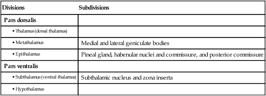
The main divisions and subdivisions of the diencephalon are listed in Table 26.1.
Table 26.1
Divisions and subdivisions of the diencephalon
| Divisions | Subdivisions |
| Pars dorsalis | |
| Medial and lateral geniculate bodies | |
| Pineal gland, habenular nuclei and commissure, and posterior commissure | |
| Pars ventralis | |
| Subthalamic nucleus and zona inserta | |
THALAMUS
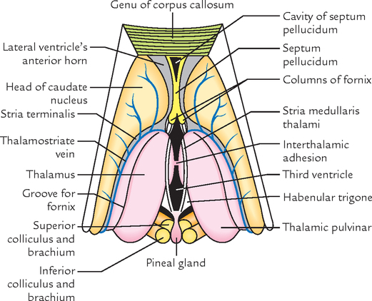
Anatomically, thalamus is a large ovoid mass of grey matter lying above the midbrain, from which it is separated by a small amount of neural tissue, the subthalamus. There are two thalami situated one on each side of a slit-like cavity, the 3rd ventricle (Fig. 26.1)
Each thalamus is 3.5 cm in length and 1.5 cm in breadth.
The long axes of the thalami are set obliquely, running backward and laterally. The pointed anterior ends are nearer to the median plane, whereas the wider posterior ends are separated from each other by pineal body, superior colliculi, and habenular triangles. The thalami are usually attached across the median plane by a narrow interthalamic connexus of grey matter (also called interthalamic adhesion). Each thalamus forms most of the lateral wall of the 3rd ventricle and floor of the central part of the lateral ventricle.
Functionally, thalamus is considered as the great sensory gateway to the cerebral cortex. It receives impulses from the opposite half of the body and transmits most of them to the sensory area of the cerebral cortex (Brodmann areas 3, 2 and 1).
Parts of the Thalamus
The thalamus is traversed anteroposteriorly by a vertical sheet of white fibres, the internal medullary lamina which bifurcates anteriorly to assume a Y-shaped configuration. This Y-shaped internal medullary lamina divides the thalamus into three main parts: anterior, medial and lateral.
The anterior part includes the anterior tubercle and lies between the ‘limbs’ of the ‘Y’. The medial and lateral parts lie on either side of the ‘stem’ of the ‘Y’. Each of these parts consists of a number of nuclei.
Nuclei of the Thalamus (Fig. 26.2)
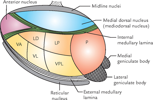
The lateral part is divided into dorsal and ventral parts.
The dorsal part is subdivided craniocaudally into three nuclei: (a) lateral dorsal (LD), (b) lateral posterior (LP), and (c) a large caudal nuclear mass, the pulvinar (P). These nuclei are termed dorsal tier of nuclei.
The ventral part is also subdivided craniocaudally into three nuclei: (a) ventral anterior (VA), (b) ventral lateral (VL) or ventral intermediate (VI), and (c) ventral posterior (VP). These nuclei are termed ventral tier of nuclei.
The ventral posterior nucleus (VP) is further subdivided into a lateral part, the ventral posterolateral nucleus (VPL) and a medial part, the ventral posteromedial nucleus (VPM).
The thalamic nuclei are summarized in Table 26.2.

Connections of the Thalamic Nuclei
The important connections of thalamic nuclei are as follows:
From a clinical point of view the connections of ventral posterior nucleus are most important because its smaller medial portion, the ventral posteromedial nucleus (VPM) receives general sensory modalities from the head and face through trigeminal lemniscus and tastes sensations from taste buds through solitariothalamic tract; and its larger lateral portion, the ventral posterolateral nucleus (VPL) receives exteroceptive sensations (pain, touch, temperature) through spinal lemniscus and proprioceptive sensations (muscle and joint sense, vibration, two-point discrimination) through medial lemniscus, from the rest of the body except face and head.
All the sensations reaching the ventral posterior nucleus are carried to the primary sensory area of the cerebral cortex by fibres passing through the posterior limb of the internal capsule (superior thalamic radiation). The vascular lesions involve posterior limb of the internal capsule which sometimes cause impairment of all forms of sensibility on the opposite side of the body.
The integrity of anterior nucleus and its connections is necessary for attention and recent memory, therefore, a lesion involving it can lead to loss of recent memory.
Since the medial dorsal nucleus is associated with ‘moods’ and emotional balance, depending on the nature of the present sensory input and past experience, the mood may be that of well-being or malaise, or of euphoria or mild depression.
Functions of the Thalamus
The main functions of the thalamus are as follows:
1. It is a sensory integration and relay station of all the sensory pathways except for the olfactory pathway which is projected directly to the cerebral cortex without being relayed in the thalamus.
2. It is capable of recognition, though poorly, of pain, thermal and some tactile sensations at its own level.
METATHALAMUS
The metathalamus consists of medial and lateral geniculate bodies. These are small rounded elevations on the inferior aspect of the posterior part of the thalamus, lateral to each side of the midbrain. The medial and lateral geniculate bodies are relay stations for the auditory and visual pathways, respectively.
Medial Geniculate Body
Medial geniculate body is an oval elevation on the inferior aspect of the pulvinar of the thalamus, lateral to the superior colliculus. It is more prominent than the lateral geniculate body. The inferior brachium runs upward, laterally and forward from inferior colliculus of the midbrain to the medial geniculate body.
The inferior brachium conveys auditory impulses to the lateral geniculate body for onward transmission to the (primary auditory area of the) cerebral cortex.
Lateral Geniculate Body
Lateral geniculate body is a small ovoid prominence visible at the terminal end of the optic tract. It is situated on the inferior surface of the pulvinar, anterolateral to the medial geniculate body. It is smaller than the medial geniculate body and connected to the superior colliculus by the superior brachium.
The fibres of superior brachium are concerned with the production of visual reflexes such as turning of head and eyes toward the sudden flash of light and constriction of pupil when light falls or is thrown on the retina.
The lateral geniculate body receives retinal fibres of both the eyes (from temporal half of the retina of the same side and nasal half of the retina of the opposite side) through optic tract and gives rise to fibres of the optic radiation which convey visual impulses to the visual cortex of the occipital lobe.
EPITHALAMUS
Pineal Gland (Epiphysis Cerebri)
Pineal gland is a midline cone-shaped reddish grey structure (only 3 mm × 5 mm in size) occupying the vertical groove between the two superior colliculi below the splenium of corpus callosum. It projects back from the posterior wall of the 3rd ventricle, below the splenium of the corpus callosum. It has a stalk which divides into two laminae. The ventral (or inferior) lamina continues with the posterior commissure and the dorsal (or superior) lamina continues with the habenular commissure. The extension of the cavity of the 3rd ventricle between the two laminae is termed pineal recess.
Structure and Functions
The pineal gland is a neuroendocrine gland and consists of parenchymal cells, called pinealocytes and neuroglial cells. The pinealocytes secrete a hormone called melatonin. The calcium phosphates and carbonates are deposited in the gland with age in the form of multilaminar corpuscles called corpora arenacea or brain sand. They are often seen as tiny shadows in radiographs of the skull. A displaced calcified pineal gland indicates a space-occupying lesion within the brain.
Pineal secretions including melatonin have an inhibitory effect on other endocrine glands and gonads.
HYPOTHALAMUS
The hypothalamus is a part of the diencephalon which lies below the thalamus. It forms the floor and the lower parts of lateral walls of the 3rd ventricle. Anatomically, hypothalamus is small in size weighing only 4 g and forming only 0.3% of the total brain mass, but physiologically there is hardly any activity in the body that is not influenced by it. Thus, the functional significance of hypothalamus is disproportionate to its size. The hypothalamus controls the autonomic and endocrine systems at the highest level, and is also involved in some affective or emotional behavior. Being the principal autonomic centre of the brain, it has been regarded as the head ganglion of the autonomic nervous system by Sherrington.
Boundaries of the Hypothalamus
The boundaries of the hypothalamus are as follows:
| Anteriorly: | Lamina terminalis (lamina terminalis extends from the optic chiasma to the anterior commissure). |
| Posteriorly | Subthalamus. |
| Inferiorly: | Structures in the floor of the 3rd ventricle, viz. tuber cinereum, infundibulum and mammillary bodies. (These structures are actually the parts of hypothalamus itself.) |
| Superiorly: | Thalamus. |
| Laterally: | Internal capsule. |
| Medially: | Cavity of the 3rd ventricle. |
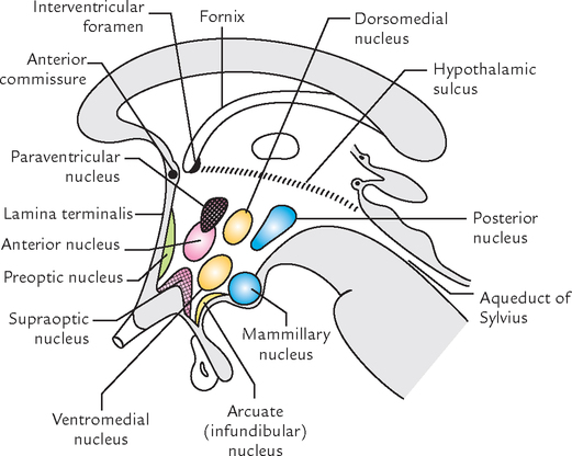
Subdivisions of the Hypothalamus
The hypothalamus is also subdivided anteroposteriorly into the following four regions:
1. Preoptic region—adjoining the lamina terminalis.
2. Supraoptic region—above the optic chiasma.
3. Tuberal region—includes the tuber cinereum, infundibulum and area around it.
4. Mammillary region—includes the mammillary bodies and area around it.
The preoptic region lies anterior to the hypothalamus between the optic chiasma and anterior commissure. Anatomically, it belongs to telencephalon but functionally it belongs to hypothalamus.
The tuber cinereum is the region bounded caudally by mammillary bodies and rostrally by optic chiasma. The infundibulum connects the posterior lobe of the hypophysis cerebri with the tuber cinereum. The tuber cinereum around the base of the infundibulum is raised to form a median eminence.
Hypothalamic Nuclei
The hypothalamus consists of numerous cell groups called hypothalamic nuclei. The nuclei present in different regions of the hypothalamus are listed in Table 26.3 and shown in Figure 26.3.

Connections of the Hypothalamus
The connections of the hypothalamus are numerous and complex, therefore, only main connections are described below:
1. The axons of supraoptic and paraventricular nuclei run in the pituitary stalk to reach the posterior pituitary (neurohypophysis) and form the hypothalamohypophyseal tract. These axons transport the neurohormones—vasopressin and oxytocin—synthesized in supraoptic nucleus and paraventricular nucleus, respectively, to the posterior pituitary.
2. The axons of other cell groups, e.g., tuberal nuclei enter the region of median eminence to deliver their neurosecretory material to the hypothalamohypophyseal portal system of blood vessels to control the secretion of hormones from the anterior pituitary (adenohypophysis).
3. The long axons from thalamus pass through the brainstem and spinal cord to synapse with the preganglionic sympathetic cells in the lateral horns of the thoracic and upper two lumbar spinal segments and with the preganglionic parasympathetic cells in the lateral horns of the S2, S3, and S4 spinal segments to form hypothalamospinal tract.
4. Mammillothalamic tract connects the mammillary body to the anterior nucleus of the thalamus, which in turn projects to the cingulate gyrus.
Functions of the Hypothalamus
1. Autonomic control: The anterior part of the hypothalamus controls the parasympathetic nervous system while posterior part controls the sympathetic nervous system.
2. Endocrine control: Regulates the hormonal secretion of the anterior pituitary by forming the releasing factors or release inhibiting factors which in turn control the endocrine activities of the body.
3. Neurosecretion: Secretes neurohormones: oxytocin and vasopressin.
4. Regulation of food and water intake: The lateral part of the hypothalamus acts as hunger centre while the medial part acts as satiety centre. A thirst centre in the lateral part regulates water intake.
5. Emotional expression: Plays an important role in the expression of autonomic emotions like laughing, crying, sweating, or blushing mediated by the integrated activity of the ANS and somatic efferent system.
6. Sexual behavior and reproduction: Regulates the sexual behavior and reproduction by influencing the secretion of gonadotrophic hormones by the pituitary gland.
7. Temperature regulation: The anterior portion of the hypothalamus prevents the rise in body temperature while posterior portion promotes heat conservation and heat production.
8. Biological clock: Regulates the cyclic activities of the body (circadian rhythm), viz. sleeping and waking cycle, but itself affected by diurnal rhythms. The circadian rhythm for many body functions is about 24 hours.
THIRD VENTRICLE
The 3rd ventricle is the cavity of diencephalon. It is a midline slit-like cavity situated between two thalami and part of hypothalamus. It extends from the lamina terminalis anteriorly to the superior end of the cerebral aqueduct of the midbrain posteriorly. The cavity of the 3rd ventricle is lined by ciliated columnar epithelium, the ependyma and traversed horizontally by a mass of grey matter, the interthalamic adhesion, connecting the two thalami. The outline of the cavity is irregular due to the presence of several diverticula or recesses.
Anteriorly on each side, the 3rd ventricle communicates with the lateral ventricle through interventricular foramen (of Monro), and posteriorly with the 4th ventricle through cerebral aqueduct (of Sylvius).
BOUNDARIES
The 3rd ventricle has anterior wall, posterior wall, roof, floor and two lateral walls (Fig. 26.4).
Anterior wall is formed from above downward by:
Posterior wall: formed from above downward by:
N.B. All structures of the floor belong to interpeduncular fossa except the optic chiasma and tegmentum of the midbrain.
Lateral wall: marked by a curved sulcus, the hypothalamic sulcus extending from the interventricular foramen to the upper end of the cerebral aqueduct. The sulcus divides the lateral wall into a larger upper part and a smaller lower part.
The larger upper part of the lateral wall is formed by the medial surface of the anterior two-third of the thalamus.
The smaller lower part of the lateral wall is formed by the hypothalamus and it is continuous with the ventricular floor.
N.B. The two lateral walls of the 3rd ventricle are normally closely approximated, hence in coronal section of the brain the cavity of the 3rd ventricle appears as a median vertical slit (Fig. 26.1).
RECESSES
The cavity of the 3rd ventricle extends into the surrounding structures as pocket-like protrusions called recesses (Fig. 26.4). These are as follows:
1. Infundibular recess: It is a deep tunnel-shaped recess extending downward through the tuber cinereum into the infundibulum, i.e., the stalk of the pituitary gland.
2. Optic (or chiasmatic) recess: It is an angular recess situated at the junction of the anterior wall and the floor of the ventricle just above the optic chiasma.
3. Anterior recess (vulva of the ventricle): It is a triangular recess which extends anteriorly in front of interventricular foramen and behind anterior commissure between the diverging anterior columns of the fornix.
4. Suprapineal recess: It is a fairly capacious blind diverticulum, which extends posteriorly above the stalk of the pineal gland and below the tela choroidea.
5. Pineal recess: It is a small diverticulum which extends posteriorly between the superior and inferior laminae of the stalk of the pineal gland.
CHOROID PLEXUS AND TELA CHOROIDEA
The tela choroidea in the roof of the 3rd ventricle is triangular in shape. The choroid plexus of the 3rd ventricle hangs downward from the tela choroidea as two longitudinal anteroposterior vascular fringes.
 Great sensory gateway to the cerebral cortex Great sensory gateway to the cerebral cortex |
Thalamus |
| Largest somatosensory relay nucleus of the thalamus |
Ventral posterior |
| Thalamus is the final relay station of all the sensory modalities except |
Olfaction |
| Seat of soul |
Pineal gland |
| Biological clock in human body |
Pineal gland |
| All the parts of the brain consist of neural cells except |
Pineal gland |
| Only part of the brain which is supplied by a nerve arising from outside the brain |
Pineal gland (it is supplied by nervus conarii which arises from superior cervical sympathetic ganglion in the neck) |
| Head ganglion of the autonomic nervous system |
Hypothalamus |
| Anatomically, all the regions of hypothalamus belong to the diencephalon except |
Preoptic region which belongs to telencephalon |


A 55-year-old patient was admitted in the neurology ward with complaints of loss of sensation on the left side of his body. After a few days the patient started improving and there was an evidence of return of sensations on the affected side. Both, the treating doctors and relatives were very happy. Few days later he suddenly started complaining of agonizing pain in his left arm and leg. The pain would start spontaneously or may be initiated by the light touch of even bed sheet or by little exposure to the cold. The pain failed to respond even to powerful analgesics. He was diagnosed as a case of thalamic syndrome.
1. What is thalamus? What is its main function?
2. Enumerate the ventral tier of the thalamic nuclei.
3. Name the nucleus of thalamus concerned with the general sensations of pain, touch, and temperature from opposite half of the body.
1. The thalamus is a large ovoid mass of grey matter lying above the brainstem from which it is separated by a small amount of neutral tissue—the subthalamus. The main function of thalamus is that it is a great sensory relay station in which all the sensory pathways relay except olfactory.
2. The ventral tier of thalamic nuclei include ventral anterior (VA), ventral lateral (VL) or ventral intermediate (VI), and ventral posterior (VP). The ventral posterior nucleus is further divided into ventral posterolateral (VPL) and ventral posteromedial (VPM) nuclei.
3. Ventral posterolateral (VPL) nucleus.
4. The thalamic syndrome is thalamic overreaction to the sensations of pain, touch, and temperature. It occurs when the patient is recovering from thalamic infarct following a vascular lesion of the thalamus.