5.2 Nerve Conduction and EMG Studies in Polyneuropathies
Nerve conduction and EMG studies have an important role in the evaluation of peripheral neuropathies; by confirming the clinical suspicion of neuropathy, identifying the predominant pathophysiology such as axonal or demyelinating; sensory or motor; and temporal course of the disease, i.e., acute, subacute, or chronic. Nerve conduction and EMG studies provide an objective and quantitative measure of nerve function and also help in predicting the prognosis of neuropathy. The electrodiagnostic battery for neuropathy evaluation should include motor and sensory nerve conduction, late responses, blink reflex, sympathetic skin responses, and needle electromyography. The nerve conduction studies include both motor and sensory conductions, preferably bilaterally, multiple nerves of both upper and lower limbs to demonstrate the characteristic symmetry or asymmetry of abnormalities.

Pathophysiological Basis of Nerve Conduction Study

The nerve conduction studies provide the information about the involvement of axon, myelin, or both. The characteristic findings in purely axonal lesions are similar to those in axonotmesis. The structural changes in interrupted nerve fibers can be classified into early axonal changes, clearance of myelin, the Schwann cell responses, and preparation for regeneration and late phase of Schwann cell atrophy and fibrosis (Bunge and Griffin, 1992) (Fig. 5.20). The nerve conduction velocity remains nearly normal throughout the Wallerian degeneration (Wilbourn, 1977). Distal latency and conduction velocities are preserved as long as a response can be obtained. In Wallerian degeneration of peripheral nerve, a length-dependent reduction in action potential takes place occurring earlier in shorter than longer nerves. The amplitude of compound muscle action potential (CMAP) declines earlier than sensory nerve action potential (SNAP). A near-normal conduction velocity is attributed to the loss of myelin sheath being secondary to axonal damage. Immediately after section of axon, the CMAP amplitude on motor nerve stimulation, SNAP on sensory nerve stimulation and distal latency remain normal up to 1 week. Needle EMG is normal although the recruitment pattern of motor unit potentials (MUPs) is reduced depending upon the degree of weakness. The CMAP and SNAP amplitudes progressively decrease and become unrecordable later. EMG changes of denervation appear by 1–3 weeks depending upon the proximity of the muscles to the site of nerve injury. On electromyography, the initial changes are prolonged insertional activity, positive sharp waves, fibrillations, and complex repetitive discharges. With partial axonal damage, the recruitment pattern is reduced, whereas no MUPs can be recruited in complete lesions. Initially the configuration of MUPs is normal; whereas in the succeeding months, the MUPs become polyphasic, high amplitude with long duration, and reduced recruitment (Table 5.18).

Table 5.18 Neurophysiological features of axonal degeneration

1. Normal or near-normal nerve conduction velocity
2. Reduction of CMAP amplitudes
3. Reduction of SNAP amplitudes
4. Fibrillations in the denervated muscles

Table 5.19 Neurophysiological features of segmental demyelination

1. Conduction block
2. Slowing of conduction across the affected segment
3. Prolonged distal latency
4. Temporal dispersion

Conduction Block and Temporal Dispersion

Conduction block and temporal dispersion are important neurophysiological findings in segmental demyelinating neuropathies. Conduction block is defined as the drop in the amplitude or area of CMAP on proximal stimulation compared to distal (Fig. 5.21A). A drop of 20–50% of the negative peak has been suggested for the diagnosis of conduction block (Brown and Feasby, 1984; Oh et al., 1994). Even in normal subjects some drop of CMAP amplitude on proximal stimulation occurs. In certain nerves such as median, ulnar, and peroneal, normally there may be a drop in CMAP amplitude up to 30% on proximal stimulation; whereas in posterior tibial nerve it is up to 41% (Oh et al., 1994). The stimulation of posterior tibial nerve in the popliteal fossa is technically difficult and may account for the greater drop of CMAP amplitude. In acquired demyelinating neuropathies, there is variable degree of demyelination, which is patchy and focal. This results in dispersion of CMAP due to phase cancellation. The negative and positive phases of action potential overlap due to variable conduction velocity in demyelinated fibers, leading to temporal dispersion. Such an amplitude drop may be erroneously diagnosed as conduction block. For interpreting the conduction block, therefore, the duration of CMAP should also be considered. A reduction of CMAP amplitude by 20% if its duration is normal; and 30% if CMAP duration is prolonged has been suggested (Brown and Feasby, 1984). To obviate this difficulty, the area measurement of CMAP employing a computer program has been recommended for diagnosis of conduction block. Peakto-peak measurement of CMAP amplitude has also been used for the diagnosis of conduction block, but the negative peak measurements are more appropriate. On proximal stimulation, the duration of CMAP increases slightly compared to that on distal stimulation, but an increase exceeding 20% is indicative of dispersion (Fig. 5.21B, Albers et al., 1985). The degree of reduction of CMAP amplitude if 20% was considered possible conduction block (Cornblath, 1990) and more than 30% as probable conduction block (Gilliatt, 1966). On histopathological correlation, the patients with even 20% reduction in CMAP amplitude were found to have definite evidence of demyelination. Hence a 20% reduction of the amplitude of proximal CMAP has been regarded as an indicator of segmental demyelination. For measurement of dispersion, although the total duration of CMAP may be measured but the duration of the negative peak is commonly employed (Oh et al., 1994).
Demyelination of distal portion of nerve may result in prolongation of distal latency. In hereditary demyelinating neuropathies, there is diffuse and uniform demyelination, which results in pronounced slowing of nerve conduction velocity without any conduction block or dispersion.

Neurophysiological Classification of Polyneuropathy

On the basis of neurophysiological findings, the neuropathies can be classified as follows:
1. Uniform demyelinating sensorimotor polyneuropathy
2. Segmental demyelinating, motor more than sensory polyneuropathy
3. Axonal, motor more than sensory polyneuropathy
4. Axonal sensory polyneuropathy
5. Axonal mixed sensorimotor polyneuropathy
6. Mixed axonal and demyelinating sensorimotor polyneuropathy
Demyelinating neuropathies have been divided into uniform and segmental neuropathies. In such a classification, some degree of overlap between various subgroups is inevitable because certain disorders can result in more than one type of neuropathy.

Segmental Demyelinating, Motor More Than Sensory Neuropathy

Table 5.21 Important causes of segmental demyelinating neuropathies motor more than sensory

1. Acute inflammatory demyelinating polyradiculoneuropathy (AIDP)
2. Chronic inflammatory demyelinating polyradiculoneuropathy (CIDP)
3. Multifocal motor neuropathy with motor block
4. Paraproteinemia–osteosclerotic myeloma, Waldenstrom macroglobulinemia, monoclonal gammopathy of undetermined significance
5. Acquired immune deficiency syndrome (AIDS) neuropathy
6. Lyme disease
7. Diphtheria
8. Penicillamine
Neurophysiological abnormalities in these disorders are related to the timing of disease. Slowing of motor and sensory conduction velocity, prolongation of terminal latency, conduction block, dispersion and prolonged or absent F-waves are the characteristic neurophysiological abnormalities (Fig. 5.23). Motor nerve conduction abnormalities are more homogeneous compared to sensory, which are patchy, being normal in some nerves, and abnormal in others.

Sensory Axonal Polyneuropathy

Sensory axonal polyneuropathies predominantly present with pain, paresthesias, and dysesthesia. Areflexia, sensory ataxia and choreoathetoid movements are found on examination. Neuropathology reveals inflammation and cell loss in dorsal root ganglia and gliosis of the posterior column of spinal cord. Nerve conduction usually reveals diminished or absent SNAP amplitude in the setting of normal motor nerve conduction velocity (Horwich et al., 1977). Sensory axonal polyneuropathy is found in carcinomatous sensory neuropathy, diabetes mellitus, hereditary sensory neuropathy type I–IV, Friedreich’s ataxia, abeta lipoproteinemia, toxins, e.g., cisplatin (Roelefs et al., 1984) and pyridoxine overdose (Schaumburg et al., 1983), rare variant of GB syndrome, vitamin E neuropathy, malabsorption, acromegaly, polycythemia, and chronic obstructive lung disease.

Axonal Type of Mixed Sensory Motor Polyneuropathy

The clinical picture of axonal type of mixed sensory motor polyneuropathies comprises paresthesia and dysesthesia of feet and distal legs. Wasting is more marked than weakness. Physical examination reveals diminished sensation with distal to proximal gradient in the lower extremities. There is loss of ankle reflex and mild weakness of toes and ankle dorsiflexors. The symptoms and signs in lower limbs when extend to knees, the symptoms may appear in hands. Pathologically there is evidence of degeneration of distal portions of axons. This group of neuropathies include majority of toxic and metabolic neuropathies, which manifest with degeneration of distal portion of axons. The important causes are nutritional deficiencies (thiamine, folic acid, B12, pyridoxine, alcoholism), metabolic disorders (diabetes mellitus, uremia, liver disease, amyloidosis, porphyria), connective tissue disorders (rheumatoid arthritis, systemic lupus erythematosus, polyarteritis nodosa), multiple myeloma, carcinoma, cryoglobulinemia, toxic neuropathies (hexacarbons, organophosphates, heavy metals such as arsenic, mercury, gold, thallium), and drugs (metronidazole, isoniazide, phenytoin, etc). Nerve conduction studies reveal reduced or absent SNAP although motor nerve conduction is normal in the early stage of illness. The CMAP amplitude becomes smaller and motor conduction velocity may also decrease slightly in the later stage (Fig. 5.24). Distal latency may be prolonged before the CMAP amplitude decreases. In contrast to demyelinating neuropathies, appreciable temporal dispersion on proximal stimulation is not found in axonal neuropathies. On EMG, fibrillations and positive sharp waves may be prominent in distal muscles. These EMG findings precede the clinical evidences of motor involvement. The neuropathies are electrodiagnostically indistinguishable from each other.

Mixed Axonal Loss and Demyelinating Neuropathy

Table 5.22 Summary of clinical picture of neuropathy
image

Limitation of Nerve Conduction Studies

Table 5.23 Limitations of nerve conduction studies

Limitations Suggested methods
Inferring symptoms of neuropathy History and examination
Small fiber neuropathy Testing pain, temperature, and autonomic functions
Biochemical and pathophysiological derangement Clinical and a variety of laboratory investigations
Inferring type of pathochange in nerve fiber, Schwann cells, and connective tissue Clinical and histopathologic logical studies

Hereditary Neuropathies

Classification of Inherited Neuropathies

Inherited neuropathies have been recognized since late 1800s when various forms were described by Charcot, Marie, Tooth, Dejerine, and Sottas. The dominantly inherited forms are designated as Charcot–Marie–Tooth (CMT) disease or hereditary motor sensory neuropathy (HMSN). These disorders have been later classified on the basis of clinical, electrophysiological, and sural nerve biopsy into HMSN (CMT) I, II, and III. CMT-I is the commonest and is characterized by earlier age of onset in the first or second decade of life, median motor nerve conduction velocity below 38m/s and nerve biopsy shows demyelination, remyelination with onion bulb formation. CMT-II has a later onset, median NCV above 38m/s and nerve biopsy reveals loss of myelinated axons. The term Dejerine–Sottas syndrome (DSS) and HMSN-III has been used to describe children with severe neuropathy. They have delayed motor development before 3 years of age and the motor abilities typically improve in the first decade followed by progressive weakness. Ventilatory failure due to phrenic nerve involvement can occur even during infancy and childhood. Sensory loss is severe resulting in sensory ataxia and areflexia. Occasional cranial nerve involvement such as miosis, reduced pupillary response to light, ptosis, facial weakness, hearing loss, and nystagmus can occur. Kyphoscoliosis, short stature, and foot deformities can occur in older children. CSF protein may be raised; NCV is very slow, below 10m/s with marked temporal dispersion without conduction block. The nerves are often enlarged and nerve biopsy reveals complete absence of axons with normal or thick myelin sheath. Onion bulbs are prominent, which are composed of Schwann cell processes or their basal lamina. The limitation of this clinical, neurophysiological, and histological classification has been brought out in recent genetic studies because of overlapping genotypic and phenotypic features. New dominant mutations in myelin protein zero (MPZ), peripheral myelin protein (PMP22), early growth response (EGR2), or other genes also can cause DSS; therefore DSS though is a meaningful label of phenotype, it lacks specificity with respect to genetic classification.
Table 5.24 Genetic classification of Charcot–Marie–Tooth disease and related disorders
image

Nerve Conduction Study in Hereditary Neuropathy

Some temporal dispersion occurs even in normal subjects on proximal stimulation and even in patients with uniform conduction slowing. Excessive temporal dispersion (>20% increase in duration) on the median, ulnar, and peroneal nerve and 30% increase in tibial nerve is suggestive of nonuniform disorders.
Some investigators although reported clear differences between CMT-I and CIDP based on abovementioned features (Lewis and Sumner, 1982), others did not confirm these results. Conduction block was reported in 60% of 22 patients in CMT-I (Oh and Chang, 1987). Possible conduction block (Hoogerndijk et al., 1992) and greater slowing of distal compared to proximal conduction velocity in CMT-I patients compared to controls have also been reported (Meer and Gilliatt, 1989). Despite these reports the concept that inherited disorders have more uniform slowing than acquired disorders remains clinically useful (Lewis et al., 2000).
Recent knowledge about the specific genetic abnormalities underlying different inherited demyelinating neuropathies has led to review of the clinical, genetic, and neurophysiological correlations. Many specific mutations in myelin gene have been shown to cause the disease, but the mechanism by which these mutations cause pathologic and neurophysiologic changes of demyelination are not fully understood. In addition, new disorders of peripheral myelin have been discovered in which slowing of NCV is nonuniform and have similarity to acquired demyelinating neuropathies. Nerve conduction studies in hereditary neuropathy have revealed three major patterns of conduction abnormalities that include uniform conduction slowing, multifocal conduction slowing, and incompletely characterized electrodiagnostic findings (Table 5.25).

Table 5.25 Electrophysiological findings of inherited demyelinating neuropathies

With uniform conduction slowing
CMT-IA
CMT-IB
Dejerine-Sottas
Metachromatic leukodystrophy
Cockayne’s disease
Krabbe’s disease
With multifocal conduction slowing
Hereditary neuropathy with liability to pressure palsies
CMTX
CMT-IB
Adrenomyeloneuropathy
Pelizaeus–Merzbacher disease with proteolipid protein null mutation
Refsum’s disease
Incompletely characterized electrophysiology
PMP22 point mutations
P0 point mutations
Adult-onset leucodystrophies
Merosin deficiency
EGR 2 mutations

CMT—Charcot–Marie–Tooth

Hereditary Motor Sensory Neuropathy Type I (HSMN I)

HMSN-I (Charcot–Marie–Tooth–CMT-I) is mostly an autosomal dominant motor sensory polyneuropathy (Fig. 5.26); however, recently autosomal recessive and X-linked varieties have also been described. After the genetic heterogeneity was discovered by linkage studies, CMT-I has been subdivided into CMT-IA (linked to chromosome17p11), CMT-IB (Chromosome 1q22–23), CMT-IC (Chromosome16p13), and CMT-IX (Xq13.1). CMT-I has an insidious onset and manifests in the first two decades of life. The patients complain of slowly progressive distal weakness, wasting predominantly affecting anterior tibial, and peroneal muscles. Foot deformity, areflexia, and distal sensory loss are common and upper limb ataxia, tremor and peripheral nerve hypertrophy occur in one-third patients. Autosomal dominant HMSN-I is the commonest type of inherited neuropathy. The characteristic neurophysiological features are:
1. Conduction velocity less than 25% of lower limit of normal in all nerves
2. Median motor forearm conduction >38m/s
3. Uniform nerve conduction velocity changes in adjacent nerves
4. Absence of conduction block and temporal dispersion
5. F response, if recordable commensurates with slowing of nerve conduction
6. Needle EMG shows minimal fibrillations in distal muscles with reinnervating MUPs and reduced recruitment
Motor conduction of upper limb nerves proves more useful than lower limbs because distal nerve fiber degeneration in legs is often complete. A conduction velocity below 38m/s in the forearm segment of the median nerve is proposed as the cut off value to distinguish CMT-I from CMT-II. Presence of conduction block in CMT-I is unusual and its presence should search for another cause such as entrapment neuropathy (Shy, 2003). Another important feature of CMT-I is exceptionally high threshold for stimulation. The changes in sensory conduction include absence of SNAP in CMT-IA. When recordable, the sensory conduction velocity is slowed to the same extent as motor conduction velocity. Sequential studies in the patients with HMSN-I have revealed that the nerve conduction velocity did not progressively slow in spite of clinical deterioration over 11–19 years in a study on 10 patients. There was, however, reduction of CMAP amplitude in eight of these patients, which correlated with clinical deterioration (Roy et al., 1989). In another longitudinal study on 69 HMSN I patients, 31 were followed up for 15 years. Ulnar conduction velocity increased by a few m/s in patients who were 5–14 or 15–19 years old at the first examination but decreased in older patients. Peroneal CMAP amplitude reduced in all. In this study, an association between conduction velocity, CMAP amplitude and neurologic disability was reported (Dyck et al., 1989). Nerve conduction studies have an important role in detection of subclinical cases and genetic counseling. The nerve conduction abnormalities have been reported as early as 6 months of age. A child with normal conduction at this age has low probability of HMSN-I. However, it is recommended that infants at risk with normal nerve conduction should be followed up yearly up to 5 years of age (Berciano et al., 1984).

Hereditary Motor Sensory Neuropathy (HMSN) Type III (Dejerine Sottas Disease)

HMSN-III is defined as a severe demyelinating or hypomyelinating type of neuropathy with extremely slow motor nerve conduction velocities and delayed motor development. In HMSN-III the conduction velocity is much slower (<10m/s) compared to HMSN-I (Fig. 5.22). In a study comprising of 11 patients with HMSN-III, the median and ulnar nerve conduction velocity was <6m/s in all except one patient. Marked temporal dispersion without conduction block was present in all the patients. Uniform slowing in adjacent motor nerves was consistent with other studies of hereditary neuropathies although pronounced temporal dispersion may make HMSN-III more difficult to distinguish from acquired neuropathies (Benstead et al., 1990). Nerve conduction studies may be used as a screening test for detecting the subclinical cases in the families of HMSN-III. Originally, the disorder was thought to be autosomal recessive, the majority of the cases are sporadic or due to dominant mutations. A comparison of HMSN-type I and III is summarized in Table 5.26.

Table 5.26 Comparison of clinical and electrophysiological features of HMSN I and III

Parameters HMSN-I HMSN-III
Inheritance Autosomal dominant/XR Autosomal recessive
Age of onset First two decades Congenital, infantile, and juvenile
Proximal weakness Uncommon Common
Sensory deficit Common Common, more severe
Progression Slow Rapid
Areflexia Partial Complete
Upper limb MNCV <38m/s <10m/s

MNCV—motor nerve conduction velocity

Congenital hypomyelination neuropathy may represent a variant of CMT-III with severely defective myelination. It manifests during infancy; nerve conduction velocity is markedly slow (below 5m/s). Nerve histology resembles Dejerine Sottas disease.

Hereditary Neuropathy with Liability to Pressure Palsies (HNPP)

The significance of conduction block in the diagnosis of HNPP is controversial. Even in the symptomatic nerve, focal conduction slowing at the site of pressure rather than the conduction block is the predominant electrodiagnostic feature. Lack of conduction block, therefore, does not preclude the diagnosis of HNPP. The frequency of conduction block in HNPP and CMT-IA was compared using two different criteria for defining conduction block, i.e., 20% and 50% drop. The frequency of conduction block was 21% in CMT-IA and 25% in HNPP on the basis of liberal criteria (20% drop). Employing more stringent criteria (50% drop) none of the CMT-IA patient had conduction block and only 6% of HNPP patients were diagnosed to have conduction block (Uncini et al., 1995).
Nerve biopsy reveals focal sausage-like thickening of myelin termed as tomacula, segmental demyelination, and axonal loss. Availability of molecular diagnosis of 17p11.2 deletions obviates the need for biopsy in the present era.

Hereditary Sensory Autonomic Neuropathy

Hereditary sensory autonomic neuropathy (HSAN) is a rare disorder, which manifests with prominent sensory loss and autonomic dysfunction without significant weakness. Sensory loss in HSAN predisposes to unnoticed recurrent trauma resulting in Charcot joints, nonhealing ulcers, infections, osteomyelitis, and acral mutilation giving the term acrodystrophic neuropathy (Fig. 5.27). Based on clinical features and type of sensory neuron involvement the HSAN is classified into five groups which are summarized in Table 5.27. There are a very few neurophysiological studies in which absence of sural SNAP and, normal median, ulnar, and peroneal motor conduction studies have been reported (Winkelman et al., 1962; Fig. 5.28).
Table 5.27 Clinical, genetic, and neurophysiological findings of different types of hereditary sensory autonomic neuropathy (HSAN)
image

Leucodystrophy

Metachromatic Leucodystrophy (MLD)

This is the commonest leucodystrophy, which is attributed to deficiency of arylsulphatase A resulting in accumulation of sulfatides in brain, peripheral nerves, and other tissues. The arylsulphatase gene is localized to chromosome 22q13 and so far more than 60 mutations have been reported. It manifests in three forms: late infantile (1–4 years), juvenile (3–21 years), and adult forms. The late infantile form is the commonest and presents with walking difficulty, hypotonia, mental retardation, ataxia, diplegia or tetraplegia, pain, and areflexia. In the later stage, there may be spasticity, seizures, decerebration, bulbar symptoms, blindness, and deafness. Rarely, the MLD patients may present with neuropathy without any cerebral symptom (DeSilva and Pearce, 1973). At times it may be difficult to separate the adult from juvenile form of MLD. There is progressive intellectual deterioration, behavioral changes including psychosis, bilateral pyramidal, extrapyramidal, and visual loss. Rarely these patients may present solely as chronic progressive demyelinating peripheral neuropathy (Thomas, 1989). The diagnosis of MLD is based on demonstration of metachromatic granules in urine and deficiency of arylsulphatase A in leucocyte or serum. The nerve conduction velocity is slowed and SNAPs amplitude reduced or unrecordable (Thomas et al., 1977). Marked uniform slowing of nerve conduction velocity is found in late infantile and juvenile forms. Slowed NCV and prolonged visual and somatosensory evoked potentials are present in most adult cases.

Adrenoleucodystrophy

Adrenoleucodystrophy is X-linked recessive disorder characterized by progressive cerebral degeneration in young males associated with adrenal insufficiency, cortical blindness, deafness, optic atrophy, spasticity, and seizures. The patients who manifest in the third or fourth decade of life usually have mild peripheral neuropathy as well. These patients are said to have adrenomyeloneuropathy or adrenoleucomyeloneuropathy (Marsden et al., 1982). Peripheral neuropathy may not be a prominent feature, especially in young patients but inclusions may be detected in Schwann cells. Nerve biopsy shows loss of myelinated and unmyelinated axons, increased endoneural collagen, and onion bulb formation. Nerve conduction study reveals reduced CMAP amplitude with mild slowing of conduction velocity. In less than 10% patients NCV may be significantly slowed, suggestive of demyelination. A significant increase of very long chain fatty acid levels in plasma, fibroblasts, or both allow reliable detection in patients and heterozygous individuals. The defective gene is located in the region Xq28.

Acute Inflammatory Demyelinating Polyneuropathy (AIDP)

AIDP refers to acute weakness of limbs due to a disorder of peripheral nerves and not due to systemic disease. The entity which was initially described as Guillain-Barré Syndrome has been redescribed in modern terms as AIDP. Pathologically, AIDP is a segmental demyelinating neuropathy manifesting with distal paresthesia, symmetric weakness of limbs, and cranial muscles usually sparing the extraocular and sphincter muscles with distal areflexia (Arnason, 1984). The variants of AIDP include pure motor, pure sensory, autonomic, relapsing, and Miller Fisher types. The symptoms of AIDP are usually approximately symmetrical and more marked initially in the lower limbs so that the paralysis appears to ascend. In children, sometimes the pain is so severe as to resemble meningism. Cranial nerves, especially facial and bulbar may be involved. The respiratory muscles are severely involved in about 25% so as to require artificial ventilation. The weakness increases for 1–4 weeks and may be associated with autonomic dysfunctions. After the first week, albuminocytological dissociation in CSF may be present. The pathological findings include inflammatory and demyelinating changes in the peripheral nerves. The diagnosis of GBS remains essentially a clinical process. The recognition of classic clinical syndrome is straight forward. Unfortunately in the early stage, the clinical features may not be clear enough and result in delay in the diagnosis. National Institute of Neurological and Communicable Disorders and Stroke in 1978 was first to provide clinical diagnostic paradigm for GBS (Albers, 1978). The core diagnostic feature is bilateral weakness and areflexia, but other supportive and exclusionary features are included (Table 5.29). Unfortunately, not all the cases fall within the inclusion and exclusion criteria especially in the early stage of disease, e.g. retained tendon reflex in weak muscles, pure motor or pure sensory variants, and early bladder involvement, which fall in the exclusion criteria of GBS. Several attempts have been made to improve the diagnostic criteria maintaining high level of specificity (Albers et al., 1985; Asbury et al., 1978; Van der Meche, 1991; These structured diagnostic protocols are mainly aimed for uniform selection of cases for treatment trials of GBS. In clinical practice these should be used as a general guideline for major diagnostic feature.

Table 5.29 Diagnostic criteria of AIDP

1. Features required for the diagnosis
(a) Progressive motor weakness of more than one limb
(b) Universal areflexia
2. Features strongly supportive
(a) Progressive weakness not exceeding 4 weeks
(b) Relative symmetry
(c) Autonomic and mild sensory symptoms
(d) Cranial nerve palsy
(e) Recovery 2–4 weeks after the progression stops
(f) Absence of fever at the onset of neuritic symptoms
(g) Albuminocytological dissociation in CSF (cells <10/mm3)
3. Features producing doubt
(a) Persistent asymmetry of weakness
(b) Bowel and bladder dysfunction
(c) More than 50 mononuclear cells/mm3 or neutrophils in CSF
(d) Definite sensory level
4. Features ruling out the diagnosis of AIDP
(a) Recent history of toxic exposure-solvent, lead, etc.
(b) Diphtheria
(c) Porphyria
(d) Pure sensory symptoms
The electrodiagnostic yield depends on the duration of AIDP. During first day or two of the disease, it may be difficult to identify the neurophysiological abnormalities. For the diagnosis of AIDP, at least three of the following neurophysiologic criteria should be fulfilled (Albers, 1989):
1. Conduction velocity <90% of lower limit of normal if amplitude exceeds 50% of lower limit of normal; <80%, if amplitude is <50% of normal in two or more nerves
2. Distal latency exceeding 115% of upper limit of normal if amplitude is normal; exceeding 125% of upper limit of normal if amplitude is reduced in two or more nerves
3. Evidence of unequivocal temporal dispersion or conduction block in one or more nerves
4. F response latency exceeding 125% of upper limit of normal
Electrodiagnostic studies provide additional information for the diagnosis though these are also of modest sensitivity in the early stage of the disease when greatest diagnostic specificity is required. The clinical presentation may have some atypical features when the electrodiagnostic tests are normal or have only nonspecific signs of polyneuropathy and CSF is still normal.
Nerve conduction abnormalities become more prominent during the initial weeks of the disease even if patient’s clinical status is improving (Albers et al., 1985; McLeod, 1995). The most comprehensive study of early electrodiagnostic testing in initially diagnosed GBS was a part of Dutch Guillain-Barré study group project assessing the therapeutic effect of IVIg (Meulstee and Van der Meche, 1995). These criteria are mentioned in Table 5.30.

Table 5.30 Dutch Guillain-Barré study group’s electrodiagnostic criteria for acute demyelinating polyneuropathy

Only one of the following abnormalities in at least two nerves
1. Increased distal motor latency >150% of ULN
2. (↓) Conduction velocity <70% of LLN
3. (↓) F-wave latency >150% of ULN
4. Abnormal CMAP amplitude drop (proximal to distal) >ULN
5. Abnormal distal CMAP duration, distal CMAP duration >300% of ULN
6. Abnormal temporal dispersion, distal to proximal CMAP duration ratio >150% ULN

Adapted from Meulstee J, Van der Meche FGA, and the Dutch Guillain-Barré study group.ULN—upper limit of normal, LLN—lower limit of normal, CMAP—compound muscle action potential

In Dutch study, sensitivity of electrodiagnostic test was 60% in identifying demyelinating neuropathy at the first test, which increased to 72% at the third test (mean 34 days).
Few studies have systematically examined the NCS features in the first week of the disease though this is the crucial time when therapeutic decisions have to be taken. Proximal conduction abnormalities manifesting with nonpersistent or absent F-waves were the most common findings in 41 patients studied in the first 6 days of illness either alone (37%) or in combination with other abnormalities (39%; Ropper and Wijdick, 1990). Conduction velocity slowing in demyelinating range and conduction block were uncommon, occurring in only 12% and 2% patients, respectively. Sensory responses are much less likely to be abnormal in the first study or any time in the illness and when abnormal are less likely to have demyelinating characteristics (Weinberg, 1999). In AIDP, normal sural with abnormal median or ulnar sensory conductions have been reported (Bansal et al., 2001).
A small but consistent number of patients have normal electrodiagnostic data in their illness; others have abnormal but without specific features of demyelination. The latter situation is more evident in patients with absent or markedly reduced distal CMAP amplitudes. With normal distal latencies, many of these patients have severe clinical syndrome with profound weakness, protracted respirator dependence and high incidence of active denervation. Some of these patients represent severe distal conduction block; others seem to have predominately or exclusively an axonal disease that spans the clinical definition of GBS. The clinical, pathological, and electrophysiological distinction between demyelinating GBS (acute inflammatory demyelinating polyneuropathy) and axonal GBS await clarification.
Electrodiagnosis plays an important role in the early detection and characterization of inflammatory demyelinating polyradiculopathies, because timely intervention reduces morbidity and disability. The diagnostic yield of various electrodiagnostic tests was studied in a retrospective analysis in 31 patients with GBS who were evaluated within 7 days of onset of weakness. H reflex was absent in 30 (97%), SNAP in upper extremity was of low amplitude or unrecordable in 19 (61%), F-waves were abnormal in 25 (84%), reduced CMAP in 22 (71%), prolonged distal latency in 20 (65%), temporal dispersion in 18 (58%), slowed motor conduction velocity in 16 (52%), and conduction blocks in 4 (13%) patients. Definite diagnosis was possible in 17 (58%) patients but not commonly until the fifth day. This study highlights that H reflex was most sensitive for diagnosis of early GBS. Upper extremity SNAPs are also frequently abnormal. Absent F responses and abnormal upper extremity SNAP with normal sural SNAP are characteristic of early GBS. If multiple nerves are tested, a definite diagnosis of GBS is possible in half the patients, but not until the fifth day after the onset of symptoms (Gordon and Wilbourn, 2001).

Distribution of Demyelination in GBS

The most common sites of nerve involvement in GBS are not randomly distributed throughout the nerve. In a study on 15 patients with GBS, conduction block was maximal in the terminal segment distal to wrist and to a lesser extent conduction block and conduction slowing were disproportionately created across the elbow and in the axila to spinal root segment. The vulnerability of these regions is attributed to relative deficiency of blood nerve barrier (Brown and Snow, 1991). The electrodiagnostic abnormalities in GBS in most studies reveal the involvement of distal and proximal nerve segments; prolongation of distal motor latencies and F-wave latencies being more common than slowed conduction velocity and conduction block (Albers et al., 1985). Others, however, have suggested more widespread involvement with sequential areas of demyelination scattered along the course of motor nerve (Asbury and Cornblath, 1990). The early preservation of motor conduction velocity in most GBS patients seems to support former position since numerous demyelinated segments along the course of the nerve should cumulatively slow conduction velocity over the conventional 100–300mm nerve segments studied. There may as well be different electrophysiological subpopulation of GBS patients reflecting varying distribution of demyelinated nerve segments (Van der Meche and Meulstee, 1988). The proposed diagnostic criteria of GBS serve as a research tool for uniform inclusion of patients. In clinical setting, one is not restricted by rigid criteria in the evaluation of possible GBS patients (Weinberg, 1999).

Subgroups and Overlap of GBS

GBS has clearly defined subgroups, which include AIDP, acute motor axonal neuropathy (AMAN) and acute motor and sensory axonal neuropathy (AMSAN). In Europe and America, the underlying pathology is AIDP (Albany et al., 1989; Griffin et al., 1995); in which secondary by stander axonal degeneration is associated with poor prognosis (Van der Meche, 1991). It has been recognized that a primary axonal neuropathy is commoner in China and causes annual epidemics in children (McKhan et al., 1991) and may also be common in India (Gupta et al., 1994). In Chinese population, there is no difference in the speed of recovery between the neurophysiologically defined axonal and demyelinating subtypes (Ho et al., 1997).
Predominantly, sensory and autonomic syndromes have been described as cranial neuropathies with ataxia and ophthalmoplegia (Miller Fisher variant).

Primary Axonal GBS

For about 20 years, epidemics of acute flaccid paralysis (AFP) have been reported from China during summer affecting children and young adults. The disease shares clinical and CSF findings with GBS but differs physiologically and pathologically. Clinically, rapid progressive ascending quadriparesis is often followed by respiratory failure. There was no fever, systemic disease, or sensory involvement. Conduction studies show normal distal motor latencies and normal motor conduction velocities but reduced amplitude of CMAP. F waves are normal when elicitable.
Rapid recovery in AMAN was related to either reversible immune-mediated changes at nodes of Ranvier, regeneration of intramuscular motor nerve terminals or both (Ho et al., 1997).

Miller Fisher Syndrome

Miller Fisher syndrome (MFS) is a variant of GBS comprising of ophthalmoplegia, ataxia, and areflexia (Fisher, 1956). It can follow campylobacter infection. IgG antibodies against ganglioside GQ1b are characteristic of this syndrome (Wilson et al., 1993). These antibodies recognize similar epitopes on C. jejuni and similar observation apply to GMI antibodies (Yuki et al., 1992). There is paucity of electrophysiological studies in MFS, that too on small number of patients. The striking feature was absence or low amplitude of SNAP and absence of H reflex with normal motor conduction velocity. These findings were regarded to be consistent with demyelinating peripheral neuropathy as seen in typical GBS (Guiloff, 1977). The H reflex studies in small muscles of hand and feet were used to study conduction in afferent fibers in a patient with MFS. The study revealed abnormalities, which correlated with degree of ataxia that contrasted with normal cutaneous sensory and motor functions and F-wave findings. This study suggested that ataxia in MFS may be due to demyelination in 1a afferent fibers (Weiss and White, 1986). Motor and sensory conduction and EMG in upper and lower limbs in 10 patients and cranial nerve conduction in seven patients were carried out. Electrophysiological abnormalities are consistent with axonal neuropathy or neuronopathy with predominant sensory nerve changes in the limbs and motor changes in cranial nerves. This feature was considered distinct from usual typical GBS (Fross and Daube, 1987). In a study on three patients with MFS, sensory motor conductions were within normal range but serial study revealed improvement in respective NCS parameters suggesting abnormalities in the initial record. H reflex elicited by stimulation of tibial nerve were absent in two, which became normally recordable at 6 months. F-waves recorded in upper and lower limbs were markedly prolonged out of proportion to their NCV slowing suggesting more proximal conduction abnormalities at the onset of illness with subsequent improvement. This study provided evidence of peripheral nerve dysfunction. On the other hand, there was no evidence of CNS abnormality by visual, ABR, and somatosensory evoked potentials as these were normal. The authors suggested that MFS should be included in the spectrum of GBS (Jamal and Ballentyne, 1988).

Chronic Inflammatory Demyelinating Polyneuropathy

Chronic inflammatory demyelinating polyneuropathy (CIDP) is a progressive, monophasic, stepwise, or relapsing disorder of peripheral nerves. The onset to peak time of the neurologic deficit is critical for the diagnosis of CIDP and it exceeds 8 weeks. Under CIDP, a number of conditions are included which can be distinguished but at times merge into each other. The disease may start at any age; however, it commonly affects adults with a peak incidence between 40 and 60 years of age. Relapsing variety is more common in younger (mean 29 years) and chronic progressive variety in older (mean 51 years) individuals. About 60% patients have a stepwise or chronic progressive course and one-third relapsing remitting course. Weakness predominates over the sensory symptoms. The lower limbs are more affected than the upper and weakness occurs both in proximal and distal muscles due to a combination of root and peripheral nerve involvement. Distal paresthesias commonly accompany the weakness at the time of presentation. The sensory symptoms are more prominent than the objective sensory loss. Facial and other cranial nerves may be involved but less frequently than in AIDP. Respiratory failure and autonomic involvement both are rare in CIDP. Relapses during pregnancy especially during third trimester are common. The diagnostic criteria of CIDP have been proposed, which may serve as a diagnostic guideline for the physician as well as a research tool. For clinical diagnosis, all the criteria may not be fulfilled at a particular time and the patient may be diagnosed as a possible or probable case (Adhoc Subcommittee of the American Academy of Neurology AIDS Task Force, 1991). The diagnostic criteria of CIDP are given in Table 5.31.

Table 5.31 Diagnostic criteria of ClDP

All the patients must have the following features (mandatory inclusion criteria):
1. Steady, stepwise, or relapsing progression of muscle weakness for 2 months
2. Symmetrical proximal and distal weakness of extremities
3. Areflexia or hyporeflexia
Patient must be devoid of following clinical and laboratory features (mandatory exclusion criteria):
Clinical Pure sensory neuropathy, mutilation of hand, and feet, retinitis pigmentosa, ichthyosis, orange toe nail, exposure to toxins and drugs
Laboratory findings Low serum cholesterol, porphyria, hyperglycemia, low serum B12, hypothyroidism, CSF cell count more than 50/mm3
Nerve biopsy Vasculitis, neurofilamentous, swollen axons, intramyelinic blebs, amyloid deposit, Fabry’s disease, leucodystrophy, Refsum disease
Neurophysiological Decremental response, myopathy or motor neuron disease
Major laboratory criteria
Nerve biopsy Evidence of segmental demyelination and remyelination, onion bulb formation, loss of nerve fiber, perivascular inflammation
Nerve conduction study Slowing of nerve conduction velocity in at least two motor nerves (70% of lower limit of normal)
CSF protein 0.45g/l or more
Definite Mandatory inclusion + mandatory exclusion + all three laboratory criteria
Probable Mandatory inclusion + mandatory exclusion + two laboratory criteria
Possible Mandatory inclusion + mandatory exclusion + one laboratory criteria
In a study on 92 CIDP patients, the onset was rapid in 16% and men were more frequently affected than women. Weakness and paresthesia were the most common symptoms but pain was a frequent feature. The age of onset ranged between 2 and 72 years, 65% patients had a relapsing and the remaining a monophasic or progressive course. The patients with relapsing type were relatively younger. History of preceding infection was present in 35% patients. All the patients had a pronounced slowing of motor conduction and impairment of sensory conduction. The patients were followed up for 10 years. On follow up, 73% patients were independent and remaining either died or were immobile (McCombe et al., 1987).

Neurophysiological Evaluation

Table 5.32 Electrodiagnostic criteria for a chronic demyelinating polyneuropathy (Ad Hoc Subcommittee of American Academy of Neurology AIDS Task Force 1991)

Must meet three of the following four criteria
1. (↓) CV in two or more nerves
a. <80% LLN if amplitude >80% of LLN
b. <70% LLN if amplitude <80% of LLN
2. Partial CB or abnormal TD in ≥ one motor nerves (not over compression sites)
a. Partial CB: <15% (δ) in duration + >20% (↓) -p area of p–p amplitude (proximal to distal sites)
b. Partial CB or abnormal TD: >15% in duration + >20% (↓) p–p area or p–p amplitude (proximal to distal sites)
3. (↑) distal latency in>two nerves
a. >125% ULN if amplitude >80% LLN
b. >150% ULN if amplitude <80% LLN
4. Absent F-waves or (↑) minimum latencies in ≥ two nerves
a. >120% ULN if CMAP amplitude >80% LLN
b. >150% ULN if CMAP amplitude <80% LLN

(↓)—decrease, (↑)—increase, LLN—lower limit of normal, ULN—upper limit of normal, CB—conduction block, TD—temporal dispersion, —changes, –p—negative peak, p–p—peak to peak, CMAP—compound muscle action potential

Revised neurophysiological criteria of CIDP (Nicolas et al., 2002) are enumerated here:
1. Conduction block (CB) or temporal dispersion (TD) present in at least three different nerves with abnormal conduction values suggesting demyelination in at least one nerve including one of the nerves with CB/TD
2. CB/TD must be present in two different nerves and abnormal conduction values in at least one nerve
3. CB/TD must be present in one nerve and abnormal conduction values in at least two other nerves
4. No CB/TD but abnormal conduction values must be present in three different nerves
In CIDP and AIDP, concurrent central abnormalities have also been reported on MRI and evoked potential studies (Mendell et al., 1987). Eighteen patients with CIDP were subjected to visual and brainstem evoked potentials and MRI studies. The evoked potentials were abnormal in 9 out of 18 patients and 5 of them had central nervous system evidences of demyelination on MRI. Visual evoked potentials were suggestive of probable anterior optic pathway involvement in four patients, which was not detected by MRI (Pakalins et al., 1988). This raises the question of chance association of diseases of central and peripheral demyelination representing the spectrum of a single demyelinating disorder.

Variants of CIDP

CIDP may be a more heterogenous condition than is generally appreciated. Majority of CIDP is sensorymotor (80%); however, 10% may be of pure motor and about 5–10% of pure sensory form. The pure sensory form although manifests with entirely sensory symptoms and signs, multifocal slowing of motor conduction is present. The disease course and response to steroids are similar to conventional sensory motor type of CIDP (Oh et al., 1992). Multifocal acquired demyelinating sensory and motor (MADSAM) neuropathy or Lewis Sumner variant showing multifocal conduction blocks with slowing of sensory motor conduction in the affected segments has also been described. It is controversial whether these are separate clinical entities or variants of CIDP.

CIDP with Diabetes Mellitus

The clinical, neurophysiological, CSF, and histopathological features simulating CIDP have been reported in diabetic patients. It may be difficult to diagnose CIDP on the basis of clinical ground alone. Some authors although highlighted predominant motor weakness (Uncini et al., 1999), others noted frequent complaints of imbalance (Gorson et al., 2000). In a recent study, however, no significant difference in clinical and neurophysiological findings were noted except older age of diabetic CIDP patients compared to nondiabetic CIDP (Haq et al., 2003). Recognition of CIDP in diabetics is important because it is amenable to IVIg or corticosteroid or both.

Subacute Idiopathic Demyelinating Polyradiculopathy (SIDP)

Motor Neuropathy with Multifocal Conduction Block or Multifocal Motor Neuropathy (MMN)

Multifocal motor neuropathy (MMN) is a rare demyelinating disorder of peripheral nerves characterized by asymmetric muscle weakness and atrophy, which is often associated with fasciculations and cramp. Clinical picture is sufficiently characteristic to allow clinical diagnosis, electrodiagnosis, however, is essential for confirmation.
Lewis et al. (1982) first described the syndrome of chronic asymmetric weakness due to persistent motor block in 5 of their 40 CIDP patients. Perry and Clarke (1985) described chronic asymmetric weakness due to persistent motor conduction block in patients who did not have sensory loss. Pestronk et al. (1988) reported the association between MMN and anti-GM1 ganglioside antibodies.

Clinical Picture

The patients with MMN are generally men and twothird are below 45 years of age. The weakness typically begins in one hand and may remain restricted to that hand for years or may even spread to involve all four limbs. There is remarkable focality of weakness and atrophy, which can be traced to individual peripheral nerve, often median and ulnar. Atrophy though is severe, in some muscles, the bulk may be preserved in spite of severe weakness unlike amyotrophic lateral sclerosis. Tendon reflex is reduced or absent in the affected limb; however, it may sometimes be normal or even brisk but is never associated with spasticity, pseudobulbar effect, or extensor plantar response (Pestronk et al., 1990). Cranial nerve involvement is rare and is seldom seen in the early stage of disease. Fasciculations and cramps are common and sensory examination normal. Clinically, MMN may simulate motor neuron disease but can be differentiated by the absence of cranial nerve palsy, absence of upper motor neuron signs and its slowly progressive course over years or decades and relative preservation of muscle bulk. The salient differentiating features between MMN and ALS are presented in Table 5.33.

Table 5.33 Differences between multifocal motor neuropathy (MMN) and amyotrophic lateral sclerosis (ALS)

  MMN ALS
Age Young adult Older age
Course Chronic (over year) Rapidly downhill
Bulbar weakness Absent Present
Upper motor neuron sign Absent Present
Weakness Profound Proportionate to wasting
Distribution of wasting Nerve Segmental
Sensory signs Absent Absent
Nerve conduction Multifocal, conduction block, slowing of conduction in the affected segment Usually normal
EMG-fibrillations Occasional, restricted Widespread and profuse
CSF protein is raised in one-third and anti-GM1 antibody is positive in 80% patients with MMN. Anti-GM1 antibody, however, is neither specific nor needed for the diagnosis of MMN as it is present in various other neurologic conditions.

Neurophysiological Evaluation

The essential electrodiagnostic feature of MMN is persistent focal conduction block in one or more motor nerves localized in the areas not prone to compression. The commonest site of block is a distal forearm but may affect proximal segments even up to nerve root. The conduction block may be restricted to 3cm or may be extensive. More than one site in a single nerve may be involved. By the time the patient reports for the study, the conduction block is relatively severe; at least in some nerves with a drop of CMAP amplitude more than 80% or even complete block (Krarup et al., 1990). In addition to conduction block, there is dispersion of proximal motor response but it is not sufficient to account for the observed changes in CMAP amplitude. The nerve conduction velocity of the blocked segment is markedly slowed; however, the conduction velocity of the remaining nerve segment is normal, possibly due to short length of conduction block. The nerve conduction abnormalities are more widespread and there may be conduction slowing, temporal dispersion, prolonged distal motor latency, and prolonged F latency in the nerves without conduction block (Katz et al., 1997). Sensory conduction is normal including the segment showing conduction block. Needle EMG is always abnormal in MMN with prominent fibrillations in the most severely involved muscles. Fasciculations are common and grouped repetitive discharges (myokymia) are also seen. Myokymia is an important finding of demyelinating neuropathy and is rare in ALS. The problems in the diagnosis of MMN are attributed to its initial description from group of CIDP and ALS patients. Different authors have used a broad range of diagnostic criteria. Consensus criteria for the diagnosis of MMN have been provided by the American Academy of Electrodiagnostic Medicine (AAEM) and are summarized in Table 5.34 (Richard et al., 2003).

Table 5.34 Criteria for the diagnosis of multifocal motor neuropathy (Richard et al., 2003)

Criteria for definite multifocal motor neuropathy
1. Weakness without objective sensory loss in the distribution of two or more named nerves. During the early stages of symptomatic weakness, the historical or physical finding of diffuse, symmetric weakness excludes multifocal motor neuropathy
2. Definite conduction block (see Table 5.35) is present in two or more nerves outside common entrapment sitesa
3. Normal sensory nerve conduction velocity across the same segments with demonstrated motor conduction block
4. Normal results for sensory nerve conduction studies on all tested nerves, with a minimum of three nerves tested. The absence of each of the following upper motor neuron signs: spastic tone, clonus, extensor plantar response, and pseudobulbar palsy
Criteria for probable multifocal motor neuropathy
1. Weakness without objective sensory loss in the distribution of two or more named nerves. During the initial weeks of symptomatic weakness, the presence of diffuse, symmetric weakness excludes multifocal motor neuropathy
2. The presence of either:
(a) Probable conduction block in two or more motor nerve segments (see Table 5.35) that are not common entrapment sites, or
(b) Definite conduction block in one motor nerve segment and probable conduction block in a different motor nerve segment, neither of which segments are common entrapment sites
3. Normal sensory nerve conduction velocity across the same segments with demonstrated motor conduction block, when this segment is technically feasible for study (that is, this is not required for segments proximal to axilla or popliteal fossa)
4. Normal results for sensory nerve conduction studies on all tested nerves, with a minimum of three nerves tested
5. The absence of each of the following upper motor neuron signs: spastic tone, clonus, extensor plantar response, and pseudobulbar palsy
Selective vulnerability of motor nerves in MMN is not well understood. Biopsy of motor nerve branch adjacent to focal conduction block revealed scattered demyelinated axons and small onion bulb formation without inflammatory changes (Kaji et al., 1992). Sural nerve biopsy, however, frequently shows subtle pathological changes of demyelination and remyelination, which is quite similar to CIDP except milder inflammatory changes in MMN. These findings suggest that sensory nerves are involved despite the lack of clinical and neurophysiological findings. MMN should be differentiated from CIDP though sufficient empiric data and knowledge are lacking. The differentiation is important because different therapeutic strategies are needed. The proposed criteria for conduction block by AAEM are summarized in Table 5.35.
Table 5.35 Proposed AAEM criteria for partial conduction block (Richard et al., 2003)
image

Neuropathy Associated with Aids

Neuromuscular diseases in AIDS occur in 30–35% of HIV-I infected patients. A wide variety of peripheral neuropathies are reported in HIV patients with onset from the seroconversion to the later stage of AIDS. Electrodiagnostic abnormalities have been found even in the patients with HIV-I infection who do not have any symptoms and signs of peripheral neuropathy (Jacobson et al., 1989). Peroneal and sural nerve action potentials are reduced in AIDS patients without any symptom of peripheral neuropathy compared to controls. Sural nerve biopsy may show up to 30% loss of myelinated fibers, but this decline in peripheral nerve functions has not been found to correlate with CD4 count, duration of symptoms, and serum B12 levels (Fuller et al., 1991). The neuropathies that occur in AIDS patients include (i) inflammatory demyelinating neuropathy, (ii) distal symmetrical polyneuropathy, (iii) multiple mononeuropathy, (iv) progressive polyradiculopathy, and (v) autonomic neuropathy.

Inflammatory Demyelinating Neuropathies

Demyelinating neuropathies have been reported in 30–33% of HIV seropositive patients referred for the evaluation of neuropathy (Cornblath et al., 1988; Miller et al., 1988a). The clinical picture of the patients with demyelinating neuropathy in association with HIV infection is similar to AIDP or CIDP. There is no difference in age of onset; sex or prodromal infection with HIV-associated GB syndrome and those without HIV infection. There is also no difference in the time to reach maximum weakness and time taken for improvement and frequency of cranial nerve palsy in both the groups (Thorton et al., 1991). The patients with AIDP have acute progressive weakness in distal and proximal muscles, which is associated with areflexia and mild sensory impairment. The neurophysiological signs of acquired demyelination are generally more prominent in CIDP compared to AIDP. The neurophysiological abnormalities include slowed motor nerve conduction velocity, conduction block, prolonged F-wave latency, and unrecordable or slowing of sensory conduction. There may be reduced or absent SNAP (Cornblath, 1988; Miller et al., 1988b). On EMG, there may be reduction of MUP recruitment proportionate to the degree of weakness. Varying amount of fibrillations may also be present. The most important abnormality in inflammatory demyelinating neuropathy associated with HIV is in CSF. The CSF protein is elevated from 50–250mg/dl, and lymphocytic pleocytosis of 20–50/mm3 is seen in majority of patients (Ebright and Crane, 1991). In a study on AIDP patients with HIV-I infection, the mean CSF cell count was 14/mm3 and 1/mm3 in those not associated with HIV-I (Thorton et al., 1991). Presence of pleocytosis in a patient with AIDP or CIDP should alert for the possibility of coexistent HIV infection.

Distal Symmetrical Polyneuropathy

Distal symmetrical axonal neuropathy manifesting with painful dyesthesias is the commonest neuropathy that occurs in patients with HIV infection. It has been reported in one-third patients with AIDS in whom other recognized causes of polyneuropathy have been excluded. This type of polyneuropathy is detected pathologically in almost all the patients dying of AIDS (Griffin et al., 1991; So et al., 1988). The chief symptoms are burning in feet, painful soles which are worsened by touch or pressure and numbness. The feet are affected more than the hands. Tendon reflexes may be absent. An elevated threshold to vibration, pinprick, and thermal sensations may be present in 85–100% patients with AIDS (Cornblath and McArthur, 1988; So et al., 1988). Mild weakness of toe movements and wasting of intrinsic foot muscles are common. Most AIDS patients who develop distal sensory polyneuropathy also have constitutional symptoms and weight loss. In distal symmetrical polyneuropathy, SNAPs are small or unrecordable. Less commonly sensory or motor amplitude of median or ulnar nerves are also reduced although nerve conduction velocities are normal or only mildly reduced (Cornblath and McArthur, 1988; Snider et al., 1983). Electromyography may demonstrate acute or chronic partial denervation and reinnervation in distal leg muscles (Cornblath and McArthur, 1988). Histopathological picture of sural nerve biopsy reveals axonal degeneration of myelinated and unmyelinated fibers (Bailey et al., 1988). Some authors have noted demyelination but it does not appear to be segmental. Epineural and endoneural perivascular infiltration has been reported in some patients (Mah et al., 1988). Distal axonopathy in HIV-I patients is attributed to distal degeneration of sensory and motor axons. Such a clinical syndrome may be caused by nutritional deficiency, alcohol, drug toxicity, diabetes mellitus, and syphilis. The cause of axonal neuropathy in HIV patients is unknown. A subset of HIV positive patients with distal symmetrical polyneuropathy have been found to have necrotizing vasculitis (Bradley and Verma, 1996).

Mononeuropathy and Multiple Mononeuropathies

The patients with multiple mononeuropathy present with multifocal sensorimotor complaints in the distribution of cutaneous nerves, mixed nerves, and nerve roots. In nine homosexual men with lymphadenopathy and multifocal mononeuropathy, five had cranial nerve involvement and five improved either spontaneously or concurrently with plasmapheresis (Lipkin et al., 1985). There are probably two forms of multiple mononeuropathies in association with HIV infection: (i) limited distribution of multiple mononeuropathy presenting with acute onset of sensory motor deficit involving one or two peripheral nerves possibly including facial nerve. These patients may have AIDS-related complex or previously asymptomatic HIV infection, which is associated with CD4 count >200. Additional neuropathies usually do not develop and the neuropathy improves over several months with or without immunosuppressive treatment (Lipkin et al., 1985). (ii) The extensive form of multiple mononeuropathy presents with the involvement of three or more nerves in two or more limbs, sometimes including laryngeal nerve. In these patients, CD4 count is <50 and cytomegalo virus infection has often been found. In the absence of CMV infection, necrotizing vasculitis should be suspected. The neurophysiological findings include reduction in amplitude of SNAP and CMAP and mild reduction of nerve conduction velocity. The EMG reveals denervation and neurogenic MUPs. The presence of asymmetric or focal axonal lesion is helpful in the diagnosis of this type of neuropathy (Lange et al., 1988). The electrodiagnostic findings may, however, be diffuse and symmetrical similar to the distal symmetric neuropathies (Gheradi et al., 1989) or may suggest demyelination (Lipkin, 1985). Histopathological changes in multifocal neuropathies include axonal degeneration in the early stage, polymorphonuclear infiltrates, and mixed axonal and demyelinating lesions and less commonly necrotizing arteritis.

Progressive Polyradiculoneuropathy

Progressive polyradiculopathy occurs late in the course of HIV disease when patients generally have advanced immunosuppression and low CD4 counts. The patients complain of lower extremity and sacral paresthesia followed by rapidly progressive paraparesis, areflexia, ascending sensory loss, and urinary retention. Pain in the distribution of cauda equina is common. On examination, usually there is minor sensory loss, lower limb areflexia, and a thoracic sensory level has occasionally been reported (de Gans et al., 1990). The other signs of pyramidal dysfunction such as hyperreflexia, spasticity, and extensor plantar are generally absent. Upper limbs remain normal till late in the course of polyradiculopathy. Cerebrospinal fluid reveals polymorphonuclear pleocytosis, elevated protein, and low glucose. Cytomegalo virus (CMV) may be isolated from CSF. On EMG, there is widespread denervation in the lower limb muscles characterized by fibrillations, sharp waves and complex repetitive discharges and neurogenic MUPs with reduced recruitment (Dalakas and Pezesckpour, 1988). Sensory and motor conductions are generally normal or only slightly affected, but F-waves may be delayed or unrecordable from the affected muscles (So et al., 1990). The amplitude of CMAP declines as the disease progresses. These neurophysiological findings are consistent with proximal axonal pathology in lumbar roots. The predominant histopathological features are marked inflammation and extensive necrosis of ventral and dorsal nerve roots (de Gans et al., 1990). The inflammation is most prominent in the lumbar region and less severe in caudal and rostral direction. Cranial nerves and spinal cord may be involved close to inflamed roots (Miller et al., 1990). In almost all the autopsied patients with polyradiculopathy, the histological picture is consistent with CMV infection; therefore, polyradiculopathy in AIDS is attributed to CMV infection. Isolation of CMV from CSF and therapeutic response to gancylovir further support the role of CMV in polyradiculoneuropathy. The other rare causes of lumbosacral polyradiculoneuropathy in AIDS include herpes zoster, syphilis, mycobacterial infection, toxoplasmosis, and leptomeningeal lymphomatosis (Gherardi et al., 1998).

Autonomic Neuropathy

The prevalence of autonomic neuropathy in AIDS is not known but orthostatic hypotension, syncope, impotence, urinary dysfunction, diminished sweating, diarrhea, and cardiac arrhythmias have been reported. In a study on 25 HIV infected patients, three had orthostatic hypotension and dizziness (Evenhouse et al., 1987). The patients with advanced disease have a higher risk of developing autonomic neuropathy. Both central and peripheral nervous system abnormalities may contribute to autonomic symptoms in AIDS patients. In an autopsy study on six patients with AIDS, abnormalities were present in all the patients in cervical sympathetic ganglia, and five had diarrhea; but autonomic functions were not evaluated in these patients (Chimelli and Scaravilli, 1991). Besides these major types of neuropathies, associated vasculitis, malignancy and drug toxicity also result in peripheral neuropathy in AIDS patients. Table 5.36 summarizes various types of neuropathies in HIV patients.

Table 5.36 Peripheral neuropathy in HIV infection

1. Neuropathies during seroconversion
AIDP and CIDP
Facial or brachial neuropathies
Bilateral brachial neuropathy
2. AIDS-related neuropathy
Distal symmetric polyneuropathy
Inflammatory demyelinating neuropathy
AIDP
CIDP
Mononeuropathies
3. Neuropathy following infection
Mononeuropathy multiplex—cytomegalo virus (CMV)
Polyradiculoneuropathy—CMV, syphilis
4. Vasculitic mononeuropathy multiplex
5. Mononeuropathy multiplex secondary to lymphoma
6. Toxic neuropathies

AIDP—acute inflammatory demyelinating polyradiculoneuropathy, CIDP—chronic inflammatory demyelinating polyneuropathy

Leprosy

Leprosy is primarily found in tropical and subtropical countries. In some parts of Asia and Africa, the prevalence of leprosy exceeds 10/1000 population and more than half million new cases are detected every year (Ooi and Srinivasan, 2004). The estimated incubation period is 3–10 (average 7) years. Particularly vulnerable tissues are those with mean daily temperature in the range of 27–30°C as this temperature is conducive to Mycobacterium leprae. M. leprae is transmitted by nasal droplet infection but can as well be transmitted through contact. The precise mechanism of entry of M. leprae to nerve is controversial. Hematogenous dissemination following infection of nasal mucosa and the invasion of perineurium and Schwann cell by the organism in cooler parts of the body has been suggested. Leprosy is a Schwannopathy and is classified on the basis of the host reaction to infection into two polar forms— tuberculoid and lepromatous. Three intermediate forms of leprosy are borderline tuberculoid, intermediate borderline, and borderline lepromatous.

Clinical Features

Three cardinal signs remain the mainstay for the diagnosis of leprosy in clinical practice—anesthetic skin lesions, enlarged peripheral nerves, and AFB in skin smear or biopsy. Disease onset is insidious; skin and peripheral nerves are the primary targets. The cutaneous lesions in leprosy are present in more than half the patients and may manifest with area of sensory loss, anhydrosis, hair loss, and atrophic cutaneous patches with painful enlargement of nerve trunks. Plantar ulcer and other trophic changes occur as late sequela. Sensory loss is the commonest finding of leprous neuropathy and is due to mixed dermal nerve and nerve trunk damage. The sensory loss in leprosy is extremely variable ranging from a small hypoesthetic patch to severe sensory loss over most of the body surface except body folds. Early skin lesions show impairment of light touch, loss of thermal, and pain sensations. Proprioception is preserved; therefore, the patient can use largely anesthetic limbs efficiently which results in trauma, trophic changes, and recurrent infections leading to further deformities. Loss of pigment in the affected nerve territory results in depigmented anesthetic patches, which are characteristic especially in dark skinned patients. Cooler areas of the body are most vulnerable to leprous neuropathy (Hastings et al., 1968). In some patients, the affected area may have dissociated sensory loss, i.e., loss of pain and temperature with preservation of touch. The sensory loss in leprosy ranges from the area of sensory nerve ending and to limited number of nerve fascicles of a nerve trunk or even large nerve trunk. Among the large nerve trunk, most commonly affected are ulnar, common peroneal, median, posterior tibial, superficial radial, greater auricular (Fig. 5.30), and facial nerves (Dastur, 1955). Nerve trunks are probably enlarged in about onethird patients with leprosy (Said, 1980). Superficial nerve such as greater auricular, supraorbital branch of trigeminal, or large nerve trunk especially ulnar nerve above elbow, peroneal and radial cutaneous nerves are thickened. Nerve hypertrophy occasionally may be associated with spontaneous tingling or pain. The other causes of hypertrophy of nerve are hypertrophic neuritis, neuroma, neurofibromatosis, and amyloidosis. In presence of isolated nerve hypertrophy the diagnosis of leprosy should be made carefully.
Motor nerve involvement in leprosy is a late feature. Amyotrophy and weakness occur together, especially in ulnar and median nerve territories resulting in characteristic claw hand. Motor weakness and wasting progresses slowly. Preservation of tendon reflexes in many patients with leprosy is attributed to the involvement of distalmost part of nerve, cutaneous nerve, and predominantly small fibers.
The pattern of nerve involvement in different types of leprosy is quite distinct and is discussed in the following section.

Lepromatous Leprosy

The neuropathy in lepromatous leprosy is relatively slow and progressive compared to other varieties. It is characterized by more widespread involvement of skin and nerves causing bilateral symmetrical distal polyneuropathy (Fig. 5.31). In the early stage sensory loss may appear in the pinna of the ear, dorsum of hand, dorsomedial forearm, dorsum of feet, and anterior calves. With progression of disease, the sensory loss spreads to malar area, nose, breast, abdomen, and buttocks. In the later stage there may be weakness. The earliest motor deficit is seen in ulnar innervated intrinsic hand muscles leading to clawing of hands. Characteristically, palms and soles are spared in spite of extensive sensory loss in other parts of the body. There is sparing of scalp “Hairline sign” due to increased temperature of scalp (Dutta et al., 1983). Skin lesions in lepromatous leprosy are numerous and include macules, papules, and nodules with infiltration and thickening of skin. In these patients AFB may be found in skin and nasal smear. The more lepromatous the findings, less marked are the symptoms (Pedley et al., 1980).

Pure Neural Leprosy

Pure neural leprosy refers to neurologic deficit and nerve thickening with or without tenderness in the absence of skin involvement. It has been reported in 10% patients attending to a leprosy center in India (Uplekar and Antia, 1986). Most of these patients present with the clinical picture simulating mononeuritis multiplex. Electrodiagnostic studies reveal demyelinating features in the early and axonal in the late stage. This group of leprosy patients is difficult to diagnose as nerve biopsy demonstrates epithelioid granulomas in only 14% cases and AFB is seen in 16% cases; however, PCR was positive in 47% (Jardin et al., 2003). Absence of skin lesions and nondiagnostic nerve biopsy often leads to delay in diagnosis of pure neural leprosy.
A symmetrical sensory motor and sensory polyneuropathy should never be a feature of Hansen’s disease although the involvement may be so widespread as to suggest a diffuse process (Fig. 5.31; Charosky et al., 1983).

Electrodiagnostic Studies

Electrodiagnostic studies are useful in confirming the segmental nature of Hansen’s disease, estimating the extent of involvement and serve as an objective index of response to treatment. Motor nerve conduction studies have been carried out in lepromatous, borderline, and tuberculoid leprosy and have confirmed the segmental slowing in nerve trunk and branches at the site of enlargement and entrapment. Maximum slowing of nerve conduction occurred in 16 of 27 ulnar nerves at the elbow and 6 of 27 median nerves just proximal to the carpal tunnel in the lower third of forearm (Divekar, 1965). In a study on lepromatous leprosy, the slowing of conduction in the elbow segment of ulnar nerve was reported (Hackett et al., 1968), whereas conduction at wrist, axilla to mid humerus and forearm segments were spared. In the lower extremities peroneal conduction studies usually reveal slowing of nerve conduction across the fibular head and distal latency prolongation at the ankle (Swift et al., 1973). In a patient with lepromatous leprosy, slowing of motor nerve conduction of ulnar, median, peroneal, and tibial nerves have been reported (Rosenberg and Lovelace, 1969). Nerve conduction velocities generally correlate with clinical weakness and nerve enlargement (NdiayeNiang et al., 1986; Verghese et al., 1970). In leprosy, the nerve conduction slowing has also been reported without any clinical evidence of neuropathy (McLeod et al., 1975). Of 71 ulnar and median nerve conduction studies in leprosy patients without any muscle weakness, nerve conduction velocity was normal in 69. Ulnar nerve conduction velocity was slow in the upper arm including the elbow segment compared to forearm. Median nerve conduction slowing occurred in distal third of forearm in three patients (41m/s, 12.2m/s and unrecordable) rather than in carpal tunnel (Verghese et al., 1970). Facial nerve conduction in leprosy has been extensively studied. Patchy and segmental involvement of the facial nerve has been reported and the zygomatic branch was found to be more frequently affected compared to the other branches (Fig. 5.32; Monrad and Krohn, 1923). In another study, orbicularis oculi was more frequently involved compared to orbicularis oris in leprosy patients. This observation was substantiated clinically, electrophysiologically and histopathologically (Dastur et al., 1968). In patients with lagophthalmos, the facial nerve latency to orbicularis oculi was most frequently prolonged; whereas in some patients the latency to frontalis and orbicularis oris was also prolonged (Divekar, 1965; Chaco et al., 1968). In leprosy patients with sensory loss, the SNAPs are unrecordable (Divekar, 1965). The lepromatous leprosy patients have normal sensory nerve conduction velocity, but the amplitude of SNAP may be reduced (Rosenberg and Lovelace, 1969). Radial nerve sensory conduction abnormality has been reported to be a sensitive indicator of neural abnormality in leprosy (Sebille, 1978). Sensory conduction in the lower extremity is more frequently abnormal compared to the upper (Ndiaye-Niang et al., 1986). Electromyography in leprosy patients commonly reveals neurogenic changes, however, occasionally short duration polyphasic low amplitude potentials suggestive of myopathy have been reported (Divekar, 1965; Magora et al., 1965) although such EMG changes may be found in acute denervation as well.

Diphtheritic Neuropathy

Diphtheritic neuropathy although is rare in western countries but is still encountered in the developing countries. The incidence of diphtheritic neuropathy is variable (8–66%) and is related to the severity of primary infection (Solders et al., 1989; Wilson, 1954). Diphtheritic neuropathy is attributed to release of an exotoxin from C. diphtheriae resulting in a two-stage neuropathy. Local neuropathy occurs after 3 weeks of infection due to local spread of toxin. Oropharyngeal infection is followed by palatal paralysis, pharyngeal numbness, difficulty in mobilizing secretions, and eventually paralysis of accommodation. Local limb paralysis follows dermal wound infection. Generalized sensory motor neuropathy begins 8–12 weeks after the infection. Recovery begins after days and weeks from the onset of symptoms. The histopathological changes are consistent with patchy segmental demyelination most marked in dorsal root ganglia, dorsal, ventral and mixed spinal nerve roots, nodose ganglia of vagus nerve and less severe changes are present in the peripheral nerves (Meyer, 1881; Veith, 1949). The neurophysiological study on 11 patients of diphtheritic neuropathy included normal or mild conduction abnormalities in first 2 weeks even in the presence of severe limb paralysis; later distal motor latency prolongation, slowing of nerve conduction velocity (15–35m/s) and on EMG fibrillations in distal muscles were noted. The nerve conduction changes were not related to recovery (Kurdi and Abdul Kadier, 1979). Diphtheritic polyneuropathy has also been reported in adults. Death and peak neuropathic deficits were similar in patients who received antitoxin within 3–6 days and those who did not receive it at all (Logina and Donaghy, 1999).

Metabolic Neuropathy

Diabetic Neuropathy

Table 5.37 Classification of diabetic neuropathy

Symmetrical Asymmetrical Combined
Distal sensory or sensory motor Cranial neuropathy Polyradiculopathy
Small fiber neuropathy; Large fiber neuropathy Truncal (thoracic radicular) Diabetic neuropathy
Autonomic neuropathy Limb neuropathy (single/multiple) Cachexia
Lumbosacral radiculopathy
Entrapment neuropathy

Distal Symmetrical Diabetic Neuropathy

Distal symmetrical polyneuropathy (DSPN) is the commonest form of diabetic neuropathy. Clinically apparent neuropathy has been reported in 8% of diabetics at the time of diagnosis, which increased to 50% after 25 years (Pirat, 1978). Most diabetic polyneuropathy involves sensory, motor, and autonomic nerve fibers. Cutaneous sensibility is reduced in the glove and stocking distribution often associated with impaired vibration and proprioceptive sensation, reduced ankle reflex, mild distal muscle weakness, and autonomic dysfunctions. The clinical manifestations of diabetic neuropathy vary considerably. Its onset may be abrupt or insidious. Its course may be static or regressive with partial recovery. Diabetic neuropathy may be subclinical or may result in severe disability.
In sensory polyneuropathy both large and small fibers may be involved. Large fiber neuropathy is characterized by painless paresthesia with impairment of vibration and joint position, touch and pressure sensation and loss of ankle reflex. In the advance stage, sensory ataxia may occur. Large fiber neuropathy results in slowing of nerve conduction, impairment of quality of life, and activities of daily living. Small fiber neuropathy on the other hand is associated with pain, burning and impairment of pain and temperature sensations, which are often associated with autonomic neuropathy. Nerve conduction studies are usually normal but quantitative sensory testing and autonomic tests are abnormal. Small fiber neuropathy is associated with increase in morbidity and mortality. Autonomic neuropathy is usually associated with DSPN, but diabetic autonomic neuropathy does not occur without sensory motor neuropathy.

Painful Diabetic Neuropathy

About 10% diabetics experience persistent pain (Low and Dotson, 1998), which may be stimulus induced or spontaneous, severe or intractable, worse at night with pins, needles, shooting, lancinating or cramping quality. Some patients develop small fiber neuropathy manifesting with pain and paresthesia early in the course of diabetes which may be associated with insulin therapy and is known as “insulin neuritis” (Tesfaye et al., 1996). It is of less than 6 months duration, symptoms are more marked in feet compared to hands and are worse at night. Sometimes acute diabetic neuropathy pain is associated with weight loss and is called as diabetic neuropathic cachexia (Van Heel et al., 1998). This syndrome can occur any time in the course of type I and type II diabetes and is more common in men.
Chronic painful diabetic neuropathy refers to painful neuropathy occurring over more than 6 months. Neuropathy can develop even before the onset of clinically diagnosable diabetes mellitus and is known as “impaired glucose tolerance neuropathy”. Symptoms, electrodiagnostic studies, and reduced nerve fiber density are consistent with small fiber neuropathy though the changes are less prominent than their florid diabetic counterparts (Sumner et al., 2003). The patients with undiagnosed painful neuropathy should, therefore, undergo glucose tolerance test (Singleton et al., 2001). The patients with newly diagnosed diabetes complain of intermittent pain and paresthesia in distal limbs is known as “hyperglycemic neuropathy”, which improves as hyperglycemia is controlled.

Asymmetric Proximal Diabetic Neuropathy

It is also referred to as diabetic amyotrophy but should better be called as diabetic proximal neuropathy (Asbury, 1977). The other examples of proximal diabetic neuropathy include thoracic radiculopathy and proximal diffuse lower extremity weakness, which should be grouped under a single term diabetic— polyradiculopathy. These are diverse manifestations of the same phenomenon, i.e., root or proximal nerve involvement. The weakness of pelvifemoral muscles occurs abruptly in a stepwise manner in individuals above 50 years of age. Most of these patients have type II diabetes but it is unrelated to the severity or duration of diabetes. The patients complain of pain in low back, hip, anterior thigh, typically unilateral but may be bilateral. Within days or weeks the weakness of thigh and leg muscles follows. Knee reflex is reduced or absent. Numbness or paresthesia is a minor phenomena. Weight loss occurs in more than half the patients. Stepwise progression occurs over months. Pain subsides long before the motor symptoms improve, which may take months though mildto-moderate weakness may persist indefinitely. In about 50% patients with diabetic proximal neuropathy DSPN may coexist. Nerve biopsy reveals multifocal nerve fiber loss suggesting ischemic injury and perivascular infiltrate pointing to an immune mechanism (Dyck and Sinnrech, 2003). Diabetic amyotrophy which was initially thought to be due to metabolic alterations was later regarded as ischemic because of biopsy changes but now is considered to be due to immunological abnormality (Dyck and Windebank, 2002). This has prompted therapy with IVIg and cyclophosphamide, which have resulted in rapid recovery (Krendel et al., 1995; Pascoe et al., 1997).
The patients with bilateral proximal diabetic neuropathy associated with DSPN may resemble CIDP if electrodiagnostic testing shows demyelinating features. In such patients, besides chronic inflammatory demyelinating polyneuropathy, monoclonal gammopathy and vasculitic neuropathy should also be considered (Krendel et al., 1995; Britland et al., 1992).
It is important to differentiate CIDP from lumbosacral radioculoplexoneuropathy probably of ischemic origin because of different therapeutic options. Diabetics are nine times more vulnerable to develop CIDP (Sharma et al., 2002) and they respond to immunodulation by corticosteroid, plasma exchange, or IVIg.

Multiple Neuropathies

Multiple neuropathies refer to the involvement of two or more nerves. As in mononeuropathy the onset is abrupt in one nerve and occurs earlier than the other nerves, which are involved sequentially or irregularly. Nerve infarctions occur due to occlusion of vasa nervosum and should be differentiated from systemic vasculitis.
Histopathologically, diabetic neuropathy is best classified as axonal neuropathy in which predominant feature is nerve fiber loss (Chopra et al., 1969; Vital and Vallat, 1980). A proximal to distal gradient of myelinated fiber abnormalities has been found at autopsy (Chopra and Fannin, 1971) and in intramuscular twig biopsy of asymptomatic diabetics (Reske Nielson et al., 1977). Denervation changes are present in distal muscles. The extent of nerve fiber loss is correlated with clinical dysfunction in diabetic neuropathy. The ultrastructural studies in diabetic neuropathy have not demonstrated distinctive features in the affected axons (Brown et al., 1976). There is also a high incidence of segmental demyelination in diabetic neuropathy and the demyelination has been suggested to be due to a selective Schwann cell disorder, which is independent of axonal loss (Thomas and Lascelles, 1966; Chopra et al., 1969). Unmyelinated fiber loss in somatic and autonomic nerves in diabetics has been demonstrated by silver staining technique and electron microscopy (Martin, 1953; Behse et al., 1977). Microvascular changes in diabetes include thickening of endoneurial capillaries and widening of perineurial basement membrane.

Diagnosis of Diabetic Neuropathy

For diagnosis of DN, bedside examination should include assessment of muscle power, sensation of pinprick, joint position, touch and temperature. Vibration test should be carried out by tunning fork of 128Hz. For touch sensation, monofilament of 1g is recommended. Sensory examination should be performed on hands and feet bilaterally. In old age (>70 years) vibration and ankle reflex may be reduced normally and considered abnormal if these are absent rather than reduced in a patient with DN. Quantitative sensory testing may be used as ancillary test but is not recommended for routine clinical practice (Shy et al., 2003). The autonomic function tests commonly used in DM are based on BP and heart rate response to a series of maneuvers. Specific tests are used in evaluating gastrointestinal, genitourinary, sudomotor function, and peripheral skin blood flow. Nerve biopsy may be useful for excluding other causes of neuropathy. Skin biopsy has been used in the diagnosis of small fiber neuropathy by quantification of PGP 9.5 when all other measures are negative (Kennedy et al., 1996). Diabetes as a cause of neuropathy is diagnosed by exclusion of other causes in patients who present with painful feet and have impaired GTT (Sumner et al., 2003). Recently, confocal corneal microscopy in the assessment of diabetic polyneuropathy has been reported. This noninvasive technique may have great potential in assessing nerve structure in vivo without need for nerve biopsy (Quattrini et al., 2003).
American Academy of Neurology recommends that diabetic neuropathy be diagnosed in presence of somatic or autonomic neuropathy when other causes of neuropathy have been excluded (AAN, 1988). About 10% of diabetics have other causes of neuropathy. Diabetic neuropathy cannot be diagnosed without careful examination because DN may be asymptomatic in a number of patients. At least one of the five criteria is needed: symptoms, signs, electrodiagnostic tests, and quantitative sensory and autonomic testing (AAN, 1988). This may be necessary in research protocols. However, for clinical practice—two of the five criteria have been recommended (Bansal et al., 2005). Underdiagnosis or misdiagnosis of diabetic neuropathy in clinical practice has been emphasized in GOAL A1C study in which 7000 patients were evaluated and only 38% with mild and 61% with severe neuropathy were detected. This study highlighted the importance of education of physician in diagnosing DN (Herman et al., 2003).

Nerve Conduction Studies

In a 10 year natural history study on 133 patients with newly diagnosed IDDM, NCV diminished in the six nerves evaluated but the maximum deficit was 3.9m/s for sural nerve (48.3–44.4m/s) and peroneal MNCV was reduced by 3m/s over the same period (Partanen et al 1995). A similar slow rate of decline was demonstrated in DCCT. A simple rule is that 1% fall in HbAc improves the conduction velocity of about 1.3m/s (Arezzo, 1997). There is, however, strong correlation between myelinated fiber density and whole sural nerve amplitude.
Fibrillations appear in the foot muscles prior to clinical evidence of atrophy, weakness or reduction of CMAP amplitude (Lamontagne and Buchthal, 1970). In uncontrolled diabetics, there may be distal paresthesia without demonstrable sensory loss, which may be associated with nerve conduction abnormalities (Ward et al., 1971). Correction of hyperglycemia in these patients results in improvement of symptoms as well as nerve conduction abnormalities (Judzewitsch et al., 1983). The pathophysiology of hyperglycemic neuropathy has been attributed to increased plasma viscosity, reduced RBC flexibility, and reduced oxygen dissociation from RBC. Nerve conduction studies have been used to monitor the effect of treatment of diabetic neuropathy.
Routine nerve conduction studies do not evaluate the small and unmyelinated fibers. Functional tests served by these fibers in somatic and autonomic systems have been developed for clinical use. These include quantitative sensory testing, measurement of heart rate, cardiac responses to Valsalva maneuver, and determination of cutaneous sweat gland sensitivity.

Uremic Neuropathy

In renal failure, polyneuropathy is common. Nearly, all the patients requiring dialysis have evidence of distal symmetrical sensorimotor polyneuropathy. In many patients, it manifests with mixed sensorimotor polyneuropathy with borderline reduction of motor and sensory evoked amplitudes (Bolton, 1980). In other patients pronounced slowing of nerve conduction velocity with reduced proximal CMAP is present suggesting segmental demyelination superimposed on axonal loss, which has been verified pathologically (Dyck et al., 1971; Roy et al., in press). On EMG, fibrillations are seen in the distal muscles particularly in foot. Femoral neuropathy following renal transplant surgery, and femoral and radial neuropathy following vascular access for hemodialysis in patients with renal failure have also been reported (Kumar et al., 1991; Roy et al., 1998; Kalita et al., 1995).

Acute Intermittent Porphyria

Acute intermittent porphyria is an autosomal dominant disorder of porphyrin metabolism with incomplete penetrance. It presents with characteristic triad of abdominal cramps, psychosis, and polyneuropathy. The exact incidence of peripheral neuropathy in porphyria is not known but is estimated to be 10–40% based on the information derived from patients diagnosed to have porphyria (Albers et al., 1978; Sorensen and With, 1971). The neurologic findings resemble AIDP in the early stage. Weakness usually develops 2 or 3 days after the onset of abdominal and psychiatric symptoms. In one study, 80% of the patients developed neuropathy within 1 month of abdominal pain (Ridley, 1969). Once neuropathy develops it may progress to the maximum deficit over one month (Sack, 1990). Weakness may begin in upper limbs and cranial nerves innervated muscles (Fig. 5.33). The proximal and distal muscles are equally affected. Unlike AIDP, the paralysis is not ascending. In a study about half the patients with porphyric neuropathy experienced onset in the arms and 80% had more marked proximal weakness than the distal. One-third patients had lower limb onset, of whom 50% had more marked proximal weakness whereas remaining patients had generalized weakness. Asymmetric weakness is a common finding (Ridley, 1969). Muscle atrophy is seen early in the course of the disease. Though several forms of porphyric neuropathy have been reported, however, typical neuropathy is considered to be a primary motor neuropathy (Leger and Salachas, 2001). Motor neuropathy is characterized by weakness, areflexia, and sensory symptoms with or without demonstrable sensory loss. Sensory loss is variable and may be either symmetric glove or stocking type or involve the upper limb and trunk (bathing suit distribution), which may be patchy and migratory. Hyporeflexia in porphyria is proportional to the degree of weakness, whereas in AIDP the reflex is lost early.
Electrodiagnostically, no distinctive changes have been noted. The electrodiagnostic findings depend on the timing of examination. In the initial stage, motor responses are generally of low amplitude with normal or slightly abnormal conduction velocity and terminal latency. The magnitude of conduction slowing is consistent with the degree of axonal degeneration or with the loss of fastest conducting motor fibers (Fig. 5.34). Partial conduction block and temporal dispersion are not a feature of porphyric neuropathy (Albers et al., 1978). At times same nerve may show abnormalities in motor conduction with relative or complete sparing of sensory conduction. Slowing and dispersion are more marked in thinly myelinated nerves (Albers et al., 1978). The EMG changes of denervation are evident by 5–10 days during recovery due to regenerating axons, which are more prominent in paraspinal and proximal muscles. These changes are patchy in nature. The reinnervating MUPs are seen during the recovery period.

image

Fig. 5.34 Nerve conduction study in a patient with porphyria having axonal motor neuropathy. (A) Reduced amplitude of median CMAP (0.3mV) with marginal slowing of NCV (43.4m/s). (B) Normal median sensory nerve conduction (46.8m/s).

Carcinomatous Neuropathy

Peripheral neuropathy has been reported in a number of malignant diseases, such as carcinoma lung, breast, gastrointestinal tract, ovary, and uterus. The carcinomatous neuropathies are mainly of two types: (i) sensory and (ii) sensorimotor. The frequency of these neuropathies is difficult to assess, however, it largely depends on the stage of disease, method, and extent of investigations used for the diagnosis. Approximate frequency of clinically apparent carcinomatous neuropathy is 5%; when quantitative sensory testing is employed its frequency is 12% and with neurophysiologic testing 30–40% (McLeod, 1993). Although sensory neuropathy was predominantly regarded as the common form, yet the sensorimotor type has now been found to be more frequent (Morton et al., 1967). The sensory neuropathy presents with numbness, dysesthesia, paresthesia spreading centripetally which has a subacute or chronic course. Orthostatic hypotension may occur as a result of autonomic dysfunction. Joint position and vibration sensations are more severely affected compared to other sensations, which results in sensory ataxia and pseudoathetosis. Internal and external ophthalmoplegia may also be associated. Tendon reflexes are depressed but weakness and wasting are late features. The symptoms may precede the tumor by 6–15 months. Sensorimotor neuropathy may be acute, subacute, chronic, and relapsing or remitting type. On nerve conduction study the SNAP is reduced or unrecordable and motor conductions normal or marginally slowed in carcinomatous sensory neuropathy (Moody, 1965). In subacute or chronic mixed sensorimotor neuropathy, distal motor latency is prolonged, motor conduction velocity is either normal or marginally slowed and sensory conduction may be abnormal. In acute and relapsing remitting type of mixed neuropathy, the motor conductions are markedly slowed suggesting an underlying demyelinating process. The EMG in distal muscles reveals denervation and reinnervation (Moody, 1965; Croft et al., 1967).

Neuropathy Associated with Paraproteinemia

Relatively mild chronic sensorimotor polyneuropathy in association with an abnormality of gamma globulin or immunoglobulin is being recognized with increasing frequency. In paraproteinemias, a clone of abnormal plasma cells produces a homogenous immunoglobulin with restricted electrophoretic mobility. This differs from dysproteinemia in which there is quantitative change in the normal components of gamma globulin, which are usually polyclonal. The paraproteinemias resulting in peripheral neuropathy include: (i) multiple myeloma, (ii) macroglobulinemia, (iii) cryoglobulinemia, (iv) monoclonal gammopathy of undetermined significance, and (v) POEMS syndrome.
In multiple myeloma of lytic type, the incidence of clinically apparent peripheral neuropathy is 13% but when electrophysiological studies are used, its incidence is as high as 39% (Walsh, 1971). The neuropathy has a subacute or chronic onset presenting as distal symmetrical mixed sensorimotor, pure sensory or pure motor type. Generally, the symptoms start in the lower limbs. Sometimes neuropathy may precede the diagnosis of myeloma by months or years (Kelly et al., 1981a). The patients may also complain of pain due to root compression. In osteosclerotic type of myeloma, peripheral neuropathy has been reported in 50%, is predominantly motor and may be the presenting feature (Kelly et al., 1981b). Neuropathy is a component of POEMS syndrome (peripheral neuropathy, organomegaly, endocrinopathy, myeloma protein, and skin pigmentation), which is also associated with osteosclerotic type of myeloma (Millers et al., 1992). Pathogenesis of peripheral neuropathy in myeloma is not well understood but hyperviscosity, metabolic, and toxic disturbances have been suggested (Rousseau et al., 1978). Nerve conduction studies generally reveal axonal type of neuropathy but sometimes the features of segmental demyelination may predominate (Fig. 5.25). Paraproteinemic neuropathy may also occur with isolated monoclonal or polyclonal gammopathies. Clones of plasma cells proliferate and produce excess quantities of IgG, IgM, and IgA or their constituent light or heavy chain polypeptides. These immunoglobulins act against myelin-associated glycoprotein in peripheral nerve. Macroglobulinemia is a result of excessive IgM in the plasma. Because of hyperviscosity these patients manifest episodic confusion, coma, stroke as well as peripheral neuropathy in 25% patients (Logothetis et al., 1960). The neuropathy in macro-globulinemia is subacute or chronic, sometimes asymmetrical affecting multiple nerve trunks; but usually it is of symmetrical distal sensorimotor type in the late stage. Histologically, the neuropathy in macroglobulinemia is demyelinating type and the nerve conduction studies are similar to CIDP. Cryoglobulins are usually IgM or IgG antibodies and are found in myeloma, lymphoma, connective tissue disorders, or chronic infection. Neuropathy has been reported in 7% patients with cryoglobulinemia (Logothetis et al., 1968). Peripheral neuropathy occasionally develops over a few days and remits rapidly; however, more commonly it is an insidious distal symmetrical sensorimotor neuropathy. The symptoms of pain and paresthesia are precipitated by exposure to cold. Rarely, there may be mononeuropathy multiplex. Pathologically, these neuropathies are axonal type and IgM antibodies have been demonstrated in perineurium. Nerve conduction studies reveal impairment of sensory and motor conduction (McLeod and Walsh, 1975).
Finding of M protein in patients with peripheral neuropathy may lead to the discovery of underlying disorders such as primary amyloidosis, multiple or osteosclerotic myeloma, macroglobulinemia, cryoglobulinemia, Castleman’s disease, lymphoma, and malignant lymphoproliferative diseases. In twothird patients having monoclonal protein, no underlying disease is found and these patients are termed as monoclonal gammopathy of undetermined significance (MGUS). The term MGUS has replaced benign monoclonal gammopathy because on longterm follow-up 25% of these patients develop plasma cell dyscrasia. The risk of progression of MGUS to a malignant plasma cell proliferative disorder is about 1% per year (Kyle et al., 2002). The frequency of MGUS increases with age and is found in 19% people over 90 years of age (Kohn, 1976). The association of MGUS with peripheral neuropathy has been reported in 10% patients; and IgG, IgM, and IgA paraproteinemia have been found in these patients (Kelly et al., 1981b). The course of these neuropathies is chronic progressive or rarely relapsing and remitting type. The neuropathy in MUGS of IgM type is demyelinating, whereas those in IgG and IgA are of axonal sensorimotor type.
Clinical, electrophysiological, and immunological features in 40 patients with IgM monoclonal gammopathy revealed symmetrical polyneuropathy in 39; of whom 13 had predominantly sensory and 17 pure sensory neuropathy. The remaining patients had multifocal mononeuropathy. By electrophysiological studies these neuropathies could be classified into demyelinating in 82.5% and axonal in 17.5%. Antimyelin-associated glycoprotein antibodies were found in 91% of demyelinating and 50% of axonal neuropathies (Chassande et al., 1998).

Nutritional Neuropathy

Neuropathy due to primary undernutrition is becoming rare with the improvement in socioeconomic status, however; epidemics of malnutrition have been reported from Cuba and Nigeria. Not only poor intake but also underlying medical diseases may also predispose to nutritional deficiencies and often individual deficiencies cannot be separated. In nutritional deficiencies, besides the peripheral nerves, other systems such as optic nerve, spinal cord, and cerebellum may also be affected. Moreover, there may be systemic manifestations such as anemia, hypoproteinemia, skin, and mucous membrane changes. In clinical setting, often multiple nutritional deficiencies are found together, but independent deficiency syndromes are described for the sake of clarity.

B-Complex Deficiency Neuropathy

Thiamine, pyridoxine, and vitamin B12 deficiencies are most commonly associated with nutritional neuropathy. Alcohol-related neuropathy is also linked to thiamine deficiency although it possibly has direct toxic effect on the nerve. Thiamine deficiency is responsible for beriberi, which results in heart failure (wet beriberi) and neuropathy (dry beriberi). Beriberi has been reported in the individuals who consume rice as a dietary staple, especially the polished rice in which the pericarp rich in thiamine has been removed. The excessive use of alcohol as a calorie source is the probable basis of thiamine deficiency in alcoholism. Patients with thiamine deficiency neuropathy, present with burning feet syndrome followed by distal symmetrical weakness and wasting. Skin is glossy, atrophic, and hairless due to autonomic dysfunction. Rarely tongue, facial, and laryngeal weakness have also been reported. Pathologically, there is Wallerian or axonal degeneration in the distal nerve segments (Windenbank, 1993). Vitamin B12 deficiency most commonly results from vitamin B12 malabsorption due to pernicious anemia although it can occur with diphyllobothrium latum infestation, strict vegetarianism, and steatorrhea (Britt et al., 1971; Badenoch et al., 1955; Misra et al., 2003). In vitamin B12 deficiency, peripheral neuropathy is rarer (5%) compared to spinal cord involvement (80%). The common neurologic signs in B12 deficiency include hyporeflexia or areflexia at the ankle with plantar extensor. The prominent sensory symptoms are due to posterior column involvement (Mayer, 1965). Electrophysiological features include slowing of nerve conduction, reduced amplitude, or unrecordable SNAP (Mayer, 1965; Gilliatt, 1966). Pyridoxine deficiency resulting in peripheral neuropathy is encountered in patients undergoing isonicotinic acid hydrazide and hydralazine therapy. The patients complain of distal symmetrical numbness and tingling, which can be reversed by discontinuation of drug (Cavalier and Gambetti, 1981; Selikoff et al., 1952).

Vitamin E Deficiency

A rare neurologic disorder of childhood consisting of spinocerebellar degeneration in association with peripheral neuropathy and pigmentary retinopathy has been related to vitamin E deficiency after prolonged intestinal malabsorption (Sokol et al., 1988). The same mechanism has been proposed to explain the neurologic disorders with abetalipoproteinemia, fibrocystic disease, and extensive intestinal resection (Harding, 1985). In these patients ataxia, loss of tendon reflexes, ophthalmoparesis, proximal muscle weakness, elevated serum CK, and decreased sensations are the usual manifestations. The neurologic functions improve after long-term correction of vitamin E deficiency. The sensory nerve conduction is either not recordable or the amplitude of SNAP is reduced. Motor nerve conduction studies are normal, however, in the later stage, the CMAP may be slightly reduced due to disuse atrophy, axonal stenosis or combination of both. Needle EMG is normal except in the later stage when fibrillations and sharp waves may be recorded in the distal muscles.
A spinocerebellar syndrome related to vitamin E deficiency in the absence of generalized fat malabsorption has been reported (Krendel et al., 1990; Sokol et al., 1988) resembling Friedreich ataxia (Ben Hanaida et al., 1993). Prominent hand tremor, loss of tendon reflex, and somatosensory evoked potentials in vitamin E deficiency spinocerebellar syndrome suggests a different pathophysiology from Friedreich ataxia. Recently inherited vitamin E deficiency has been mapped to chromosome 8q (Ben Hamida et al., 1993).

Critical Care Neuropathy

The clinical picture of CIP may not be apparent in ICU because of limitation of neurologic examination in an intubated patient with several lines. The tendon reflexes though are absent in severe CIP, they may be preserved in moderately severe cases. This condition should be suspected when there is difficulty in weaning off from ventilator in spite of stabilization of respiratory and cardiac functions in a patient who had sepsis and multiorgan failure. CIP can be suspected when in response to deep painful stimuli the flaccid and areflexic limbs do not move in spite of strong grimacing of face. Comprehensive neurophysiological studies help in diagnosis of CIP.
Microcirculatory disturbances following septicemia and multiorgan failure have been suggested to be responsible for CIP (Zochodne et al., 1987). Neuromuscular blocking agents and corticosteroids may singly or in combination have toxic effects on peripheral nerves and muscles, however, prospective studies have failed to demonstrate this effect (Berek et al., 1996; Wilt et al., 1991). Sepsis may also induce change in microvascular circulation and permeability resulting in entry of various toxic metabolites and drugs (corticosteroid and neuromuscular blocking agents) into the muscles and peripheral nerves.

Neurophysiological Findings

Electrophysiological findings in CIP are typical of primary axonal degeneration of motor and sensory fibers in multiple nerves (Bolton, 1995). Since reduced CMAP, fibrillations and positive sharp waves are also seen in primary myopathies, CIP can be diagnosed with certainty only if SNAPs are reduced. In the presence of limb edema SNAP recording may be flawed and may necessitate near nerve needle recording or sequential studies. Phrenic nerve conduction and needle EMG of diaphragm should be performed to establish if CIP is the cause of difficulty in weaning from ventilator (Bolton, 1993).
Differential diagnosis of CIP includes pure axonal motor neuropathy as a complication of neuromuscular blocking agent (Gooch et al., 1991). Acute myopathy leading to difficulty in weaning patients from ventilator has also been reported in patients receiving high dose of corticosteroid but without neuromuscular blocking agent (NMBA) (Henson et al., 1997). In denervation and disuse atrophy, the number of cytoplasmic corticosteroid receptors has been shown to increase (Du Bois and Alman, 1981). In denervated muscles of rats treated with dexamethasone and NMBA there were increase in glucocorticoid receptors, which also increased in disuse atrophy. It seems that reduced muscle activity may underline acute corticosteroid myopathy with or without NMBAs. Disuse myopathy and necrotizing myopathy of intensive care may have somewhat similar clinical picture. On nerve conduction studies CMAP amplitude may be reduced and SNAP normal. On EMG, spontaneous activity is present with poor recruitment of MUPs.
In a critical care setting, the spectrum of weakness may not be restricted to nerve, neuromuscular junction and muscle but may even involve encephalopathy and diverse metabolic alterations. Some authorities have even suggested the term critical illness weakness instead of CIP (Breuer et al., 1999). Therefore, for diagnosis and prognosis of CIP, the overall clinical setting, neurophysiological changes and laboratory data have to be considered and sometimes sequential studies undertaken.
In patients with GB syndrome on respirator, there may be septicemia and multiorgan failure. The worsening of GB syndrome may need to be differentiated from critical care neuropathy. If neurophysiological tests reveal worsening due to axonal degeneration, it is more likely to be critical illness neuropathy and management of sepsis should take upper hand rather than further immunosuppression (Bolton, 1995).

Toxic Neuropathy

A large number of environmental toxins, industrial chemicals, and drugs result in neuropathies. Environmental and industrial toxins may affect a number of persons in a locality and even result in epidemic outbreaks of neuropathy. A recent epidemic of toxic neuropathy occurred in Spain. Spanish toxic oil syndrome was due to ingestion of adulterated rapeseed oil. The clinical picture was of sensory loss, diffuse weakness, and myalgia (Ricoy et al., 1983). Industrial neuropathies are recognized because of occurrence of the neuropathy in the coworkers and improvement while away from work. The measurement of toxins or drug in blood or other biological fluids may help in the diagnosis of toxic neuropathies. Most of the toxic neuropathies result in peripheral distal axonopathies, which result in distal sensory motor symptoms (Schaumberg and Spencer, 1979). The relative degree of motor and sensory involvement, the distribution of muscle weakness, extent of loss of tendon reflex, presence of paresthesia, pain and autonomic disturbances vary in different toxic neuropathies. A few exceptions to this generalization are diphtheritic neuropathy (Kaplan, 1980), suramin (Shaumburg and Kaplan, 1995), amiodarone (Schaumburg, 1991), and lead, which result in demyelinating neuropathy. Lead neuropathy results in characteristic wrist or foot drop. Dysautonomia is a feature of acrylamide, arsenic, and thallium neuropathy.
Table 5.38 Clinical features of toxic neuropathies
image

Drug-Induced Neuropathy

A large number of drugs result in peripheral neuropathy. Most of the drug-induced neuropathies are distal axonopathies and due to amiodarone, disulfiram, gold, hydralazine, INH, metronidazole, nitrofurantoin, nitrous oxide, phenytoin, pyridoxine, taxol, thalidomide, and vincristine. The clinical picture of drug-induced neuropathies is that of distal paresthesias, impairment of sensation, and loss of ankle reflex. The nerve conduction velocity is either normal or marginally slowed and SNAP amplitudes may be reduced. Chloramphenicol and ethambutol result in similar type of clinical picture but may have associated optic neuritis. Metronidazole and cisplatin result in a neuropathy with predominant large fiber involvement. In these patients impaired joint position and vibration sensations may be associated with absent ankle reflex. There may be prominent sensory ataxia or athetosis. Milder degree of pyridoxine intoxication results in distal axonopathy. Dapsone results in a pure motor or predominantly motor neuropathy following high-dose long-term treatment. Nucleosides used in treatment of AIDS result in dosedependent sensory neuropathy. Doxorubicin causes widespread degeneration of sensory and Gasserian ganglia. Suramin, a chemotherapeutic agent, results in sensory motor paralysis resembling GB syndrome. Chloroquine and colchicine result in a syndrome of neuromyopathy. The nerve conduction and EMG studies result in the changes corresponding to the predominant dysfunction. The neurophysiological changes, however, are not diagnostic of any subtype of toxic neuropathy.

Ischemic Neuropathy

Ischemia has been implicated in the pathology of diverse disorders of peripheral nerves including chronic compressive mononeuropathy, vasculitic neuropathy, and diabetic neuropathies. Peripheral nerves are relatively resistant to ischemia compared to central nervous system. The final pathology depends on the degree, mode of onset, and duration of hypoxia. Experimental studies on ischemic neuropathy have revealed length-dependent slowing of motor and sensory nerve conduction (Nielsen and Kardel, 1974). Amplitude of SNAP declines immediately after ischemia but temporal dispersion does not develop for at least 6min. Following severe ischemia, axonal loss is expected and in experimental animal, focal conduction block, conduction slowing, and demyelination have also been demonstrated within 5–15min of initial arterial blockade reaching maximum by 30min and persisting as long as 2h. There is drop of amplitude up to 80% and slowing of conduction as slow as 10m/s without dispersion (Levin, 2002).
In clinical setting, axon loss following acute ischemia has been reported in polyarteritis nodosa and Churg–Strauss syndrome. Nerve trunk axon loss gives rise to mononeuropathy multiplex and overlap polyneuropathy. Ischemia in a major limb artery results in ischemic monomelic neuropathy with nerve infarct in a distal to proximal gradient, often affecting only hand or fist (Wilbourn et al., 1983).
Irrespective of the cause, the clinical picture in ischemic neuropathy is dominated by painful sensory loss and weakness in peripheral nerve trunk distribution. Demyelination is not the pathological or electrophysiological feature. However, nerve conduction studies performed before completion of Wallerian degeneration distal to the site of nerve infarction, may reveal transmission failure across the infarcted segment giving rise to conduction block (Briemberg et al., 2002).
There are limited studies on definite chronic ischemia and it is confounded by associated other conditions such as age, diabetes. Despite advance chronic ischemia many patients do not complain of sensory and motor symptoms although distal sensory and motor deficit on examination could be detected. Associated skin and soft tissue ischemic changes are predominant. Sensory and motor nerve conduction study shows mild changes. In a study, 44 limbs with intermittent claudication, 16% had rest pain or skin ulceration, 50% reported sensory symptoms only on questioning, all had loss of vibration on great toe, 50% had loss of ankle reflex; 4.6% impaired touch and pinprick, and 2.3% had distal weakness. Sural nerve conduction study was not significantly reduced although CMAP of peroneal and posterior tibial was reduced and F latency prolonged (Weber and Ziegler, 2002). The prolongation of F latency in chronic ischemia may be due to the presence of associated segmental demyelination, loss of fastest conduction axons, decreased excitability of motor neurons and metabolic changes, which can slow the conduction.

Neuropathy Associated with Connective Tissue Disorder

Vasculitic Neuropathy

Vasculitic neuropathy is due to infarction of one or more nerves produced by inflammatory occlusion of blood vessels (Albert et al., 1988). The pathological hallmark of vasculitis is segmental fibrinoid necrosis of a vessel wall and transmural inflammatory cell infiltration (Bouche et al., 1986). The underlying mechanism of vasculitic neuropathy is nerve infarction due to occlusion of vasa nervosa of 75–200μm diameter epineural arteries. This results in axonal degeneration with a central fascicular distribution at the level of infarction (Dyck et al., 1972). Sometimes demyelination and conduction block may also occur but are transient and not prominent (Perry et al., 1981; Dyck et al., 1972). In experimental studies, the large myelinated fibers have been found to be less vulnerable to ischemia compared to small fibers (Perry and Brown, 1982); but this has not been consistently found in clinical studies on polyarteritis nodosa and rheumatoid arthritis (Conn et al., 1972; Vital and Vital, 1985).
The clinical picture of vasculitic neuropathy may be distinctive and may or may not be associated with other manifestation of connective tissue disease. In 34–67% patients with histologically proven necrotizing vasculitis, there was no evidence of systemic necrotizing vasculitis or connective tissue disease (Dyck et al., 1987; Kissel et al., 1985; Davies et al., 1996). In electrodiagnostically confirmed cases of mononeuropathy multiplex, 60% of 35 patients did not have a connective tissue disease (Hellmann et al., 1988). In a recent study, 25 patients with isolated vasculitic neuropathy without any evidence of systemic vasculitis have been reported. Most of these patients had mononeuropathy multiplex or asymmetrical neuropathy; only six of them had symmetric neuropathy. In contrast to vasculitic neuropathies associated with systemic vasculitis, the prognosis was better in the patients with isolated vasculitic neuropathy without systemic involvement (Davies et al., 1996). Vasculitic neuropathy may also develop in the setting of malignancy and infections including HIV (Said et al., 1988).
The clinical presentation of vasculitic neuropathy may include mononeuropathy multiplex; overlap syndrome and generalized polyneuropathy (Fig. 5.36). Partially and fully confluent patients are as common as mononeuropathy in vasculitis patients with multiple nerve involvement (Bouche et al., 1986; Kissel et al., 1985; Said et al., 1988). Review of 164 patients from five different series on distribution of vasculitic neuropathy revealed mononeuropathy multiplex in 49% patients, asymptomatic polyneuropathy in 12%, and distal symmetrical polyneuropathy in 38% (Olney, 1992). In mononeuropathy multiplex, the distribution of nerve infarction is not random. In a study on 94 patients with vasculitic neuropathy, peroneal nerve was involved in 76%, ulnar in 28%, tibial in 11%, and median nerve in 9% (Said et al., 1988). The nerve infarctions are typically located at mid-thigh level for the peroneal division of sciatic nerve and at mid-upper arm level for ulnar nerve (Conn and Dyck, 1984). This predilection for nerve infarction is due to lack of overlapping vascular supply in these regions. Nerve ischemia due to vasculitis results in a deep aching pain, which is poorly localized but proximally located in the affected limb. The weakness and burning pain in the cutaneous distribution of the affected nerve occurs after a few hours or days of the onset of proximal deep pain. Most patients have abnormal pain and temperature sensation but some have joint position and vibration sense impairment also (Hellmann et al., 1988). The neurophysiological findings in vasculitic mononeuritis multiplex are those of multifocal axonal degeneration. Nerve conduction reveals low amplitude of SNAP and CMAP in multiple nerves with normal or minimal reduction of conduction velocities (Bouche et al., 1986; Gocke et al., 1970; Ropert and Metral, 1990).
A multifocal distribution of amplitude reduction may be supported by the following criteria:
1. Right to left asymmetry of amplitude between same nerves
2. Low amplitude response in one but not in the other nerve of same limb
3. Low amplitude response for an upper limb nerve while the amplitude for lower limb nerve is normal (nonlength dependent)
Rare reports of transient conduction block in vasculitic neuropathy may be due to either temporal dispersion or an early examination before the effect of acute Wallerian degeneration has manifested (Olney, 1992). Electromyography reveals acute partial denervation (fibrillation, sharp waves, normal MUP with reduced recruitment) in a multifocal distribution. The EMG also helps in diagnosing acute partial denervation in proximal muscles, which appear clinically normal. In vasculitic neuropathies, the proximal muscles reveal the abnormalities as frequently as the distal muscles. Among the diseases associated with vasculitic neuropathy polyarteritis nodosa and rheumatoid arthritis (18% each) are the commonest; other diseases are systemic lupus erythematosus, systemic sclerosis, Sjögren’s syndrome (1% each), undifferentiated connective tissue disease (9%), other systemic diseases, and nonsystemic vasculitis (46%) (Olney, 1992).

Necrotizing Vasculitis

This group comprises polyarteritis nodosa (PAN), Churg–Strauss syndrome, and an overlap syndrome. The diagnosis rests on demonstrating necrotizing vasculitis at least in two organ systems. In this group, peripheral neuropathy occurs in 44–60% patients (Cohen et al., 1980; Frohnert and Sheps, 1967; Travers et al., 1979). The presence of vasculitic neuropathy itself satisfies the criteria of involvement of one organ system. Most of the neuropathies in patients with PAN are vasculitic in nature and comprise mononeuropathy multiplex or asymmetrical polyneuropathy (Bouche et al., 1986). Neuropathy is the major presenting feature in more than one-third patients with PAN, which usually develops in the first year of illness. Patients generally have the signs and symptoms of systemic disease and laboratory evidences of multiple organ involvement. Weight loss, fever (66–76%), cutaneous sign and arthralgia (44–58%), hypertension (28–34%), renal failure, and gastrointestinal involvement (8–31%) have been reported (Frohnert and Sheps, 1967; Guillevin et al., 1988). The abnormalities in laboratory findings include raised ESR, leucocytosis; anemia, abnormal urinalysis, and presence of HBsAg. The diagnosis of PAN group of disorders requires histopathologic documentation of necrotizing vasculitis or angiographic documentation of aneurysm at the bifurcation or segmental narrowing in renal, hepatic, or mesenteric vessels. Angiography has been reported to suggest the diagnosis of PAN in 60% cases (Albert et al., 1988; Travers et al., 1979). The yield of nerve and muscle biopsy is higher especially if directed at the site of electrodiagnostic abnormalities (Albert et al., 1988); therefore muscle nerve biopsy should be the initial investigation for confirming the diagnosis of PAN. Churg–Strauss syndrome is differentiated from PAN by the presence of asthma, peripheral eosinophilia, eosinophilic infiltrates, or granulomata. The peripheral nerve involvement is similar to PAN. There is controversy regarding independent status of PAN and Churg–Strauss syndrome (Guillevin et al., 1988; Moore and Cupps, 1983).

Rheumatoid Arthritis

Rheumatoid arthritis is the commonest connective tissue disorder and systemic arteritis occurs in 8–24% patients of long standing rheumatoid arthritis for an average of 14 years (Conn et al., 1984; Sokoloff et al., 1951). Systemic arteritis is associated with weight loss, rheumatoid nodules, and cutaneous lesions in more than 80% patients. Of these patients vasculitic neuropathy is present in 40–50% (Cruickshank, 1954; Scott et al., 1981). Neuropathy in rheumatoid arthritis is clinically, pathologically, and electrophysiologically similar to that in PAN. Although two-third patients with rheumatoid arthritis may develop mild sensory neuropathy, clinically significant vasculitic neuropathy however develops only in 1–10% (Conn et al., 1984). In rheumatoid vasculitic neuropathy, ESR and rheumatoid factor titer are high and associated with a poor prognosis.
Vasculitic neuropathy may be associated with other connective tissue disorders, e.g., SLE in 6–21% patients and rarely with Sjögren’s syndrome, Wegner’s granulomatosis, giant cell arteritis, and systemic and mixed cryoglobulinemia.

Distal Symmetrical Polyneuropathy

Distal symmetrical polyneuropathy manifests with subacute or chronic evolution of sensory symptoms, which are attributed to axonal degeneration. In a study on seven consecutive patients with SLE, the pathological evidence of vasculitis was subtle with perivascular mononuclear infiltrates and intimal thickening. In these patients immunological factors other than the vasculitis leading to axonal degeneration were suggested (McCombe et al., 1987). This type of neuropathy is common in rheumatoid arthritis occurring in up to 75% patients when detailed clinical and electrodiagnostic studies have been undertaken (Conn et al., 1984; Good et al., 1965). These histological changes were also found in autopsy of the patients with rheumatoid arthritis who did not have clinically apparent neuropathy (Conn et al., 1984). Peripheral neuropathy has been reported in 10–15% patients with Sjögren’s syndrome. The most frequent type of neuropathy is mixed distal symmetrical but pure sensory and asymmetrical pattern may also be present (Mellgren et al., 1989). In giant cell arteritis, peripheral neuropathy has been reported in 14% patients with about half having diffused peripheral neuropathy. In majority of patients the neuropathy improves with steroid treatment (Caselli et al., 1988).

Trigeminal Neuropathy

Trigeminal sensory neuropathy is characterized by unilateral or bilateral facial numbness and is occasionally associated with pain. Trigeminal neuropathy has been reported with Sjögren’s syndrome, SLE, mixed connective tissue disorders, and dermatomyositis. Trigeminal neuralgia may be the presenting feature of progressive systemic sclerosis and mixed connective tissue disorders (Farrel and Medsger, 1982; Lecky et al., 1987). Blink reflex study has revealed an efferent delay (delayed ipsilateral R1 and R2) or an absent response in about half of the patients (Lecky et al., 1987). The available pathological data support degeneration of peripheral myelinated axons from a lesion distal to Gasserian ganglion. The cause of sensory neuropathy is attributed to either vasculitis or fibrosis (Lecky et al., 1987).

Overview of Investigations in Neuropathy

With advances in genetics, immunology, and other areas, a number of diagnostic tests are available. In spite of these developments, about 20% of chronic axonal neuropathies remain undiagnosed (Lubec et al., 1999). To investigate neuropathy with patients in a cost-effective manner, a rational approach is necessary. In most patients, the diagnosis can be established or the list of differential diagnosis considerably shortened after history and clinical examination. When the diagnosis is obvious (diabetes, alcoholism, leprosy, and drug toxicity) further investigations are not necessary. Routine ordering of heavy metals, porphyria, and thyroid function tests are not recommended unless indicated by the clinical picture. When the etiology of neuropathy is not apparent clinically, the extent of investigations depend on various clinical and laboratory variables; such as onset of neuropathy (acute, subacute, or chronic), course, (progressive, relapsing, or regressive), distribution (generalized symmetric, asymmetric, or multifocal), type (sensory, motor, mixed, axonal or demyelinating involving large, small or mixed fibers), and presence of autonomic dysfunctions (Pourmand, 2002).
If history, physical examination, and electrodiagnostic evaluation do not establish the cause, the initial basic tests include blood counts, ESR, serum chemistry, serum B12, serum electrophoresis, immuno-electrophoresis, and chest radiography. The additional tests will depend on the results of earlier tests, e.g., homocysteine and methylmalonic acid in patients with low serum B12 level and bone marrow examination in a patient with M protein. If these evaluations are normal, it may be prudent to treat the patients symptomatically and keep them in follow-up. The later evaluation depends on the course of neuropathy (McLeod, 1995).
The following tests are useful but indicated in selected cases on the basis of history, clinical examination and electrodiagnostic tests and initial laboratory testing. Anti-nerve antibodies are expensive and have a low diagnostic yield if applied to unselected patients with neuropathy moreover these have little effect on patient management. Myelin-associated glycoprotein (MAG) antibodies in patients suspected to have IgM neuropathy have sensitivity of about 50%. GQ1B antibodies have reported sensitivity and specificity of 90% in patients with Miller Fisher syndrome. Anti-Hu antibodies in elderly patients with ataxic sensory paraneoplastic neuropathy have a sensitivity of 90%. Genetic tests are rapidly evolving and should be ordered when hereditary neuropathy is suspected.
CSF examination is useful in demyelinating and infectious (Lyme) neuropathy but not in axonal neuropathies. Sural nerve biopsy is of questionable value because in most chronic axonal neuropathies the histologic changes are nonspecific, moreover, it is invasive. Sural nerve biopsy should be performed in specialized laboratories where histological techniques and interpretation expertise is available and when vasculitic or Hensen’s neuropathy is suspected. Majority of neuropathy patients (89%) can be diagnosed with noninvasive tests, whereas invasive tests (CSF and biopsy of nerve or muscle) establish the diagnosis in a minority (17%) of patients (Lubec, 1999).

References

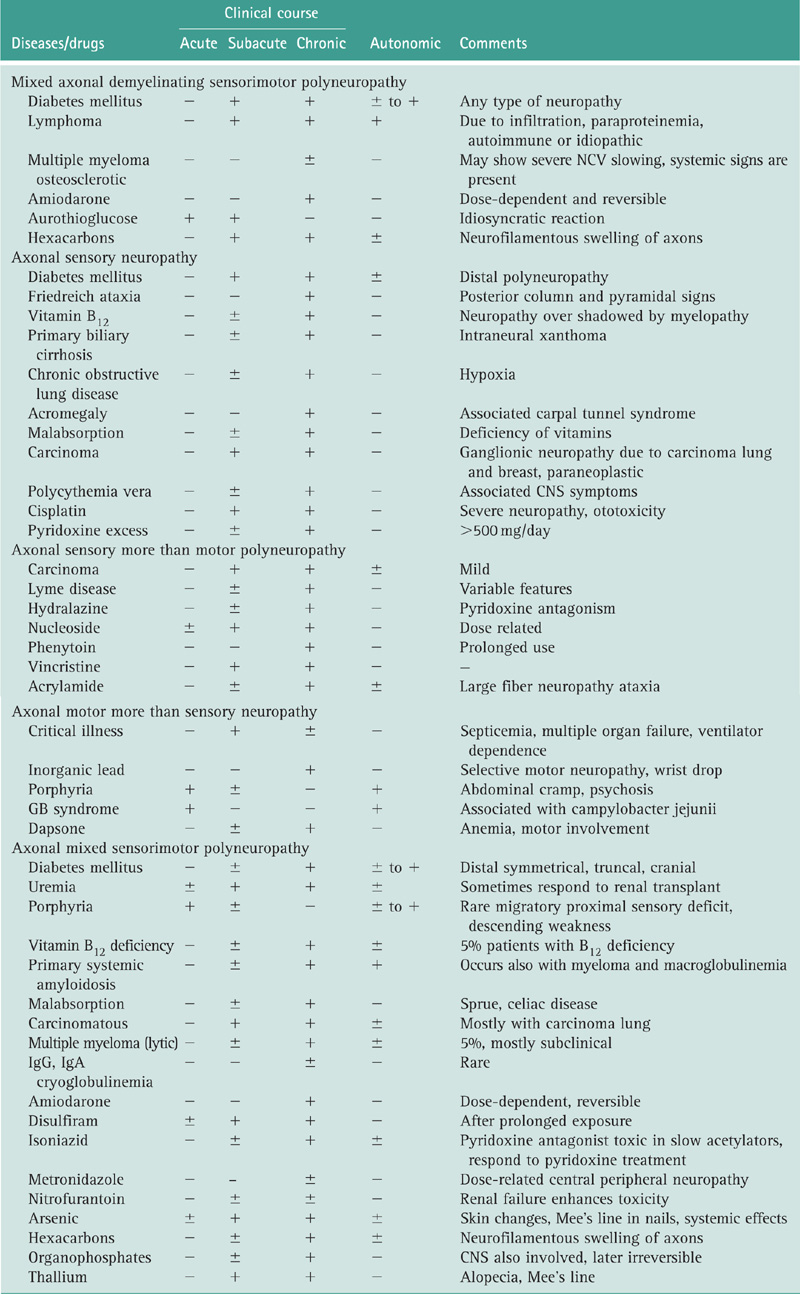
AAN. Consensus Report. Report and recommendations of the San Antonio Conference on diabetic neuropathy. American diabetes Association. American Academy of Neurology. 1988;11:592-597. Diabetes Care
Albers JW. AAEM Case report #4. Guillain Barre Syndrome. Muscle Nerve. 1989;12:705.
Albers JW, Donofrio PD, McGonagle TK. Sequential electrodiagnostic abnormalities in acute inflammatory demyelinating polyradiculoneuropathy. Muscle Nerve. 1985;8:5128.
Albers JW, Kelly JJ. Acquired inflammatory demyelinating polyneuropathies: clinical and electrodiagnostic features. Muscle Nerve. 1989;12:435.
Albers JW, Robertson WC, Daube JR. Electrodiagnostic findings in acute porphyric neuropathy. Muscle Nerve. 1978;1:292.
Ad Hoc Subcommittee of the American Academy of Neurology AIDS Task Force. Criteria for diagnosis of chronic inflammatory demyelinating polyneuropathy (CIDP). Neurology. 1991;41:617.
Albert DA, Rimon D, Silverstein MD. The diagnosis of polyarteritis nodosa: a literature based decision analysis approach. Arthritis Rheumatoid. 1988;31:1117.
Arezzo JC. The use of electrophysiology for the assessment of diabetic neuropathy. Neurosci Res Comm. 1997;21:13.
Arnason BGW. Acute inflammatory demyelinating polyradiculoneuropathy. In: Dyck PJ, Thomas PK, Lambert EH, Bunge R, eds. Peripheral Neuropathy. Philadelphia: WB Saunders; 1984:2050. Vol. II
Asbury AK. Proximal diabetic neuropathy. Ann Neurol. 1977;2:179-180.
Asbury AK, Amason BG, Karp KR, et al. Criteria for diagnosis of Guillain Barre Syndrome. Ann Neurol. 1978;3:565.
Badenoch J, Bedford PD, Evans JR. Massive diverticulosis of the small intestine with steatorrhea and megaloblastic anemia. Q J Med. 1955;24:321.
Bailey RO, Baltch AL, Venkatesh R, et al. Sensory motor neuropathy associated with AIDS. Neurology. 1988;38:886.
Balagtas-Balmaseda OM, Grabois M, Balmaseda PF, et al. Cubital tunnel syndrome in rheumatoid arthritis. Arch Phys Med Rehabil. 1983;64:163.
Bansal R, Kalita J, Misra UK. Pattern of sensory conduction in Guillain–Barre syndrome. Electromyogr Clin Neurophysiol. 2001;41:433.
Bedlack RS, Wu T, Hammons S, et al. MINGE neuropathy: five cases mimicking chronic inflammatory demyelinating polyneuropathy. Muscle Nerve. 2004;29:364.
Behse F, Buchthal F, Carlsen F. Nerve biopsy and conduction studies in diabetic neuropathy. J Neurol Neurosurg Psychiat. 1977;40:1072.
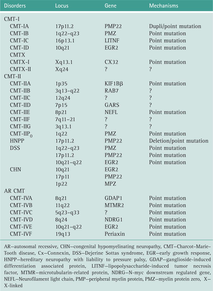
Ben Hamida C, Doerfinger N, Blel S, et al. Localisation of Friedrich’s ataxia phenotype with selective vitamin E deficiency to chromosome 8q by homozygosity mapping. Nature Genetics. 1993;5:195.
Benstead TJ, Kuntz NL, Miller RG, et al. The electro physiologic profile of Dejerine Sotta’s disease (HMSN III). Muscle Nerve. 1990;13:586.
Berciano J, Combarros O, Callija J, et al. The application of nerve conduction and clinical studies to genetic counselling in hereditary motor and sensory neuropathy type I. Muscle Nerve. 1984;12:302.
Berek K, Margreiter J, Willeit J, et al. Polyneuropathies in critically ill patients: a prospective evaluation. Intensive Care Med. 1996;22:849.
Bolton CF, Gilbert JJ, Hahn AF, et al. Polyneuropathy in critically ill patients. J Neurol Neurosurg Psychiat. 1984;87:1223.
Bolton CF. Clinical neurophysiology of the respiratory system. AAEM minimonograph #40. Muscle Nerve. 1993;16:809.
Bolton CF. Critical illness polyneuropathy. In: Asbury AK, Thomas PK, eds. Peripheral Nerve Disorders. Oxford: Butterworth Heinemann; 1995:262. Vol. 2
Bolton CF. Peripheral neuropathies associated with chronic renal failure. Can J Neurol Sci. 1980;7:89.
Bouche P, Leger JM, Travers MA. Peripheral neuropathy in systemic vasculitis: clinical and electrophysiologic study of 22 patients. Neurology. 1986;36:1598.
Bradley WG, Verma A. Painful vasculitic neuropathy in HIV-I infection: relief of pain by prednisone therapy. Neurology. 1996;47:1446.
Breuer AC. Critical care neuropathy an outdated concept. Muscle Nerve. 1999;22:419.
Briemberg H, Levin KH, Amato AA. Multifocal conduction block in peripheral nerve vasculitis. J Clin Neuromuscl Dis. 2002;3:1353-1358.
Britt RP, Harper C, Spray GH. Megaloblastic anemia among Indians in Britain. Q J Med. 1971;40:499.
Brittand ST, Young RJ, Sharma AK, et al. Acute and remitting diabetic polyneuropathy: a comparison of peripheral nerve fibre pathology. Pain. 1992;48:361.
Bromberg MB, Albers JW. Patterns of sensory nerve conduction abnormalities in demyelinating and axonal peripheral nerve disorders. Muscle Nerve. 1993;16:262.
Brown MJ, Greene DA. Diabetic neuropathy: pathophysiology and management. In: Asbury AK, Gilliatt RW, eds. Peripheral Nerve Disorders. London: Butterworth; 1984:126.
Brown MJ, Pleasure DE, Asbury AK. Painful diabetic neuropathy. a morphometric study. Arch Neurol. 1976;33:164.
Brown WF, Feasby TE. Conduction block and Guillain Barre polyneuropathy. Brain. 1984;107:219.
Brown WF, Snow R. Patterns and severity of conduction abnormalities in Guillain Barre syndrome. J Neurol Neurosurg Psychiat. 1991;54:768.
Bruggen JP, Vander Meche FGA, de Gager AEJ, et al. Ophthalmoplegic and lower cranial nerve variants merge into each other and into classical Guillain–Barre syndrome. Muscle Nerve. 1998;21:239.
Bruyn GW, Garland H. Neuropathies of endocrine origin. In: Vinken PJ, Bruyn GW, eds. Handbook of Clinical Neurology. Amsterdam: North Holland; 1970:29.
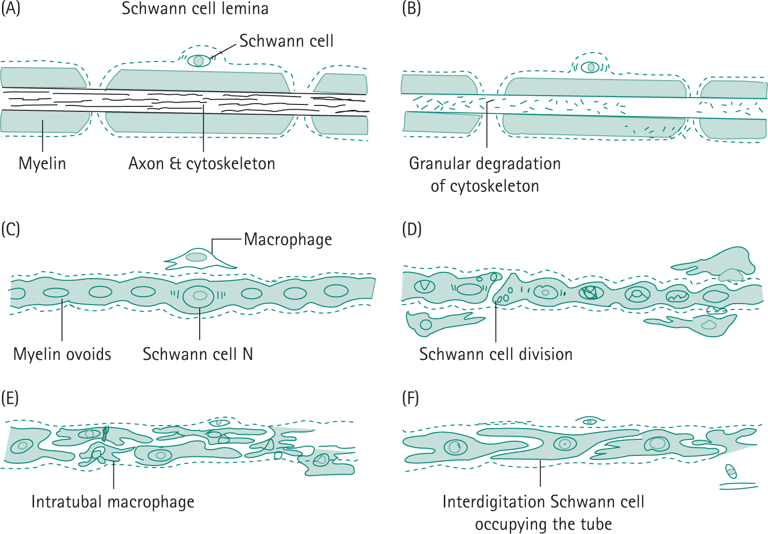
Bunge R, GritIin JW. The cell of Schwann. In: Asbury AK, Mckhann GM, McDonald WI, eds. Diseases of the Nervous System: Clinical Neurology. Philadelphia: WB Saunders; 1992:87.
Caselli RJ, Daube JR, Hunder GG, et al. Peripheral neuropathic syndrome in giant cell (temporal) arteritis. Neurology. 1988;38:685.
CaValier SJ, Gambetti P. Dystrophic axons and spinal cord demyelination in cystic fibrosis. Neurology. 1998;31:714.
Chaco J, Magora A, Jauberman H, et al. An electromyographic study of lagophthalmos in leprosy. Int J Lepr. 1968;36:288.
Charosky CB, Gatti JC, Cardama JE. Neuropathies in Hansen’s disease. Int J Lepr. 1983;51:576.
Chassande B, Leger JM, Younes-Chennoufi AB, et al. Peripheral neuropathy associated with IgM monoclonal gammopathy correlations between clinical, electrophysiological features in 40 cases. Muscle Nerve. 1998;21:55.
Chimelli L, Scaravilli F. Morphological changes in autonomic nervous system of patients with AIDS (abstract). Proceedings of the Seventh International Conference on Neuroscience of HIV Infection. Italy: Pandova; 1991. 89
Chopra JS, Fannin T. Pathology of diabetic neuropathy. J Pathol. 1971;104:175.
Chopra S, Hurwitz LJ, Montogomery DAD. The pathogenesis of sural nerve in diabetes mellitus. Brain. 1969;92:391.
Cohen RD, Conn DL, Ilstrup DM. The clinical features and response to treatment in polyarteritis. Mayo Clin Proc. 1980;55:146.
Conn DL, Dyck PI. Angiopathic neuropathy in connective tissue diseases. In: Dyck PJ, Thomas PK, Lambert EH, eds, et al. Peripheral Neuropathy. Philadelphia: Saunders; 1984:2027. Vol. 2.
Conn DL, McDuffie FC, Dyck PJ. Immunopathological study of sural nerves in rheumatoid arthritis. Arthritis Rheum. 1972;15:135.
Cornblath DR. Treatment of neuromuscular complications of human immuno deficiency virus infection. Ann Neurol. 1988;23:(S)88.
Cornblath DR. Electrophysiology in Guillain Barre syndrome. Ann Neurol. 1990;27:S17.
Cornblath DR, McArthur JC. Predominantly sensory neuropathy in patients with AIDS and AIDS related complex. Neurology. 1988;38:794.
Cornblath DR, Mellitis ED, Griffin JW, et al. The Guillian Barre syndrome study group, motor conduction studies in Guillain Barre syndrome: description and prognostic value. Ann Neurol. 1988;23:354.
Croft PB, Eurich H, Wilkinson M. Peripheral neuropathy of sensorimotor type associated with malignant disease. Brain. 1967;90:31.
Cruickshank B. The arteritis of rheumatoid arthritis. Ann Rheum Dis. 1954;13:136.
Dalakas MC, Pezesckpour GH. Neuromuscular diseases associated with human immunodeficiency virus infection. Ann Neurol. 1988;23:(S)38.
Dastur DK, Anita NH, Divekar SC. The facial nerve in leprosy II: pathology, pathogenesis, electromyography and clinical correlation. Int J Lepr. 1968;34:118.
Dastur DK. Cutaneous nerve in leprosy: the relationship between histology and cutaneous sensibility. Brain. 1955;78(6):1-3.
Davies L, Spies JM, Pollard ID, et al. Vasculitis confined to peripheral nerves. Brain. 1996;II:1441.
de Gans J, Portegies P, Tiessens G, et al. Therapy for cytomegalo virus polyradiculopathy in patients with AIDS. Treatment with ganciclovir. AIDS. 1990;4:421.
De Jong JGY. Over families met hereditary disposite tot het optreten van neuritiden, gecorreleard met migraine. Psychiat Neurol BI. 1947;50:60.
De Silva KL, Pearce J. Neurology of metachromatic leucodystrophy. J Neurol Neurosurg Psychiat. 1973;36:30.
Divekar SC. Electrodiagnostic studies in leprosy. In: Antia NH, Dastur DK, eds. Symposium on Leprosy (University of Bombay). Bombay: JJ Group of Hospitals; 1965:72. February–March
Donofrio PD, Albers JW. AAEM mini monograph #34: Polyneuropathy: classification by nerve conduction studies and electromyography. Muscle Nerve. 1990;13:889.
DuBois DC, Almon RR. A possible role for glucocorticoid in denervation atrophy. Muscle Nerve. 1981;4:370.
Dulbourg O, Mouton P, Brice A, et al. Guidelines for diagnosis of hereditary neuropathy with liability to pressure palsies. Neuromuscul Disord. 2000;10:206.
Dutta AK, Mandal SB, Jopling WH. Surface temperature bald and hairy scalp in reference to leprosy affection. Ind J Dermatol. 1983;28:1.
Dyck PJ. Detection, characterization and staging of polyneuropathy: assessed in diabetes. Muscle Nerve. 1988;11:21.
Dyck PJ, Benstead TJ, Conn DL, et al. Nonsystemic vasculitic neuropathy. Brain. 1987;110:843.
Dyck PJ, Chance PF, Lebo RV, et al. Hereditary motor and sensory neuropathies. In: Dyck PJ, Thomas PK, Griffin JW, eds, et al. Peripheral Neuropathy. Philadelphia: WB Saunders; 1993.
Dyck PJ, Conn DL, Okazaki H. Necrotizing angiopathic neuropathy. Mayo Clin Proc. 1972;47:461.
Dyck PJ, Johnson WJ, Lambert EH, et al. Segmental demyelination secondary to axonal degeneration in uremic neuropathy. Mayo Clin Proc. 1971;46:400.
Dyck PJ, Karies JL, Lambert EH. Longitudinal study of neuropathic deficits and nerve conduction abnormalities in hereditary motor and sensory neuropathy type I. Neurology. 1989;39:1302.
Dyck PJ, Lambert EH. Lower motor and primary sensory neuron diseases with peroneal muscular atrophy (i) neurologic, genetic and electrophysiologic findings in hereditary polyneuropathies (ii) neurologic, genetic and electrophysiologic findings in various neuronal degenerations. Arch Neurol. 1968;18:603.
Dyck PJB, Sinnreich M. Diabetic neuropathies. Continuum. 2003;9:19.
Dyck PJB, Winderbark AJ. Diabetic and non diabetic lumbosacral radiculoplexus neuropathies. New insights into pathophysiology and treatment. Muscle Nerve. 2002;25:477.
Evenhouse M, Hans E, Snell E, et al. Hypotension in infection with the human immune deficiency virus. Ann Int Med. 1987;107:598.
Farrel DA, Medsger TA. Trigeminal neuropathy in progressive systemic sclerosis. Ann J Med. 1982;73:57.
Fisher M. An unusual variant of acute idiopathic polyneuritis (syndrome of ophthalmoplegia, ataxia and areflexia). N Engl J Med. 1956;255:57.
Friedman SA, Schulman RH, Weiss S. Hearing and diabetic neuropathy. Arch Int Med. 1975;135:573.
Frohnert PP, Sheps SG. Long term followup study of polyarteritis nodosa. Am J Med. 1967;43:8.
Fross RD, Daube JR. Neuropathy in the Miller Fisher syndrome: clinical and electrophysiologic findings. Neurology. 1987;37:1493.
Fuller GN, Jacoba JM, Guiloff RJ. Subclinical peripheral nerve involvement in AIDS. An electrophysiological and pathological study. J Neurol Neurosurg Psychiat. 1991;54:318.
Gheradi R, Lebargy F, Gaulard P, et al. Necrotising vasculitis and HIV replication in peripheral nerves. N Engl J Med. 1989;321:685.
Gherardi RK, Chretien F, Delfau-Larue MH, et al. Neuropathy in diffuse infiltrative lymphocytosis syndrome. An HIV neuropathy not a lymphoma. Neurology. 1998;50:1041.
Gilliatt RW, Goodman HV, Willison RG. The recording of lateral popliteal nerve action potential in man. J Neurol Neurosurg Psychiat. 1961;24:305.
Gilliatt RW. Nerve conduction in human and experimental neuropathies. Proc R Soc Med. 1966;58:989.
Gocke OJ, HSu K, Morgan C, et al. Association between polyarteritis and Australia antigen. Lancet. 1970.1149.
Gooch JL, Suchyta MR, Balbierz JM, et al. Prolonged paralysis after treatment with neuromuscular blocking drugs. Crit Care Med. 1991;19:1125.
Good AE, Christopher RP, Koepke GH, et al. Peripheral neuropathy associated with rheumatoid arthritis. Ann Intern Med. 1965;63:87.
Gordon PH, Wilbourn AJ. Early electrodiagnostic findings in Guillain-Barré syndrome. Arch Neurol. 2001;58:913.
Gorson KC, Ropper AH, Adelman LS, et al. Influence of diabetes mellitus on chronic inflammatory demyelinating polyneuropathy. Muscle Nerve. 2000;23:37.
Griffin JW, Cornblath DR, Alexander E, et al. Ataxic sensory neuropathy and dorsal root ganglionitis associated with Sjögren’s syndrome. Ann Neurol. 1990;23:304.
Griffin JW, Crawford TO, Tyor WR, et al. Predominantly sensory neuropathy in AIDS. Distal axonal degeneration and unmyelinated fibre loss. Neurology. 1991;41:(S)374.
Griffin JW, Ho TW. The Guillain–Barre syndrome at 75: the campylobacter connection. Ann Neurol. 1993;34:125.
Guillevin L, Dul LTH, Godeu P, et al. Clinical findings and prognosis of polyarteritis nodosa and Churg–Strauss angitis: a study of 165 patients. Brit J Rheumatoid. 1988;27:258.
Guiloff RJ. Peripheral nerve conduction in Miller Fisher syndrome. J Neurol Neurosurg Psychiat. 1977;40:801.
Gupta SK, Taly AB, Suresh TG, et al. Acute idiopathic axonal neuropathy (AIAN): a clinical and electrophysiological observation. Acta Neurol Scand. 1994;89:220.
Hackett ER, Shipley DE, Livengood E. Motor nerve conduction velocity studies in patients with leprosy. Int J Lepr. 1968;36:282.
Hanson P, Dive A, Brucher JM, et al. Acute corticosteroid myopathy in intensive care patients. Muscle Nerve. 1997;20:1371.
Haq RU, Pendlebury WW, Fries TJ, et al. Chronic inflammatory demyelinating polyradiculoneuropathy in diabetic patients. Muscle Nerve. 2003;27:465.
Harding AE. Spinocerebeller degeneration associated with a selective defect of vitamin E absorption. N Engl J Med. 1985;313:32.
Hastings RC, Brand PW, Mansfield ER, et al. Bacterial density in skin in lepromatous leprosy as related to temperature. Leprosy Rev. 1968;39:71.
Hellmann DB, Laing TJ, Petri M, et al. Mononeuritis multiplex: the yield of evaluations for rheumatic diseases. Medicine. 1988;67:145.
Herbison GL, Teng C, Martin JH, et al. Carpal tunnel syndrome in rheumatoid arthritis. Am J Phys Med. 1973;52:68.
Herman KH, Kennedy L. For the GOAL AIC study group. Physician perception of neuropathy in a large diabetes type 2 population (GOAL AIC study) confirms under diagnosis of neuropathy in everyday clinical practice. Diabetologia. 2003;46(S2):A71.
Hoogedijk JE, DeVisser M, Bolhuis PA, et al. Hereditary motor and sensory neuropathy Type 1: clinical and neurophysiological features of the 17p duplication subtype. Muscle nerve. 1994;17:85.
Horwich MS, Cho L, Porro RS, et al. Subacute sensory neuropathy. A remote effect of carcinoma. Ann Neurol. 1977;2:7.
Hughes RAC, Saunders E, Hall S, et al. Subacute idiopathic demyelinating polyradiculopathy. Arch Neurol. 1992;49:612.
Infante J, Garcia A, Combarros O, et al. Diagnostic strategy for familial and sporadic cases of neuropathy associated with 17p 11.1 deletion. Muscle Nerve. 2001;24:1149.
Jacobson J, Smith T, Gaub J, et al. Progressive neurological dysfunction during latent HIV infection. Brit Med J. 1989;299:225.
Jamal GA, Ballantyne JP. The localization of the lesion in patients with acute ophthalmoplegia ataxia and areflexia (Miller Fisher syndrome). Brain. 1988;111:95.
Jardim MR, Antunes SL, Santos AR, et al. Criteria for diagnosis of pure neural leprosy. J Neurol. 2003;250:806.
Judzewitsch RG, Jaspan JB, Polonsky KS, et al. Aldose reductase inhibition improves nerve conduction velocity in diabetic patients. N Engl J Med. 1983;308:119.
Kaji R, Oka N, Tsuji T, et al. Pathological findings at the site of conduction block in multifocal neuropathy. Ann Neurol. 1993;33:152.
Kaku DA, Parry GJ, Malamut A, et al. Nerve conduction studies in Charcot–Marie Tooth poly neuropathy associated with a segmental duplication of chromosome 17. Neurology. 1993;43:1806.
Kalita J, Misra UK, Sharma RK, et al. Femoral and radial neuropathy following vascular access cannulation for hemodialysis. Nephron. 1995;69:362.
Kaplan JG. Neurotoxicity of selected biological toxins. In: Spencer PS, Schaumburg HH, eds. Experimental and Clinical Neurotoxicology. Baltimore: Williams and Wilkins; 1980:631.
Katz J, Barohn R, Kojan S, et al. Axonal multifocal motor neuropathy without conduction block or other features of demyelination. Neurology. 2002;58:615.
Katz J, Wolfe G, Bryan W, et al. Electrophysiological findings in multifocal motor neuropathy. Neurology. 1997;48:700.
Kelly JJ, Kyle RA, Miles JM, et al. The spectrum of peripheral neuropathy in myeloma. Neurology (NY). 1981;31:24.
Kelly JJ, Kyle RA, O’Brien PC, et al. The prevalence of monoclonal gammopathy in peripheral neuropathy. Neurology (NY). 1981;31:1480.
Kennedy WR, Wendelshafer-Crabb G, Johnson T. Quantification of epidermal nerves in diabetic neuropathy. Neurology. 1996;47:1040.
Kissel JT, Slivka AP, Warmolts JR, et al. The clinical spectrum of necrotising angiopathy of the peripheral nervous system. Ann Neurol. 1985;18:251.
Klein CJ, Cunningham JM, Atkinson EJ, et al. The gene for hereditary motor and sensory neuropathy type 2C maps to 12q24. Ann Neurol. 2002;52:S1.
Kobayashi T, Yamanaka T, Jacobs JM, et al. Twitcher mouse an enzymatically athenic model of human globoid cell leucodystrophy (Krabbe disease). Brain Res. 1980;202:479.
Kohn J. Benign paraproteinemias. J Clin Pathol. 1976;8(S6):77.
Krarup C, Stuwart JD, Sumner AJ, et al. A syndrome of asymmetric limb weakness with motor conduction block. Neurology. 1990;40:118.
Krendel DA, Gilchrist JM, Johnson A, et al. Isolated deficiency of vitamin E with progressive neurological deterioration. Neurology. 1990;40:118.
Krendel DA, Lostigan DA, Hopkins LC. Successful treatment of neuropathy in patients with diabetes mellitus. Arch Neurol. 1995;52:1053.
Kumar A, Dalela D, Bhandari M, et al. Femoral neuropathy—an unusual complication of renal transplantation. Transplantation. 1991;51(6):1305.
Kurdi A, Abdul Kadier M. Clinical and electrophysiological studies of diphtheric neuritis in Jordan. J Neurol Sci. 1979;42:243.
Kuritsky A, Yerginer YM, Korezyn AD. Peripheral neuropathies in cerebrotendinous xanthomatosis. Neurology (Miniap). 1979;29:880.
Kyle RA, Dyck PJ. Amyloidosis and neuropathy. In: Dyck PJ, Thomas PK, Griffin JW, eds, et al. Peripheral Neuropathy. 3rd ed Philadelphia: WB Saunders; 1993:1294. Vol. II
Kyle RA, Therneau TM, Rajkumar SV, et al. A longterm study of prognosis in monoclonal gammopathy of undetermined significance. N Engl J Med. 2002;346:564.
Lamontagne A, Buchthal F. Electrophysiological studies in diabetic neuropathy. J Neurol Neurosurg Psychiat. 1970;33:442.
Lange DJ, Britton CB, Younger DS, et al. The neuromuscular manifestations of human immunodeficiency virus infection. Arch Neurol. 1988;45:1084.
Lecky BRF, Hughes RAC, Murray NMF. Trigeminal sensory neuropathy. Brain. 1987;110:1463.
Leger JM, Salachas F. Diagnosis of motor neuropathy. Eur J Neurol. 2001;8:201.
Levin KH. Neuropathy of ischemia. Muscle Nerve. 2002;26:435.
Lewis RA, Sumner AJ, Brown MJ, et al. Multifocal demyelinating neuropathy with persistent conduction block. Neurology. 1982;32:958.
Lipkin WI, Parry G, Kiprov D, et al. Inflammatory neuropathy in homosexual men with lymphadenopathy. Neurology. 1985;35:1479.
Logina I, Donaghy M. Diphtheritic polyneuropathy: a clinical study and comparison with Guillain Barre syndrome. J Neurol Neurosurg Psychiat. 1999;67:433.
Logothetis J, Kennedy WR, Ellington A, et al. Cryoglobulinemic neuropathy: incidence and clinical characteristics. Arch Neurol. 1968;19:389.
Logothetis J, Silverstein P, Coc J. Neurologic aspects of Waldenstrom’s macroglobulinemia. Arch Neurol (Chicago). 1960;5:564.
Low P, Dotson R. Symptom treatment of painful neuropathy. JAMA. 1998;280:1863-1864.
Low PA, Walsh JC, Huang CY, et al. The sympathetic nervous system in diabetic neuropathy—a clinical and pathological study. Brain. 1975;98:341.
Lubec D, Meillbacher WC, Fimsterer J, et al. Diagnostic workup in peripheral neuropathy: an analysis of 171 cases. Postgrad Med J. 1999;73:723.
Magora A, Sagher F, Chaco J, et al. An electrodiagnostic study of lower motor unit in leprosy. Int J Lepr. 1965;33:829.
Mah V, Yartavarian LM, Akers MA, et al. Abnormalities of peripheral nerve in patients with human immunodeficiency virus infection. Ann Neural. 1988;24:713.
Marsden CD, Obeso JS, Lang AE. Adrenoleukomyelo neuropathy presenting as spinocerebellar degeneration. Neurology (NY). 1982;32:1031.
Martin MM. Diabetic neuropathy: a clinical study of 150 cases. Brain. 1953;76:594.
Mayer RF. Peripheral nerve function in vitamin B 12 deficiency. Arch Neurol. 1965;B:355.
McCombe PA, Pollard JD, Mcleod JG. Chronic inflammatory demyelinating polyradiculopathy. Brain. 1987;110:1617.
Mcleod JG. Paraneoplastic neuropathies. In: Dyck PJ, Thomas PK, Griffin JW, eds, et al. Peripheral Neuropathy. Philadelphia: WB Saunders; 1993:1583.
McLeod JG. Investigations in peripheral neuropathy. J Neuro Neurosurg Psychiat. 1995;58:274.
McLeod JG, Hargrave JC, Walsh JC, et al. Nerve conduction studies in leprosy. Int J Lepr. 1975;43:21.
McLeod JG, Walsh JC. Neuropathies associated with paraproteinemias and dysproteinemias. In: Dyck PJ, Thomas PK, Lambert EH, eds. Peripheral Neuropathy. Philadelphia: WB Saunders; 1975:1012.
Meer J, Gilliatt RW. Proximal and distal conduction velocity in the motor nerves of patients with hereditary motor and sensory neuropathy (abstract). Muscle Nerve. 1989;12:762.
Mellgren SI, Conn DL, Stevens JC, et al. Peripheral neuropathy in primary Sjögren’s syndrome. Neurology. 1989;39:390.
Mendell JR, Kolkin S, Kissel JT, et al. Evidence for central nervous system demyelination of chronic inflammatory demyelinating polyradiculoneuropathy. Neurology. 1987;37:1291.
Meulstee J, Van der Meche FGA. The Dutch Guillain–Barre Study Group. Electrodiagostic criteria for polyneuropathy and demyelination: application in 135 patients with Guillain–Barre syndrome. J Neurol Neurosurg Psychiatry. 1995;59:482-486.
Meyer P. Anatomische untersuchungen uber diphtheritische Lahmung. Yirchows Arch Pathol Anat Physiol. 1881;85:181.
Miller RG, Parry GJ, Pfaeftl W, et al. The spectrum of peripheral neuropathy associated with ARC and AIDS. Muscle Nerve. 1988;11:857.
Miller RG, Peterson GW, Daube JR, et al. Prognostic value of electrodiagnosis in Guillian Barre syndrome. Muscle Nerve. 1988;11:769.
Miller RG, Storey JR, Grecoc M. Ganciclovir in the treatment of progressive AIDS related polyradiculopathy. Neurology. 1990;40:569.
Millers GD, O’Fallon JR, Tally JN. Plasma cell dyscrasia with polyneuropathy—the spectrum of POEM syndrome. N Engl J Med. 1992;327:1919.
Misra UK, Kalita J, Pandey R. Primary amyloid neuropathy—a case report. Neurol India. 1994;42:32.
Misra UK, Kalita J, Das A. Vitamin B12 deficiency syndrome. Electromyogr Clin Neurophysiol. 2003;43:57.
Molina R, Provost IT, Alexander EL. Peripheral inflammatory vascular disease in Sjögren’s syndrome. Arthritis Rheum. 1985;28:1341.
Monrad-Krohn BH. The neurological aspects of leprosy. Jacob Dybwad: Christiana; 1923.
Moody JF. Electrophysiological investigations in the neurological complications of carcinoma. Brain. 1965;88:1023.
Moore PM, Cupps TR. Neurological complications of vasculitis. Ann Neurol. 1983;14:155.
Morton DL, Itubashi HH, Grimes OF. Nonmetastatic neurological complications of bronchogenic carcinoma. The carcinomatous neuromyopathies. J Thorac Cardiovasc Surg. 1967;5:14.
Mossa A, Dubowitz V. Peripheral neuropathy in Cockayne’s syndrome. Arch Des Child. 1970;45:674.
Mouton R, Tardieu S, Gouider R, et al. Spectrum of clinical and electrophysiologic features in HNP patients with the 17p11.1 deletion. Neurology. 1993;52:1440.
Mulder DW, Bastron JA, Lambert EH. Hyperinsulin neuropathy. Neurology. 1956;6:627.
Mulder DW, Lambert EH, Dastron JA, et al. The neuropathies associated with diabetes. A clinical and electromyographic study of 103 unselected diabetic patients. Neurology. 1961;11:275.
Nakano KK. The entrapment neuropathies of rheumatoid arthritis. Orthop Clin North Ann. 1975;6:837.
Ndiaye-Naiang M, Diagne M, Ndiaye IP, et al. The value of electromyographic studies in leprosy. Acta Leprol (Geneva). 1986;100:51.
Nicolas G, Massonobe T, Le Forestier NN, et al. Proposed revised electrophysiological criteria for chronic inflammatory demyelinating polyradiculoneuropathy. Muscle Nerve. 2002;25:25.
Nielsen VK, Kardel T. Decremental conduction in normal human nerves subjected to ischaemia?. Acta Physiol Scand. 1974;92:249-262.
Noordeen SK. A look at world leprosy. Leprosy Rev. 1991;62:72.
Oh SJ, Chang CW. Conduction block and dispersion in hereditary motor and sensory neuropathy (abstract). Muscle Nerve. 1987;10:656.
Oh SJ, Kim DE, Kuruoglu HR. What is the best diagnostic index of conduction block and temporal dispersion?. Muscle Nerve. 1994;17:489.
Oh SJ, Joy JL, Kuruoglu R. “Chronic sensory demyelinating neuropathy”: chronic inflammatory demyelinating polyneuropathy presenting as a pure sensory neuropathy. J Neurol Neurosurg Psychiat. 1992;53:677.
Olney RK. Neuropathies in connective tissue disease. AAEM mini monograph #38. Muscle Nerve. 1992;15:531.
Ooi WW, Srinivasan J. Leprosy and the peripheral nervous system: basic and clinical aspects. Muscle Nerve. 2004;30:393.
Pakalins A, Drake ME, Barotin RJ, et al. Evoked potentials in chronic inflammatory demyelinating polyneuropathy. Arch Neurol. 1988;45:1014.
Partanen J, Niskanen L, Lehtinen J, et al. Natural history of peripheral neuropathy in patients with non-insulin dependent diabetes mellitus. N Engl J Med. 1995;333:89-94.
Pascoe MK, Low PA, Windebank AJ, et al. Subacute diabetic proximal neuropathy. Mayo Clin Proc. 1997;72:1123.
Pedley JC, Harman OJ, Waudby H, et al. Leprosy in peripheral nerves. Histopathological findings in 119 patients in Nepal. J Neurol Neurosurg Psychiatr. 1980;43:198.
Perry GJ, Clarke S. Pure motor neuropathy with multifocal conduction block masquerading as motor neuron disease. Muscle Nerve. 1985;8:617.
Perry GJ, Comblath DR, Brown MJ. Transient conduction block in acute peripheral nerve ischemia. Muscle Nerve. 1981;4:441.
Pestronk A, Chaudhry V, Feldman EL, et al. Lower motor neuron syndromes defined by patterns of weakness, nerve conduction abnormalities, and high titers of antiglycolipid antibodies. Ann Neurol. 1990;27:316.
Pestronk A, Cornblath DR, Illys AA, et al. Antibodies to GMI ganglioside. Ann Neurol. 1988;24:73.
Pestronk A, Cornblath DR, Ilyas AA, et al. A treatable multifocal motor neuropathy with antibodies to GM1 ganglioside. Ann Neurol. 1988;24:73.
Pfeifer MA, Cook D, Brodsky J, et al. Quantitative evaluation of cardiac parasympathetic activity in normal and diabetic man. Diabetes. 1982;31:339.
Pirat J. Diabetes mellitus and its degenerative complications, a prospective study of 4400 patients observed between 1947 and 1973. Diabetes Care. 1978;1:168. 252
Pourmand R. Evaluation of patients with suspected peripheral neuropathy: do the right thing and not everything. Muscle Nerve. 2002;26:286.
Quattrini C, Jeriorska M, Gawkrodger D, et al. Dermal nerve depletion and angiogenesis in diabetic neuropathy. Diabetologia. 2003;46(S):A72.
Reske Nielson E, Harmsen A, Vorre P. Ultrastructure of muscle biopsies in recent, short term and long term juvenile diabetes. Acta Neurol Scand. 1977;55:345.
Richard KO, Richard AL, Timothy DP, et al. Consensus criteria for the diagnosis of multifocal motor neuropathy. Muscle Nerve. 2003;27:117.
Ricoy JR, Cabello A, Rodriguez J, et al. Neuropathological studies related to the toxic syndrome related to adulterated rapeseed oil in Spain. Brain. 1983;106:817.
Ridley A. The neuropathy of acute intermittent porphyria. Q J Med. 1969;28:307.
Roelefs RI, Hrusheshy W, Rogin J. Peripheral sensory neuropathy and cisplatin chemotherapy. Neurology. 1984;34:934.
Ropert A, Metral S. Conduction block in neuropathies with necrotizing vasculitis. Muscle Nerve. 1990;13:102.
Ropper AH, Wijdicks EEM, Shahani BT. Electrodiagnostic abnormalities in 113 consecutive patients with Guillain–Barre syndrome. Arch Neurol. 1990;47:881-887.
Rosenberg RN, Lovelace RE. Mononeuvitis multiplex in lepromatous leprosy. Arch Neurol. 1969;20:257.
Rousseau LL, Frank G, Grisar T, et al. Osteosclerotic myeloma with polyneuropathy and ectopic secretion of calcitonin. Cancer. 1978;14:133.
Roy A, Kalita J, Gayathri N, et al. Reversible neuropathy in chronic renal failure. Nephron. 1998;80:63.
Roy EP, Gutmann L, Riggs JE. Longitudinal conduction studies in hereditary motor and sensory neuropathy type. Muscle Nerve. 1989;12:52.
Sack GH Jr. Acute intermittent porphyria. JAMA. 1990;264:1290.
Said G. A clinicopathologic study of acrodystrophic neuropathies. Muscle Nerve. 1980;3:491.
Said G, Lacroix-Ciaudo C, Fujimura H, et al. The peripheral neuropathy of necrotising arteritis: a clinicopathological study. Ann Neurol. 1988;23:46J.
Saito M, Hayashi Y, Suzuki T, et al. Linkage mapping of the gene for Charcot-Marie-Tooth disease type 2 to CMT2A. Neurology. 1997;49:1630.
Schaumburg HH, Spencer PS. Clinical and experimental studies of distal axonopathy: a frequent form of brain and nerve damage produced by environmental chemical hazards. Ann NY Acad Sci. 1979;329:14.
Schaumburg HH, Caplan J, Windebank A, et al. Sensory neuropathy from pyridoxine abuse: a new megavitamin syndrome. New Engl J Med. 1983;309:445.
Schaumburg HH, Kaplan JQ. Toxic neuropathies. In: Asbury AK, Thomas PK, eds. Peripheral Nerve Disorders. Oxford: Butterworth Heinnemann; 1995:238.
Schaumburg HH. Occupational environmental and biologic agents. In: Schaumburg HH, Berger AR, Thomas PK, eds. Disorder of Peripheral Nerves. Philadelphia: FA Davis; 1991:274.
Scott DGL, Bacon PA, Tribe CR. Systemic rheumatoid vasculitis: a clinical and laboratory study of 50 cases. Medicine. 1981;60:288.
Sebille A. Respective importance of different nerve conduction velocities in leprosy. J Neurol Sci. 1978;38:89.
Selikoff IJ, Robitzek EH, Ornstin GG. Treatment of pulmonary tuberculosis with hydrazine derivatives of isonicotinic acid. JAMA. 1952;150:251.
Sellman MS, Mayer RF. Conduction block in hereditary neuropathy with susceptibility to pressure palsies. Muscle Nerve. 1987;10:621.
Sharma K, Lross J, Farronay O, et al. Demyelinating neuropathy in diabetes mellitus. Arch Neurol. 2002;59:758.
Shy ME. Inherited peripheral neuropathies. Continum. 2003;9:117.
Sidiq M, Kirsner AB, Sheon RP. Carpal tunnel syndrome. First manifestation of the systemic lupus erythematosus. JAMA. 1972;222:1416.
Singleton JR, Smith AG, Bromberg MB. Painful sensory polyneuropathy associated with impaired glucose tolerance. Muscle Nerve. 2001;24:1225-1228.
Skillman TG, Johnson EW, Hamwi GJ, et al. Motor nerve conduction velocity in diabetes mellitus. Diabetes. 1961;10:46.
Snider WD, Simpson DM, Nielsen S, et al. Neurological complications of acquired immune deficiency syndrome: analysis of 50 patients. Ann Neurol. 1983;14:403.
So YT, Engstrom JW, Olney RK. The spectrum of electrodiagnostic abnormalities in patients with human immunodeficiency virus infection. Muscle Nerve. 1990;13:855.
Sokol RJ, Kaydent H, Bettis DB, et al. Isolated vitamin deficiency in the absence of fat malabsorption familial and sporadic cases: characterisation and investigation of causes. J Lab Clin Med. 1988;111:548.
Sokoloff L, Wilens SL, Bunim LL. Arteritis of striated muscle in rheumatoid arthritis. Am J Pathol. 1951;27:157.
Solders G, Nennesmo I, Persson A. Diptheritic neuropathy: an analysis based on muscle and nerve biopsy and repealed neurophysiological and autonomic function test. J Neurol Neurosurg Psychiat. 1989;52:876.
Sorensen AWS, With TK. Persistent paresis after prophyric attacks. S Afr Med J. 1971;45(special issue):101.
Stewart PM, Hersley WJ. An acute attack of variegate porphyria complicated by severe autonomic neuropathy. Aust NZ Med. 1981;11:82.
Sumner CJ, Sheth S, Griffin JW, et al. The spectrum of neuropathy in diabetes and impaired glucose tolerance. Neurology. 2003;60:108.
Swift TR, Hackett ER, Shipley DE, et al. The peroneal and tibial nerves in lepromatous leprosy. Clinical and lectrophysiologic observations. Int J Lepr Other Mycobact Dis. 1973;41:25.
Tesfaye S, Malik R, Harris N, et al. Arterio venous shunting and proliferating new vessels in acute painful neuropathy of rapid glycemic control (insulin neuritis). Diabetologia. 1996;39:329-335.
Thomas PK. Central and peripheral demyelination. In: Serratrice G, Pellisiel JF, Desnuelle D, eds, et al. Myelopathies. Paris expansion scientifique Francaise: Neuropathies et Myopathy; 1989:14. I
Thomas PK. Diabetic neuropathy. In: Kin H, Jarrett J, eds. Complications of Diabetes. London: Edward Arnold; 1982.
Thomas PK. Understanding neuropathies: the impact of ancillary investigations. Baitheres Clin Neurol. 1996;5:157.
Thomas PK, Eliasson SG. Diabetic neuropathy. In: Dyck PJ, Thomas PK, Lambert EH, eds, et al. Peripheral Neuropathy. Philadelphia: WB Saunders; 1984:1773.
Thomas PK, King RHM, Koeen RS, et al. Comparative ultrastructural observations on peripheral nerve abnormalities in the late infantile, juvenile and late onset forms of metachromatic. I. Eucodystrophies. Acta Neuropathol (Berl). 1977;39:237.
Thomas PK, Lascelles RG. The pathology of diabetic neuropathy. Q J Med. 1966;35:489.
Thorton CA, Latif AS, Emmanuel JC. Guillain Barre syndrome associated with human immunodeficiency virus infection in Zimbabwe. Neurology. 1991;41:812.
Travers RL, Allison DJ, Brettle RP, et al. Polyarteritis nodosa: a clinical and angiopathic analysis of 17 cases. Semin Arthritis Rheum. 1979;8:184.
Uncini A, De Angelis MV, Di Muzio A, et al. Chronic inflammatory demyelinating polyneuropathy in diabetics: motor conductions are important in the differential diagnosis with diabetic polyneuropathy. Clin Neurophysiol. 1999;110:705.
Uncini A, DiGuglielmo G, DiMuzio A, et al. Differential electrophysiological features of neuropathy associated with 17p11.2 deletion and duplication. Muscle Nerve. 1995;18:628.
Uplekar MW, Antia NH. Clinical and histopathological observations on pure neuritic leprosy. Ind J Lepr. 1986;58:513.
Van der Meche FGA, Meulstee J, Kleyweg RP. Current diagnostic criteria for Guillain–Barre syndrome. Ann Neurol. 1991;30:851-852.
Van der Meche FGA, Meulstee J. Guillain–Barre syndrome, a model for random conduction block. J Neurol Neurosurg Psychiat. 1988;55:1158.
Van Heel DA, Levitt NS, Winter TA. Diabetic neuropathic cachexia—the importance of positive recognition and early nutritional support. Int J Clin Pract. 1998;52:591.
Van Heel DA, Levitt NS, Winter TA. Diabetic neuropathic cachexia—the importance of positive recognition and early nutritional support. Int J Clin Pract. 1998;52:591-592.
Veith G. Untersuchnger uber die histologie der polyneuritis diptheria. Beitr Pathol Anat. 1949;110:567.
Verghese M, Ithiamani KV, Satyanarayan KR, et al. A study of conduction velocity of ulnar and median nerves in leprosy. Int J Lepr. 1970;38:271.
Vital C, Vallat JM. Ultrastructural Study of Human Diseased Peripheral Nerve. New York: Masson; 1980. 76
Vital A, Vital C. Polyarteritis nodosa and peripheral neuropathy. Acta Neuropathol (Berlin). 1985;71:136.
Waldinger TP, Siegle RJ, Weber W, Voorhees JJ. Dapsone induced peripheral neuropathy case report and review. Arch Dermatol. 1984;120:356.
Walsh JC. Neuropathy of multiple myeloma. Arch Neurol. 1971;25:404.
Ward JD, Barnes CG, Fisher DJ, et al. Improvement in nerve conduction following treatment in newly diagnosed diabetes. Lancet. 1971;1:428.
Weber F, Ziegler A. Axonal neuropathy in chronic peripheral arterial occlusive disease. Muscle Nerve. 2002;26:47.
Weiss JA, White JC. Correlation of a 1a afferent conduction with the ataxia of Fisher syndrome. Muscle Nerve. 1986;9:327.
Widenbank AJ. Metal Neuropathy. In: Dyck PJ, Thomas PK, Griffin JW, eds, et al. Peripheral Neuropathy. Philadelphia: WB Saunders; 1993:1549.
Wilbourn AJ. Serial conduction studies in human nerve degeneration. Electroencephalogr Clin Neurophysiol. 1977;43:616.
Wilbourn AJ, Furlan AJ, Hulley W, Ruschhaupt W. Ischemic monomelic neuropathy. Neurology. 1983;33:447.
Willison HJ, Veitch J, Patterson G, et al. Miller Fisher syndrome is associated with serum antibodies to GQ1b ganglioside. J Neurol Neurosurg Psychiat. 1993;56:204.
Wilt NJ, Zochodne DW, Bolton CF, et al. Peripheral nerve functions in sepsis and multiple organ failure. Chest. 1991;99:176.
Winkelman RK, Lambert EH, Hayles AB. Congenital absence of pain report of a case and experimental studies. Arch Dermatol. 1962;85:325.
Yao JK, Herbert PN. Lipoprotein deficiency and neuromuscular manifestations. In: Dyck PJ, Thomas PK, Griffin JW, Low PA, Poduslo JF, eds. Peripheral Neuropathy. 3rd ed. Philadelphia: WB Saunders; 1993:1179. Vol. II
Yuki N. Infectious origins of and molecular mimicry in Guillain–Barre and Fisher syndromes. Lancet Infect Dis. 2001;1:29.
Zochodne DW, Bolton CF, Wells, et al. Critical illness polyneuropathy: a complication of sepsis and multiple organ failure. Brain. 1987;110:819.