THE ARCHITECTURE AND DESIGN OF HUMAN BODY
After studying this chapter, the student should be able to:
 classify humans in the Animal Kingdom
write the characteristic features of humans
state the levels of structural organization of the human body
identify the various regions of the human body
outline the body cavities and their contents
notify the disposition of body structures encountered during dissection
correctly solve the review questions provided at the end of the chapter
INTRODUCTION
The human body is complex like a highly technical and sophisticated machine. Therefore the knowledge of its organization is essential to comprehend.
Before beginning to study the organization of human body one should know the taxonomic position of humans in the Animal Kingdom because we share many characteristics with all living animals. Like all other living animals we breathe, eat and digest food, excrete body wastes, locomote and reproduce our own kind. We are subject to disease, injury, pain, aging, mutations and death. The processes by which our bodies produce, store, and utilize energy are also similar to those found in all living organisms. The fundamental patterns of development and growth of many nonhuman animals are also seen in human development.
In the scheme of classification of Animal Kingdom established by biologists to organize the structural and evolutionary relationship of living organisms, each category of classification is referred to as a taxon. The highest taxon is the Kingdom and the most specific category is Species. Human beings are species belonging to the Animal Kingdom.
Phylogeny is the origin and evolutionary development of animal species.
CLASSIFICATION OF HUMANS IN THE ANIMAL KINGDOM
Human beings are biological creatures that are classified into eight main taxonomic groupings.
The scientific classification of human beings and the characteristic features of each group are as follows:
| Group | Features |
| Kingdom: Animalia | Eukaryotic cells that lack walls and photosynthetic pigments |
| Phylum: Chordata | Presence of notochord and pharyngeal pouches |
| Subphylum: Vertebrata | Presence of vertebral column |
| Class: Mammalia | Presence of mammary glands and hair |
| Order: Primata | Presence of well developed brain and prehensile hands |
| Family: Hominidae | Large cerebrum, bipedal locomotion |
| Genus: Homo | Flattened face, prominent chin and nose with inferiorly directed nostrils |
| Species: sapiens | Largest cerebrum |
CHARACTERISTICS OF HUMANS
Human beings possess certain anatomical features which separate them from other animals. These are as follows:
1. A large well developed brain: The average adult human brain weighs between 1350 and 1400g. This gives humans a large brain-to-body weight ratio. The human brain accounts for emotions, thought, reasoning, memory and precise coordinated movements.
2. Bipedal locomotion: Human beings stand and walk on two limbs and the upper limbs swing with movements of lower limbs.
3. An opposable thumb: The human thumb is tremendously versatile. It is opposable to fingers, hence adapted to grasp the objects.
4. Well developed articulated speech: Human beings have well developed articulated speech. Hence they can communicate with each other, which led to the development of culture and language.
5. Stereoscopic vision: The human eyes are directed forwards so that when we focus on an object, we view it from two angles. The stereoscopic vision gives us the depth of perception a three-dimensional image.
ORGANIZATION OF THE HUMAN BODY
Within the body there are different levels of structural organization. The five main levels of organization (Fig. 3.1) are:

1. Cellular level: The cell is the structural and functional unit of life. The human is a multicellular organism composed of 60–100 trillion cells. It is at cellular level that metabolism, growth, repair, etc. are carried out. The human body contains many distinct types of cells each of which is specialized to perform specific functions. The examples of specialized cells are: (a) bone cells, (b) muscle cells, (c) fat cells, (d) blood cells, and (e) nerve cells. Each of these cell types has a unique structure related to its function.
2. Tissue level: The tissue is a group of cells with similar structure and function including extracellular substance between them. The examples are: (a) epithelial tissue, (b) connective tissue, (c) muscle tissue, and (d) nervous tissue.
3. Organ level: The organ is composed of two or more types of tissues that are integrated to perform a particular function. The examples of organs are: (a) heart, (b) stomach, (c) spleen, (d) eye, (e) skin, etc.
4. System level: A body system consists of various organs that have similar or related functions. These are 11 major systems in the body, viz.: (a) integumentary, (b) skeletal, (c) muscular, (d) nervous, (e) endocrine, (f) cardiovascular, (g) lymphatic, (h) respiratory, (i) digestive, (j) urinary, and (k) reproductive systems.
5. Organism level: An organism is any living thing considered as a whole whether it is composed of one cell, viz. bacteria or of trillions of cells such as human being.
The human organism is a complex of organ systems which are mutually dependent on each other, interrelated and function together.
BODY REGIONS
The human body is divided into several regions. The major body regions are: head, neck, trunk (trunk is frequently divided into thorax and abdomen), upper extremity, and lower extremity:
1. Head is divided into (a) a facial region, which includes the eyes, nose and the mouth, and (b) cranial region, which encloses brain.
2. Neck is the region that extends between head and trunk and is roughly cylindrical. The neck supports the head and allows it to move.
3. Thorax is the upper part of trunk and commonly referred to as the chest. The thoracic region includes nipples and sexually mature breasts on its front aspect.
4. Abdomen is located below the thorax. It presents navel (umbilicus) in its center on the front.
The lower portion of the abdomen forms the pelvis. The inferior aspect of the trunk between the two thighs is called the perineum.
5. Upper extremity includes shoulder region, arm (brachium), elbow, forearm (antebrachium), wrist and hands, and fingers.
6. Lower extremity includes gluteal region, thigh, knee, legs, ankle, foot, and toes.
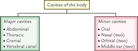
BODY CAVITIES
For functional and protective purposes, the viscera are located within the cavities of the body. The body cavities contain organs and systems that have related functions.
The major cavities of the body (Fig. 3.2) are as follows:

1. Abdominal cavity: It is the largest cavity in the body. It is divided into large upper portion called abdominal cavity proper and small lower portion called pelvic cavity.
Abdominal cavity proper: It is oval in shape and situated in the main part of the trunk. It is bounded superiorly by diaphragm which separates it from thoracic cavity, anteriorly by muscles forming the anterior abdominal wall, posteriorly by lumbar vertebrae and muscles forming the posterior abdominal wall, laterally by lower ribs and parts of the muscles of the abdominal wall, and inferiorly it is continuous with the pelvic cavity.
The abdominal cavity proper contains organs and glands involved in digestion and absorption of the food:
(a) Stomach, small intestine, and most of large intestine
Pelvic cavity: It is roughly funnel shaped and extends from the lower end of the abdominal cavity. It is bounded anteriorly by pubic bones, posteriorly by sacrum and coccyx, laterally on either side by innominate bone, inferiorly by muscles of pelvic floor, and superiorly it is continuous with the abdominal cavity.
(a) Terminal portion of large intestine, i.e. sigmoid colon, rectum and anus
2. Thoracic cavity: This is situated in the upper part of the trunk. It is bounded anteriorly by sternum and costal cartilages of the ribs, laterally by 12 pairs of ribs and intercostal muscles, posteriorly by thoracic vertebrae and intervertebral discs between the bodies of the vertebrae.
3. Cranial cavity: It is the cavity within the skull and therefore bounded by the bones of the skull as follows: anteriorly by frontal bone, posteriorly by occipital bone, laterally on each side by temporal bone, superiorly by parietal bones and inferiorly by sphenoid, ethmoid and parts of frontal, temporal and occipital bones. The cranial cavity contains:
4. Vertebral canal: It is located within the vertebral column. It contains:
The smaller/minor cavities of the body are:
1. Oral (buccal cavity): it contains teeth and tongue
2. Two nasal cavities: these are concerned with respiration and sense of smell
3. Two orbital cavities: each of them houses eyeball and its associated muscles, nerves and vessels
4. Two middle ear cavities: each of them contain ear ossicles
The various cavities of the body are summarized in Flowchart 3.1.
BODY MEMBRANES
The membranes are sheets of epithelial cells and their supporting connective tissue layers. They cover or line the internal structures or cavities. The three main membranes are:
1. Mucous membranes: secrete a thick, viscid substance called mucus which lubricates or protects the associated organs.
They line the various cavities and tubes that enter or exit from the body, viz., oral and nasal cavities, tubes of respiratory, reproductive, urinary and digestive systems.
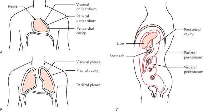
2. Serous membranes: secrete a thin watery fluid called serous fluid which acts as a lubricant. The serous membranes cover the organs with the thoracic and abdominal cavities.
The serous membranes are invaginated by the viscera and become divided into two layers: (a) visceral layer adhering to the outer surface of the organ; and (b) parietal layer lining the wall of the body cavity.
The space between the visceral and parietal layer forms a serous cavity such as pleural cavities, pericardial cavity and peritoneal cavity (the largest serous cavity in the body).
The pericardial cavity surrounds the heart (Fig. 3.3 A), pleural cavity surrounds the lung (Fig. 3.3 B), and peritoneal cavity surrounds the abdominal and pelvic viscera (Fig. 3.3 C).
The space between the visceral and parietal serous membranes (serous cavity) is normally filled with a thin lubricating film of serous fluid produced by the membranes. As organs rub against the body wall or against another organ, the serous fluid and serous membranes reduce the friction.

The serous membranes sometimes become inflamed, usually as a result of infection. The inflammation of pericardium is called pericarditis, that of pleura is called pleuritis and that of peritoneum is called peritonitis. When inflamed, the serous membranes produce an increased amount of serous filled within serous cavities leading to clinical conditions like pericardial effusion (accumulation of fluid in the pericardial cavity), pleural effusion (accumulation of fluid in the pleural cavity), and ascites (accumulation of fluid in the peritoneal cavity).
The important serous membranes and serous cavities of the body are summarized in Table 2.1.
BODY FLUIDS
Above 60% of body weight is formed by fluids present in the body. The body fluid is of two types:
1. Extracellular fluid: accounts for 22% of body weight. The extracellular fluid consists of blood, plasma, lymph, cerebrospinal fluid and fluid in the interstitial spaces of the body.
The interstitial fluid, i.e. intercellular fluid (also called tissue fluid) bathes all the cells of the body except the outer layers of the skin. It is the medium through which substances pass from blood to the body cells, and from cells to the blood.
The well-being of every cell is dependent upon the composition of the intercellular fluid, which is therefore maintained at constant level by many control mechanisms in the body. This is called homeostasis.
2. Intracellular fluid: accounts for 38% of body weight. The composition of intracellular fluid is largely controlled by the cell itself, because there are selective uptake and discharge mechanisms present in the cell membrane.
GENERAL DISPOSITION OF THE BODY STRUCTURES
During dissection, following structures are encountered from superficial to deep.
1. Skin: is the outermost covering of the body
2. Superficial fascia: is made up of loose connective tissue and filled with variable amount of fat in different regions.
3. Deep fascia: is thin tough inelastic fibrous membrane deep to the superficial fascia and superficial to the muscles.
4. Muscles (skeletal muscles): are present deep to the deep fascia as red fleshy masses of different size and shape.
5. Blood vessels and nerves: are present between the muscles embedded in loose connective tissue. When traced distally, they give branches.
6. Bones and joints: bones are hard structures of different sizes and shapes deep to muscles. The junctions between the bones are called joints. The bones along with joints form the skeletal framework of the body.
INTRODUCTION TO STUDY OF ILLNESS
In order to understand the anatomical basis of specific diseases described in later chapters, it is necessary to adopt a systematic approach as outlined below:
1. Etiology: the cause of the disease
2. Pathogenesis: the nature of disease process and its effect on normal body functioning
3. Complications: the other consequences which might arise if the disease progresses
Etiology: The disease is caused by one or more factors. The common factors causing the disease are:
1. Genetic abnormalities, inherited or acquired
2. Infection by microbes or parasites, viz., worms, bacteria or viruses
Pathogenesis: The main processes causing the illness are as follows:
1. Inflammation, the tissue’s response to its damage is due to trauma or invasion by microbes.
2. Tumors, that arise when the rate of cell production exceeds that of normal cell destruction causing a mass to develop.
3. Abnormal immune mechanisms, sometimes the normal protective immune mechanisms of the body may cause undesirable effects.
4. Thrombosis, embolism and infarction, the effects and consequences of abnormal changes in the blood and/or walls of blood vessels.
5. Degeneration, which leads to impaired function.
6. Metabolic abnormalities, such as phenylketonuria cause undesirable effects.
Terminology associated with the disease
• Acute: a disease with sudden onset
• Chronic: a long standing disorder
• Congenital: a disorder which one is born with
• Acquired: a disorder which develops any time after birth
• Symptom: an abnormality or disorder described by the patient
• Sign: an abnormality or disorder noted by the doctor
• Syndrome: a group of signs and symptoms which together constitute a disease
Prognosis: It is a forecast of the probable course and outcome of a disease, e.g. the prognosis of benign tumors is generally good, whereas those of malignant tumor is poor.
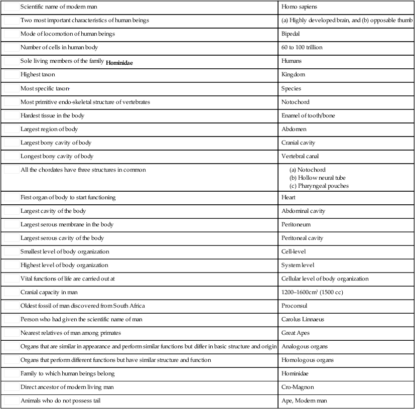
 Scientific name of modern man Scientific name of modern man |
Homo sapiens |
| Two most important characteristics of human beings |
(a) Highly developed brain, and (b) opposable thumb |
| Mode of locomotion of human beings |
Bipedal |
| Number of cells in human body |
60 to 100 trillion |
| Sole living members of the family Hominidae |
Humans |
| Highest taxon |
Kingdom |
| Most specific taxon* |
Species |
| Most primitive endo-skeletal structure of vertebrates |
Notochord |
| Hardest tissue in the body |
Enamel of tooth/bone |
| Largest region of body |
Abdomen |
| Largest bony cavity of body |
Cranial cavity |
| Longest bony cavity of body |
Vertebral canal |
| All the chordates have three structures in common |
|
| First organ of body to start functioning |
Heart |
| Largest cavity of the body |
Abdominal cavity |
| Largest serous membrane in the body |
Peritoneum |
| Largest serous cavity of the body |
Peritoneal cavity |
| Smallest level of body organization |
Cell-level |
| Highest level of body organization |
System level |
| Vital functions of life are carried out at |
Cellular level of body organization |
| Cranial capacity in man |
1200–1600cm3 (1500 cc) |
| Oldest fossil of man discovered from South Africa |
Proconsul |
| Person who had given the scientific name of man |
Carolus Linnaeus |
| Nearest relatives of man among primates |
Great Apes |
| Organs that are similar in appearance and perform similar functions but differ in basic structure and origin |
Analogous organs |
| Organs that perform different functions but have similar structure and function |
Homologous organs |
| Family to which human beings belong |
Hominidae |
| Direct ancestor of modern living man |
Cro-Magnon |
| Animals who do not possess tail |
Ape, Modern man |
*In the classification, scheme established by biologists to organize the structural and evolutionary relationships of organisms, each category of classification is called “taxon”.