Uses of L-Thyroxine
An average replacement dose is 1.6 µg/kg body weight/day for adults, up to 4.0 µg/kg body weight/day for children, and lower doses for older individuals (1.0 µg/kg body weight/day) (Sawin et al, 1983; Davis et al, 1984). The initial dose and the optimal time needed to establish the full replacement dose are dependent upon the age, weight, and cardiac status of each patient. The requirement might increase during pregnancy and in postmenopausal women starting hormonal replacement (Arafah, 2001). A serum TSH between 0.5 and 2.0 µU/mL is the therapeutic goal level for L-T4 replacement in primary hypothyroidism. A serum FT4 concentration in the upper third of the reference interval is the therapeutic target in central hypothyroidism.
TSH should be used to monitor patients receiving thyroid hormone replacement therapy, as well as those treated with hormone, to suppress malignant thyroid diseases (Spencer, 1990). Both TSH and FT4 should be used to monitor hypothyroid patients suspected of intermittent noncompliance. At least 6 weeks is needed before retesting of TSH following a change in the dose of L-T4. Annual TSH measurement is recommended in patients on a steady dose of T4. If FT4 is being assayed, patients should withhold their levothyroxine dose on the day of the test, as serum FT4 will be increased (about 13%) above baseline for 9 hours after the last dose is ingested (Ain et al, 1993); the TSH, however, is unlikely to be affected. Ideally, L-T4 should be taken before meals, at the same time every day and at least 4 hours from any other medications or vitamins/dietary supplements.
L-T4 is used to suppress TSH in patients with well-differentiated thyroid carcinoma, for which thyrotropin is considered a trophic factor (Dulgeroff & Hershman, 1994). It is recommended to use a TSH target of 0.05 to 0.1 µU/mL for low-risk patients and a TSH value <0.1 µU/mL for high-risk patients. If the thyroglobulin level is undetectable and no evidence of recurrence is noted 5 to 10 years after thyroidectomy, the dose of L-T4 can be reduced to give low-normal TSH values (<0.4 µU/mL).
Calcitonin
Medullary thyroid carcinoma (MTC) originates from the C cells of the thyroid. It accounts for 1% to 2% of thyroid cancers and 0.57% of thyroid nodules (Pacini et al, 1994).
Among cases of MTC, 25% are hereditary (multiple endocrine neoplasia types 2A and 2B) (Dunn, 1994; Brandi et al, 2001; Cobin et al, 2001). These are autosomal dominant inherited multiglandular syndromes. An important, recurring MTC genetic mutation is that of the RET oncogene located on the chromosome subband 10q11.2 (Mulligan et al, 1993; Hofstra et al, 1994). The recommended method of initial testing for MEN2A is either a single or multi-tiered analysis to detect RET mutation in exon 10 (codon 609, 611, 618, 620), exon 11 (codon 630, 634), and exons 8, 13, 14, 15, and 16 (Donis-Keller et al, 1993).
The current ATA risk categories for Heriditary MTC are:
• Highest risk (patients with MEN2b AND RET codon M918T mutation (HST)
• High risk (patients with MEN2a and RET codon c634 mutation (H)
• Moderate risk (patients with hereditary MTC and RET codon mutations other than M918T and c634
The C cell secrete several hormones and biogenic amines: calcitonin, ACTH,
β-melanocyte stimulating hormone, somatostatin, chromogranin, histaminase, neurotensin, and carcinoembryonic antigen (CEA) (Abe et al, 1977).
Both calcitonin and CEA are valuable tumor markers for patients with MTC. An elevated level of calcitonin in circulating blood indicates the presence of MTC. Mature calcitonin results from posttranslational modification of a larger 141–amino acid precursor (preprocalcitonin) within the parafollicular C cells. Preprocalcitonin undergoes cleavage of a single peptide to form procalcitonin; the latter has 116 amino acid residues. The immature calcitonin peptide consisting of 33 amino acids is located centrally within the procalcitonin molecule. The mature, active, 32–amino acid calcitonin is produced from immature calcitonin by the enzyme peptidylglycine-amidating monooxidase. Measurement of calcitonin is done by two-site immunometric assays using monoclonal antibodies: One recognizes the N-terminal region, and the other the C-terminal region. This method is more sensitive and more specific (Motte et al, 1988; van Heyningen, 1994; Becker et al, 1996). This method eliminates any cross-reactivity with procalcitonin that can be elevated during sepsis or inflammatory conditions (Becker et al, 2004). The cutoff level in healthy adults is about 10 ng/L, and it is higher in children <3 years of age.
Serum calcitonin measurements are used as tumor markers for detecting residual thyroid tissue or metastasis in patients with MTC. It should be measured before and 6 months after surgery. The presence of residual tissue or a recurrence of MTC can be ruled out only if both basal and postpentagastrin or calcium-stimulated calcitonin are undetectable. Provocative stimuli, such as calcium and pentagastrin (Pg) or omeprazole, have been used to detect C cell abnormalities, as they increase calcitonin levels at all stages of MTC (Wells et al, 1978; Barbot et al, 1994; Gagel, 1996; Erdogan et al, 1997; Wion-Barbot et al, 1997; Vieira et al, 2002; Vitale et al, 2002).
In the Pg stimulation test for the diagnosis of MTC, an intravenous infusion of Pg (0.5 µg/kg body weight) is given over 5 seconds; blood samples are collected at baseline and 1, 2, 5, and 10 minutes after the start of the infusion. Interpretations of results are summarized in Table 24-9.
TABLE 24-9
Interpretation of the Pentagastrin (Pg) Test
| Peak Calcitonin (CT) ng/L (pg/mL) | Interpretation |
| <10 | Normal (80% of adults) |
| >30 but <50 | 5% of normal adults |
| >50 but <100 | Possible MTC or other thyroid pathology |
| >100 | Probable MTC |
| Basal or post-Pg CT value >10 pg/mL | C cell pathology or residual tissue in MEN 2 patients and MTC patients after surgery |
In the calcium stimulation test, intravenous injection of 2.5 mg/kg of calcium gluconate is given over 30 seconds; blood samples are then collected at baseline and at 1, 2, and 5 minutes. An increase in the plasma calcitonin level above 100 ng/L is an indication of C cell hyperplasia. The calcium infusion test has been reported to be less sensitive than the Pg test for the diagnosis of MTC, but, if combined with the Pg test, it enhances the sensitivity of the Pg test (Wells et al, 1978).
Calcitonin may also be elevated in other conditions unrelated to thyroid neoplastic conditions, as summarized in Table 24-10.
TABLE 24-10
Conditions in Which Calcitonin May Be Elevated Other Than MTC
| Neuroendocrine tumors | Small cell lung cancer, intestinal and bronchial carcinoid, all neuroendocrine tumors |
| Benign C cell hyperplasia (HCC) | Autoimmune thyroid disease, differentiated thyroid cancer |
| Other diseases | Kidney disease, hypergastrinemia, hypercalcemia |
Adrenal Function
The adrenal glands are pyramidal structures located above each kidney, each weighing approximately 4 to 6 g. Anatomically, the adrenal gland is divided into two distinct parts, the medulla (inner layer) and the cortex (outer layer). The medulla, which is of neural crest origin (ectoderm), stores and secretes catecholamines. The cortex is of mesenchymal origin and is further divided into three zones: The outermost zona glomerulosa, which produces mineralocorticoids; the zona fasciculata, which is responsible for glucocorticoid production; and the inner zona reticularis, which synthesizes androgens. The cortex makes up about 80% to 90% of the adrenal gland. The glands have a very rich arterial supply that forms a subcapsular plexus and empties into a central vein. By weight, they have the highest perfusion of blood per gram of tissue—a feature that ensures rapid dissemination of hormones throughout the body in response to stress.
Hormones of the Adrenal Medulla
The adrenal medulla is part of the sympathoadrenal axis. Being of neural crest origin, it possesses the capability of synthesizing catecholamines through the process of amine precursor uptake and decarboxylation. The initial and rate-limiting step in catecholamine synthesis is the conversion of tyrosine to 3,4-dihydroxyphenylalanine (dopa) by the enzyme tyrosine hydoxylase. Through a series of steps, L-dopa is subsequently converted to dopamine (D), norepinephrine (NE), and epinephrine (E) (Fig. 24-9). Epinephrine is almost exclusively produced and secreted by the adrenal medulla, where the ratio of NE/E is about 1 : 4. However, because all three catecholamines are also synthesized within the central and sympathetic nervous systems, the peripheral NE/E ratio is more like 9 : 1.
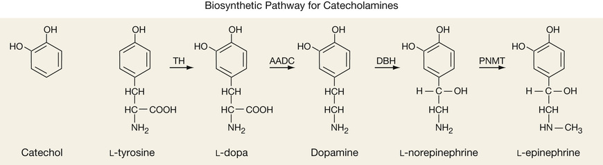
The catecholamines are metabolized by either catechol-O-methyltransferase (COMT) or monoamine oxidase (MAO). COMT converts D to methoxytyramine, E to metanephrine, and NE to normetanephrine, all of which in turn can be oxidized to vanillylmandelic acid (VMA) by MAO. MAO can also convert E and NE to 3,4-dihydroxymandelic acid, which is acted upon by COMT to form VMA. 3-Methoxy-4-hydroxyphenylacetic acid (homovanillic acid [HVA]) is the final product of dopamine metabolism.
Pheochromocytoma
Catecholamine-producing tumors fall into two categories: pheochromocytomas and paragangliomas. Pheochromocytomas arise from the chromaffin cells of the adrenal medulla and account for 90% of these tumors. Paragangliomas are extra-adrenal in origin, arising in the paravertebral sympathetic ganglia of the chest, abdomen, and pelvis, and the parasympathetic chains along the vagus and glossopharyngeal nerves. Pheochromocytomas have an incidence of about 500 to 1600 per year (Pacak et al, 2001a) and account for <1% of all secondary causes of hypertension. Although 90% of pheochromocytomas are benign, they are almost invariably lethal if not diagnosed and properly treated (Pacak et al, 2001a). Most pheochromocytomas are sporadic; however, 10% to 20% are familial, occurring as part of multiple endocrine neoplasia type 2A or 2B (MEN 2A, MEN 2B), von Hippel–Lindau (VHL) disease, neurofibromatosis type 1 (NF-1), or familial paraganglioma (FP). The hereditary forms tend to present at a younger age, and except for FP, the tumors are usually intraadrenal and bilateral.
Sustained or paroxysmal hypertension is the most common manifestation of this disease and is present in about 90% of patients. Remarkably, 10% of patients are normotensive. More than 90% will present with paroxysmal attacks characterized by at least two of the three following symptoms: headache associated with palpitations and diaphoresis (Sheps et al, 1994). Other symptoms include orthostatic hypotension, labile blood pressure, excessive sweating, anxiety, nervousness, weight loss, fatigue, pallor, and tremor. These symptoms can last from a few seconds to several hours, with the interval between attacks being highly variable—from several times a day to once every few months. Indications for screening for pheochromocytoma are listed in Box 24-6.
There is ongoing debate as to which test is the best for diagnosing pheochromocytoma. COMT is present in adrenal chromaffin cells and in tumors derived from these cells; it is absent from sympathetic nerves. As a result. the products of COMT, metanephrine and normetanephrine, serve as specific markers of chromaffin tumors (Lenders et al, 2014). Thus the most recent guidelines recommend the measurement of either plasma-free metanephrines or 24-hour urine collection for fractionated metanephrines via high-pressure liquid chromatography, with tandem mass spectrometry (HPLC/MS-MS) as the initial test (Lenders et al, 2014). Plasma normetanephrines, and urine for fractionated catecholamines and VMA may also be obtained.
The diagnosis of pheochromocytoma is made if the plasma concentration of either free metanephrine or normetanephrine is about four times the upper reference limit. Further testing is required in patients with high levels that are less than four times the upper reference limit (Sheps et al, 1994; Eisenhofer & Pacak, 2004; Eisenhofer et al, 2004). It is recommended that the samples be collected after the patient has been supine for at least 30 minutes. If a “seated” specimen returns as elevated, it should be confirmed by either repeating the test with the patient supine or obtaining a 24-hour urine sample for fractionated metanphrines. In centers where this assay is not available, the initial test should be the chromatographic measurement of a 24-hour urine collection for normetanephrine, metanephrine, fractionated free catecholamines (epinephrine, norepinephrine, dopamine), and creatinine (Lenders et al, 2002; Eisenhofer & Pacak, 2004; Eisenhofer et al, 2004). Metanephrine is the most sensitive and specific of these metabolites (Heron et al, 1996). Urinary creatinine should be measured in all 24-hour collections to assess for completion of the collection.
The diagnosis of pheochromocytoma can also be made by measuring plasma catecholamines; however, given their short half-life and episodic secretion, it is of use only if the sample is collected during a paroxysm. Values from the 24-hour urine collection or plasma catecholamines that are two to three times the upper limit of normal are usually diagnostic of pheochromocytoma.
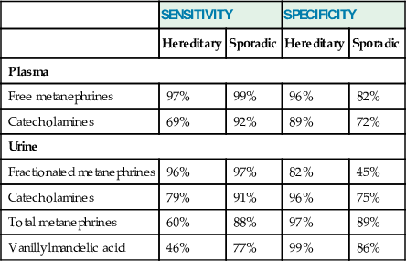
In cases of sporadic pheochromocytoma, the choice and interpretation of diagnostic tests depend on the pretest level of suspicion for disease. In this setting, the 24-hour urinary fractionated metanephrine and fractionated catecholamine measurements provide clinically acceptable sensitivity and significantly better specificity than fractionated plasma free metanephrine values (Sawka et al, 2003). Given the difficulties in collecting a complete 24-hour urine sample from pediatric patients, fractionated plasma free metanephrines should be considered the biochemical test of choice in that population (Weise et al, 2002) (Table 24-11).
TABLE 24-11
Sensitivity and Specificity of Hormone Levels for Diagnosing Pheochromocytoma
| SENSITIVITY | SPECIFICITY | |||
| Hereditary | Sporadic | Hereditary | Sporadic | |
| Plasma | ||||
| Free metanephrines | 97% | 99% | 96% | 82% |
| Catecholamines | 69% | 92% | 89% | 72% |
| Urine | ||||
| Fractionated metanephrines | 96% | 97% | 82% | 45% |
| Catecholamines | 79% | 91% | 96% | 75% |
| Total metanephrines | 60% | 88% | 97% | 89% |
| Vanillylmandelic acid | 46% | 77% | 99% | 86% |
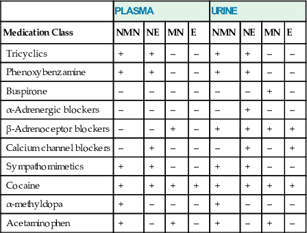
Several factors can cause false-positive test results, either by stimulating catecholamine secretion and/or by interfering with the assay (Table 24-12). When assaying for plasma free metanephrines, patients should abstain from caffeinated beverages and alcohol for 24 hours before testing. They should also avoid acetaminophen, tricyclic antidepressants, phenoxybenzamine, α agonists (e.g., methyldopa [Aldomet]), and monoamine oxidase inhibitors for at least 5 days before testing (Lenders et al, 1995). During testing for catecholamines, in addition to the list of items to avoid when testing for metanephrines, the patient should avoid nicotine, sympathomimetics (theophylline, pseudoephedrine), α agonists (e.g., albuterol), and levodopa/carbidopa. If antihypertensive medications are needed, angiotensin-converting enzyme inhibitors (ACEIs), angiotensin receptor blockers, and selective α1-adrenoceptor blockers (e.g., prazosin) can be used without fear of causing false-positive results (Eisenhofer et al, 2003).
TABLE 24-12
Effects of Medications on Testing for Pheochromocytoma
| PLASMA | URINE | |||||||
| Medication Class | NMN | NE | MN | E | NMN | NE | MN | E |
| Tricyclics | + | + | – | – | + | + | – | – |
| Phenoxybenzamine | + | + | – | – | + | + | – | – |
| Buspirone | – | – | – | – | – | – | + | – |
| α-Adrenergic blockers | – | – | – | – | – | + | – | – |
| β-Adrenoceptor blockers | – | – | + | – | + | + | + | + |
| Calcium channel blockers | – | + | – | – | – | + | – | + |
| Sympathomimetics | + | + | – | – | + | + | – | – |
| Cocaine | + | + | + | + | + | + | + | + |
| α-methyldopa | + | – | – | – | + | – | – | – |
| Acetaminophen | + | – | + | – | + | – | + | – |
Although urinary catecholamine levels may be elevated in renal insufficiency and renal failure, the measurement of plasma free metanephrines can be used to reliably diagnose pheochromocytoma in both conditions (Eisenhofer et al, 2004). Stressors such as an acute myocardial infarction, congestive heart failure, surgery, and acute cerebrovascular accident are all associated with elevated levels of catecholamines. In these situations, one can treat empirically and test once the patient has stabilized. Plasma normetanephrine concentrations increase with age; as a result, older adult patients are particularly susceptible to having false-positive tests. The use of fractionated urinary metanephrines and catecholamines may be more suitable for this population (Sawka et al, 2003).
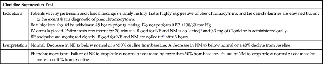
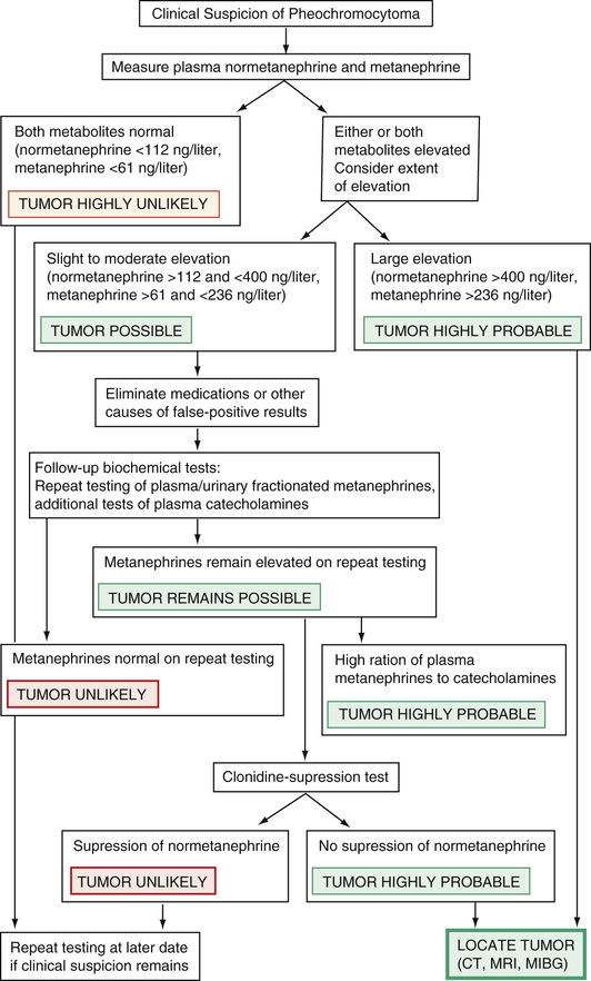
Plasma levels of normetanephrine less than 112 ng/L (0.61 nmol/L) and of metanephrine less than 61 ng/L (0.31 nmol/L) virtually exclude pheochromocytoma, so that no immediate further testing for the tumor should be necessary. With plasma concentrations of normetanephrine above 400 ng/L (2.19 nmol/L) or of metanephrine above 236 ng/L (1.20 nmol/L), the probability of pheochromocytoma is so high that the immediate task is to locate the tumor (Eisenhofer et al, 2003). More often than not, test results are returned as equivocal, requiring the need for confirmatory tests such as the clonidine suppression test or the glucagon stimulation test, or the measurement of urinary fractionated catecholamines. Clonidine is a centrally acting α-adrenergic agonist; it suppresses catecholamine release from the nervous system, but has no effect on its release by the tumor (Bravo et al, 1981). In those with pheochromocytoma, clonidine fails to adequately suppress plasma levels of norepinephrine to less than 40% from baseline. The clonidine suppression test is indicated only if the plasma catecholamines are greater than 1000 pg/mL (5.9 nmol/L). It is unreliable in those with normal or mildly elevated plasma catecholamines (Taylor et al, 1986; Elliott & Murphy, 1988; Sjoberg et al, 1992). Eisenhofer and colleagues (2003) showed that measuring plasma normetanephrines before and after clonidine testing increased the sensitivity and specificity of this test, especially in those who have only modest elevations of norepinephrine. The glucagon stimulation test can lead to dangerous rises in blood pressure and is rarely used. It should be performed only in patients whose blood pressure is well controlled, and a physician must be present throughout the test. A rise in plasma norepinephrine to greater than threefold, or greater than 2000 pg/mL, is diagnostic of pheochromocytoma (Table 24-13). The proposed mechanism of action is stimulation of glucagon-sensitive adenylate cyclase receptors expressed on these tumors. Unless the history is compelling, or the patient falls into one of the categories of genetically inherited disorders, it is often unnecessary to repeat testing in those with slightly positive results.
TABLE 24-13
Pharmacologic Tests for Diagnosing Pheochromocytoma
| Clonidine Suppression Test | |
| Indications | Patients with hypertension and clinical findings or family history that is highly suggestive of pheochromocytoma, and the catecholamines are elevated but not to the extent that is diagnostic of pheochromocytoma. Beta blockers should be withdrawn 48 hours prior to testing. Do not perform if BP <100/60 mm Hg. IV cannula placed. Patient rests recumbent for 20 minutes. Blood for NE and NM is collected,* and 0.3 mg of Clonidine is administered orally. BP and pulse are monitored closely. Blood for NE and NM are collected* after 3 hours. |
| Interpretation | Normal: Decrease in NE to below normal or a >50% decline from baseline. A decrease in NM to below normal or a 40% decline from baseline. |
| Pheochromocytoma: Failure of NE to drop below normal or decrease by more than 50% from baseline. Failure of NM to drop below normal or decrease by more than 40% from baseline. | |
Among catecholamine-secreting tumors, 10% to 20% are familial. Genetic testing should be considered if the patient is younger than 50 years of age at presentation, has physical traits suggestive of one of the familial disorders, has multifocal disease, or has a positive family history. Tests include assaying for mutations in the gene for menin in MEN 1, the RET oncogene in MEN 2A (Sipple's disease) and MEN 2B, neurofibromin in NF-1, and VHL in von Hippel–Lindau syndrome. Familial paragangliomas are associated with various defects in the gene for succinate dehydrogenase. As with all genetic testing, pretest and posttest counseling is mandatory.
Chromogranin A (CgA) is a protein that is stored and secreted along with the catecholamines from the adrenal medulla and sympathetic nervous system. Although it is elevated in more than 80% of pheochromocytomas, it is not specific for this disorder, being secreted by other chromaffin tissues (Hsiao et al, 1991). CgA was initially thought to be useful in the diagnosis of pheochromocytoma, since medications typically used to treat it had no impact on CgA secretion or measurement. Despite a relatively high sensitivity of 86%, it has poor diagnostic specificity. This is due, in large part, to the fact that the kidneys play a major role in the clearance of CgA from the circulation, so that even mild degrees of renal impairment (e.g., creatinine clearance [CrCl] <80 mg/mL/min) can lead to significant increases in serum concentration of CgA (Bravo & Tagle, 2003). Among hypertensive patients with CrCl less than 80 mL/min, overall sensitivity, specificity, and accuracy and positive and negative predictive values of serum CgA dropped to 85%, 50%, 59%, 38%, and 90%, respectively. However, when combined with elevated plasma catecholamines in patients with CrCl at least 80 mL/min, the diagnostic specificity and positive predictive values improved to 98% and 97%, respectively (Canale & Bravo, 1994). Its major use is in postoperative monitoring for recurrence of these tumors.
Testing Procedures
For 24-hour urine collections, creatinine is measured to verify the adequacy of the collection. To preserve the specimen adequately, urine should be collected in a container to which 25 mL of 6 N HCl has been added.
Plasma catecholamines are collected after an overnight fast (water permitted). The patient is placed in a reclining position in a quiet environment, and a heparin lock is inserted intravenously. After 20 to 30 minutes, blood is collected in a prechilled ethylenediaminetetraacetic acid (EDTA) lavender-top tube. The whole blood sample should be kept in ice water until centrifuged (preferably at 4° C). Separation of plasma should take place within 2 hours of phlebotomy; the sample should then be frozen immediately.
Both urine and plasma specimens should be analyzed using high-performance liquid chromatography with tandem mass spectrometry, as this technique greatly eliminates problems caused by interfering substances (Taylor & Singh, 2002). For the urine metabolites, it is important to use age-appropriate reference ranges when interpreting the results. It is also important to be aware that reference ranges can vary widely not only from one laboratory to another, but even within the same laboratory, as newer methods are introduced.
Additional Follow-Up Testing
Once the diagnosis is confirmed biochemically, the tumor should be localized by a CT scan or MRI of the adrenal glands. If this is negative, imaging studies of the abdomen, chest, and pelvis should be performed. CT has greater sensitivity, and MRI, greater specificity. MRI is superior to CT in detecting extraadrenal lesions and has the advantage of not requiring ionizing radiation or needing ionic contrast. If a tumor cannot be located by CT or MRI, or if metastatic disease is suspected, scanning with 131I- or 123I-labeled meta-iodobenzylguanidine should be performed. Octreoscanning and positron emission tomography are reserved for when the other techniques have failed. 18F-FDG PET/CT scanning is especially useful in patients with paragangliomas or metastatic disease (Pacak et al, 2001b).
Following successful tumor resection, the prognosis is generally excellent. Urinary metanephrines should be retested several weeks after surgery to ensure that the resection was complete, and should be measured periodically thereafter as an early marker of disease recurrence (Werbel & Ober, 1995).
An algorithm to evaluate pheochromocytoma has been proposed by Eisenhofer and colleagues (2003) (Fig. 24-10).
Neuroblastoma
Similarly to pheochromocytoma, neuroblastoma is of neural crest origin, arising within the adrenal glands or the sympathetic chain. It is the second most common solid malignant tumor in childhood, usually occurring before 3 years of age. Symptoms relate primarily to tumor mass rather than to hypertension, which is often mild or absent. At the time of diagnosis, 70% of cases will have distant metastases. About 90% of patients have elevated urinary homovanillic acid (HVA) levels at the time of diagnosis, whereas almost 75% have increased urinary vanillymandelic acid (VMA) levels (Tuchman et al, 1985). Both tests should be ordered when screening for the disease. In healthy children, at least up until about 15 years of age, urinary VMA and metanephrines tend to be higher (per milligram of creatinine) and more variable than in adults. Urinary metanephrines also can be elevated in neuroblastoma patients, but are not a sensitive measure of residual tumor. Urinary HVA is increased in familial dysautonomia (Riley-Day syndrome) and in some cases of pheochromocytoma.
Hormones of the Adrenal Cortex
The adrenal cortex is composed of three distinct zones: The outermost zone, the zona glomerulosa, is followed by the intermediate zona fasciculata, which surrounds the innermost zona reticularis. In the broadest terms, each zone is responsible for the synthesis and secretion of a unique set of hormones: The zona glomerulosa—mineralocorticoids (aldosterone); the zona fasciculata—glucocorticoids (cortisol); and the zona reticularis—sex steroids (dehydroepiandrosterone sulfate and androgens). However, under certain pathologic and physiologic conditions, these distinctions become blurred.
Mineralocorticoid Axis
The chief mineralocorticoid is aldosterone, which promotes the reabsorption of sodium and water by the kidney to help maintain blood pressure and tonicity. Expression of the enzyme CYP11B2 (aldosterone synthetase) within the glomerulosa is site-specific; as a result, the synthesis of aldosterone and its intermediary 18-hydroxylated metabolites is restricted to the zona glomerulosa. The precursor molecules to aldosterone, 11-deoxycorticosterone (DOC) and 11-deoxycortisol, similarly possess mineralocorticoid activity. However, unlike aldosterone, they can be synthesized within the zona fasciculata, as well as in the zona glomerulosa, which explains the hypertension and electrolyte disturbances seen in some forms of congenital adrenal hyperplasia. Although aldosterone will respond to acute changes in ACTH, it is mainly under the control of the renin-angiotensin system.
The zona fasciculata makes up 75% of the cortex, and is responsible for the synthesis and secretion of glucocorticoids and to a lesser extent androgens and estrogens. The glucocorticoids are 21-carbon steroid compounds with a hydroxyl group on carbon 17, hence the synonym 17-hydroxycorticosteroids (17-OHCS). Cortisol is the key glucocorticoid, regulating its own secretion through negative feedback on the hypothalamic-pituitary-adrenal (HPA) axis and inhibiting corticotropin-releasing hormone (CRH) from the hypothalamus and ACTH release from the pituitary gland. Both CRH and AVP (ADH) are produced by the parvocellular neurons of the paraventricular nuclei of the hypothalamus. ACTH secretion is stimulated by CRH and, to a much lesser extent, by AVP. ACTH in turn stimulates cortisol production by the adrenal glands. Cortisol is needed in times of stress to maintain blood pressure and blood sugar, and to prevent shock. Although cortisol is the most important glucocorticoid, corticosterone, which is a hormone of the mineralocorticoid pathway, also possesses glucocorticoid activity.
Androgens and estrogens are produced by the zona reticularis. The androgens are 18-carbon steroids with saturated A rings, in contrast to the estrogens, which are 17-carbon steroids with unsaturated A rings.
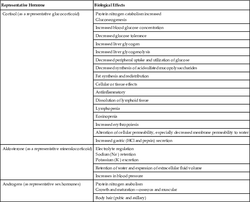
The functions of these hormones are summarized in Table 24-14.
TABLE 24-14
Physiologic Effects of Steroids
| Representative Hormone | Biological Effects |
| Cortisol (as a representative glucocorticoid) | Protein nitrogen catabolism increased Gluconeogenesis |
| Increased blood glucose concentration | |
| Decreased glucose tolerance | |
| Increased liver glycogen | |
| Increased liver glycogenolysis | |
| Decreased peripheral uptake and utilization of glucose | |
| Decreased synthesis of acid-sulfated mucopolysaccharides | |
| Fat synthesis and redistribution | |
| Cellular or tissue effects | |
| Antiinflammatory | |
| Dissolution of lymphoid tissue | |
| Lymphopenia | |
| Eosinopenia | |
| Increased erythropoiesis | |
| Alteration of cellular permeability, especially decreased membrane permeability to water | |
| Increased gastric (HCl and pepsin) secretion | |
| Aldosterone (as a representative mineralocorticoid) | Electrolyte regulation Sodium (Na+) retention Potassium (K+) excretion |
| Retention of water and expansion of extracellular fluid volume | |
| Increases in blood pressure | |
| Androgens (as representative sex hormones) | Protein nitrogen anabolism Growth and maturation—osseous and muscular |
| Body hair (pubic and axillary) |
Congenital Disorders of Adrenal Cortical Enzyme Deficiencies
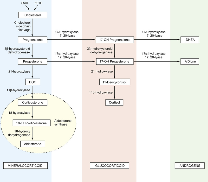
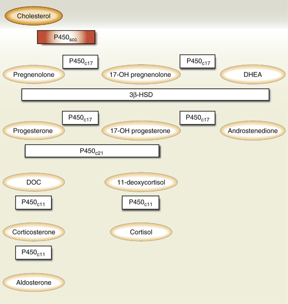
The hormones of the adrenal cortex are steroid derivatives, synthesized from low-density lipoprotein (LDL) cholesterol. LDL is delivered to the adrenal glands, where it is taken up by LDL receptors. Evidence also supports local synthesis of LDL from acetyl coenzyme A. Steroid acute regulatory protein (StAR) shuttles LDL across the mitochondrial membrane, where it begins its journey down the steroidogenic pathway (Fig. 24-11). The enzymes that catalyze these synthetic reactions are of four general types: hydroxylases, dehydrogenases, desmolases, and isomerases. Because most of the inborn errors of metabolism affecting steroid hormone synthesis in the adrenal cortex involve deficiencies of hydroxylases, they constitute the most clinically important group of enzymes.
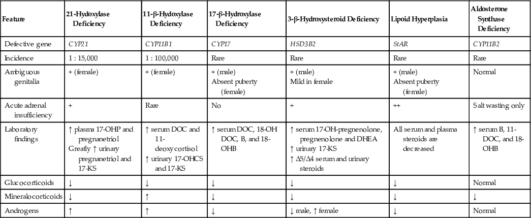
At least eight different metabolic defects in the synthesis of cortisol and aldosterone have been described, each characterized by a deficiency of a specific adrenal enzyme. A vast majority of these enzymatic deficiencies are inherited as autosomal recessive traits with variable degrees of penetrance. Those enzymatic defects that uniquely affect the biosynthesis of cortisol are grouped together under the rubric of congenital adrenal hyperplasia (CAH). These five enzymes are P450scc (defect in StAR), 3-β-hydroxysteroid dehydrogenase, 21-hydroxylase, 11-hydroxylase, and 17-hydroxylase. CRH and ACTH synthesis and secretion are normally under the negative-feedback control of cortisol. In CAH, defects in the enzymes necessary for cortisol production lead to cortisol deficiency; cortisol deficiency in turn results in disinhibition of negative feedback on CRH and ACTH production. As a consequence, CRH and ACTH levels rise, inducing adrenal hyperplasia and a forward push in steroidogenesis, as the body tries to compensate and normalize cortisol production. Not only does this result in a buildup of the hormonal precursors directly preceding the enzymatic defect, it also causes massive shunting of these precursors down the remaining functional pathways. The clinical manifestations of CAH are heterogeneous, depending on the severity and location of the enzymatic defects, which hormones are deficient, and which are produced in excess. Symptoms range from shock, salt wasting, and anomalous sexual development in infancy, to hirsutism and infertility in the adult. Sometimes, as in partial enzymatic deficiencies of cortisol synthesis, near-adequate hormone synthesis is possible if hypersecretion of ACTH is able to stimulate adrenal hyperplasia to compensate for the deficiency. The clinical manifestations of various adrenal cortical enzyme deficiencies and their associated laboratory findings are summarized in Table 24-15.
TABLE 24-15
Congenital Adrenal Hyperplasia: Clinical and Biochemical Features
| Feature | 21-Hydoxylase Deficiency | 11-β-Hydoxylase Deficiency | 17-β-Hydroxylase Deficiency | 3-β-Hydroxysteroid Deficiency | Lipoid Hyperplasia | Aldosterone Synthase Deficiency |
| Defective gene | CYP21 | CYP11B1 | CYP17 | HSD3B2 | StAR | CYP11B2 |
| Incidence | 1 : 15,000 | 1 : 100,000 | Rare | Rare | Rare | Rare |
| Ambiguous genitalia | + (female) | + (female) | + (male) Absent puberty (female) | + (male) Mild in female | + (male) Absent puberty (female) | Normal |
| Acute adrenal insufficiency | + | Rare | No | + | ++ | Salt wasting only |
| Laboratory findings | ↑ plasma 17-OHP and pregnanetriol Greatly ↑ urinary pregnanetriol and 17-KS | ↑ serum DOC and 11-deoxycortisol ↑ urinary 17-OHCS and 17-KS | ↑ serum DOC, 18-OH DOC, B, and 18-OHB | ↑ serum 17-OH-pregnenolone, pregnenolone and DHEA ↑ urinary 17-KS ↑ Δ5/Δ4 serum and urinary steroids | All serum and plasma steroids are decreased | ↑ serum B, 11-DOC, and 18-OHB |
| Glucocorticoids | ↓ | ↓ | ↓ | ↓ | ↓ | Normal |
| Mineralocorticoids | ↓ | ↑ | ↓ | ↓ | ↓ | ↓ |
| Androgens | ↑ | ↑ | ↓ | ↓ male, ↑ female | ↓ | Normal |
The diagnosis is made by measuring the various serum hormone levels and assessing which steroids are produced in excess and which are deficient, and by calculating the precursor/product ratio and comparing these results to age- and sex-matched normative data. Levels of hormones distal to the block (product hormones) may be elevated as a result of peripheral conversion of the markedly elevated precursor hormones; the use of precursor/product ratios mitigates the risk for misdiagnosis due to misleading elevations of product hormones (Levine, 2002). If hormone levels return as borderline, but the clinical suspicion for CAH remains high, the steroid levels should be remeasured 60 minutes after intravenous administration of 0.25 mg of ACTH. ACTH drives steroidogenesis forward, accentuating the block. When there is a proband, the diagnosis can be more accurately determined by genotyping.
CAH is categorized according to severity of disease into classic (neonatal, severe) and nonclassic (late-onset, cryptogenic) forms. The classic form is further subdivided into salt-losing and non–salt-losing (simple virilizing) variants.
21-Hydroxylase Deficiency
The enzyme 21-hydroxylase (also referred to as CYP21, CYP21A2, and P450c21) is located within the mitochondrial endoplasmic reticulum. 21-hydroxylase deficiency is the most common cause of CAH, accounting for about 95% of all cases. Screening of newborns using capillary heel blood on paper filter disks has identified this disorder in about 1 in 14,000 persons in North America, and in as many as 1 in 300 Yupik Eskimos of Alaska (Pang et al, 1988, 1982). The classic form is detected in about 1 in 16,000 live births; the nonclassic form is seen in about 0.2% of the general Caucasian population, and in 1% to 2% of those of Eastern European Jewish ancestry (Speiser et al, 1985; Therrell, 2001).
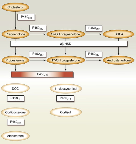
21-OH catalyzes the conversion of 17-hydroxyprogesterone (17-OHP) to 11-deoxycortisol (compound S), and progesterone to DOC. Thus, 21-OH deficiency leads to elevated levels of the steroid precursors 17-OHP and pregnanetriol in the urine and 17-OHP in the serum. These precursors are shunted toward the pathway, leading to excess androstenedione and testosterone production (Fig. 24-12). Clinical presentation often correlates with the severity of enzymatic dysfunction. The classic variety presents in the newborn period or in early childhood with adrenal insufficiency and virilization, with or without salt wasting. The nonclassic form presents in late childhood as early adrenarche, or in young adulthood as hirsutism, amenorrhea, and infertility. The presentation in women is very similar to that of polycystic ovarian disease. Males may develop precocious puberty, adrenal rests within the testes, and infertility.
A close functional relationship exists between the adrenal cortex and the adrenal medulla. Dysplasia of the medulla and catecholamine hyposecretion have been described in classic 21-OH deficiency. In a study of 38 children with classic (CYP21A2) disease, levels of plasma epinephrine and metanephrine and urinary epinephrine were 40% to 80% lower in affected individuals than in normal persons (Merke et al, 2000). In another study, those with CYP21A2 showed a significantly decreased catecholamine response to exercise that was unaffected by the administration of stress doses of glucocorticoids (Weise et al, 2004). It has been suggested that the degree of medullary impairment may be a biomarker for CAH severity (Merke et al, 2002).
Diagnosis.
Prenatal diagnosis is important because suppressive treatment with steroids can abrogate the development of virilization of the female fetus. Diagnosis is made by measuring the level of 17-OHP in amniotic fluid or by genotyping cells obtained by chorionic villous sampling. The genes responsible for 21-OH deficiency, CYP21 (CYP21A2) and CYP21P (CYP21A), are located on chromosome 6. Of these two homologous genes, only CYP21 is active; deleterious mutations within CYP21P interfere with normal gene expression. These mutations can be identified by PCR and Southern blotting on chorionic villous samples (White et al, 1994a; New, 1995). The 2010 consensus statement on the management of CAH due to 21-OH deficiency recommends that prenatal treatment of CAH be regarded as experimental and that the diagnosis should rest on clinical and hormonal data, with genotyping being reserved for equivocal cases and genetic counseling (Speiser et al, 2010).
Neonatal screening, which is now mandatory across the United States and in several other countries, is performed by measuring 17-OHP or by genotyping blood that has been obtained from a heelstick and collected on filter paper. The sample for 17-OHP is analyzed using automated time-resolved dissociation-enhanced lanthanide fluoroimmunoassay (DELFIA) (Speiser et al, 2010). Causes for false-positive screens include prematurity, illness, stress, and possibly the antenatal administration of glucocorticoids used to induce fetal lung maturation. The initial positive screen is followed up by checking the 17 OHP level by LC-MS/MS (Speiser et al, 2010). In regard to genotyping, aside from its being the most definitive test for diagnosing CAH, the fact that the genotype correlates fairly well with disease severity means that it can also be used as a prognostic tool (Nordenstrom et al, 1999).
In the newborn with salt wasting, unstimulated 17-OHP levels are typically greater than 8000 ng/dL, rising to 100,000 ng/dL (3000 nmol/L) following administration of ACTH. Levels in the simple virilizing variant range from 10,000 to 30,000 ng/dL (300 to 1000 nmol/L). Those with nonclassic disease typically have 17-OHP levels ranging from 1500 to 10,000 ng/dL (50 to 300 nmol/L) (New et al, 1983). It is of note that randomly drawn hormone levels may be normal in those with nonclassic disease; therefore it is important to test during the early morning. If the results are equivocal, the diagnosis can be confirmed by comparing serum 17-OHP levels before and 60 minutes after administration of 0.25 mg ACTH (Cortrosyn). ACTH acts to stimulate steroidogenesis, serving to dramatically increase the bottleneck at the site of the enzymatic block, resulting in a dramatic increase in precursors, in this case, 17-OHP. Post-ACTH 17-OHP values <330 ng/dL are normal, 330 to 1000 ng/dL indicates a heterozygote carrier, and levels greater than 2000 ng/dL are diagnostic for nonclassic CAH. If a proband is available, genotyping is superior to these older biochemical tests in identifying heterozygotes (Honour & Rumsby, 1993).
The goals of glucocorticoid and mineralocorticoid replacement therapy in children are the attainment of normal growth, weight, and pubertal development, and optimization of final adult height. In adults, the major treatment goals include lessening of signs of virilization and resumption of fertility. Glucocorticoid replacement is titrated to keep the 17-OHP level partially suppressed to between 100 and 1000 ng/dL (3 to 30 nmol/L) and the ACTH under 100 ng/L, thereby preventing shunting toward testosterone synthesis, and normalizing levels of androstenedione and testosterone. Normalization of 17-OHP should not be attempted, because this requires supraphysiologic levels of glucocorticoids and may result in Cushing's syndrome (Speiser & White, 2003).
Varying degrees of hypoaldosteronism accompany all forms of 21 OH deficiency. This is biochemically apparent as an elevated plasma renin activity (PRA) and a decreased aldosterone/PRA ratio. The objective of mineralocorticoid replacement is to normalize the plasma renin activity.
11-β-Hydroxylase Deficiency
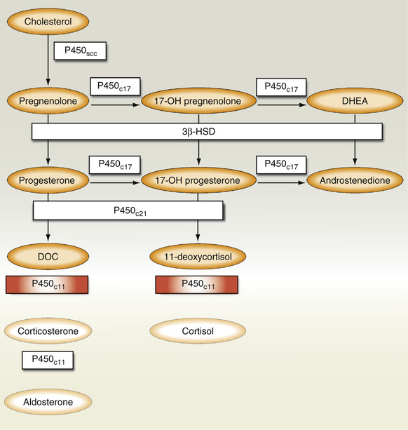
The second most common enzyme deficiency of the adrenal cortex, accounting for about 7% of all cases of CAH, is the 11-β-hydroxylase (11-OH) deficiency. A defect in this enzyme blocks the final conversion of 11-deoxycortisol to cortisol and DOC to corticosterone. As with 21-OH deficiency, a compensatory increase in ACTH secretion leads to adrenal hyperplasia and a mass action shunting of precursor steroids toward testosterone synthesis, resulting in signs of virilization. This block also results in the accumulation of DOC; the mineralocorticoid activity of DOC leads to the development of hypertension and hypokalemia, similar to what is seen with hyperaldosteronism (Fig. 24-13).
11-OH deficiency is an autosomal recessive disorder caused by mutations of the genes CYP11B1 and CYP11B2, located on chromosome 8q21-q22 (White et al, 1994b). Diagnosis in the neonate is established by the presence of a high basal and a high ACTH-stimulated 11-deoxycortisol. Concentrations of urinary tetrahydro-11-deoxycortisol in the urine are also elevated. During childhood and in young adults, the diagnosis of 11-OH deficiency is made by the presence of elevated early-morning and ACTH-stimulated serum levels of 11-deoxycortisol that are more than three times the upper limit for age-matched normals. Levels of DOC and adrenal androgens (androstenedione, dehydroepiandrosterone, and dehydroepiandrosterone sulfate) are also elevated. Plasma renin activity and aldosterone are often suppressed as a result of salt and water retention induced by elevations of DOC. Unlike the heterozygotes with 21-OH deficiency, those with 11-OH deficiency often fail to show a rise in precursors following ACTH stimulation (Pang et al, 1980). However, an exuberant response was seen in those who had hirsutism (Gabrilove et al, 1965).
Prenatal diagnosis of 11-OH deficiency is made by measuring levels of tetrahydro-11-deoxycortisol (THS) in the maternal urine or amniotic fluid. These levels begin to rise in the first trimester. In addition to THS, elevation of 11-deoxycortisol levels and of the THS to tetrahydrocortisol plus tetrahydrocortisone ratio is seen (Rosler et al, 1988).
Treatment consists of the replacement of glucocorticoids, causing normalization of DOC and plasma renin activity.
3-β-Hydroxysteroid Dehydrogenase Deficiency
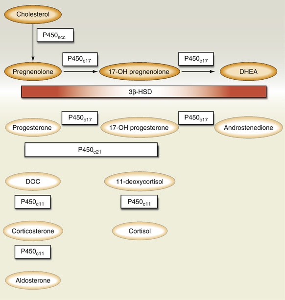
3-β-hydroxysteroid dehydrogenase (3-β-HSD), which catalyzes the second enzymatic step in steroidogenesis, is coded by two genes, HSD3BI and HSD3BII. HSD3BII is expressed in the adrenals and gonads; HSD3BI is expressed in placenta, skin, and other peripheral tissues and usually remains functionally intact at these sites in CAH. A defect in 3-β-HSD leads to a block in the conversion of Δ5 steroids (pregnenolone, 17-OH pregnenolone, and dihydroepiandrosterone) to Δ4 steroids (progesterone, 17-OHP, androstenedione), resulting in an increase in circulating levels of Δ5 steroids (Fig. 24-14). However, because HSD3BI is usually intact, levels of the Δ4 steroids may be normal or even elevated.
Patients with classic disease will have manifestations of glucocorticoid deficiency, with or without the accompaniment of salt wasting. Affected males have incomplete masculinization, and females may be normal or may have ambiguous genitalia. A late-onset variant has been described that is associated with features typical of polycystic ovarian disease, such as hirsutism, oligomenorrhea, and infertility (Pang et al, 1985).
Previous criteria for the diagnosis of 3-β-HSD deficiency examined the basal and ACTH-stimulated Δ4/Δ5 steroid ratios, 17-OH-pregnenolone (17-OHP)/cortisol, and levels of pregnenolone, 17-OH-pregnenolone, and dehydroepiandrosterone in urine and blood. The standard for diagnosing this disorder has been recently revised to correlate more closely with genotypic studies. The ACTH-stimulated Δ5-17 P levels and Δ5-17 P/cortisol ratios have been shown to be the best indices for definitively diagnosing 3-β-HSD deficiency (Lutfallah et al, 2002). Applicability of these new criteria to patients with the nonclassic variant is still debatable.
Treatment consists of glucocorticoids and mineralocorticoids, as well as sex steroids, in accordance with normal growth and development.
17-Hydroxylase Deficiency
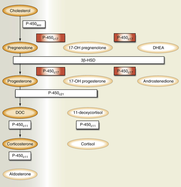
17-Hydroxylase (CYP17, P450c17) is expressed in the adrenal glands and the gonads and encodes two enzymes—17α-hydroxylase and 17,20-lyase. 17α-hydroxylase catalyzes the conversion of pregnenolone and progesterone to their respective 17-OH derivatives. 17,20-Lyase converts 17-OH pregnenolone to dihydroepiandosterone, and 17-OH progesterone to androstenedione. CYP17 deficiency blocks the conversion of pregnenolone and progesterone to the 17-hydroxy derivatives, causing shunting from testosterone and cortisol synthesis to aldosterone (Fig. 24-15). Accordingly, these patients develop hypertension and hypokalemic alkalosis in association with incomplete masculinization (in the male) and decreased testosterone and cortisol levels. 17-OH deficiency is diagnosed by demonstrating high DOC, pregnenolone, and progesterone levels, along with decreased urinary 17-ketosteroids and 17-hydroxycorticosteroids. The gene associated with this condition (CYP17) has been located on chromosome 10q, the same gene as 17,20-desmolase (Kater & Biglieri, 1994).
Congenital Lipoid Adrenal Hyperplasia
Congenital lipoid adrenal hyperplasia (lipoid CAH) is the most severe form of CAH, in which the synthesis of all gonadal and adrenal cortical steroids is markedly impaired. Lipoid CAH may be caused by a defect in the StAR or the P450 side chain cleavage (scc) (Fujieda et al, 2003). StAR, located on chromosome 8p11, controls the rate-limiting step in steroidogenesis. It is responsible for the shuttling of cholesterol from the outer to the inner mitochondrial membrane. 20,22-Desmolase (CYP11A1, P450scc) converts cholesterol to pregnenolone (Fig. 24-16). Pathologically, the adrenal cortex shows marked accumulation of cholesterol and other lipids, which is the primary distinguishing feature from congenital adrenal hypoplasia.
The presentation of this extremely rare disorder is that of severe adrenal insufficiency with hypotension, salt wasting, and feminization of external genitalia in males. Occasionally, females may not present until the onset of puberty. Diagnosis is made by the presence of extremely low cortisol and aldosterone concentrations and elevated ACTH and plasma renin activity.
Aldosterone Synthetase Deficiency
Aldosterone synthetase (CYP11B2) is the final step in the steroid synthetic pathway leading to the production of aldosterone. It stimulates a multistep process: 11-hydroxylation of DOC to corticosterone, 18-hydroxylation of corticosterone to 18-hydroxycorticosterone, and, finally, 18-dehydrogenation to aldosterone. Isolated enzyme deficiency leads to salt wasting, hyperkalemia, and metabolic acidosis. This condition may be diagnosed by demonstrating the presence of metabolites of corticosterone and 11-deoxycorticosterone in the urine, elevated serum DOC, and deficiency of corticosterone, 18-hydroxycorticosterone, or aldosterone in the serum.
Cortisol and the Glucocorticoids
The adrenal cortex secretes cortisol in response to ACTH, a diurnal rhythm, and stress. ACTH, synthesized in the adenohypophysis, is formed from the cleavage of a much larger precursor molecule: POMC. In addition to ACTH, cleavage of POMC releases β-LPH, which in turn is cleaved to yield γ-LPH and β-endorphin. Within the ACTH sequence are α-MSH and the corticotropin-like intermediate lobe protein. Endorphins, which act on neurons in the brain, constitute a distinct peptidergic system related to pain perception. Although β-endorphin is secreted in parallel with ACTH, the significance of this remains unknown.
ACTH consists of 39 amino acid residues, of which residues 1 to 24 at the amino terminal possess full hormonal activity. Occasionally, POMC is incompletely processed; this leads to the formation of other forms of ACTH that usually have little biological activity, although they may retain immunoreactivity. These forms may predominate in malignant conditions such as ectopic production by primary or metastatic lung cancer, and in some patients with Nelson's syndrome, a disorder characterized by the occurrence of a pituitary tumor and skin hyperpigmentation following bilateral adrenalectomy. Defects in POMC cleavage enzymes may also be responsible for the formation of rare forms of isolated ACTH deficiency (Nussey et al, 1993).
ACTH is secreted in response to several factors, of which CRH and AVP are the most important. Among the other moieties reported to stimulate ACTH secretion are atrial natriuretic factor (ANF), angiotensin II, IL-6, IL-1, and tumor necrosis factor-α (Rivier & Vale, 1983; Chrousos, 1998).
CRH is synthesized and released from the hypothalamus; it acts to stimulate the synthesis and release of ACTH from the pituitary gland. CRH is released in a circadian pattern and in response to physiologic stimuli such as stress and hypoglycemia. The HPA axis consists of various feedback loops that control cortisol synthesis and secretion. When plasma cortisol increases, it suppresses the release of CRH, ACTH, and AVP, which, in turn, leads to lowering of the cortisol level. Conversely, when serum cortisol reaches a nadir, the hypothalamus and pituitary gland respond by increasing CRH and ACTH production, leading to stimulation of cortisol formation and secretion. By this mechanism, ACTH and cortisol control the concentration of each other within a very narrow range, and a small change in one results in a concomitant change in the other. When the adrenal gland is unable to respond to ACTH because of damage or disease, cortisol levels are low and ACTH levels are high. In those conditions in which the pituitary gland is destroyed, ACTH is not formed and cortisol levels are consequently low. Damage to the hypothalamus is also associated with low ACTH and cortisol levels; testing with CRH may permit the distinction between these two entities. Synthetic forms of CRH are used in testing the anterior pituitary gland's reserve of ACTH by comparing plasma ACTH and cortisol before and 1 hour after CRH stimulation (Grodum et al, 1993). This test is useful in distinguishing between lesions affecting the hypothalamus, pituitary gland, and adrenal glands (Fukata et al, 1993). If the lesion is in the hypothalamus, after a time delay, ACTH levels rise following CRH administration. If it is in the pituitary gland, no significant ACTH response occurs. With primary adrenal insufficiency, administration of CRH causes a further rise in an already elevated ACTH level, but little or no rise in cortisol level.
If the HPA axis is interrupted by the administration of large quantities of exogenous glucocorticoids, they will exert an inhibitory effect on the hypothalamus and pituitary gland, suppressing CRH and ACTH secretion. If this suppression continues over a period of weeks, it leads to atrophy of the adrenal glands; as a result, the HPA axis becomes unable to secrete cortisol in times of stress. The HPA axis can fully recover after tapering off of steroids.
The second influence on plasma cortisol levels is the diurnal pattern, which is due to the circadian pattern of ACTH release. Major increases in secretion occur at between 4 AM and 8 AM, followed by a decrease in ACTH during the rest of the day. In subjects with a normal sleep-wake cycle, the lowest ACTH concentrations are found shortly after midnight. Sudden changes in sleep-wake patterns have little effect on the diurnal pattern, but permanent changes in daily sleep habits result in a gradual change in diurnal secretory patterns. Superimposed on the circadian periodicity is an ultradian rhythm of 10 to 18 secretory bursts per 24 hours (Horrocks et al, 1990).
The third important influence on cortisol secretion is stress. Stimuli such as surgical trauma, pyrogens, hypoglycemia, and hemorrhage are capable of bringing about an acute increase in ACTH and cortisol secretion. This response to stress may be absent or decreased in magnitude in patients in whom large doses of steroids have been administered for some time. The initiation of any stress response is dependent on an intact nervous system. For example, trauma normally results in the acute release of ACTH and cortisol; however, in patients with spinal cord transections, the normal transmission of neurologic stimuli is interrupted, and as a result, the same trauma applied to an extremity will not elicit any ACTH or cortisol response. Evidence suggests that the stress response of cortisol is mediated through excitatory and inhibitory inputs that integrate at the level of the hypothalamus and modulate CRH secretion. Cortisol levels also rise after meals, especially those high in protein; it also rises in depression (Linkowski et al, 1987).
Most disorders of cortisol secretion can be classified by the patterns of response of the following three hormones to suppression and stimulation: ACTH, plasma cortisol, and urinary free cortisol (Snow et al, 1992).
Laboratory Measurement of ACTH
Plasma ACTH is measured using a two-site immunoradiometric assay or an immunochemiluminometric assay. To prevent degradation of ACTH, it is best to collect the sample in a prechilled EDTA lavender-top tube. The specimen should be kept in an ice bath and should be processed as soon as possible. Following centrifugation in a refrigerated centrifuge, the specimen should be separated, transferred to a plastic tube, and kept frozen at –20° C until time of analysis. The normal reference range is 2 to 12 pmol/L (9 to 52 pg/mL) between 7 AM and 10 AM. Plasma ACTH is a useful tool for distinguishing primary (adrenal) from secondary (pituitary) or tertiary (hypothalamic) adrenal insufficiency. In primary adrenal insufficiency, low cortisol concentrations are found, along with increased ACTH levels. In secondary or tertiary adrenal insufficiency, both ACTH and cortisol are expected to be low. ACTH levels best discriminate between healthy individuals and those with adrenal insufficiency when specimens for ACTH are collected between 8 AM and 10 AM.
ACTH is less useful in diseases of cortisol excess. Up to 50% of patients may have normal ACTH levels in Cushing's disease. Although the values tend to run higher (>20 pmol/L [>90 pg/mL]) in Cushing's syndrome due to ectopic ACTH production, the values overlap with those seen in Cushing's disease in 30% of cases (Findling, 1992). Because of the normal diurnal variation in ACTH and cortisol secretion, measurement of these values during their expected nadir, from 11 PM to 1 AM, is helpful in confirming the diagnosis of ACTH-dependent Cushing's disease. An elevated midnight ACTH of greater than 5 pmol/L (23 pg/mL) in the face of an elevated serum cortisol confirms the diagnosis of ACTH-dependent Cushing's disease. Those patients with ectopic ACTH-secreting tumors characteristically have markedly elevated plasma ACTH (usually >200 pg/mL) and elevated serum cortisol. Measurement of ACTH by plasma extraction, which detects ACTH precursors and fragments, may be useful in distinguishing patients with cancer-related syndromes or ACTH-secreting tumors from those with Cushing's disease, as the former entities are more likely to produce these other forms of ACTH (White & Clark, 1993). ACTH-secreting neoplasms may be occult, creating diagnostic difficulties; in this instance, ACTH measurement using selective venous sampling has proven useful in localization of the lesion.
In patients with increased levels of circulating glucocorticoids due to an adrenal adenoma or carcinoma, and those surreptitiously taking steroids, ACTH secretion is inhibited and levels are low or undetectable. In patients with pituitary-induced adrenal hyperplasia (Cushings' disease), plasma ACTH may be at or above the upper reference interval at 9 AM, and fail to show the expected fall after midnight. Another use of ACTH assays is in the determination of adequacy of cortisol replacement in congenital adrenal hyperplasia. When replacement therapy is optimal, ACTH values are similar to those seen in a reference population.
Plasma Corticotropin-Releasing Hormone
CRH measurements are performed by liquid chromatography and tandem mass spectrometry (LC/MS-MS) and remain largely a research tool.
CRH circulates in one of two forms: free, or bound to CRH-binding protein. The reference range for plasma CRH in men and nonpregnant women is <34 pg/mL. CRH is increased in Cushing's syndrome due to ectopic production of CRH. CRH-binding protein increases during pregnancy; as a result the normative range for CRH varies throughout pregnancy: first trimester <40 pg/mL; second trimester <153 pg/mL; and third trimester <847 pg/mL (Goland et al, 1986).
Serum Cortisol Measurements
About 90% of circulating cortisol is bound to serum protein, of which 10% to 20% is loosely bound to albumin, and the remainder is bound to the glycoprotein transcortin (cortisol-binding globulin [CBG]). The remaining 10% of circulating cortisol is the unbound, free hormone. It is believed that only free cortisol is active, and that the protein-bound fraction is metabolically inert, probably serving as a reservoir for free cortisol. Protein binding also may protect cortisol from deactivation by the liver or filtration by the kidney.
One of the earliest and simplest methods used to determine the serum cortisol concentration was a fluorometric assay developed by Nelson and Samuels that was based on a technique first developed by Porter and Silber (Porter & Silber, 1950; Nelson & Samuels, 1952). The subsequent development of antibody-based immunoassays provided a more specific method for cortisol estimation with the added advantages of requiring a smaller specimen volume and a more rapid turnaround time. The problem was that some of the antibodies used showed a large degree of cross-reactivity with other steroid species, resulting in spuriously elevated cortisol concentrations. In chronic renal disease, various steroids and their glucuronides accumulate in the blood; cross-reactivity between these steroids and their conjugates with the antibodies used in the assay can yield significantly erroneous cortisol concentrations. Similarly, in CAH, high concentrations of cortisol precursors are seen in the serum because of an enzyme defect. Because these precursors cross-react with assay antibodies, artificial elevations of cortisol are found. The degree of interference varies with the assay used and cannot be easily predicted. Nonisotopic immunoassay methods using organometallic tracers, fluorescence polarization, and enzyme immunoassay techniques have also been developed for cortisol determinations (Bacarese-Hamilton et al, 1992; Lentjes et al, 1993; Philomin et al, 1994). The major disadvantage of all of these cortisol assays continues to be lack of specificity.
The measurement of cortisol by radioimmunoassay (RIA) and chemiluminescent techniques has largely been supplanted by high-performance liquid chromatography (HPLC) with tandem mass spectrometry, which appears to offer the ultimate in specificity. Serum cortisol is collected in a no-additive (red-top) tube. Reference values for serum cortisol for men and women roughly range from 5 to 25 µg/dL (140 to 690 nmol/L) at 8 AM to 10 AM, dropping to about 3 to 12 µg/dL (80 to 330 nmol/L) by 4 PM. Because of wide swings in basal cortisol levels resulting from its diurnal and ultradian pattern of secretion, serum cortisol assays are most useful when evaluated in the context of dynamic manipulation (i.e., adrenal stimulation or suppression).
Salivary Cortisol
Up to 30% of urinary free cortisol (UFC) and dexamethasone suppression screening tests may return an incorrect result. Recent studies have shown that the use of a midnight salivary cortisol (MSC) is a viable alternative. CBG is not present in saliva; therefore the results may be more useful in situations in which CBG concentration is altered. Salivary cortisol is highly stable and easy to collect, making it a useful tool for screening and for diagnosing instances of increased cortisol secretion. It does not appear to be affected by the rate of saliva production and can reflect a change in serum cortisol in as rapid as a few minutes (Read et al, 1990). Because of alterations in circadian rhythm, it may not be an appropriate test for shift workers or those with highly variable bed times. One study compared the sensitivity for the detection of Cushing's syndrome by nighttime salivary cortisol levels versus that for simultaneous inpatient serum cortisol levels and urine glucocorticoid excretion. It was found that the salivary cortisol measurements worked as well as plasma measurements and better than urine glucocorticoid excretion. The authors concluded that measurement of bedtime salivary cortisol was a practical and accurate screening test for the diagnosis of Cushing's syndrome (Papanicolaou et al, 2002). Another study compared the diagnostic performance of MSC measurement versus that of midnight serum cortisol (MNC) and UFC in differentiating 41 patients with Cushing's syndrome from 33 with pseudo-Cushing's states, 199 with simple obesity, and 27 healthy normal weight volunteers. In the whole study population, no statistically significant differences in terms of sensitivity, specificity, diagnostic accuracy, and predictive values were observed among tests. In particular, the overall diagnostic accuracy for MSC was similar to those of UFC and MNC (Putignano et al, 2003; Elamin et al, 2008). A recent review of the literature has shown that elevated late-night (11 PM to 12 AM) salivary cortisol has greater than 90% sensitivity and specificity for the diagnosis of endogenous Cushing's syndrome (Raff, 2009). It is recommended to have the patient avoid tobacco on the day of specimen collection.
Urinary Free Cortisol Measurements
Only 1% of the total adrenal secretion appears in the urine as cortisol, but it is this fraction that provides valuable aid in the diagnosis of adrenal disease. In the kidney, glomerular filtration of free cortisol (unbound to CBG and albumin) is followed by passive tubular reabsorption without a demonstrable reabsorption maximum. At serum cortisol levels of about 20 to 25 µg/dL (the upper 8 AM reference value), the binding capacity of transcortin (CBG) is exceeded, leading to a very rapid and disproportionate increase in the unbound fraction compared with the total serum cortisol. Doubling of serum cortisol from 20 to 40 µg/dL results in at least a fivefold increase in unbound cortisol. At these levels, free cortisol clearance by the kidneys is directly proportional to the unbound serum cortisol concentration, and a steep rise in cortisol clearance is seen. Serum cortisol reflects the sum of free, CBG-bound and albumin-bound cortisol. Only free (unbound) cortisol is filtered by the kidneys; therefore unlike serum cortisol, UFC is not affected by conditions and medications that alter CBG and albumin concentrations. Thus, when UFC rather than serum cortisol is used, it is easier to differentiate patients with adrenal hyperfunction from a reference population.
UFC is the best specimen to submit because it provides an integrated profile of total cortisol secretion over a 24-hour period, which is most helpful in those with sporadic excess cortisol production. The collection should also be assayed for creatinine to ensure that an adequate specimen has been submitted. The reliability of the test may be further improved by submitting urine collected over 2 or 3 days because day-to-day fluctuations in cortisol excretion are known to occur. Urinary free cortisol levels are unaffected by alterations in hepatic metabolism of cortisol. Although total cortisol production and urinary 17-OHCS may be increased, serum cortisol and urinary free cortisol remain within the reference interval. Because renal clearance of cortisol is dependent on normal kidney function, it is not surprising that patients with renal disease have low UFC values. UFC is unreliable when CrCl is <20 mL/min, and is of reduced reliability when CrCl is <60 mL/min (Chan et al, 2004). Increased serum concentration of transcortin during pregnancy and with estrogen therapy results in increased serum cortisol levels. This increase is not reflected by an elevation of cortisol metabolites in urine, but urinary free cortisol may be increased. Conditions in which spuriously elevated levels occur include starvation, use of topical steroids, and perhaps hydration in the form of water loading.
Method.
HPLC with mass spectrometry is considered the current reference method for measuring UFC; it has diagnostic sensitivity of 100% and specificity of 98% for distinguishing patients with Cushing's syndrome from normal individuals (Rudd, 1985). Prior assays (like RIA) were less specific with a tendency to overestimate the amount of cortisol present.
The normal range varies according to the assay technique used; however, values that are four times the upper reference limit are diagnostic for Cushing's syndrome. A low urinary free cortisol value is suggestive of adrenal hypofunction; however, because there is great overlap with the normal reference interval, this test is not used to make the diagnosis of adrenal insufficiency.
Hypercortisolism: Cushing's Syndrome
Cushing's syndrome is composed of a group of clinical and metabolic disorders resulting from prolonged exposure to elevated concentrations of glucocorticoids. The excessive levels of glucocorticoids may be of endogenous (secreted by the adrenal zona fasciculata), or far more commonly, of exogenous (e.g. pharmacologically administered steroids) origin. Patients with severe forms of the syndrome are easily recognizable by their florid presentation (e.g. striae, facial plethora, proximal muscle weakness, and stereotypical accumulations of fat). Many of the signs and symptoms due to hypercortisolism are common medical complaints (e.g. fatigue, weight gain, obesity, diabetes, and hypertension), however it is important to suspect Cushing's in certain situations as these have an increase in morbidity and mortality. Persons who may fall into this category and who should undergo screening include those with osteoporosis, hypertension, or diabetes occurring at a younger-than-expected age; patients with multiple, progressive features suggestive of Cushing's; and those with incidentally discovered adrenal adenomas (Nieman et al, 2008). Patients with ectopic ACTH-producing tumors have elevated ACTH and glucocorticoid levels. However, because of the rapid growth of these tumors, they usually die before clinical signs of the syndrome can manifest. Features most commonly appearing in this group include hypokalemia and proximal muscle weakness (Salgado, et al 2006).
Laboratory findings in Cushing's syndrome include: (1) excessive production of cortisol measured as elevated serum cortisol, urinary free cortisol, or midnight salivary cortisol, (2) loss of circadian rhythm of ACTH and cortisol, and (3) loss of suppression of cortisol production by administration of the synthetic glucocorticoid dexamethasone.
Cushing's disease specifically refers to hypercortisolism due to an ACTH-secreting pituitary adenoma. Cushing's syndrome is a global term that encompasses a wide variety of entities associated with hypercortisolemia.
Cushing's syndrome is most commonly iatrogenic in origin; however, it may also be due to a primary adrenal malignancy or ectopic production of CRH or ACTH by a tumor. In adrenal Cushing's, excess cortisol is autonomously produced by the adrenal glands, resulting in suppression of the hypothalamic-pituitary axis. Adrenal Cushing's (adenoma or carcinoma) accounts for less than 20% of cases, whereas pituitary Cushing's accounts for about 68%, and ectopic production of ACTH (outside the pituitary-adrenal axis) is the cause in about 12% of cases (Orth, 1995). Because the treatment and prognosis differ depending on the underlying etiology, it is important that a specific diagnosis be reached.
Evaluation of a patient suspected of having Cushing's syndrome begins with use of one of the three screening tests. If one of the tests returns as borderline, or the suspicion for the diagnosis remains high, a different screening test should be performed. The second phase includes confirmation of the diagnosis and identification of the pathophysiologic process causing the hypercortisolism.
Tests Used for the Diagnosis of Cushing's Syndrome
Screening Tests.
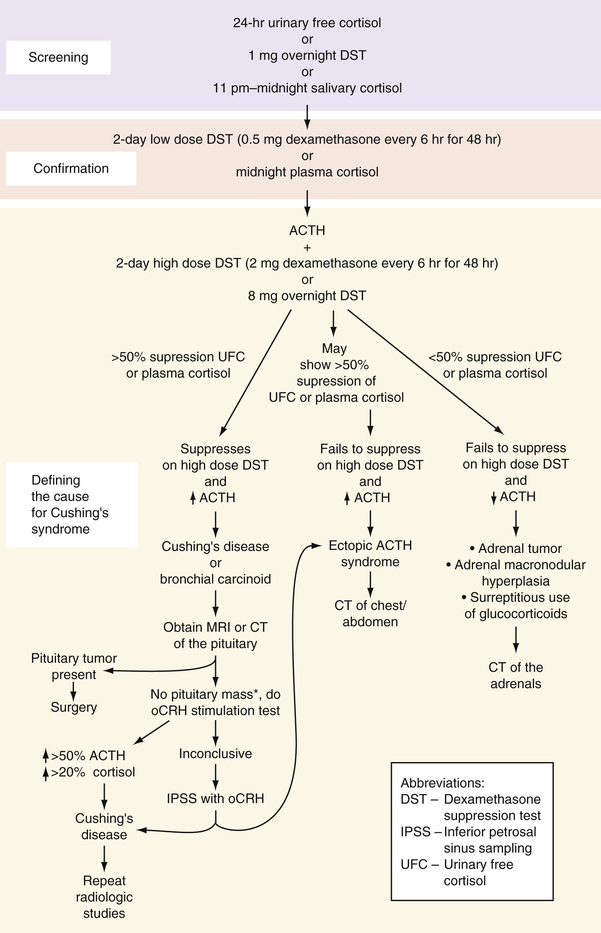
Three screening tests are used in the evaluation of a patient suspected of having Cushing's syndrome (Fig. 24-17): the 24-hour urinary free cortisol, the overnight dexamethasone suppression test (DST), and the plasma or salivary midnight cortisol (MSC) level. The 24-hour urinary free cortisol is a reflection of the unbound circulating cortisol that is freely filtered by the glomerulus. Unlike serum cortisol, it is unaffected by the level of circulating CBG. HPLC or gas chromatography coupled with tandem mass spectrometry provides the best specificity for measuring urinary free cortisol. Unlike RIA or enzyme-linked immunosorbent assay, these techniques are not affected by cross-reactivity with steroid metabolites or synthetic glucocorticoids. The upper range of normal with these methods is 110 to 138 nmol/24 hours (40 to 50 µg/hour) (Raff & Findling, 2003). The creatinine should be measured in all collections to ensure the adequacy of the specimen. Urinary cortisol excretion is decreased when the glomerular filtration rate is <30 mL/minute, and thus may be normal despite the presence of excessive cortisol production (Arnaldi et al, 2003). It has been suggested that the 1-mg overnight DST should be the preferred screening test used for these individuals (Nieman et al, 2008). Because most pediatric patients being evaluated for Cushing's are of adult weight (>45 kg), adult normal ranges may be used (Nieman et al, 2008).
As per the latest guidelines for the diagnosis of Cushing's syndrome, any UFC concentration above the upper limit of normal for that particular assay should be considered a positive test (Nieman et al, 2008). Values greater than four times the upper limit of normal are diagnostic of Cushing's syndrome. From 10% to 15% of patients with Cushing's syndrome will have at least one in four 24-hour urine collections for free cortisol return as normal (Nieman & Cutler, 1990). If cortisol excretion is normal but clinical suspicion is high, the study should be repeated or a different screening method should be used. Milder elevations of UFC can be seen in pseudo-Cushing's and during normal pregnancy. Pseudo-Cushing's is an entity characterized by HPA axis overactivity but without true Cushing's syndrome. It has been described in depression, anxiety disorders, alcoholism, poorly controlled diabetes, and morbid obesity. Refer to the Pseudo-Cushing's section later in the chapter for more details. The overnight dexamethasone suppression test is a much simpler test to perform. The patient takes 1 mg of dexamethasone orally between the hours of 11 PM and 12 midnight. The plasma cortisol is drawn the following morning between 8 AM and 9 AM, respectively. The original criterion for an abnormal response was failure to suppress the morning cortisol level to <5 µg/dL (138 nmol/L); this has been revised downward to <1.8 µg/dL (50 nmol/L) (Findling & Raff, 1999; Arnaldi et al, 2003; Nieman et al, 2008). Using the lower cutoff of < 1.8 µg/dL (50 nmol/L) provides a greater than 95 % sensitivity and 80% specificity, and serves to minimize the number of false-positive results (Nieman et al, 2008). Failure to suppress could be due to Cushing's syndrome or pseudo-Cushing's, and may even occur in some patients who are normal. The false-positive rate can be as high as 30% for a variety of reasons: Dexamethasone was taken too early; the patient was on phenobarbital, phenytoin, or another medication known to accelerate the metabolism of dexamethasone; malabsorption; alcoholism; or morbid obesity. Pregnancy and drugs such as estrogen, which increase serum transcortin (aka CBG), may also result in elevated cortisol levels. Because of these and possibly other factors, about 1% of healthy individuals, 13% of obese patients, 50% of women on estrogens, and 25% of hospitalized and chronically ill patients show false-positive overnight dexamethasone suppression tests (DSTs). False-negative results, on the other hand, occur in less than 2% of patients.
The midnight salivary cortisol (MSC) concentration is a simple, convenient, and accurate means by which to diagnose Cushing's syndrome. MSC carries a high diagnostic sensitivity and specificity, and has been demonstrated to have an excellent correlation with the plasma free cortisol concentration (Papanicolaou et al, 2002; Yaneva et al, 2004; Alwani et al, 2014). Using a cutoff of 9.3 nmol/L on a commercial ELISA kit, MSC had a sensitivity of 100%, specificity of 83%, PPV of 94%, and NPV of 100% (Alwani et al, 2014). It performs slightly better than the 24-hour UFC in distinguishing patients with Cushing's syndrome from obese subjects (Yaneva et al, 2004).
Several studies describe the use of a late-night (11 PM to 12 AM) salivary cortisol (LNSC); others use the terms MSC and LNSC interchangeably, although the methodologies describe the samples as being collected at midnight. There are no studies comparing MSC with LNSC; however, because the data for MSC is more robust, it is the preferred test.
The patient can collect the sample at home. Two methods can be used to collect the sample: The patient can chew on a cotton pledget for 2 to 3 minutes and then place the pledget into a plastic tube; alternatively, the patient can passively drool directly into a test tube. The sample is then mailed to the reference laboratory; because salivary cortisol is very stable, there is little concern about degradation during transport. The sample can be analyzed either by immunoassay or liquid chromatography–mass spectrometry (LC-MS/MS). While immunoassay is more widely used, LC-MS/MS is useful in evaluating for surreptitious use of steroids and for steroid excess in those using inhaled steroids (Raff, 2013). Normative values vary according to the reference laboratory. Results should be interpreted with caution in those who may have blunting of their circadian rhythm (e.g., shift workers, patients with depression, critical illness). Falsely elevated results may be seen in smokers and users of chewing tobacco, in which case refraining from use for 24 hours prior to collection is recommended.
Screening in special populations.
The preferred screening test during pregnancy is the UFC. UFC levels normally rise during the second and third trimesters, therefore UFC values greater than three times the upper limit of normal are considered consistent with Cushing's syndrome (Nieman et al, 2008).
Because of the decrease in cortisol filtration with declining renal function, the 1-mg DST is the recommended screen in patients with renal failure. For those suspected of having cyclic Cushing's, use of serial UFC or MSC is suggested. The diagnostic thresholds for MSC during pregnancy and in renal disease remain to be established.
For an incidentally discovered adrenal mass, the 1-mg DST has been shown to be superior to the UFC and MSC (Raff, 2013).
Confirmatory Tests for the Diagnosis of Cushing's Syndrome.
For equivocal screening results, one option would be to repeat testing in 3 months. A more definitive way to confirm the diagnosis of Cushing's syndrome is with a midnight plasma cortisol or 2-day low-dose DST performed alone or with the administration of CRH.
Midnight plasma cortisol requires hospital admission for at least 48 hours, insertion of a line for IV access before 10 PM, the patient sleeping at the time of the blood draw, and availability of staff to draw the blood at midnight. A midnight plasma cortisol greater than 7.5 µg/dL (207 nmol/L) is diagnostic of Cushing's; this has a 100% specificity for normals and those with pseudo-Cushing's (Newell-Price et al, 1995; Papanicolaou et al, 1998). The low-dose (2-day 2 mg) dexamethasone suppression test (LDDST) requires the collection of two baseline 24-hour urine samples for UFC. The patient is then given 0.5 mg dexamethasone orally every 6 hours, starting at 9 AM for 2 days. On day 2 of dexamethasone, another 24-hour urine sample is collected for UFC. All 24-hour urine collections should include the measurement of creatinine to assay for adequacy of the collection. A normal response is a decrease in UFC to less than 10 µg (27 nmol) per 24 hours on the second day of dexamethasone. This has a sensitivity of 97% to 100% for discriminating patients with Cushing's syndrome from normal individuals (Newell-Price et al, 1998). Instead of measuring UFC, serum cortisol can be measured at baseline (9 AM day 1) and at 48 hours following the first dose of dexamethasone. A normal response consists of suppression of the plasma cortisol to less than 1.8 µg/dL (50 nmol/L), which has a sensitivity and specificity of more than 95% (Newell-Price et al, 1998). Performance of the LDDST with CRH has been purported to better distinguish Cushing's syndrome from pseudo-Cushing's (Yanovski et al, 1993). For this test, 1-µg/kg or 100 µg ovine CRH is administered IV 2 hours after the last dose of dexamethasone. Cortisol is measured prior to the first dose of dexamethasone on day 1, immediately prior to the administration of synthetic ovine CRH (oCRH), and then 15 minutes following oCRH. A dexamethasone level should be measured at the time of CRH administration to exclude false-positive results. Adherence to the time schedule is important for both LDDST and LDDST with CRH. The normal response for both tests is a serum cortisol <1.8 µg/dL (<50 nmol/L).
Tests for Defining the Cause of Cushing's.
Once the diagnosis of Cushing's syndrome is established, the next step is to determine its cause. Cushing's is categorized as being ACTH dependent or ACTH independent; this is accomplished by measuring the plasma ACTH, or by performing a high-dose DST or a CRH stimulation test (Table 24-16).
TABLE 24-16
Differential Diagnosis of Hormonal Values Seen in Cushing's Syndrome
| Cause | Plasma ACTH | Plasma Cortisol (PM) | High-Dose or Overnight Dexamethasone Suppression |
| Pituitary-dependent | N—slightly ↑ | ↑ | Yes |
| Adrenal disease | ↓—undetectable | ↑ | No |
| Ectopic Cushing's* | ↑↑↑ | ↑↑ | Usually no |
| Pseudo-Cushing's | N—slightly ↑ | N–↑ | Usually yes |
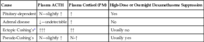
ACTH-independent Cushing's syndrome results from autonomous production of cortisol, which would feed back on the hypothalamus and pituitary gland, causing suppression of CRH and ACTH secretion. The technique for measuring plasma ACTH has been described previously. Values below the level of detection or below 10 pg/mL (2 pmol/L) at 9 AM with concomitantly elevated cortisol support an ACTH-independent cause for Cushing's syndrome. Plasma levels greater than 20 pg/mL (4 pmol/L) in the face of hypercortisolism are strongly suggestive of an ACTH-dependent cause. A CRH stimulation test with measurement of ACTH is indicated for values between 10 and 20 pg/mL (2 to 4 pmol/L) (Orth, 1995). Although patients with ectopic ACTH production tend to have markedly elevated ACTH levels, there is great overlap with the levels seen in Cushing's disease; therefore, it cannot be used to reliably distinguish between these entities.
The high-dose (2-day 8-mg) DST is useful for distinguishing Cushing's disease from adrenal or ectopic Cushing's. It is of note that cortisol levels in neuroendocrine tumors such as bronchial carcinoid may also suppress on high-dose dexamethasone. For the high-dose DST, a baseline 24-hour urine sample for UFC is collected. Beginning on day 1, the patient receives 1 mg of dexamethasone orally every 6 hours for 48 hours; urine for UFC is collected on day 1 and on day 2. Suppression of UFC or plasma cortisol by 50% or more from baseline is diagnostic of Cushing's disease. As an alternative to measuring UFC, plasma cortisol can be collected before, during, and 6 hours after the last dose of dexamethasone. Most patients with adrenal adenomas, carcinomas, or ectopic ACTH syndromes do not show suppression. This test has a sensitivity and specificity of 60% to 85%; the greater the degree of suppression, the greater the specificity.
The 8-mg overnight DST has been reported by several authors to share similar sensitivity and specificity to the high-dose DST (Dicheck et al, 1994). An 8 AM plasma cortisol is obtained, and 8 mg of dexamethasone is administered orally at 11 PM. An 8 AM plasma cortisol is obtained the next morning. Reduction of cortisol to less than 5 µg/dL is strongly supportive of Cushing's disease (see Fig. 24-17).
Most pituitary tumors and a few ectopic ACTH-secreting tumors will respond to CRH stimulation with an increase in plasma ACTH and cortisol, whereas little response is noted in those with adrenal Cushing's. For the CRH stimulation test, two basal plasma cortisol and ACTH levels are collected about 5 minutes apart, and then 1 µg/kg of body weight or a single dose of 100 µg of oCRH is injected intravenously. Blood is sampled every 15 minutes for 1 to 2 hours for cortisol and ACTH. Normal subjects will show a rise in ACTH and cortisol of approximately 15% to 20%, while those with Cushing's disease typically show a greater than 50% rise in ACTH and a greater than 20% rise in cortisol above baseline (Kaye & Crapo, 1990; Newell-Price et al, 2002; Stewart, 2003). Ovine CRH is superior to human CRH in distinguishing among the different causes of Cushing's syndrome (Nieman et al, 1989). Vasopressin stimulates ACTH release through the V3 receptors in the anterior pituitary gland. Administration of 10 units of vasopressin intramuscularly normally causes doubling of ACTH levels and an increase in serum cortisol of 150 µg/L over baseline. This offers no advantage over testing with CRH. It may prove useful, however, in differentiating Cushing's disease from pseudo-Cushing's syndrome and in the postoperative assessment of Cushing's disease (Newell-Price et al, 2002).
In rare instances, it may be necessary to use selective sampling of inferior petrosal sinus blood to document that the pituitary gland is the source of excess ACTH. This invasive, technically difficult procedure carries a relatively high cost and complication rate and should be performed only at a center well experienced with this technique. The ACTH concentration in the petrosal sinus blood should exceed twice that of peripheral venous blood to ensure diagnostic sensitivity and specificity of 100% (Orth, 1995). In those with ectopic production, the ratio is usually less than 1.4 : 1. Use of CRH during sampling increases the sensitivity and specificity.
Given that the incidence of incidentaloma of the adrenal glands and pituitary gland has been reported to be as high as 10%, imaging studies should be performed only after the hormonal diagnosis of Cushing's disease or syndrome has been made. In Cushing's disease, visualization of the sella is performed by MRI or CT, with and without contrast, with MRI being the preferred study. For adrenal Cushing's, the test of choice is a CT of the abdomen; for ectopic disease, CT of the chest may also be needed.
Pseudo-Cushing's Syndrome
Excess activity of the hypothalamic-pituitary axis, similar to that seen in pituitary Cushing's syndrome, has been demonstrated in some patients with alcoholism, major depression, poorly controlled diabetes, and obesity. Resolution of the underlying problem results in normalization of the HPA axis. These patients may not suppress on a low-dose DST and may have elevated UFC. Depressed patients who fail to suppress cortisol secretion in response to dexamethasone also show an impaired ACTH response to exogenous CRH, but will usually retain a normal cortisol response (Gold et al, 1986; Thalen et al, 1993). A single serum cortisol above 7.5 µg/dL at midnight has been shown to discriminate Cushing's syndrome from pseudo-Cushing's with 100% specificity (Papanicolaou et al, 1998). A more definitive test for distinguishing between these two entities is the combined dexamethasone-oCRH test. 1 µg/kg or 100 µg oCRH is infused 2 hours after a low-dose dexamethasone test. A plasma cortisol concentration greater than 38 nmol/L measured at 15 minutes following the administration of oCRH correctly identifies all cases of Cushing's syndrome and all cases of pseudo-Cushing's, with a specificity, sensitivity, and diagnostic accuracy of 100% (Yanovski et al, 1993).
Adrenal Insufficiency
Adrenal insufficiency is categorized according to the key site of dysfunction within the HPA axis: primary (adrenal), secondary (pituitary), and tertiary (hypothalamic). A major distinction between primary adrenal insufficiency and either of the central causes is that primary disease is associated with mineralocorticoid deficiency. In the Western world, primary adrenal insufficiency, also known as Addison's disease, is most commonly due to autoimmune adrenalitis (70% to 90% of all cases); other causes include tuberculosis (most common cause worldwide), granulomatous disorders, metastatic disease, hemorrhage, human immunodeficiency virus, acquired immunodeficiency syndrome, and infection. The most common cause of central adrenal insufficiency is HPA axis suppression due to prolonged treatment with pharmacologic doses of steroids.
Tests for the Diagnosis of Adrenal Insufficiency
Basal Hormone Measurements
An 8 AM to 9 AM plasma cortisol of less than 3 µg/dL (83 nmol/L) is indicative of adrenal insufficiency and obviates the need for further testing. Although most patients with hypocortisolism have low serum cortisol levels, a level drawn during a time of stress that falls within the reference range does not exclude the diagnosis. Rather, it may support this diagnosis, as a suboptimal cortisol level may have risen into the reference interval in response to a very high ACTH level induced by stress. A random cortisol of greater than 25 µg/dL (700 nmol/L) during critical illness makes it unlikely that the patient is adrenally insufficient (Burke, 1985), whereas a random cortisol of less than 10 µg/dL is strongly suggestive of adrenal insufficiency (Fleseriu & Loriaux, 2009).
ACTH Stimulation Test
The most convenient procedure for studying patients suspected of having hypocortisolism is the ACTH stimulation test. This test, which can be performed at any time of day, consists of drawing a baseline serum cortisol and then administering 250 µg of Cosyntropin (a commercially available ACTH analog) intravenously or intramuscularly. Serum for cortisol is again collected at 30 and 60 minutes post-Cosyntropin. A normal response is a cortisol level greater than 18 to 20 µg/dL (500 to 550 nmol/L) at either time point (Speckart et al, 1971; Burke, 1985). Drawing an ACTH level at baseline may help in distinguishing primary adrenal insufficiency (associated with an elevated ACTH usually greater than 50 to 100 pg/mL) from secondary or tertiary forms (which would have a low ACTH level less than 10 pg/mL) (Oelkers et al, 1992) (refer to the Laboratory Measurement of ACTH section earlier in the chapter). The aldosterone response to ACTH may also help in making this distinction; failure of aldosterone to increase by more than 4 ng/dL over baseline suggests primary adrenal dysfunction.
Steroids should never be withheld from a critically ill patient suspected of having adrenal insufficiency; instead a random sample for cortisol and ACTH should be drawn and treatment initiated with dexamethasone, which does not interfere with the cortisol assay. Formal testing should take place within 72 hours, before HPA axis suppression occurs. About 90% of cortisol circulates bound to CBG and albumin, and approximately 10% circulates freely (unbound). CBG declines during critical illness, resulting in an increase in the fraction of unbound (free) cortisol compared with the bound fraction. Because the serum total cortisol concentration does not accurately reflect this alteration in free/bound cortisol, it is common to overdiagnose adrenal insufficiency in the critical care setting. Since access to laboratories that readily and reliably measure free cortisol is limited, some have advocated use of the free cortisol index (FCI) and calculated free cortisol (CFC) to better delineate those patients in true need of steroid replacement therapy (Beishuizen et al, 2001; Hamrahian et al, 2004; Ho et al, 2006). To obtain the FCI, it is necessary to measure the serum CBG. CFC is the total cortisol (nmol/L)/CBG (mg/L), with normal being a value above 12. Given that for the vast majority of institutions, determination of CBG requires that the sample be sent to a reference laboratory, it may be more prudent and certainly more expedient to simply send aliquots from t-0, t-+30, and t-+60 minutes post–ACTH administration for serum free cortisol, and provide steroid coverage pending these results (Fleseriu & Loriaux, 2009). Checking the ACTH level at t-0 will help to further classify adrenal insufficiency as being primary versus secondary or tertiary. Salivary cortisol, which is unaffected by changes in CBG, may eventually prove useful in this setting (Raff, 2009).
Some have supported the use of a 1-µg dose of ACTH instead of the full 250-µg dose for diagnosing adrenal insufficiency. The premise is that the massively supraphysiologic dose of 250 µg will cause those with partial adrenal insufficiency to mount a normal response, and that these individuals will fail to show an adequate rise in cortisol in response to a 1-µg test dose. This procedure has not gained wide acceptance, nor has the normal range been standardized; more importantly, the diagnosis of partial adrenal insufficiency remains in contention.
The ITT has long been considered the gold standard for assessing the HPA axis. Unlike the ACTH stimulation test, the ITT assesses the integrity of the entire HPA axis. If the HPA axis is intact, insulin-induced hypoglycemia stimulates the hypothalamus and the pituitary gland to secrete CRH and ACTH, respectively, which, in turn, leads to a rise in cortisol. This test is contraindicated in individuals with ischemic heart disease, in older adults (>70 years of age), during pregnancy, and in those with a history of seizures. Patients highly suspected of having central adrenal insufficiency (i.e., prior pituitary surgery or radiation therapy) should receive a lower dose (0.05 U/kg) of insulin, rather than 0.1 to 0.15 U/kg. Glucose and cortisol are measured at –15 minutes, at baseline, and then at 15, 30, 45, 60, 90, and 120 minutes following insulin administration. For the test to be valid, adequate hypoglycemia must be attained (signs and symptoms of hypoglycemia and glucose less than 40 mg/dL [2.2 mmol/L]). A normal response is a rise in the cortisol level to greater than or equal to 18 µg/L or 20 µg/L at any time during the test (Nelson & Tindall, 1978; Burke, 1985; Grinspoon & Biller, 1994). Preliminary work suggests there was no difference in peak GH or cortisol level attained when analog insulin was substituted for regular insulin for the ITT (Yuen et al, 2008).
The cortisol response to hypoglycemia can be predicted reliably by the response to acute ACTH stimulation—a safer, cheaper, and quicker test (Stewart, 2003). The ITT is used mainly as a second-line measure to further evaluate those patients who had a borderline response to the ACTH stimulation test.
In the days before the commercial availability of a reliable assay to measure ACTH, it was common to perform a prolonged ACTH stimulation test (ACTH infusion over a 2-day period) to distinguish central from primary adrenal insufficiency. In other hormone systems, lack of stimulation normally leads to upregulation of the end-organ receptors; however, the adrenal glands downregulate their ACTH receptors in response to lack of stimulation. A short ACTH stimulation test would typically produce an inadequate response. By priming the system with prolonged exposure to ACTH, those with hypothalamic disease display a delayed but normal rise in cortisol.
Overnight Metyrapone Test
Metyrapone has also been used to assess the integrity of the HPA axis. Metyrapone inhibits 11-β-hydroxylase, preventing the conversion of 11-deoxycortisol (compound S) to cortisol. Under normal circumstances, the metyrapone-induced drop in cortisol is detected at the level of the hypothalamus and pituitary gland; a compensatory increase in CRH and ACTH secretion occurs, stimulating steroidogenesis and leading to a buildup of 11-deoxycortisol, the hormone preceding the block. Patients with central adrenal insufficiency fail to increase CRH and/or ACTH, steroidogenesis is not stimulated, and 11-deoxycortisol fails to increase. Metyrapone 30 mg/kg is administered orally at midnight; blood for cortisol and 11-deoxycortisol is collected the following morning at 8 AM. A rise in 11-deoxycortisol to greater than 7 µg/dL (200 nmol/L) is normal (Fiad et al, 1994; Grinspoon & Biller, 1994). An abnormal response is defined by 11-deoxycortisol less than 7 µg/dL accompanied by cortisol less than 5 µg/dL. This test should be performed with caution as it may provoke an addisonian crisis in those with central hypocortisolism. Metyrapone is currently only available on a compassionate basis; as a result this test is rarely performed.
Corticotropin-Releasing Hormone Test
The oCRH test can be used not only to diagnose hypocortisolism, but to localize the site of damage (Schulte et al, 1984). It is a safe test; however, it is expensive. After blood is drawn for baseline ACTH and cortisol, 100 µg oCRH is administered intravenously. Blood is then collected for cortisol and ACTH every 15 minutes for 60 to 90 minutes. A normal response is seen as cortisol greater than 20 µg/dL. Basal ACTH levels and their change in response to oCRH are used to localize the site of pathology. Plasma corticotropin usually peaks at 15 to 30 minutes, and cortisol usually peaks at around 30 to 40 minutes post oCRH injection (Oelkers, 1996).
Both the bound and free cortisol concentrations show a progressive rise throughout pregnancy. With this in mind, it has been recommended that higher cutoffs be used to properly interpret the ACTH stimulation results in pregnancy (Jung et al, 2011). A random morning cortisol level less than 11 µg/dL, 16 µg/dL, and 22 µg/dL when measured during the first, second, and third trimester of pregnancy, respectively, should raise the suspicion of adrenal insufficiency (Jung et al, 2011). A serum cortisol 60 minutes post-ACTH of less than 25 µg/dL, 29 µg/dL, and 32 µg/dL for the first, second, and third trimester of pregnancy, respectively, is diagnostic of adrenal insufficiency (Lebbe & Arlt, 2013; Yuen et al, 2013).
Antibodies against 21-hydroxylase antigen should be assayed in those whose Addison's disease (primary adrenal insufficiency) does not have an obvious explanation. Positive antibodies confirm autoimmune adrenalitis; these patients should be screened and monitored for the development of other autoimmune disorders. The absence of antibodies does not rule out an autoimmune etiology, as titers usually drop in concurrence with adrenal destruction.
The use of steroids in the treatment of malignant, inflammatory, and immunologic disorders is a common iatrogenic cause of adrenal insufficiency. The degree of adrenal suppression is dependent on the specific glucocorticoid dose, duration, frequency, and route of administration. Assessment of adrenal function in patients being tapered off of exogenous steroids can be performed once they are tapered to the equivalent of a daily dose of 10 mg hydrocortisone. Have them omit the morning glucocorticoid dose, and measure the 8 AM cortisol level. If it is greater than 10 µg/dL, routine supplementation of steroids can be ended. Because the adrenal cortex lags behind the pituitary gland in recovery from steroid suppression, complete adrenal recovery can also be demonstrated by an appropriate rise in serum cortisol following an 8 AM Cosyntropin infusion.
Renin-Aldosterone Axis
At least 20 million people in the United States have hypertension, and 90% to 98% of cases are classified as essential hypertension. The mortality and morbidity from associated myocardial, cerebrovascular, and renal complications necessitate aggressive treatment of this disorder. Investigation into the etiology of hypertension revealed the importance of the renin-angiotensin-aldosterone system, not only in the origin and persistence of hypertension but also as a guide to its treatment. The role of the renin-aldosterone axis is to maintain blood pressure within normal limits by sensing and responding to changes in plasma volume, salt balance, and renal perfusion pressure.
Renin is a proteolytic enzyme which, although formed by other tissues throughout the body, is mainly formed and stored by juxtaglomerular (JG) cells within the macula densa of the kidney. Renin is synthesized from a larger precursor protein, prorenin (big renin), and is converted to its active form within the JG cells. The release of renin into the circulation occurs in response to a variety of triggers: a drop in renal hydrostatic pressure, hyponatremia, hypokalemia, or a decrease in catecholamines, angiotensin II, or atrial natriuretic hormone. Both suppression of plasma renin activity induced by potassium administration and stimulation of renin activity following potassium depletion occur independently of changes in aldosterone secretion or in sodium balance (Brunner et al, 1970).
Several isoenzymes of renin are known. Their release is regulated by cAMP. Renin acts on angiotensinogen (renin substrate), an α2-globulin formed by the liver, converting angiotensinogen to angiotensin I. Angiotensin I in turn is converted into angiotensin II by the angiotensin-converting enzyme (ACE). ACE is found in the pulmonary and vascular endothelia, as well as in cell membranes of the kidney, heart, and brain (Brewster & Perazella, 2004). It is believed that angiotensin II is the peptide responsible for the physiologic effects on target tissue. Angiotensin II not only stimulates the release of aldosterone, but it is also a very potent vasoconstrictor that can stimulate the release of catecholamines from the adrenal glands and norepinephrine from the sympathetic nervous system, and stimulate the release of vasopressin. The octapeptide angiotensin II is further split to a heptapeptide, angiotensin III; in addition, angiotensin I may be changed directly to angiotensin III without being converted to angiotensin II. Although speculation about the functions of angiotensin III is ongoing, it appears to modulate aldosterone secretion in a similar manner to angiotensin II. The active angiotensins are rapidly cleared by various aminopeptidases (angiotensinases) within the circulation and during transit through tissues. These relationships are shown in Figure 24-18.
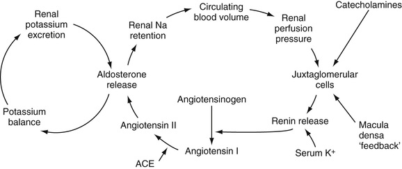
Renin, through its product, angiotensin II, directly stimulates the synthesis and secretion of aldosterone by the adrenal zona glomerulosa. Renin release, the rate-limiting step in the renin-aldosterone axis, is dependent on changes in effective plasma volume, which, in turn, are dependent on tubular reabsorption of sodium by the kidney. Low plasma volume and low serum sodium stimulate the secretion of renin, resulting in aldosterone release, which, in turn, causes sodium retention along with an increase in plasma volume and blood pressure, and potassium loss. Conversely, an increase in effective blood volume or acute elevation in blood pressure results in low renin, low angiotensin, low aldosterone, and subsequent sodium loss. In contradistinction to aldosterone, renin secretion is inhibited by hyperkalemia (Fig. 24-19).
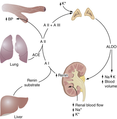
Aldosterone and glucocorticoids act at the mineralocorticoid receptor located in the cells lining the collecting ducts of the kidney to cause reabsorption of sodium, leading to an increase in intravascular volume. Aldosterone also causes renal potassium loss. Studies have suggested a role for aldosterone in the development and perpetuation of vasculopathy; it has been shown to stimulate genes for collagen synthesis, tissue growth factors, inflammatory factors, and plasminogen activator inhibitor type 1 (Brilla et al, 1994; Schunkert et al, 1997; Brown et al, 2000; Rocha et al, 2000; Luft, 2002). The synthesis of aldosterone is restricted to the zona glomerulosa; here the enzyme aldosterone synthetase (CYP11B2) converts corticosterone to aldosterone. The major stimuli for aldosterone secretion are angiotensin II and hyperkalemia. ACTH is a much weaker stimulus. Somatostatin, atrial natriuretic hormone, dopamine, and heparin directly inhibit aldosterone synthesis. Cortisol circulates at a much higher concentration than aldosterone, and although it binds with equal affinity to the mineralocorticoid receptor, it does not normally function as a major mineralocorticoid. The reason is that the enzyme 11-β-hydroxysteroid dehydrogenase II (11-HSD-2, also known as CYP11B2), expressed by the mineralocorticoid targets cells in the collecting tubules acts to convert cortisol to cortisone, an inactive metabolite.
Renin and Hypertension
The work of Laragh and colleagues has indicated that essential hypertension can be classified on the basis of renin measurements as high, low, or normal renin, and that drug selection can be based on this classification (Laragh et al, 1972; Laragh, 1993). About 15% of patients with essential hypertension have high-renin hypertension. Hyperreninemia resulting from renal parenchymal disease or renal vasculopathy leads to increased aldosterone production and the subsequent retention of sodium and enhanced potassium excretion. Patients with this condition are hypervolemic, intensely vasoconstrictive, and more prone to ischemic injury. Renin profiling has shown renin to be an independent risk factor for myocardial infarction. Even if pressures are controlled by antihypertensives, if they do not address plasma renin activity, such patients fare worse than individuals with lower renin levels (Alderman et al, 1991). Increased aldosterone (secondary aldosteronism) may contribute significantly to the symptoms and course of high-renin hypertension. Some of the causes of high-renin hypertension are listed in Box 24-7.
Renin-secreting tumors are extremely rare and can be difficult to diagnose. They can be benign or malignant, renal or extrarenal. The most common location is in the juxtaglomerular apparatus. A young patient with markedly elevated plasma renin activity, hyperaldosteronism, and hypokalemia in the absence of a renovascular lesion typifies the classic clinical presentation (Corvol et al, 1994). Malignant hypertension is also associated with high plasma renin activity and aldosterone. When such patients are given medications that interrupt the renin-aldosterone axis, such as ACEIs, blood pressure often normalizes. In renal vascular hypertension, the renin and aldosterone levels are also elevated. However, in contradistinction to malignant hypertension, treatment with ACEs usually results in further deterioration of renal function. Patients suspected of having renal artery stenosis should undergo further testing (e.g., captopril-induced renogram, renal duplex ultrasound, magnetic resonance angiography). A positive test leads to renal vein angiography and catheterization for selective blood sampling. In unilateral renal disease, both plasma renin and aldosterone are elevated. Asymmetry in the renin levels obtained during renal vein catheterization offers one of the best measurements to judge the likelihood of blood pressure response to corrective surgery. When the ratio of plasma renin in the renal vein of the affected to the nonaffected side is at least 1.5 : 1, surgery may lead to improvement. With suppression of renin release from the nonaffected side, renal vein renin levels approximating those found in blood specimens obtained from the inferior vena cava also indicate probable success of curative surgery. Almost 40% of these patients have peripheral blood plasma renin activity that is within the reference interval (Streeten et al, 1979); consequently, peripheral plasma renin is a poor predictor of response to surgery. Renin secretion can be found in lung cancer, hepatic hamartoma, and other rare conditions (Anderson et al, 1989).
When an acceleration of hypertension occurs, renin is usually markedly increased; however, with chronic renal failure, almost any renin level can be expected. A small number of hypertensive patients on dialysis have intractable, accelerated hypertension. In those patients in whom dialysis cannot control the hypertension, markedly elevated plasma renin levels can be lowered by nephrectomy or, in the case of renal artery stenosis, angioplasty or revascularization (Whitehouse et al, 1981; Sonkodi et al, 1990; Tullis et al, 1999). ACEIs can be used to mitigate the rise in aldosterone; however, in the setting of renal disease, they can precipitate or worsen hyperkalemia. In renal transplant patients, elevated plasma renin may be indicative of renal ischemia and rejection.
Systemic hypertension occurs in patients with Cushing's syndrome. Some of these patients have increased plasma renin and renin substrate. Other Cushing's syndrome patients have hypokalemia associated with secretion of a minor mineralocorticoid (such as DOC or corticosterone) that causes hypertension (Krakoff et al, 1975). In still other patients, severely elevated cortisol levels overwhelm the capacity of 11-hydroxysteroid dehydrogenase (11-HSD) to convert cortisol to its metabolically inactive form, cortisone, leading to a picture of excess mineralocorticoid activity with suppressed plasma renin activity. Other causes of high-renin hypertension include treatment with medications such as diuretics, vasodilators, or other antihypertensives. Hormonal agents such as glucocorticoids, as well as some estrogen-containing oral contraceptives, have been found to increase renin substrate activity.
Even though plasma renin activity, aldosterone, and urinary sodium excretion may be normal in 60% of hypertensive patients, evidence indicates that renin-angiotensin plays a significant part in normal-renin hypertension. The response of many hypertensive patients with normal renin to angiotensin-converting enzyme inhibitors or angiotensin II antagonists (saralasin) has implicated renin and angiotensin II in sustaining hypertension in these patients. Moreover, Ames showed that once blood pressure was increased by angiotensin infusion, it could be maintained with only one fifth of the original dose (Ames et al, 1965).
It was found that low-renin essential hypertension, which involves chronic expansion of plasma and extracellular fluid volume, is characterized by aldosterone oversecretion and responds to diuretic therapy. At least 25% of patients with essential hypertension are found to have low-renin hypertension. Most investigators have characterized this state as hyporesponsive, meaning that low-renin hypertensive patients fail to respond as well as healthy subjects do with upright posture, sodium restriction, diuretics, vasodilators, or a combination of these. Atrial natriuretic peptide may be elevated in these patients, and the response of aldosterone to renin may be exaggerated (Sergev et al, 1990). Renin suppression increases with age and appears to be more common in women and in the black population.
Listed in Box 24-8 are a group of syndromes associated with low levels of plasma renin. These have been divided into a subgroup that is of adrenal origin (primary) and a subgroup that is nonadrenal or secondary in origin. Primary aldosteronism is uncommon compared with renin hypertension. It is characterized by (1) systolic and diastolic hypertension caused by oversecretion of aldosterone by an adrenal adenoma (aldosterone-producing adenoma [APA]) or hyperplasia (idiopathic hyperaldosteronism [IHA]), (2) low renin or a high aldosterone/renin ratio (>50), (3) renal potassium wasting, and (4) sodium retention. It should be suspected in patients with hypertension and spontaneous or diuretic-induced hypokalemia, resistant hypertension requiring more than three medications, onset younger than 30 years of age, or an incidentally found adrenal mass. Several potential screening tests have been used to detect primary aldosteronism and to separate unilateral aldosterone-producing adenomas from other causes of primary aldosteronism. A low-salt diet (<2 g/day), stress, upright posture, and diuretics all increase plasma aldosterone, whereas a high-salt diet and lying in a supine position suppress aldosterone secretion in healthy subjects. Combinations of these maneuvers have been used in the diagnosis of excessive aldosterone secretion; however, they do not reliably distinguish APA from IHA. When healthy subjects are placed on a high-salt diet and lie in the supine position, they suppress their plasma aldosterone levels to less than 10 ng/dL (278 pmol/L).
Primary Hyperaldosteronism: Screening and Confirmation Tests
The algorithm proposed by Blumenfeld and colleagues for evaluating hypertensive patients suspected of having primary hyperaldosteronism is to initially measure the serum potassium (Blumenfeld et al, 1994). If it is less than 3.6 mmol/L, then plasma renin activity is measured. Plasma renin activity of less than 1.0 ng/mL/hour leads to 24-hour urine collection for aldosterone and potassium excretion. Findings of 24-hour urinary potassium greater than 30 mmol/24 hours in the face of hypokalemia and of aldosterone greater than 15 mg/24 hours lead to the localization of the underlying pathology to the adrenal gland.
A more recent set of guidelines for the detection and diagnosis of primary hyperaldosteronism, as proferred by the Endocrine Society, include the following (Funder et al, 2008): Although patients with primary hyperaldosteronism classically present with spontaneous or easily provokable hypokalemia, many in fact will have serum potassium levels that are within the normal range. Thus, the preferred screening test is a plasma aldosterone concentration/plasma renin activity (PAC/PRA) ratio, PAC expressed in ng/dL, and PRA in ng/mL/hour. This is performed after the patient has remained upright for at least 2 hours. The patient should have stopped spironolactone and eplerenone for 4 to 6 weeks and other diuretics for at least 2 weeks before testing. Hypokalemia should be corrected (as it may suppress aldosterone secretion), and the patient should not be on a sodium-restricted diet. A PAC/PRA ratio greater than 30 is suggestive of primary hyperaldosteronism; a value greater than 50 is virtually diagnostic (Weinberger & Fineberg, 1993; Blumenfeld et al, 1994). It is of note that the lower limit of normal for the PRA assay varies by laboratory; therefore, the diagnosis is supported only when both the PAC/PRA ratio and the PAC are elevated. The preferred technique for measuring PRA is a validated immunometric assay. HPLC tandem mass spectrometry is preferred for measuring plasma and urine aldosterone (Funder et al, 2008). The PAC/PRA ratio is highly dependent on the renin concentration; as such, it is important to use an assay with sufficient sensitivity in the lower ranges (e.g., <0.2 to 0.3 ng/mL/hour).
The diagnosis of primary hyperaldosteronism is confirmed by performing one of the four following confirmatory tests: oral sodium loading test, saline infusion test, fludrocortisone suppression test, or the captopril challenge test (Funder et al, 2008). Saline suppression can be accomplished by infusing 2 L of 0.9% saline over 4 hours, or by administering sodium chloride tablets 10 to 12 g daily for 3 days. PAC at the end of the 4-hour infusion of less than 5 ng/dL (140 pmol/L) makes the diagnosis of hyperaldosteronism unlikely. A PAC greater than 10 ng/dL (280 pmol/L) strongly supports the diagnosis of primary hyperaldosteronism, whereas values greater than 5 ng/dL (140 pmol/L) but less than 10 ng/dL (280 pmol/L) are considered indeterminate and may be seen with IHA. On the last day of oral sodium administration, a 24-hour urine sample is collected for aldosterone, creatinine (to assess for adequacy of the collection), and sodium (>200 mmol/day confirms adequate sodium intake). An aldosterone greater than 10 mg/24 hours (28 nmol/24 hours) following 3 days of sodium loading confirms the diagnosis (Bravo et al, 1983, Bravo, 1994; Holland et al, 1984). For the fludrocortisone suppression test, 0.1 mg fludrocortisone is given orally every 6 hours for 4 days, together with slow-release potassium chloride supplements (in doses sufficient to maintain the plasma potassium close to 4.0 nmol/L) and slow-release sodium chloride (30 mmol three times a day) and liberalization of dietary sodium intake (enough to maintain a urinary sodium excretion of at least 3 mmol/kg). On day 4, an upright plasma cortisol is measured at 7 AM and 10 AM, and PAC and PRA are measured at 10 AM. A PAC greater than 6 ng/dL with a PRA less than 1 ng/mL/hour confirms the diagnosis, provided that the 10 AM cortisol is less than the 7 AM value. For the captopril challenge, baseline blood is collected for PRA, PAC, and cortisol; then 25 to 50 mg of captopril is administered orally. PRA, PAC, and cortisol are again collected at 1 and 2 hours post-captopril. The patient is kept seated throughout the entire test. PAC is normally suppressed to greater than 30%; however in primary hyperaldosteronism, the PAC remains elevated and the PRA suppressed. The choice of test depends on several factors, including patient compliance, presence of uncontrolled hypertension, renal insufficiency, congestive heart failure, and local expertise.
Differentiating Among the Different Causes of Primary Hyperaldosteronism
APA differs from IHA by more extreme blood pressures, greater potassium wasting, and higher atrial natriuretic peptide levels. Also, urinary and serum levels of 18-oxocortisol, 18-hydroxycortisol (serum >300 ng/dL), and 18-hydroxycorticosterone (serum >100 ng/dL) tend to be higher. A positive postural stimulation test (plasma aldosterone: ambulating rising to less than 130% of the supine level) has a moderate sensitivity and specificity for adenoma, but not for hyperplasia. Once the diagnosis of primary hyperaldosteronism is confirmed, the next step is to image the adrenal glands by CT or MRI, looking for bilateral hyperplasia or a unilateral hypodense adenoma. The specificity of imaging to correctly categorize patients as having APA versus IHA varies from 67% to 84% (Doppmann et al, 1992; Gleason et al, 1993; Harpe et al, 1999; Magill et al, 2001). Removal of the adenoma is curative in 35% of cases and leads to improvement in another 55% (Blumenfeld et al, 1994). Surgery can cure or improve hypertension in patients with unilateral disease. For those in whom the disease is bilateral, medical treatment with drugs such as spironolactone leads to improvement in 76% of cases. Hyperplasia is not always bilateral.
As a result of multiple factors, including the increased incidence of nonfunctioning adrenal adenoma after 40 years of age, and the fact that hyperplasia may present as a unilateral adenoma on imaging, it is highly recommended that bilateral adrenal vein sampling be performed in individuals over 40 years of age who are contemplating surgery. Bilateral adrenal vein sampling is technically demanding and entails risk; however, as performed by an experienced radiologist, it is the gold standard for distinguishing adenoma from hyperplasia. An intravenous infusion of ACTH at 50 µg/hour is started 30 minutes before sampling and is continued throughout the procedure. This serves to mitigate any stress-induced fluctuations in aldosterone during the sampling, accentuate the gradient in cortisol from adrenal vein to inferior vena cava, and maximize the difference in secretion of aldosterone between the right and left adrenal glands in instances of APA. Samples for aldosterone and cortisol are obtained from each adrenal vein and from the periphery. An adrenal vein/peripheral vein cortisol of greater than 10 : 1 ensures that the samples are taken from the adrenal veins. There should be a less than 20% difference in cortisol measurements between both adrenal glands.
To correct for dilutional effects, the cortisol-corrected aldosterone ratio (i.e., aldosterone/cortisol) is utilized. In APA, the cortisol-corrected aldosterone ratio is usually at least fivefold higher on the side with the adenoma, and no gradient is seen in hyperplasia (Doppmann & Gill, 1996). As per the recent Endocrine Society guidelines, a cortisol-corrected aldosterone ratio from high side to low side of greater than 4 : 1 indicates APA, a ratio of less than 3 : 1 suggests IHA, and values between 4 : 1 and 3 : 1 are indeterminate and need to be interpreted in conjunction with imaging studies and clinical settings. Using these cutoffs, a sensitivity of 95% and a specificity of 100% are achieved (Funder et al, 2008).
Several studies have shown a discrepancy between radiologic findings and surgical findings in up to one third of all cases. Kempers found that when compared with adrenal vein sampling, CT and MRI misdiagnosed the cause of primary hyperaldosteronism in 37.8% of patients (Kempers et al, 2009). Given the increased prevalence (5% to 10%) of adrenal incidentalomas (nonfunctioning adrenal tumors) with increasing age, many authorities are recommending adrenal vein sampling for patients with hyperaldosteronism who are older than 40 years of age and have a unilateral hypodense adrenal nodule greater than 1 cm on CT scan (Young, 1999). Based on the findings of large prospective studies, others have suggested that the major strength of this technique may be its use in evaluating those patients with a high probability of APA but with normal findings or bilateral nodular disease on CT. It should be stressed that this test is technically difficult, and even in the very best of hands is successful in only approximately 92% to 96% of cases (Doppmann et al, 1992; Radin et al, 1992; Young, 1999).
The syndrome of inapparent mineralocorticoid excess is due to dysfunction of 11-β-hydroxysteroid dehydrogenase type 2 (11-β-HSD2). Cortisol can function as a potent mineralocorticoid, binding to the mineralocorticoid receptor in the kidney with the same affinity as aldosterone. 11-β-HSD2 mitigates the mineralocorticoid action of cortisol by converting it to the metabolically inactive form, cortisone. When activity of this enzyme is decreased because of hereditary factors, the ingestion of large quantities of licorice (which has a high content of glycyrrhizic acid) or chewing tobacco, hypertension, hypokalemia, low PRA, and low PAC may result. An elevated cortisol/cortisone concentration on a 24-hour urine collection confirms the diagnosis.
Renin may be suppressed by excessive sodium intake. It is also suppressed in the syndrome of hyporenin hypoaldosteronism, which is most commonly seen in patients with diabetes mellitus and renal disease. Secondary aldosteronism results from nonadrenal disease, in which both adrenal glands are stimulated (Table 24-17). Typically, these patients are not hypertensive.
TABLE 24-17
Differentiating the Various Causes of Hyperaldosteronism
| Disorder | Aldosterone | Renin | Serum K+ |
| Primary hyperaldosteronism | ↑ | ↓ | ↓ |
| Renin-secreting tumor | ↑ | ↑ | ↓—N |
| Dexamethasone-suppressible hyperaldosteronism | ↑ | ↓ | ↓ |
| Renovascular hypertension | N—↑ | N—↑ | ↓—N |
| Bartter's syndrome | ↑ | ↑ | ↓ |
| Diuretics, congestive heart failure, cirrhosis, nephrotic syndrome | ↑ | ↑ | N—↓ |
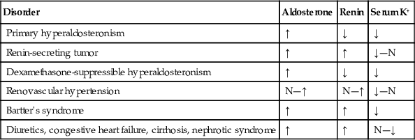
Conditions such as nephrosis, cirrhosis, and heart failure are associated with decreased renal perfusion, causing a compensatory increase in renin and aldosterone levels (secondary hyperaldosteronism). The response of the renin-aldosterone system in pregnancy is especially complex; renin, renin substrate, angiotensin II, and aldosterone appear to be increased.
There are two variants of familial hyperaldosteronism (FH), FH-1, which is also known as glucocorticoid remedial hyperaldosteronism (GRA), and FH-2. GRA is an autosomal dominant inherited disorder characterized by hyperaldosteronism, hyporeninemia, and severe, usually refractory, hypertension, with onset typically occurring in those younger than 20 years of age with a positive family history of early hypertension and/or hemorrhagic stroke. It is the result of translocation of aldosterone synthetase (CYP18, CYP11B2) onto the ACTH-sensitive promoter sequence of the 11-β-hydroxylase (CYP11B1) gene within the zona fasciculata. Thus, aldosterone synthesis is stimulated by ACTH. In addition to increased aldosterone, these individuals produce excess 18-OH cortisol and 18-oxycortisol. Diagnosis can be confirmed by normalization of aldosterone following 4 to 6 weeks of steroid suppression, or by genetic testing using Southern blot or long polymerase chain reaction (Lifton et al, 1992; Kurtz & Spence, 1993). FH-2 is an autosomal dominant condition that is associated with APA, IHA, or both. Unlike FH-1, aldosterone does not suppress in response to glucocorticoid treatment.
Liddle syndrome is a rare autosomal dominant disorder characterized by hypertension and hypokalemia; however, unlike in primary hyperaldosteronism, PRA and PAC are both low. The defect is due to a mutation in the amiloride-sensitive sodium channel within the renal collecting ducts, leading to enhanced sodium reabsorption and increased potassium secretion. Liddle syndrome can be diagnosed by genetic testing or, if genetic testing is unavailable, by good clinical response to amiloride or triamterene, and by lack of response to dexamethasone or aldosterone antagonists.
Aldosterone Measurements
An isolated aldosterone measurement with no attention to patient preparation is of little clinical value. Even when time of sampling, posture, and dietary sodium and potassium are controlled, it is difficult to discriminate with certainty between primary aldosteronism and other forms of hypertension with the use of plasma aldosterone measurements alone. Interpretation of the aldosterone level must be done in the context of a simultaneously collected plasma renin and serum potassium.
Aldosterone assays are performed on plasma using extraction to remove aldosterone from plasma proteins, followed by chromatography and immunoassay or, preferably, LC-MS/MS. Urine is assayed following acid hydrolysis and extraction. Normative values exist for different age groups, postures, and sodium intakes (Table 24-18). HPLC with tandem mass spectrometry affords greater consistency and is gradually replacing older methods.

Renin Measurements
Important technical differences have been noted in assessment of renin using current methods. Renin measurements are of two types: PRA and PRC. The PRA is a bioassay wherein a plasma specimen containing renin is allowed to react with its substrate, angiotensinogen; after a specified period of time, the reaction is terminated, and generated angiotensin I is measured by RIA. For estimation of PRA, the endogenous substrate is not eliminated. Therefore, the rate of generation of angiotensin I is influenced by the concentrations of endogenous renin and its substrate. This type of assay is the most widely used method for the determination of renin. Comparison of results among laboratories is not possible because of procedural differences (e.g., variations in pH, ionic strength, length of the assay, the angiotensinase inhibitor, the conditions under which the specimen was obtained) and because of the lack of a specific reference preparation. In addition, the literature on renin assays reveals confusion regarding units of measurement employed; even when an attempt is made to express the many arbitrary units in the same terms (nanograms of angiotensin liberated per milliliter per hour), wide ranges are reported for human plasma renin activity in reference populations.
When PRC rather than PRA is measured, the effect of substrate is eliminated. The PRC assay is an immunoassay directed to the renin molecule itself.
Assays of PRA and PRC provide similar information, except in a few clinical situations. With oral contraceptive administration, PRC remains within the reference interval, whereas PRA increases owing to the increase in substrate. Other procedures such as freezing, thawing, and acidification have been found to convert prorenin to rennin, thereby increasing the values in PRC assays.
Because renin release is controlled by many physiologic and pharmacologic variables, it is extremely important to know the conditions under which the blood specimen was obtained. Such conditions as upright posture, the administration of diuretics, and low-sodium diets are potent stimuli of renin release and should be adequately controlled before plasma renin is measured. Renin also appears to be extremely labile, so the variables involved in specimen processing should be vigorously controlled. Blood should be drawn into an iced EDTA tube, which inactivates the enzymes (e.g., angiotensinases); it should be centrifuged at 4° C, and the plasma should be separated promptly from the cells, frozen immediately, and kept frozen until ready to be analyzed; with this technique, the specimen is stable for several months at –20° C.
Direct measurement of renin substrate, angiotensin I, or angiotensin II is not used widely in clinical practice because of tedious extraction or concentration steps, as well as difficulty in eliminating formation or degradation of these compounds by proteases and other enzymes involved in the renin system. Although ACE levels can be measured easily, they are of little use in the diagnosis of hypertensive disorders.