「重要なお知らせ:日本語書籍をご購入いただき、eLibraryをご利用の皆さまへ」
エルゼビアは、より快適にサービスをご利用いただくため、システムの重要なアップ
デートを実施いたしました。
現在eLibraryで日本語電子書籍をご利用のお客様は、今後より高いアクセシビリティとセ
キュリティを備えた新しいプラットフォーム「eBooks+」へアカウントが移行されてい
ます。eBooks+のご利用については
こちらよりご利用・ご登録ください。
 (0 rating)


Book Description
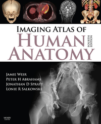
Imaging Atlas of Human Anatomy, 4th Edition provides a solid foundation for understanding human anatomy. Jamie Weir, Peter Abrahams, Jonathan D. Spratt, and Lonie Salkowski offer a complete and 3-dimensional view of the structures and relationships within the body through a variety of imaging modalities. Over 60% new images-showing cross-sectional views in CT and MRI, nuclear medicine imaging, and more-along with revised legends and labels ensure that you have the best and most up-to-date visual resource. This atlas will widen your applied and clinical knowledge of human anatomy.
- Features orientation drawings that support your understanding of different views and orientations in images with tables of ossification dates for bone development.
- Presents the images with number labeling to keep them clean and help with self-testing.
- Features completely revised legends and labels and over 60% new images-cross-sectional views in CT and MRI, angiography, ultrasound, fetal anatomy, plain film anatomy, nuclear medicine imaging, and more-with better resolution for the most current anatomical views.
- Reflects current radiological and anatomical practice through reorganized chapters on the abdomen and pelvis, including a new chapter on cross-sectional imaging.
- Covers a variety of common and up-to-date modern imaging-including a completely new section on Nuclear Medicine-for a view of living anatomical structures that enhance your artwork and dissection-based comprehension.
- Includes stills of 3-D images to provide a visual understanding of moving images.