Superficial Thermotherapy

Description

Heating modalities are generally classified by their depth of effective heating and are considered to be superficial or deep.1-5,66,107,111 Superficial thermotherapy, or the use of superficially applied heat, is an extremely common modality in the rehabilitative setting. In fact, it is so common that it may be our most overused rehabilitative modality. Like all modalities, superficial thermotherapy should be used to accomplish a specific goal and not be used as a “cure-all“ modality. The goals most appropriately treated with superficial thermotherapy include improving range of motion, increasing circulation, and reducing pain or the sense of tightness that is frequently associated with injured tissues.

Clinical Pearl #8

Thermotherapy should be used only when examination of the patient suggests that the acute inflammation has passed and the patient is in the waste removal and repair phases or beyond.

How We Think It Works

Range of motion

First coloquial, the range-of-motion effects for all thermal modalities are not a direct function of the modality itself. Instead, the thermal modality should be seen as an adjunct that allows other techniques, such as stretching or joint mobilization, to be more effective. In isolation, thermal modalities are not effective in altering limitations in range of motion. However, combining thermal modalities with specific techniques to improve range of motion can be effective when used correctly.

Correct use of thermal modalities for range of motion involves elevating the temperature of the tissue that is limiting the range of motion into a range where it becomes more elastic and its length can be effectively altered. This means that first, the choice of modality must be based on the depth of the limiting tissue and, second, the modality needs to be capable of producing adequate tissue temperatures. Superficial thermal modalities are generally capable of elevating tissue temperatures down to a depth of roughly 1 to 2 cm, thus making them appropriate for only the most superficial of tissues.3,5,107 The tissue temperature required depends on the type of tissue. For collagenous tissues, such as tendon, ligament, and scar, tissue temperatures of between 39°C and 45°C (102°F and 113°F) are required.8,12 Muscle tissue, in contrast, because it is able to change length easily, causes limitations in range of motion as a function of resting muscle tone rather than structural elements, such as with collagen fibers. To date, the appropriate temperature to best facilitate residual changes in muscle length have not been described, but they are probably less than those required for collagenous tissues.

Achieving an elevated temperature is only a piece of the equation. The key to being effective is to apply the stretching or joint mobilization while the tissue is in the target temperature range, typically just 2 to 3 minutes.112-114

Increasing the circulation

Circulatory changes are observed with all thermal modalities, hot or cold. The degree of circulatory change is dependent on the tissue temperature achieved and the quantity of tissue being heated. Superficial thermotherapy increases tissue temperature and produces a vasodilation effect in small arteries and veins, arterioles, and venules. These vascular effects are seen only in superficial vessels.60 There is some debate about the actual mechanism by which the vessels are induced to dilate, but the most commonly cited rationales involve local metabolites, nitric oxide signaling, and spinal reflexes.60,115,116 A strong argument for spinal reflexes as an explanation is that some degree of dilation occurs in the contralateral limb during these treatments even though the metabolic demand of these contralateral tissues does not change.60

Dilation of vessels allows increased perfusion of the capillary beds fed by the arterioles. Note, however, that vasodilation does not occur in the capillary beds themselves. Capillaries have a vessel wall that is a single cell thick, and these cells are endothelium and not smooth muscle. Because capillaries have no muscular layer, they cannot actively dilate or constrict.

Reducing pain

Pain reduction with superficial thermotherapy is perhaps the primary reason why this modality is so popular with patients. Hot packs simply feel good, and most patients enjoy them. This can lead to problems when athletes think that all they need is a hot pack before every practice. As is the case with any modality, superficial thermotherapy should be used only for a specific therapeutic goal rather than finding a goal to apply so the athlete can use thermotherapy.

Thermotherapy has many of the same effects on nerve function as cryotherapy treatments. They decrease peripheral nerve conduction velocity, inhibit most nerve receptors, and alter spinal nerve conduction.2,3 Even with these changes, the most likely explanation for the pain-reducing effects of thermotherapy lies in their stimulation of cutaneous temperature receptors, which may help reduce pain through a gait control mechanism.2,3

Techniques and Dosage

Hot packs

The most common form of superficial thermotherapy is the application of moist heat packs, typically hot Hydrocollator packs. These canvas packs are partitioned into cells filled with silica gel or a similar substance that is capable of absorbing a large quantity of water. The packs are immersed in hot water (160°F to 170°F). By absorbing the hot water, the packs are able to retain heat for an extended period. Application of these packs necessitates some type of barrier between them and the skin because they will cause burns if applied directly. Generally, a terrycloth pack is used; however, towels also work nicely. Even with a Hydrocollator pack, towels are often used initially and are removed as the pack cools. The duration of application has not been well studied, but typical applications are in the range of 20 to 30 minutes and produce skin temperatures in excess of 40°C to 41°C and muscle temperatures in the range of 38°C.

Clinical Pearl #9

It is unlikely that Hydrocollator packs will produce adequate temperature to improve the elasticity of collagen in any tissues except the most superficial connective tissues with little overlying adipose tissue.

Paraffin bath

Paraffin baths, or melted paraffin with a small amount of mineral oil (seven parts paraffin, one part mineral oil), are another common form of superficial thermotherapy. Paraffin, by nature of its low specific heat, allows considerably greater thermal conduction than does water of the same temperature. The mineral oil is used to lower the melting temperature of the paraffin to a point at which it can be used safely with patients. Paraffin baths are typically used to treat the hands or feet and are best for areas that can be dipped into the paraffin. Although some have suggested that paraffin can be applied with a brush to larger areas, these areas are better treated with hot packs. Paraffin bath temperatures are generally in the range of 118°F to 126°F, and the most common application technique involves dipping the hand or foot into the paraffin 7 to 12 times to form a wax “glove“ and then covering the glove with a plastic bag and wrapping it with towels to help retain the heat. The duration with this method is generally 15 to 20 minutes. A less used but more effective alternative involves immersing the hand or foot directly in the bath for 5 to 15 minutes. This method is used less frequently because it presents an increased risk for burns.

Whirlpools

Warm whirlpools, like their cold counterparts, are extremely common in athletic health care settings. They are most beneficial in reducing the perception of soreness that occurs a few days after strenuous exertion or the stiffness in a recently immobilized body part, but they can be used for any superficial thermal modality purpose. The temperature of a warm whirlpool generally depends on the body part to be treated. Extremity whirlpool temperatures are usually hotter (102°F to 106°F) than whole-body immersion whirlpools (98°F to 102°F). They are sometimes used for cleaning skin wounds, and an anti-infective agent such as povidone-iodine solution is generally added for this purpose. When used for wound care, great diligence must be used in cleaning the whirlpool between patients.

Fluidotherapy

Although less common than moist heat techniques, such as hot packs or paraffin, fluidotherapy is another effective superficial thermal modality. Fluidotherapy involves the circulation of heated air and dry cellulose particles through a cabinet into which the body part is inserted through a nylon sleeve. The air and cellulose act as a fluid, with the body part floating as though it were in water and transferring heat to it through convection. The temperature used in fluidotherapy is typically in the range of 100°F to 118°F, and treatment durations are usually 20 minutes. Fluidotherapy devices come in a variety of sizes and configurations to accommodate different body parts, and many also have sleeves through which practitioners can insert their hands to perform manual therapy during the treatment.

Future Questions

Although the use of superficial thermal modalities is very common, very little good research about their specific physiologic effects has actually been conducted and even less about their efficacy in improving patient outcomes. The common 15- to 30-minute applications are more a matter of tradition than a data-driven guideline. The appropriate temperatures to use and the tissue target temperature sought are also an area for which few definitive data exist. These modalities are prime targets for future research to investigate their duration, frequency of use, and appropriate tissue temperatures.

Ultrasound

Description

Aside from cryotherapy, probably no other modality has had more research completed on it than therapeutic ultrasound. The unfortunate reality about this research, however, is that we still do not know a great deal about therapeutic ultrasound and there is a good amount of controversy about clinical outcomes.13,14,117-120 Ultrasound, simply defined, is the use of acoustic energy at a frequency beyond the audible range (above 30,000 Hz). Ultrasonic energy is used for many different purposes, some of which, such as ultrasonic cleaning, have little to do with healing. Medical ultrasound is the application of ultrasonic energy for medical purposes, and it has two general categories. The first is imaging ultrasound, for which ultrasonic energy at frequencies between 1,000,000 Hz (1 MHz) and 10 MHz is used to image deep structures such as the heart or major blood vessels. A MEDLINE search using the term ultrasound will produce predominantly imaging ultrasound literature. Therapeutic ultrasound, in contrast, is the use of ultrasonic energy to cause specific changes in tissues in an effort to improve healing or alter their function.

Although therapeutic ultrasound can theoretically use any of thousands of frequencies between 800 and 3 MHz, two predominant frequencies, 1 and 3 MHz, are by far the most commonly used in the United States. A newer variant on ultrasound is long wave ultrasound, sometimes called kilohertz ultrasound to differentiate it from the more common megahertz ultrasound frequencies. Kilohertz ultrasound most commonly uses a frequency of 45 kHz instead of 1 or 3 MHz. The acoustic energy is created when an alternating electrical current is applied to a neutral crystal, usually lead zirconate-titanate, which causes the crystal to vibrate through a piezoelectric effect. The vibrating crystal is tightly adhered to a metal plate on the transducer of the ultrasound device, and sound waves are transmitted from the crystal through the metal plate and into the tissue. Generally, a coupling medium is used to facilitate the transmission of acoustic energy.

We tend to conceptualize therapeutic ultrasound as being either thermal or nonthermal based on the intensity and duty cycle parameters selected. In reality, the combination of intensity and duty cycle produces a continuum with thermal ultrasound at one end and nonthermal at the other. Most of the time, our treatments are somewhere in between. We collectively characterize thermal ultrasound as a deep heating modality, although this characterization may be misplaced for 3-MHz ultrasound because it is commonly thought to heat to depths between 0.8 and 1.6 cm.11,107,121,122 For kilohertz ultrasound, the depth of heating is even less (almost no heating deeper than 1 cm),123,124 which makes it essentially equivalent to a moist heat pack for thermal purposes.125

How We Think It Works

To answer the question about how we think therapeutic ultrasound works is somewhat complex because ultrasound is used for several different goals. We use thermal ultrasound for goals related to circulation, range of motion/tissue extensibility, reabsorption of calcium deposits, and driving medications through the skin. Nonthermal ultrasound is gaining popularity for goals related to resolution of edema, regeneration of tissue, and healing of fractures.

Thermal ultrasound

Because it is more commonly used, we will begin with thermal ultrasound, which is using ultrasound to cause an increase in temperature in tissues. For ultrasound to cause a rise in tissue temperature, the acoustic energy must be absorbed. Absorption of acoustic energy is greater in tissues with higher protein content and at the interface between different types of tissue, particularly between bone and muscle.7-9 For the temperature to increase, the rise in temperature produced by ultrasound must be greater than removal of that heat by conduction to other tissues and by the influx of unheated blood and carrying away of heated blood. Typically, thermal ultrasound is produced by a combination of higher ultrasound intensity and greater duty cycle (the percentage of time that acoustic energy is being produced by the transducer).

The most common use of thermal ultrasound is to augment techniques to improve range of motion. As is the case with superficial thermal modalities, ultrasound by itself is not adequate to effect a change in range of motion. Instead, ultrasound is used in an attempt to alter the elasticity of restricting tissues so that efforts to stretch them will be more effective.112-114,120

Clinical Pearl #10

Ultrasound is not effective in producing changes in range of motion by itself; it is effective only as an adjunct to techniques, such as stretching or joint mobilization, and even then only if these techniques are performed while the tissue temperature is still elevated in the therapeutic range.

Many studies have investigated this therapeutic range and described the clinical parameters needed to produce therapeutic temperatures. Most of the early work was completed by Lehman et al in the 1960s through the early 1980s.7-9,107 In several papers, they and others8,12,107 attempted to identify the range of temperatures that produce changes in the elasticity of collagenous tissues and reported that the therapeutic range was somewhere between 39°C or 40°C and 45°C. Temperatures below 39°C or 40°C did not produce significant changes in elasticity, and temperatures higher than 45°C frequently caused tissue damage. Later, Lehmann paraphrased this range in relative terms; that is, he described it in terms of the change in temperature rather than absolute temperatures.107 A very important and often overlooked aspect of this early temperature-defining work was that it was not studied in vivo or in humans and that the initial paper examining changes in elasticity and temperature did not use ultrasound!12 In reality, the therapeutic temperature range was determined by using excised sections of rat tail tendon in a heated bath at various temperatures. Although the data from this study are commonly applied to therapeutic ultrasound, we are actually making a relatively significant leap of faith that most clinicians do not realize they are making.

The relative description of change in temperature first suggested by Lehmann107 has gained a great deal of popularity with the extensive work of Draper et al,11,112-114,117,126-128 whose papers often cite that a “vigorous heating“ temperature increase of 3°C to 4°C is required for changes in elasticity. As a general rule, thinking of relative change in temperature as a clinical goal may be problematic, however. Many tissues commonly treated with ultrasound have baseline temperatures in the neighborhood of 35.5°C, and some baseline temperatures have been recorded that are below 35°C.120 For these tissues, a 4°C change in temperature barely reaches the lower bound of the therapeutic range or may not reach it at all. Merrick et al121 observed that neurologically normal subjects reported that a distinct heating sensation was experienced with thermal ultrasound and that this sensation became uncomfortable to the point of discontinuing the treatments when temperatures exceeded 41°C. They speculated that this observation, though unconfirmed, may eventually form the basis of a clinical guideline for thermal ultrasound treatments, where the beginning of patient discomfort might serve as an indicator that the temperature has reached the therapeutic range.

The period when the temperature is in the therapeutic range has been described as the “stretching window.“113,114 The stretching window with ultrasound has been reported to last only 2 to 3 minutes, and researchers suggest that stretching should begin even before the conclusion of the ultrasound treatment. It has been clearly demonstrated that stretching at a lower magnitude for a longer period is more effective that shorter stretches of greater magnitude.8,12,129 It has also been reported that the stretching window is briefer for superficial structures (3 MHz)113 than it is for deeper structures (1 MHz).114

The literature directly examining ultrasound and stretching is very sparse, and in the small amount of research on the topic, very little promise is seen. Ultrasound applied to the calf of nonpathologic subjects produced little benefit during stretching when compared with stretching alone (only a 3° difference in dorsiflexion).112 In addition, the benefits of ultrasound were not residual; that is, no difference was noted at the beginning of the session the next day. The lack of good findings here may be a function of how ultrasound was used in the study rather than failure of the modality.118,120 The treatment area for ultrasound is normally limited to twice the effective radiating area of the crystal, and this generally represents a volume of tissue roughly the same as two rolls of 35-mm photographic film for 1-MHz ultrasound. This is a relatively small volume of tissue when compared with the total volume of the ankle plantar flexors. By heating only a portion of the total group and using subjects without an existing limitation in range of motion, it is quite possible that the lack of effect in this study was related to the methodology. Similarly, the heating took place mostly in muscle tissue rather than collagenous tissues such as the Achilles tendon, and there are presently no data to suggest how heating of muscle affects range of motion, particularly in normal subjects. For these reasons and because we know that temperature does indeed affect the elasticity of collagenous connective tissues, ultrasound should be further examined in range-of-motion studies in which the volume of tissue and the type of tissue are best suited to ultrasound treatments.

Thermal ultrasound, via its increase in temperature, can also lead to circulatory changes through vasodilation, and this increased blood flow may last as long as 45 to 60 minutes.130 These circulatory changes, coupled with the case study–based notion that continuous ultrasound tends to cause reabsorption of calcium from bony deposits, has made ultrasound popular for the treatment of bone spurs and other bony deposits. This is particularly the case with bursitis and a variety of tendinopathies, as well as myositis ossificans. In reality, the actual efficacy of ultrasound for this purpose is still in question and has not been examined aside from the case study literature, where no specific cause for the resolution could be identified.1

Nonthermal ultrasound

Nonthermal ultrasound is somewhat less familiar to most practitioners than its thermal counterpart, but it is gaining in popularity. By convention, most nonthermal ultrasound treatments are accomplished by using a pulsed duty cycle. That is, the acoustic energy emitted by the transducer is “pulsed“ so that it has an “on“ period in which acoustic energy is emitted and an “off“ period in which no energy is emitted. A 20% duty cycle with relatively normal intensity is probably the most common protocol, but some machines allow a number of pulsed options. The idea is that the heat produced by ultrasound is allowed to dissipate before it induces a meaningful rise in tissue temperature. An alternative and increasingly more popular means of producing nonthermal ultrasound is to use a continuous (100% on time) duty cycle with a very low ultrasound intensity. To date, however, the data are inadequate to compare the two approaches.

Nonthermal ultrasound is used primarily when the goal is augmentation of a repair or regeneration of damaged tissue. Although the work is preliminary, a number of strong studies appear to support this use. Nonthermal ultrasound has been suggested to increase the regeneration of muscle and bony tissue and aid in the healing of slow-to-heal skin ulcers.131-135 This work is still very preliminary, with much of it conducted in animal models or in patients who are very different from the athletic patients whom we typically see. Therefore, caution should be used in applying these findings to the sports medicine area. Likewise, we do not yet know whether nonthermal ultrasound is most effective when used in a pulsed protocol or a low-intensity continuous protocol, although investigations of this topic are under way.

Kilohertz ultrasound

The newest trend in ultrasound devices is to use much lower frequencies. These devices produce sound waves in the kilohertz range rather than the more common megahertz range. Even so, the most common 45-kHz frequency is still more than twice the upper limit of human hearing. Because frequency and wavelength are inversely related, these kilohertz devices have much longer wavelengths than their megahertz-based cousins. In fact, typical wavelengths for 1- and 3-MHz ultrasound devices are 1.5 and 0.5 mm, respectively, whereas the wavelength for 45-kHz ultrasound is in the vicinity of 30 cm. Hence, they are often called long wave ultrasound.

Very little research123-125,136,137 has been conducted on these devices, and therefore we know precious little about their physiology or clinical efficacy. The most commonly touted effect is that the long wavelength permits deeper penetration. However, it is important to recognize that these devices have an extremely short near field and much longer far field.124,125,136 The far field is divergent, which means that it produces little to no thermal effects. Consequently, the thermal effects of kilohertz ultrasound appear to be limited to around 1 cm in depth and are much smaller in magnitude (≈︀ 0.4°C) than are the thermal effects of megahertz ultrasound (≈︀ 10°C or higher). In fact, they are very similar to simply applying a hot water bottle.125 It is likely that kilohertz ultrasound has little or no redeeming value as a thermal modality. However, some very interesting and compelling research suggests that it may be quite useful for stimulating bone healing.137 Although most kilohertz devices are single frequency and not available from conventional ultrasound devices, multifrequency ultrasound devices, such as the Duo Son (S.R.A. Developments Ltd., South Devon, UK) are beginning to come to market.

Phonophoresis

Another common use for therapeutic ultrasound is transcutaneous delivery of medications, a technique known as phonophoresis. Since its introduction in 1954, phonophoresis has become a very popular clinical technique for the management of musculoskeletal injuries in athletes.116 Unlike its cousin iontophoresis, phonophoresis is thought to drive whole molecules through the skin and into the underlying tissue and bloodstream.130 If effective, phonophoresis would have the benefit of providing local delivery of medication without the problems related to injection or the side effects often associated with oral medications. Transport of a drug across the skin barrier is limited by its ability to cross the outermost layer of the skin, the stratum corneum. Because this layer is composed of dead stratified squamous epithelial tissue, its permeability is greatly dependent on its level of hydration. Removal of a portion of the stratum corneum by abrasion greatly increases drug absorption until the layer is reestablished in 2 to 3 days. The easiest path for drug passage through the skin is through hair follicles, sebaceous glands, and sweat ducts, with the follicles serving as the primary route of transmission. Heating the skin before phonophoresis increases the rate of drug transmission, thereby enhancing local delivery.119 Conversely, heating immediately following phonophoresis increases the rate of drug absorption by the vascular system, thereby decreasing local delivery but enhancing systemic delivery.

Phonophoresis is somewhat controversial, however.119,138-142 In several studies, phonophoresis has been shown to increase the diffusion of hydrocortisone across the skin and into skeletal muscle and nervous tissue, and several studies have shown positive clinical effects. On the other hand, most hydrocortisone preparations for phonophoresis have been suggested to be poor transmitters of ultrasound (Table 8-8). In one abstract, however, ultrasound with hydrocortisone preparations was reported to produce intramuscular temperatures similar to those with standard ultrasound.143 This leads to ongoing confusion about the efficacy of phonophoresis, and clearly, additional investigation is direly needed in this area.

Table 8-8 Ultrasound Transmission by Phonophoresis Media

Product Transmission Relative to Water (%)
Media that Transmit Ultrasound (US) Well
Lidex gel, fluocinonide 0.05%a 97
Thera-Gesic cream, methyl salicylate 15%b 97
Mineral oilc 97
US geld 96
US lotione 90
Betamethasone 0.05% f in US geld 88
Media that Transmit US Poorly
Diprolene ointment, betamethasone 0.05%g 36
Hydrocortisone (HC) powder 1%h in US geld 29
HC powder 10%h in US geld 7
Cortril ointment, HC 1%i 0
Eucerin creamj 0
HC cream 1%k 0
HC cream 10%k 0
HC cream 10%k mixed with equal weight US geld 0
Myoflex cream, trolamine salicylate 10%l 0
Triamcinolone acetonide cream 0.1%k 0
Velva HC cream 10%h 0
Velva HC cream 10%h with equal weight US geld 0
White petrolatumm 0
Other
Chempad-Ln 68
Polyethylene wrapo 98

a Syntex Laboratories Inc, 3401 Hillview Ave, PO Box 10850, Palo Alto, CA 94303.

b Mission Pharmacal Co, 1325 E Durango, San Antonio, TX 78210.

c Pennex Corp, Eastern Ave at Pennex Dr, Verona, PA 15147.

d Ultraphonic, Pharmaceutical Innovations Inc, 897 Frelinghuysen Dr, Newark, NJ 07114.

e Polysonic, Parker Laboratories Inc, 307 Washington St, Orange, NJ 07050.

f Pharmfair Inc, 100 Kennedy Dr, Hauppauge, NY 11788.

g Schering Corp, Galloping Hill Rd, Kenilworth, NJ 07033.

h Purepac Pharmaceutical Co, 200 Elmora Ave, Elizabeth, NJ 07207.

i Pfizer Labs Division, Pfizer Inc, 253 E 42nd St, New York, NY 10017.

j Beiersdorf Inc, PO Box 5529, Norwalk, CT 06856.

k E Fougera & Co, 60 Baylis Rd, Melville, NY 11747.

l Rorer Consumer Pharmaceuticals, Div of Rhône-Poulenc Rorer Pharmaceuticals Inc, 500 Virginia Dr, Fort Washington, PA 19034.

m Universal Cooperatives Inc, 7801 Metro Pkwy, Minneapolis, MN 55420.

n Henley International, 104 Industrial Blvd, Sugar Land, TX 77478.

o Saran Wrap, Dow Brands Inc, 9550 Zionsville Rd, Indianapolis, IN 46268.

Reprinted from Cameron, M.H., and Monroe, L.G. (1992): Relative transmission of ultrasound by media customarily used for phonophoresis. Phys. Ther., 72:147. With the permission of the American Physical Therapy Association.

Techniques and Dosage

To understand this section, a few ultrasound parameters should be discussed in relation to their effect on treatment. First, the frequency of the acoustic energy (usually 1 or 3 MHz) determines the effective depth of the treatment. Lower-frequency ultrasound (i.e., 1 MHz) has a more collimated acoustic energy beam that results in a greater depth of heating than higher frequency (i.e., 3 MHz) does. We generally describe the effective depth of heating in terms of half-value depths. A half-value depth is the depth at which 50% of the ultrasound energy has been absorbed by the tissue. Ultrasound at 1 MHz has a greater half-value depth (2.3 cm) than 3 MHz (0.8 cm) does.122 Ultrasound devices have been shown to produce effective heating at depths of at least up to twice the half-value depth (i.e., around 5 cm for 1 MHz and around 2 cm for 3 MHz).11,122

Other important parameters for ultrasound include the spatial averaged intensity, often referred to simply as intensity. The spatial averaged intensity is the total amount of acoustic energy emitted by the transducer averaged over the effective radiating area (ERA) of the transducer. The ERA is simply the area of the transducer that is actually emitting the acoustic energy. The ERA is related to the size of the crystal and not the area of the sound head that contacts the patient. In reality, the ERA is always smaller than the patient contact area of the sound head. The intensity of the ultrasound is one of two major factors that determine the rise in temperature. Higher intensities translate into higher temperatures. The other major determinant of rise in temperature is the duty cycle already discussed.

A final important parameter for ultrasound is the beam nonuniformity ratio (BNR). The BNR of an ultrasound transducer is simply the ratio of the peak intensity at any point on the sound head to the average intensity. Because ultrasound crystals do not have perfect structures, “hot spots“ are produced on the crystal where more energy is emitted than in other spots. The lower the BNR, the more uniform the crystal and the more comfortable the treatment to the patient. BNRs greater than 5:1 are generally considered to be unacceptable in modern equipment, and BNRs lower than 4:1 should be sought. The BNR and ERA of the ultrasound device are generally found on a label on the transducer head or lead wire. Most ultrasound manufacturers report only the average BNR for a sample of their devices rather than reporting the BNR for each device.

Although not really considered a treatment parameter, another important consideration in ultrasound application is the coupling medium selected. Coupling media are used between the tissue being treated and the patient contact surface of the ultrasound transducer in an effort to facilitate the transfer of acoustic energy. Ultrasound is not well propagated through air, and without a coupling medium, a large amount of the energy is actually reflected at the transducer surface and may in fact cause damage to the ultrasound transducer. Although many types of coupling media are available, not all are equally effective. To allow comparison, the transmission capacity for media is usually expressed in terms relative to the transmission with distilled water (see Table 8-8). Some commonly used media, such as hydrocortisone powder in ultrasound gel, have actually been shown to exhibit poor transmission of ultrasound. Interestingly, although 10% hydrocortisone cream in ultrasound gel has been shown to transmit only 7% as much ultrasound as distilled water does, ultrasound treatments coupled with this medium appear to produce similar tissue temperature as ultrasound gel alone.143 This apparent contradiction is puzzling and requires further study. Similarly, indirect ultrasound, in which the transducer and the body part are both immersed in water, has been shown to produce smaller temperature effects than directly coupled ultrasound. This may be a function of the temperature of the water and needs further exploration.

Perhaps more than any other research group, Draper et al have reported a great deal of information with regard to establishing guidelines for the use of thermal ultrasound.11,113,114,117,127,144,145 Their findings and those of other laboratories are summarized in Box 8-9. In an often cited paper by Draper, Castel, and Castel,11 changes in temperature with continuous ultrasound at both 1 MHz and 3 MHz, at different depths, and at different intensities were described. The observations from this study were that for 1-MHz ultrasound, an intensity of 2.0 W/cm2 for 10 minutes was required to reach the therapeutic range and, for 3 MHz, an intensity of 2.0 W/cm2 required only 3 minutes to reach the therapeutic range. An interesting set of observations was reported separately by Holcomb and Joyce147 and by Merrick et al,121 who used identical parameters to compare different brands of ultrasound devices. They each reported that not all devices produce the same results and that one brand in particular (Omnisound 3000) produced substantially greater increases in temperature than the others. Merrick et al121 went on to suggest that because the commonly accepted parameters described by Draper et al to produce therapeutic changes in temperature were determined with an Omnisound, these parameters may not be adequate for other brands and that either greater intensities or durations are probably required with other devices. Recommendations for effective ultrasound treatments are presented in Table 8-9.

Box 8-9 Pertinent Research Findings Regarding the Clinical Use of Ultrasound for Thermal Purposes

Subcutaneous fat plays little or no role in determining the ultrasound dosage.146
Many (if not most) clinicians do not use an adequate intensity of ultrasound.11,117
Indirect (underwater) ultrasound does not produce the same temperature effect as direct ultrasound with coupling gel does.145
Precooling of tissues negates the thermal effects of ultrasound.128

Table 8-9 Recommendations for Effective Thermal Ultrasound Treatments

Parameter Why It Is Important Recommended Value
Sound frequency It controls the depth of heating. Use 1 MHz for tissues between 2.5 and 5 cm deep and 3 MHz for tissues up to 2.5 cm deep.
Duty cycle It helps determine whether the heat can accumulate. Continuous (100% duty cycle) should be used.
Treatment area Diluting the treatment over too large an area negates the heating effect. It is like using a candle to heat a bathtub full of water. The treatment area should be no larger than twice the effective radiating area (ERA) of the crystal. Note: the ERA is smaller than the patient contact area of the sound transducer.
Spatial averaged intensity It determines the degree of heating. Higher intensities produce greater heating. For 1-MHz ultrasound, the intensity should be at least 1.5 W/cm2, with 2.0 W/cm2 being recommended. For 3-MHz ultrasound, 1.5 W/cm2 is recommended.
Treatment duration It determines whether a thermal effect can be expected. For 1-MHz treatments at 2.0 W/cm2, the duration should be roughly 10 minutes. For 3-MHz treatments at 1.5 W/cm2, the duration should be roughly 4-6 minutes. It may be possible to use patient sensation as a guide to duration. The patient should feel a heating sensation that approaches discomfort when the therapeutic range is reached.
Beam nonuniformity ratio (BNR) It determines the patient’s comfort and may contribute to the rate of heating. Devices with lower BNRs are more comfortable and appear to heat tissue more quickly. Look for a BNR of 4:1 or less, and lower is better.

Future Questions

Although several major questions still need to be answered regarding ultrasound, none are more important right now than the question of clinical outcome data. To date, virtually no quality outcome data are available for therapeutic ultrasound, as highlighted in a review by Robertson and Baker13 earlier in the chapter (see Table 8-1). Even though they are to be commended for their attempts to describe the literature on outcomes with ultrasound, Robertson and Baker have created somewhat of a problem in that their review suggested that the data do not support the clinical efficacy of therapeutic ultrasound. Although they reviewed the available literature, the only studies available for their use had serious methodologic flaws that included dramatic problems in the size of the treatment area, the intensity used, and the duration used.14 These flaws were of such magnitude that positive clinical outcomes could not have been expected to occur. Thus, when using data strictly from these problematic studies, the only logical conclusion would be that ultrasound is not effective. Since the publication of this review, a number of papers, editorials, and policy statements calling for the end of ultrasound as a clinical treatment have been made. However, these calls for the death of ultrasound are probably a bit premature. Evidence is mounting that when used appropriately, ultrasound does indeed cause some significant physiologic changes. However, quality clinical trials with good methodology are desperately needed to document whether these effects seen in the laboratory translate into positive outcomes for patients.

Short Wave Diathermy

Description

Short wave diathermy (SWD) is another deep-heating thermal modality, and it is probably the best thermal modality available to the practitioner.148 It is also a modality about which many practitioners have significant reservations, some of which are well founded and others are not. SWD uses short wave (10 to 100 MHz) electromagnetic energy to cause an increase in tissue temperature. To avoid radiofrequency interference with communications frequencies, the Federal Communications Commission regulates the frequencies of SWD available and has allocated three frequencies for medical use (13.56, 27.12, and 40.68 MHz).

SWD devices are not commonly found in athletic health care facilities, and we rarely spend much time on these modalities in our education programs. In fact, many of those teaching modality courses have never used diathermy on a patient. In many cases, diathermy education consists of a brief discussion of indications and effects and a more pointed discussion of the risks, contraindications, and precautions. The net result is that many practitioners are unfamiliar with diathermy and are apprehensive about using it on their patients. Likewise, we have been taught (incorrectly) that ultrasound can produce similar effects with considerably more safety.

Clinical Pearl #11

SWD is probably safer than most practitioners suspect and appears to be considerably more effective than the other deep thermal modalities at our disposal.

How We Think It Works

All types of diathermy produce changes in temperature through resistance to the passage of electromagnetic energy through the tissue being treated.127,148-152 In the case of SWD, the passage of energy can lead to therapeutic changes in temperature to depths up to 6 to 8 cm. As is the case with ultrasound, continuous SWD produces greater increases in temperature than does pulsed SWD.152 However, unlike ultrasound, pulsed SWD can indeed cause therapeutic changes in temperature and is actually the most common form.

The effects of diathermy are essentially the same as those for any other deep thermal modality addressed in this chapter and include changes in temperature and their resulting changes in nerve function, circulation, tissue repair, and tissue elasticity. The real beauty of SWD is that it accomplishes all these effects to a much greater degree than do the other deep thermal modalities that we use.151 For example, a typical 1-MHz therapeutic ultrasound treatment can cover a treatment area roughly the volume of two rolls of 35-mm photographic film. This is fine if something small is being treated, but it becomes a significant limitation if trying to treat an entire low back region or the hamstring of a running back. SWD, in contrast, is able to treat a volume of tissue roughly equivalent to a full bowl of breakfast cereal. The dramatic difference in the volume of tissue treated allows SWD to be effective in places where ultrasound cannot be.112,144,151

Techniques and Dosage

Although several application systems are available for pulsed SWD, the most common is the induction method. In this setup, an electromagnetic field is generated by passing an electrical current through a coiled cable electrode, and the patient is placed into this field. Unlike the conductance method of diathermy, the patient is not actually part of the electrical circuit. A tissue’s resistance to the passage of this electromagnetic field causes the increase in temperature. The inductance method has two main configurations. One uses a cable electrode that is coiled on top of or around the body part. The other has the cable precoiled into a “drum“ that is usually on a swing arm attached to the unit. The drum setup is very popular because it is the easiest and safest to use.

The most common frequency for pulsed SWD is 27.12 MHz, and quite a few devices are available on the market, although most are quite expensive and typically cost up to 10 times as much as a top-of-the-line ultrasound device. The parameters for most units are consistent and include a 20-minute treatment duration and pulsed delivery at 800 bursts per second with a 400-μsec burst width. Average outputs of less than 38 W are considered to be nonthermal, whereas higher outputs are thermal.

SWD is not without risks and drawbacks. Aside from the hefty price for the device, many practitioners have safety concerns related to both the patient and the clinician. Many safety concerns involve inadvertent burning of the patient. Patient burns typically result from clinician errors, including not checking the precautions and contraindications such as metal jewelry or implants, lack of sensation in the treatment area, or accumulation of perspiration (water heats preferentially). Burns are also more common with continuous SWD than with pulsed SWD, particularly when using capacitance-type electrodes, where the patient becomes part of an electrical circuit. Microwave diathermy, which is less common and not discussed in this chapter, can cause burns because the energy is reflected at tissue interfaces and forms standing waves that result in hot spots. Fortunately, most of the newer diathermy devices are pulsed SWD units operating at 27.12 MHz with induction electrodes that are no more likely to cause burns than are hot Hydrocollator packs.

A more pertinent safety-related concern with diathermy involves stray electromagnetic energy from the units.148,153,154 Diathermy uses electromagnetic fields to produce thermal changes in the treated tissues. Unfortunately, these fields can extend beyond the area being treated. Martin et al154 examined stray electromagnetic energy from both SWD and microwave diathermy units. They reported that continuous SWD units and microwave diathermy units have stray electromagnetic fields above the recommended levels for a distance of about 1 m surrounding cables and electrodes. Pulsed SWD units, which are more common, had stray fields above the recommended levels for a distance of about 0.5 m surrounding the electrodes. It has been suggested that repeated exposure to these stray fields may cause adverse health effects in clinicians, and thus appropriate care should be taken.

Electrical Stimulation

Description

People have been passing electrical currents through their bodies for healing purposes for thousands of years. In just the last century a systematic attempt has been made to describe the therapeutic effects of electricity and to organize them in such a way to make them useful. Much like the case with cryotherapy, we have collectively learned that there are a number of different forms and therapeutic uses of electricity, and their popularity in athletic health care facilities is growing. The recent growth in the use of electrotherapy is probably related to recent manufacturing improvements in electrotherapy devices that make them easier than ever to use and provide more treatment options than ever before. Although this has certainly bolstered the clinical use of electrotherapy, it has also created a strong tendency to use “cookbook“ electrotherapy protocols rather than protocols based on specific goals for the patient. In fact, many clinicians now learn to use only preset protocols that are factory programmed into the machine, and they have great difficulty in creating a custom protocol to accomplish their goals. Worse yet, many of the factory preset protocols for electrotherapy devices have little or no basis in basic research or outcomes data and may not be effective at all. Similarly, because of a great deal of inconsistency in manufacturers’ terminology, practitioners must first translate the instruction manuals into a common set of terms before they can understand them. For these reasons, it is important that practitioners have a good understanding of the basics of electrotherapy and the ability to apply these basics to produce the outcomes desired. This section provides a basic framework and description of electrotherapy, but it can not substitute for a comprehensive course in therapeutic modalities.

How We Think It Works

Electricity fundamentals and terminology

A basic familiarity with electricity is assumed for this discussion, but a brief review of a few essential concepts that influence the clinical use of electrotherapy is provided here.58 First, electricity is the flow of electrons from an area of high concentration to an area of lower concentration. Because electrons carry a negative charge, the area of high electron concentration has a negative charge or negative polarity, and the area of low concentration has a positive charge or positive polarity. Therefore, electricity flows from a negatively charged area, called the negative pole or cathode, to a positively charged area, called the positive pole or anode. An electrically conductive pathway connecting the negative pole to the positive pole is called a circuit. Electrotherapy treatments work by making the targeted tissues a part of this circuit. The flow of electricity along a circuit is known as current. Electrical currents can be either continuous, like water constantly running through a garden hose, or interrupted, which is like turning the spigot for our garden hose on and off quickly and repeatedly so that separate spurts of water travel through the hose. The amount of electricity flowing along the circuit is measured in amperes and is analogous to the volume of water in the garden hose. The force that moves the electrons along the circuit is referred to as voltage, and it is analogous to water pressure in our garden hose. The relationship between force and flow (voltage and current) is describe by Ohm’s law (Box 8-10).

Box 8-10 Ohm’s Law

I = image
I = current flow (in amperes)
V = driving force (in volts)
R = resistance to current flow (in ohms).

Note: The greater the driving force, the greater the flow of current.

For most of the things that clinical electrotherapy is used for, the direction of the current flow (i.e., which end of the circuit is positive or negative) is seldom as important as ensuring that current actually flows through the target tissue in a sufficient amount to cause the physiologic response being sought. The flow of current is always unidirectional, from the negative pole to the positive pole. If the two poles at the ends of the circuit never change polarity while the current is on, the direction of current flow is constant and is called direct current (DC). If the two poles at the ends of the circuit switch polarity, the direction of current flow also switches and is called alternating current (AC). AC is the type of current available from electrical wall outlets, and it switches direction at a constant rate of 60 cycles per second (60 Hz) in North America. DC is the type of current available from a battery.

By connecting the electrical circuit to an oscilloscope, we can visualize the shape, or waveform, of the current (Fig. 8-5). The waveform for DC would be entirely on one side of the horizontal baseline and would continue along indefinitely until the current is turned off (A). Because the current is always moving in one direction, the charge would always have the same polarity and would remain on one side of the baseline. DC can therefore be said to have a single phase and is often called monophasic current. With AC, the waveform would initially be on one side of the baseline and then switch to the other side when the direction of the current and therefore the polarity alternated (B). The graph would repeat this switching as long as the current is flowing. Thus, AC can be said to have two phases (one positive and one negative) and is therefore often called biphasic current. A third type of current, polyphasic, actually has three or more phases and is typically produced by simultaneously overlaying an interrupted current over a continuous biphasic current called a carrier frequency. Common examples of polyphasic waveform devices are interferential stimulators and Russian stimulators.155

image

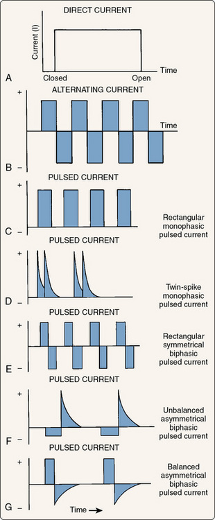
Figure 8-5 Graphic representation of the three types of electrical current. A, Direct current. B, Alternating current. C through G, Pulsed currents.

(Modified from Robinson, A.J. [1989]: Basic concepts and terminology in electricity. In: Snyder-Mackler, L., and Robinson, A.J. [eds.]. Clinical Electrophysiology. Baltimore, Williams & Wilkins, pp. 9, 11, 13.)

If the current is turned on and off repeatedly, individual phases with periods of no current flow (no charge) between them (C) would occur, and this is often called pulsed or interrupted current. The majority of electrotherapy devices use interrupted current, although a few exceptions are discussed later in the chapter. Electrotherapy devices allow the practitioner to control the number of these individual pulses per second (pulse rate). With low pulse rates (below 30), pulsing muscle contractions can be induced. By increasing the pulse rate to somewhere between 30 and 50 pulses per second, the muscle contraction appears smooth and sustained. A muscle that is contracting in a smooth and sustained fashion is said to be in tetany. Even higher pulse rates are often used and also cause tetanic contractions. It should be noted (D to G) that by using different combinations of polarity, voltage, and phase duration, the shape of each phase can be controlled. Common phase shapes are rectangular or square (E), spiked or twin spiked (D), asymmetric (F and G) in which the positive phase and negative phase have different shapes, and sinusoidal (Fig. 8-6). If the positive phase and negative phase have the same voltage (height), the waveform is said to be balanced (G).

image

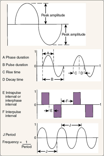
Figure 8-6 Characteristics of electricity.

(Modified from Robinson, A.J. [1989]: Basic concepts and terminology in electricity. In: Snyder-Mackler, L., and Robinson, A.J. [eds.]. Clinical Electrophysiology. Baltimore, Lippincott, Williams & Wilkins, p. 15.)

In examining a waveform, a specific set of terms is used to describe the phases and pulses (see Fig. 8-6). A phase is a single positively or negatively charged bolus of current, whereas a pulse is several consecutive phases that are continuous. All currents have at least one phase, but pulses are seen only with interrupted current and are separated from each other by brief intervals when no phase is present. The height, or amplitude, of each phase represents the voltage (the driving force for the current). The width of each phase represents the phase duration or phase width, usually in milliseconds. A related concept is pulse width, or the combined duration of all of the phases within a single pulse of interrupted current. Related to phase and pulse widths are phase and pulse intervals, or the duration between phases when there is no current (phase interval) or between pulses where there is no current (pulse interval). Phase interval and pulse interval are terms that are sometimes used interchangeably.

The lack of consistency in terminology across manufactures and textbooks is an ongoing problem in electrotherapy, which leads us to a fourth and sometimes confusing waveform concept called frequency. The frequency of a waveform can mean two very different things. Technically, it represents the number of cycles per second for the current. A cycle, also called a period, is one complete waveform including all of its phases. In clinical usage, the term frequency more commonly means the number of pulses per second and is also called the pulse rate. Remember that a pulse consists of several consecutive cycles of a current in an interrupted current. For example, if you use the electricity coming from your wall outlet, it is a continuous sinusoidal biphasic current with a frequency of 60 Hz. Now, if you are using an outlet connected to a switch and you manually turn the current on and off 10 times per second, the resulting current would be an interrupted sinusoidal biphasic current with a frequency of 60 Hz and a pulse rate of 10 Hz. Obviously, this terminology can be somewhat confusing, and most clinicians and manufacturers generally use “frequency“ to mean “pulse rate“ and the actual frequency of the current being interrupted is ignored.

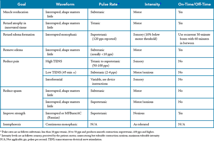
Although scientific study of electrotherapy is progressing, not as much is known about the physiology of this modality as is known about cryotherapy, superficial thermotherapy, or ultrasound, and the precise mechanisms by which electrotherapy is effective have not yet been described. As is the case for these better-studied modalities, electrotherapy is also characterized by a conspicuous lack of outcomes research. Even though the literature is still sparse, electrotherapy has been suggested to be effective in achieving a number of rehabilitative goals (Table 8-10), most of which are related to the ability of electricity to depolarize nerves. In addition, some the effects proposed are related to the electrical charge fields around the electrodes and do not rely on nerve depolarization.

Table 8-10 Common Therapeutic Goals for Electrotherapy

Goal Rationale
Muscle reeducation Retrain firing patterns or overcome neuromuscular inhibition in intact muscles following injury or pathology. The mechanism is thought to be related to increasing the quantity of motor units recruited or decreasing the inhibition of the motor nerves that is preventing normal function
Retard atrophy Cause the muscle to contract in an effort to reduce the effects of immobilization or paralysis on atrophy.
Retard edema formation Limit the formation of edema during acute inflammation by inhibiting the increase in vascular permeability with sensory-level stimulation.
Remove edema Remove edema that is already present through a muscle pump mechanism with motor-level stimulation.
Reduce pain Interfere with the transmission or perception of pain through a variety of different electrotherapy approaches.
Reduce spasm Reduce acute spasms by either decreasing the muscle’s contraction frequency or by fatiguing the muscle until it fails (very uncomfortable). Electrotherapy can also be used to manage the spasticity associated with neuromuscular diseases or spinal cord trauma.
Increase strength Increase muscle force output in nonpathologic tissue by causing hypertrophy of the muscle. Although muscular strength can be improved with motor-level stimulation, the protocols required are very uncomfortable and are not nearly as effective as resistance exercise. This is not generally an appropriate goal for electrotherapy.
Increase range of motion A commonly cited but misleading goal. Electrotherapy can be effective in reducing muscle spasticity or edema and thereby countering loss of ROM associated with these conditions, but electrotherapy is not an effective means of improving ROM by itself.
Transport medications Iontophoresis: deliver medications by driving electrically charged ions through the skin.
Tissue healing Microcurrent: some evidence has shown that microcurrent may augment tissue repair in individuals with fractures or slow-healing skin ulcerations. The mechanism has yet to be described.

ROM, Range of motion.

Muscle reeducation

Neuromuscular function is altered through inhibition following injury as discussed previously with regard to rehabilitative cryotherapy. In fact, the great majority of early muscular strength and power loss following an injury is thought to be the result of neurologic inhibition rather than more morphologic causes, such as loss of muscle mass. Loss of muscular strength and power is seen immediately following injury before loss of muscle mass has even occurred. In fact, loss of muscle mass as an explanation for the loss of strength becomes valid only after several weeks have passed. The goal of muscle reeducation is to counteract postinjury inhibition in an attempt to allow more normal use of the surrounding musculature. By reducing the degree of inhibition seen in these muscles in the early period following injury, earlier and more functional exercise can begin, and it is this exercise that is the most important tool in returning the athlete to competition. This is sometimes thought of as helping a muscle “remember“ how to contract. Similarly, reeducation can be used to help reestablish neuromuscular pathways after periods of immobilization or even to help correct pathologic neuromuscular patterns such as seen when a patient compensates for gait or postural abnormalities. An additional means by which electrotherapy can help in reeducation is by overcoming the inhibition associated with the injury.90

The primary means by which electrotherapy is thought to be an effective tool for muscle reeducation is through the recruitment of motor units that are not otherwise being recruited. The muscle tissue is not directly induced to contract, however. Instead, the electrical current stimulates motor neurons to depolarize and thereby causes contraction of their respective muscle fibers. By artificially recruiting these motor units through stimulation of the motor nerves it is thought that we may somehow overcome the inhibitory stimuli that are interfering with their voluntary recruitment at some location up the neurologic tree.94 Despite common anecdotal agreement about the efficacy of muscle reeducation, very little research has directly examined its effects.

Retard atrophy

As is the case with muscle reeducation, the use of electrotherapy to retard atrophy is based on the ability of electrical current to induce muscle contraction.156,157 During disuse, immobilization, or paralysis, the relative inactivity of the muscles leads them to atrophy. Likewise, lack of muscle contraction–induced stress on the bones can also lead them to atrophy, eventually to the point where they can become fragile if the disuse is of sufficient duration. Electrotherapy is frequently used in cases in which prolonged immobilization is anticipated, such as with casting for fractures or with paralysis. The premise is that electrotherapy can be used to induce low-intensity isometric contractions of the muscle that can retard the progression of atrophy without compromising the immobilization. Note that the word retard was used rather than prevent. Prevention implies that we can completely counteract the atrophy that is occurring (Box 8-11). Instead, we are more likely to slow its progression.

Box 8-11 Three Reasons Why Electrotherapy Cannot Completely Prevent Atrophy

The scenario in which electrotherapy is typically used to retard atrophy involves prolonged and purposeful immobilization. In such situations, strong muscle contractions are generally to be avoided so that displacement of the immobilized structures does take place. Therefore, we tend to use mild isometric contractions that will retard atrophy, but not prevent it.
Second and probably more important, the pattern of motor unit recruitment with electrical stimulation appears to be quite different from that seen with voluntary contraction. In volitional contractions, smaller-diameter motor neurons supplying small muscle fibers of small motor units are recruited first, and larger neurons with larger and stronger motor units are recruited later as necessitated by the need for greater muscle force. With electrical stimulation, this order appears to be reversed, with larger-diameter motor neurons being recruited first and smaller-diameter fibers being recruited less and only when sufficient voltage is used.1,2,4,5 This different pattern of muscle recruitment leads to a variable rate of atrophy in which the smaller motor neuron units, which are responsible for fine motor tasks, atrophy at a greater rate than do the larger motor neuron units, which are responsible for gross motor tasks.
A third reason why we appear to be unable to completely prevent atrophy is related to the athletic ability and training level seen in athletes. Even if we were not concerned about the consequences of displacing immobilized tissue with strong contractions, it is unlikely that strong enough contractions could be produced to prevent all muscle loss in injured athletes. By the nature of their extensive training and conditioning programs, athletes have considerably larger muscle mass and force-producing capacity than general patients do. The differential recruitment of motor units with electrical stimulation implies that we are unlikely to be able to generate sufficient muscle force with electrical stimulation to provide an adequate stimulus to retain the muscle mass that has been produced by extensive resistance training.

Edema management

Management of edema with electrotherapy can take two different forms. The first is retarding the formation of edema. The second is removing edema that is already present. The technique is different for each strategy and should be used only in the correct situation. For example, electrotherapy should be used to retard the formation of edema only in the period while edema is forming immediately following the injury. Use of this approach after a large amount of edema is already present may actually inhibit removal of the edema.

Sensory-level, high-voltage pulsed electrical stimulation applied directly to the area of the injury has been shown to limit the volume of edema following uniform injuries in an animal model.88,90-93 Two mechanisms have been proposed for this retarding of edema through the application of electrotherapy. The first is to combat the increasing permeability of the capillaries during the initial acute inflammatory response and thereby reduce the efflux of fluid from the circulatory system into the injured tissues. As explained in Chapter 2, one of the major events of the acute inflammatory response is a marked increase in capillary permeability that results from the release of numerous chemical mediators. The increase in permeability occurs when adjacent endothelial cells in capillaries do not adhere to each other as tightly as they normally do. This makes the capillaries “leaky“ and allows the transcapillary Starling forces to exert an even greater influence. The second—and less accepted—of the proposed mechanisms is that pulsed monophasic current causes vascular spasm that limits delivery of fluid to the injured area. Regardless of the suggested physiologic explanations for the effectiveness of the technique, the protocol used is very specific, requires a specific waveform, and is outlined in the later section on technique and dosage. One of the key elements of the protocol is the timing in relation to injury. This approach is effective only when it is begun before meaningful edema has developed. Therefore, the time window for initiating this modality is quite literally the first few minutes following the injury.

In addition to retarding the formation of edema during acute inflammation, electrotherapy can also be a valuable adjunct in removal of the edema that is already present. The proposed mechanism by which existing edema is removed is somewhat different from that for retarding the formation of edema, however. Although retarding the formation of edema is based on limiting the permeability of the vasculature, such a strategy may actually hinder the ability to remove edema. When present, edema is removed by the lymphatic system rather than the circulatory system. Removal requires that the edematous fluid be absorbed into lymph capillaries, where it flows to larger collecting vessels and eventually to one of the lymphatic ducts, and the fluid is then returned to the circulatory system. If the permeability of the lymphatic capillaries is limited, a potential outcome of the edema retardation electrotherapy protocol, movement of fluid into the lymphatic system may actually be hindered and thus reduce the effectiveness of edema removal.

Instead of using protocols aimed at altering permeability,t edema removal protocols focus on moving fluid into the lymphatic system and then moving it along the lymphatic vessels and away from the injury site.158-163 This is accomplished by motor-level electrical stimulation in which the muscles are induced to contract in a pulsing fashion with interrupted current. Each muscle contraction exerts external pressure on the lymphatic vessels. Squeezing of the lymphatic vessels causes the fluid in them to move. This strategy for removal of edema is sometime called the “muscle pump“ strategy. When fluid reaches the collecting vessels of the lymphatic system, its flow essentially becomes unidirectional because of the presence of one-way valves within the vessels. Much like the valves located in veins, pressure exerted on the lymphatic vessel from muscular contractions causes the upstream valve to close and the downstream valve to open, thereby allowing the fluid to flow back only toward the circulatory system. Because fluid moves with each muscular contraction, electrotherapy pulse rates that are below the level needed for tetany are used. Tetanic contractions would produce only a single pressure pulse and would not be expected to move as much fluid as would repetitive contractions.

Clinical Pearl #12

Even though electrotherapy can be a useful adjunct for removal of edema, the role of exercise in removing edema should not be overlooked. Exercise also induces muscle pumping, typically in more muscles than are used with electrotherapy alone.

Pain management

Management of pain with electrotherapy is among the most common, best documented, and most successful uses of this modality.164-174 Much of the pain reduction literature deals with a subform of electrotherapy called transcutaneous electrical nerve stimulation (TENS). TENS typically involves the use of pulsed, sensory-level stimulation to interfere with the transmission of pain signals in the spinal cord through a mechanism known as gait control. Gait control uses sensory information on A-β afferent nerves to interfere with the transmission of pain on A-δ and C afferent fibers. To be effective, adequate stimulation of A-β fibers must occur. The literature suggests that this is most likely to occur at rates between 60 and 150 pulses per second, and rates of 100 to 150 pulses per second are most common.1,2,5 The high pulse rates have led this protocol to be called high-frequency TENS. High-frequency TENS units generally use a short pulse duration (20 to 60 μsec) combined with a 50- to 100-Hz frequency of stimulation. Additionally, the combination of sensory-level TENS and cryotherapy has been shown to provide greater pain relief than with either of these modalities used individually.

Although the gait control strategy is certainly the most common, it is not the only strategy for controlling pain with electrotherapy. Motor-level stimulation with a high-voltage (> 150 V) stimulator can be used to stimulate the release of endogenous opiate-like substances from nerve fibers. Sometimes this protocol is referred to as “low-frequency TENS“ because of its low pulse rate (2 to 4 pulses per second). It is frequently uncomfortable during application but results in relief of pain following the treatment. This is somewhat different for conventional high-frequency TENS, in which pain relief is usually experienced during the treatment and for a short time thereafter. The release-of-opiates strategy is not as common as the gate control strategy; however, it may provide longer-lived pain relief.

Another pain relief technique with electrotherapy involves the use of a sensory-level polyphasic current known as interferential current.168,170,171 Interferential current is actually the combination of two different biphasic currents that are out of phase with each other. The two currents each have different frequencies and are carried on two different circuits (or channels) that are applied more or less perpendicular to each other. The differing frequencies of the currents and the fact that they are out of phase with each other produce a constantly changing waveform in the region where the currents cross. The key feature of interferential electrotherapy is this ever-changing waveform. Because the waveform is constantly changing, the body has a very difficult time accommodating to it. Accommodation is the process by which the sensory system learns to “ignore“ sensory stimuli that are unchanging. For example, anyone married for more than a few months no longer senses the wedding ring against their finger. The body has a reasonably good ability to accommodate to electrotherapy, particularly sensory-level electrotherapy. This accommodation reduces the efficacy of the treatments. With interferential electrotherapy, the constantly changing waveform reduces the accommodation and allows the treatment be more effective than fixed-waveform treatments. The mechanism of pain relief is thought to be the same as for high-frequency TENS. In fact, many TENS units now use modulated waveforms that change throughout the treatment in an attempt to overcome accommodation.

Spasticity management

Management of spasticity is among the more commonly cited uses of electrotherapy, although it is generally less applicable to athletic injuries than to other conditions, such as neurologic lesions. Management of the spasticity associated with neurologic lesions frequently involves stimulating the antagonist muscles and relying on reciprocal relaxation. In fact, the vast majority of the literature on the use of electrotherapy for spasticity focuses on the type of spasticity resulting from spinal cord trauma, cerebral vascular accidents, brain trauma, and disease.175 Because these lesions are not common with athletic injuries, they will not be discussed further in this chapter. On the other hand, management of muscle spasm associated with athletic injuries is a common use of electrotherapy and needs to be addressed, although the literature describing such use is very sparse.

Two strategies are predominantly used for managing athletic injury–related acute muscle spasm with electrotherapy, although only one of them is very tolerable to the patient. The more tolerable of the two strategies involves using motor-level stimulation of the spastic muscles in an attempt to alter their frequency of contraction. Recall that the rate of firing of the motor units determines whether a muscle’s contraction will be pulsing or smooth (tetanic). Smooth contractions occur with firing rates higher than 30 to 50 pulses per second. When a muscle is in spasm, it is generally contracting smoothly and continuously. By using a muscle stimulator with relatively high intensity and less than a tetanic pulse rate, it may be possible to slow the firing rate and cause the spasm to abate. The other and probably more effective strategy involves using the stimulator to fatigue the muscle to the point where the spasm ceases. This strategy has been shown to be effective, but it is not very comfortable and most patients may not tolerate it well. Generally, the technique involves using high–pulse rate, maximum tolerable intensity stimulation with a high-voltage stimulator to recruit as many motor units as possible. This will eventually lead to muscular fatigue, and the spasm will lessen or stop altogether. Although this technique is uncomfortable, it can be made more comfortable and more effective if it is combined with static stretching of the affected muscles. Cryotherapy is also a common adjunct with this technique.

Increasing strength

Even though electrotherapy can indeed be used to improve strength, this is among the most often misused forms of this modality. To understand this use of electrotherapy, a distinction must first be made between using electrotherapy for muscle reeducation versus electrotherapy for increasing strength. As discussed earlier, muscle reeducation involves using electrotherapy to overcome the neuromuscular inhibition that is interfering with normal muscle function. Said another way, muscle reeducation uses an electrical stimulator in an attempt to improve pathologic muscle function by reestablishing the normal and appropriate neurologic pathways for muscle contraction. This is different from using a stimulator to improve muscle strength.

When a stimulator is used to improve strength, the goal is not to overcome inhibition or other neuromuscular pathology. Instead, electrotherapy is used to strengthen tissue that has normal neuromuscular function but is not as strong as desired. Said another way, muscle strengthening with electrotherapy involves causing the contractile elements within muscle to overload in an attempt to induce them to adapt by becoming stronger. This is precisely the same goal as used during resistance training to improve muscle strength, but the catch is that resistance training is considerably more effective in achieving this goal than electrotherapy is.

To strengthen muscle tissue, regardless of the method, the muscle must be made to exert more force than it is accustomed to exerting. This principle is known as overload. Overload of a muscle can be accomplished in two ways: increasing the rate of firing of motor units and recruiting a larger number of motor units.60 During resistance training, the body’s strategy is to do both, but to predominantly favor recruiting more motor units. In fact, during exercise the body varies its motor unit recruitment pattern. At the beginning of an exercise, the smaller-diameter motor neurons of the fine control motor units are recruited. If more force is required, the larger-diameter motor neurons controlling the stronger but less finely controlled motor units are recruited. As the muscle begins to fatigue, the fine control motor units fail first and additional large motor units are recruited until they eventually fail. This is why some shaking and less coordination are observed as someone fatigues during resistance training. When a muscle stimulator is used, however, a different pattern of recruitment takes place.1,2,5

Use of a muscle stimulator to improve strength also depends on overload, and both a high rate of motor unit firing and measures aimed at recruiting more motor units are used. In strengthening with electrotherapy, a pulse rate at or near the maximum available for the stimulator is used in an effort to produce as high a rate of motor unit firing as possible. In addition, as high an intensity (voltage) as tolerable is used because higher intensities lead to better penetration of the nerve by the electrical current and therefore recruitment of an increased number of motor units. The unfortunate catch in this strategy, however, is that recruitment of motor units with electrotherapy appears to be the reverse of normal voluntary recruitment. With electrotherapy, motor units that are of larger diameter and closer to the surface of the nerve are preferentially recruited. These same motor units appear to be stimulated over and over rather than recruiting different motor units as some of them fatigue, as is the case with normal exercise. This repeated firing of the same motor units is the reason that muscular fatigue is experienced so quickly with electrical stimulation and not as quickly with exercise. The repeated recruitment of a limited set of motor units also limits the effectiveness of electrotherapy-based strengthening programs. Strengthening adaptations are essentially limited to the motor units that are being recruited, and electrotherapy recruits fewer motor units than active exercise does. Although some degree of strengthening of normal tissue can occur with electrotherapy, the strengthening is not as effective as with active exercise in a resistance training program. Therefore, the use of electrotherapy for muscle reeducation is recommended in athletic therapy, but electrotherapy as a tool to strengthen muscles that have normal function is not.

Increasing range of motion

Many modalities texts suggest that electrotherapy can be used to improve range of motion, and it most certainly can do so, but not in the way that we are usually seeking with athletic injuries. The use of electrotherapy to improve range of motion is appropriate and effective in only a few limited situations. The most common involves a patient with neurologic trauma or disease that results in spasticity. For example, it is not uncommon to see spasticity in the gastrocnemius and soleus of a spinal cord–injured patient. Such spasticity leads to lack of ankle dorsiflexion range of motion. Electrotherapy can be applied to the dorsiflexors to both stimulate them and inhibit the gastrocnemius and soleus. This would allow greater dorsiflexion range of motion because of the reduction in spasticity of the plantar flexors. Though useful in some cases for neurologic injuries and disease, this type of improvement in range of motion is of little value in the rehabilitation of common athletic injuries. Another limited situation that is of more use with athletic injuries would be improving range of motion that is limited by pain, edema, or both. As discussed earlier, electrotherapy can be used for the management of these conditions and may result in an indirect improvement in range of motion as well.

Iontophoresis of pharmaceuticals

Iontophoresis, or the use of an electrical current to drive medications through the skin, is another common form of electrotherapy. This technique has some real advantages in that it can be used to deliver medications locally without having to inject them. This can be particularly useful for patients with fear of needles or for pediatric patients. In fact, iontophoresis with local anesthetics is gaining popularity as a preinjection technique to lessen the discomfort of pediatric immunizations. Though potentially advantageous, there is also some controversy over iontophoresis in the literature.130,176-182 Reports conflict regarding whether iontophoresis delivers enough medication to a deep enough tissue depth to be effective for many conditions. Moreover, outcome data for iontophoresis are limited and have not yet adequately demonstrated that the technique is of much benefit to patients with musculoskeletal injuries.

Iontophoresis requires a very specific type of electrical current and is not possible with typical muscle stimulators. To use electrical current to move medications, the medications must dissociate into electrically charged ions in solution. When an electrical current is applied to the medication solution, the charge at each electrical pole repels the medication ions with like charges and attracts the ions with opposite charges. Therefore, it is critical to apply the medication to the electrode that has the same polarity as the ion of interest in the medication. Likewise, only direct (monophasic) current can be used because alternating (biphasic) current would both repel and retract the medication and produce no net transport through the skin. The current used also needs to be continuous. Interrupted currents do not repel the drug long enough for it to travel through the skin. For these reasons, iontophoresis stimulators are different from other electrotherapy devices and are designed expressly for use in iontophoresis. These devices are used with single-use electrodes and deliver low-intensity DC in a monopolar setup as described later. The dosage is typically the product of the duration of treatment and the amount of current used and is expressed in milliampere minutes (mA • min). The specific dosage depends on the medication being used, and treatments generally last between 10 and 20 minutes, although they can be longer if lower amounts of current must be used because the patient does not tolerate DC well. A new iontophoresis device, the IontoPatch, was introduced in April 2001 and is somewhat different. Instead of using the typical 10- to 20-minute treatment with low to moderate amounts of current, the IontoPatch uses a much longer duration (usually 24 hours) and an extremely low level of current delivered from a self-contained battery in the patch. The lower level of current means fewer complications in terms of skin burns, and anecdotal reports have been very favorable, although the device is new enough that research literature is lacking.

A relatively small number of medications, usually corticosteroids, are commonly used with iontophoresis in athletic medicine settings. However, from a technical standpoint, any medication that dissociates into ions in solution and produces the desired effects could be used. The most common medications are presented in Table 8-11.

Table 8-11 Nonsteroidal Ions and Radicals

Ion or Radical (Charge) Features*
Magnesium (+) From magnesium sulfate (Epsom salts), 2% aqueous solution; excellent muscle relaxant, good vasodilator, mild analgesic
Mecholyl (+) Familiar derivative of acetylcholine, 0.25% ointment; powerful vasodilator, good muscle relaxant and analgesic; used for discogenic low back radiculopathies and sympathetic reflex dystrophy
Iodine (−) From Iodex ointment, 4.7%; bactericidal, fair vasodilator, excellent sclerolytic agent; used successfully for adhesive capsulitis (“frozen shoulder“), scars
Salicylate (−) From Iodex with methyl salicylate, 4.8% ointment (if desired without the iodine, can be obtained from Myoflex ointment—trolamine salicylate, 10%—or from a 2% aqueous solution of sodium salicylate powder); a general decongestant, sclerolytic, and antiinflammatory agent; used successfully for frozen shoulders, scar tissue, warts, and other adhesive or edematous conditions
Calcium (+) From calcium chloride, 2% aqueous solution; believed to stabilize the irritability threshold in either direction, as dictated by the physiologic needs of the tissues; effective for spasmodic conditions, tics, “snapping joints“
Chlorine (−) From sodium chloride, 2% aqueous solution; good sclerolytic agent; useful for scar tissue, keloids, burns
Zinc (+) From zinc oxide ointment, 20%; trace element necessary for healing; especially effective for open lesions and ulcerations
Copper (+) From 2% aqueous solution of copper sulfate crystals; fungicide, astringent, useful for intranasal conditions (e.g., allergic rhinitis—hay fever), sinusitis, and dermatophytosis (athlete’s foot)
Lidocaine (+) From Xylocaine, 5% ointment; anesthetic and analgesic, especially for acute inflammatory conditions (e.g., bursitis, tendinitis, tic douloureux, and temporomandibular joint pain)
Lithium (−) From lithium chloride or carbonate, 2% aqueous solution; effective as an exchange ion for gouty tophi and hyperuricemia
Acetate (−) From acetic acid, 2% aqueous solution; dramatically effective as a sclerolytic exchange ion for calcific deposits
Hyaluronidase (+) From Wydase crystals in aqueous solution, as directed; for localized edema
Tap water (+/−) Usually administered with alternating polarity, sometimes with glycopyrronium bromide for hyperhidrosis
Ringer solution (+/−) With alternating polarity; used for open decubitus lesions
Citrate (+) From potassium citrate, 2% aqueous solution; reported effective for rheumatoid arthritis
Priscoline (+) From benzazoline hydrochloride, 2% aqueous solution; reported effective for indolent ulcers
Antibiotics: gentamicin sulfate (+) 8 mg/mL; for suppurative ear chondritis

* All solutions are 2%; ointments are also low-percentage compounds. The literature and clinical reports agree that the lower the percentage, the more effective the ionic exchange and transfer. Whether this is purely a physical chemistry phenomenon or an example of the Arndt-Schultz law, which states that “the smaller the stimulant, the greater the physiologic response,“ remains to be proved.

The lithium ion replaces the weaker sodium ion in the insoluble sodium urate tophus and converts it to soluble lithium urate.

The acetate radical replaces the carbonate radical in the insoluble calcium carbonate calcific deposit and converts it to soluble calcium acetate.

From Kahn, J. (1987): Non-steroid iontophoresis. Clin. Manage., 7:15. Reprinted from Clinical Management with the permission of the American Physical Therapy Association.

Clinical Considerations

The specific waveform chosen for electrotherapy can be absolutely critical in some cases and can make very little difference in others. One of the hallmarks of a skilled practitioner is to understand the difference. To understand this difference, some familiarity with the electricity fundamentals and terminology discussed earlier is required, as is understanding of the following concepts. First, it has already been discussed that waveforms can be monophasic (also known as DC current), biphasic (also known as AC current), or polyphasic (a mixed waveform, such as interferential current). It has also been discussed that waveforms can be either continuous or interrupted. These features can be combined to produce some general classes of waveforms that have an impact on clinical treatments.

Continuous versus interrupted current

Continuous currents are found in only very few devices and are used in very specific situations. The first of these situations is for iontophoresis, where a continuous monophasic current is used. Continuous currents must be used in this case because interrupted currents do not have a sufficient duration of current flow to move ions across the skin. A second situation in which continuous current is used involves the stimulation of denervated muscle to prevent disuse atrophy. In this case, continuous current is used because the longer current duration makes it easier to induce depolarization of what little remains of the motor nerves or perhaps even the muscle itself. Aside from these two situations, continuous currents are normally found only as biphasic carrier frequencies in polyphasic currents. In these situations, they are normally used to help create a perpetually changing waveform to help overcome accommodation, as is the case with both interferential current and a waveform used for strengthening known as MFBurstAC, or Russian current.155

Polarity

Electrotherapy devices that offer monophasic waveforms can readily be distinguished from biphasic or polyphasic waveforms by nature of the ability to select a polarity for the treatment electrodes. Because biphasic and polyphasic waveforms have both positively and negatively charged phases that alternate, no specific polarity can be assigned to the electrodes. In reality, only in very few situations does the specific polarity of the electrodes make a clinical difference; however, it makes a very big difference in a few. For example, iontophoresis can be accomplished only with a continuous monophasic current and, even then, only when the drug is applied to the correct electrode. The other situation in which polarity clearly matters involves the stimulation of denervated muscle for preventing disuse atrophy; however, this application is less important in the rehabilitation of athletic injuries. Another situation in which polarity has been suggested to make a difference involves the use of electrotherapy to retard the formation of edema. In this case, cathodal (negative polarity) stimulation has been shown to be effective, as discussed later. There is little evidence to suggest that polarity is an important consideration in the management of pain or in the ability to produce contractions in innervated muscle tissue.

Unipolar versus bipolar

One of the easiest to understand yet most misunderstood application technique related to electrotherapy is unipolar versus bipolar electrode configuration. A unipolar electrode configuration means that the active effects of the stimulator are seen only in the electrodes attached to one of the two electrical poles. Bipolar configuration means that the active effects of the stimulator are seen in the electrodes attached to both poles. Recall that to have an electrical circuit, the electricity must have a pathway to flow from one pole (negative) to another pole (positive). In DC these poles maintain constant polarity, and in AC they switch polarity. For purposes of electrode configuration, the actual polarity of the electrode does not matter except in the cases discussed earlier. Whether an electrode displays “active effects“ of the current depends on the current density under the electrode, and this is determined by the combination of current intensity and size of the electrode. For a given amount of current, a bigger electrode will have the current spread over a larger area, and a smaller electrode will have the current spread over a smaller area. The amount of current per unit of area is the current density. To have an active effect, such as inducing sensory or motor stimulation, the current density must be adequate. The smaller the electrode, the greater the current density, and therefore greater stimulation effects will be seen. Conversely, the larger the electrode, the smaller the current density, and therefore little or no stimulation effect will occur.

In a unipolar configuration, current density is manipulated by using electrode size. One pole has a relatively small electrode (or several small electrodes), and the other pole has a relatively large electrode, usually called a dispersive electrode. Because a dispersive electrode has a large area, it has a small current density that does not produce active effects. Unipolar configurations are standard on monophasic high-voltage stimulators and are very useful in situations in which you want to move the active electrode, such as with trigger point stimulation. Bipolar configurations use similarly sized electrodes at both poles, so similar effects are seen at both electrodes. Bipolar configurations are more common on newer devices and biphasic stimulators. Bipolar configurations are useful for situations in which you do not plan to move the electrodes or when exposure of enough skin to use a dispersive electrode can compromise a patient’s modesty.

The names unipolar and bipolar can sometimes be confusing because they sound similar to polarity. In reality, unipolar and bipolar configurations have absolutely nothing to do with the polarity of the electrical current under the electrode. Although unipolar arrangements are typically the standard electrode setup on monophasic high-voltage stimulators, this is actually a matter of convention rather than necessity. In reality, any stimulator can be used in either a unipolar or bipolar configuration. The choice is really a matter of clinical convenience rather than association with a specific stimulator. To convert a unipolar configuration to a bipolar configuration, the large dispersive electrode would simply be replaced with an electrode similar in size to the other active electrode. Similarly, a greater effect can be produced with the stimulator by merely using smaller electrodes. To convert a bipolar configuration to a unipolar configuration, one of the active electrodes would simply be replaced with a larger dispersive electrode. One unique case of a unipolar setup involves using electrotherapy while immersed in water, such as a whirlpool or ice slush. In this case, a relatively small electrode is attached to a motor point of a body part that is immersed in water (e.g., the gastrocnemius). This small (active) electrode must be attached to a motor point that is out of the water. The other electrode lead wire is immersed in the water along with the body part. The water acts as a very large dispersive electrode, and the small electrode acts as an active electrode. This technique is generally used for edema retardation or removal protocols.

Current modulation

In addition to the manipulations of electrical currents already discussed, a number of other current modulations are also common. Current modulation is simply an alteration in the current’s waveform. It includes applications already discussed, such as using interrupted current, as well as a few other alterations that will be discussed here. It is used for a variety of reasons, such as counteracting accommodation, increasing patient comfort, minimizing fatigue, or making contraction easier.

Among the most common modulations is varying the pulse rate of an interrupted current to minimize accommodation. Most contemporary stimulators have built-in presets to modulate the pulse rate. Typically, these presets vary the pulse rate up and down within a specific band of frequencies that corresponds to a desired effect. For example, among the more common presets is one that varies the pulse rate up and down between 1 and 10 pulses per second, obviously in the subtetanic range if used with muscle contraction. With sufficient intensity, this setting would cause the muscle to visibly twitch at a varying rate that would not become a tetanic contraction. Similar presets can be found in the pulse rate range just above the threshold for tetany and also at much higher frequencies such as those commonly used for pain management or muscle strengthening.

Another common modulation is ramping the intensity with an interrupted current. When using a ramp setting, the current is not at its maximum intensity when it first comes on. Instead, each successive pulse of current increases slightly in intensity until the desired maximum is reached. The ramp setting allows the user to specify the time that it takes for the maximum to be reached. Some devices also allow a ramp setting for when the current is ending. Ramp settings are used to increase patient comfort, with it generally being more comfortable to ramp up to the maximum rather than being hit with it all at once.

Another modulation related to patient comfort is controlling the pulse width. There is an indirect relationship between the pulse duration (width) and the pulse amplitude (the intensity) when inducing a muscle contraction. For patients who have a hard time tolerating high amplitudes, the amplitude can be reduced to tolerable levels and the pulse width increased to still induce muscle contraction. Increasing pulse width can also improve the ability to induce a contraction in other situations as well.

The use of “on-time” and “off-time” is another common modulation with electrotherapy. Recall that muscle stimulators induce recruitment of the same set of motor units over and over. Because this does not match the normal physiologic recruitment pattern for motor units, it often leads to rapid fatigue in the muscle. Although in some protocols fatigue is desirable, such as when trying to overcome spasticity, muscle fatigue is to be avoided with most electrotherapy protocols. For this reason, many stimulators provide the practitioner with the ability to have either “continuous” current flow or current flowing for a certain period followed by a rest period with no flow of current. This type of modulation is often called interrupted, but it should not be confused with interrupted current as discussed previously. Normally, when we speak of current interruption, we mean that discreet pulses of current alternate with brief (microseconds to milliseconds) intervals of no current. When on and off modulation is used, current flows during the on-time, and this current is almost pulsed at whatever pulse rate setting is chosen. Likewise, the continuous setting on most stimulators means that the flow of interrupted (pulsed) current is constant rather than meaning a noninterrupted current. Obviously, the terminology gets to be confusing and becomes worse when different manufacturers use their own terminology. There is little consensus on the correct durations for on-time or off-time or even the correct on-off ratio. Even though research is required to better explore this parameter, many clinicians anecdotally report using a 1:1 ratio or less of on-time to off-time.

Intensity

The intensity setting controls the amplitude (voltage) of the waveform and therefore controls the quantity of current flowing through the circuit. Recall that the relationship between force and flow (voltage and current) is describe by Ohm’s law (see Box 8-10). The greater the intensity, or driving force, the greater the flow of current. Although most contemporary stimulators provide the user with a readout of intensity or current levels, use of a numerical value can be misleading. Because of variability in electrode placement, electrode size, skin conductivity, moisture, and other factors, the voltage used for one treatment to induce muscle contraction would not necessarily induce the same degree of contraction with a different treatment for the same patient. For this reason, it is more useful to think of intensity levels in terms of their effects rather than their numerical value. Perhaps the easiest scheme involves classifying intensity progressively as subsensory, sensory, motor, and noxious (Table 8-12). Subsensory levels are obviously not perceived by the patient and are rarely used outside microcurrent electrotherapy. Sensory levels of intensity imply that the patient can feel the current but the current does not cause muscle contraction. Motor levels of intensity cause muscle contractions. As the name implies, noxious levels of intensity are rather uncomfortable and are rarely if ever used, but they provide the maximum tolerable level of muscle contraction.

Table 8-12 Classification of Intensity Level by Its Effects

Subsensory level The level is not perceived by the patient and is rarely used outside microcurrent electrotherapy.
Sensory level The patient can feel the current but the current does not cause muscle contraction.
Motor level Motor levels of intensity cause muscle contractions. As the name implies, noxious levels of intensity are rather uncomfortable and are rarely if ever used, but they provide the maximum tolerable level of muscle contraction.

Techniques and Dosage

This chapter presents only a general framework for electrotherapy techniques. For more complete and detailed information, a full text on modalities should be consulted. To simplify this section, general strategies and parameters are summarized in Table 8-13. Very few data are available regarding appropriate treatment durations for most protocols. By convention, most are 15 to 30 minutes in duration unless otherwise noted.

Table 8-13 General Electrotherapy Parameters Common in Rehabilitation of Athletic Injuries

image

Future Questions

Many questions are still unanswered about electrotherapy because very little research has actually has examined specific combinations of parameters to determine their efficacy. Similarly, very few quality outcomes studies are available. Currently, some very promising work is being conducted in the areas of retarding edema formation and in the management of pain. Similarly, iontophoresis also receives sporadic and conflicting examinations. As a general statement, electrotherapy as a whole remains a vast and little explored area with many areas for further study.

Conclusion

Introduction

Modalities have specific uses in specific situations and are of little benefit when used for the wrong reason, with the wrong technique, or at the wrong time. At their best, therapeutic modalities are an exceptionally useful complement to the rehabilitative process but are not a replacement for it.
The key to using modalities appropriately is to match the specific physiologic effects of the modality with the specific rehabilitative goal for the patient.
Practitioners should familiarize themselves with the specific details of their state practice acts before using any therapeutic modalities.

Research Regarding Modalities

A number of clinically common modalities may not be as effective as once thought, and the clinical efficacy of modalities that are known to be useful can easily be compromised by incorrect application techniques.
The general trend is that clinical practice almost always precedes scientific research in regard to the use of modalities.
Most research concerning the therapeutic value of modalities has concentrated on clinically relevant types of data, which are indirect measures of a modality’s effectiveness and important in establishing outcomes data, versus the direct effect, which is important in establishing modality theory.
A number of modality effects are commonly accepted by clinicians but have little scientific support. In some cases there is even scientific evidence to the contrary, yet these widely held clinical beliefs still persist.

Modalities for Acute Care

The two primary goals of using modalities for acute injures are restricting the total quantity of tissue damage associated with an injury and limiting sequelae of the acute inflammatory response.

Cryotherapy

Data support the application of cold in reducing local skin and intramuscular temperature, the metabolic rate of the cooled tissue, and blood flow; inhibiting inflammation; retarding the formation of edema/effusion; and helping manage pain.
We do not yet have a definitive answer about the most effective tissue temperature, duration of cryotherapy, or on-off ratio for using cryotherapy for the treatment of acute injury.
Cold-induced vasodilation is a misnomer; in reality, cryotherapy does not increase blood flow above baseline levels at all.
Cryotherapy used in combination with compression has been shown to produce greater reductions in temperature than cryotherapy used alone.
Ice bags made from ice stored in unrefrigerated hoppers should be applied directly to the skin without the addition of an insulating layer. Frozen gel packs or ice stored in a freezer, however, should have some type of appropriate barrier between them and skin.

Compression

It is believed that compression is effective in managing acute injuries by increasing the cooling efficacy of cryotherapy, manipulating Starling forces, and reducing bleeding from vessels damaged during the injury.
Little scientific evidence supports a specific pressure and duration, intermittent versus continuous compression, intermittent cycling parameters, and whether compression or elevation is a more important factor in retarding edema formation.

Elevation

The premise for elevation is based on gravity limiting the amount of blood delivered to an acutely injured area, which would result in several positive physiologic events. Unfortunately, the magnitude of the actual benefits from elevation has not been described in the literature.

Modalities for Rehabilitation

In postacute rehabilitation, the goals are focused mostly on removing the unwanted remnants of inflammation, repairing tissue, and restoring more normal physiologic function of the repaired tissue.
When a modality is used to accomplish a specific goal, the modality can and should be discontinued when that goal has been accomplished or when the modality is no longer proving to be effective for the patient.

Cryotherapy

Cold reduces pain and pain is one cause of inhibition; therefore, cold should help overcome inhibition and allow the patient to begin controlled rehabilitative exercise at an earlier point in the rehabilitative process. This is also the primary basis for the efficacy of cryokinetics.
Some early evidence indicates that cryotherapy facilitates the motor neuron pool, which helps overcome neuromuscular inhibition following injury.

Superficial Thermotherapy

Superficial thermotherapy should be used as an adjunct, along with stretching or joint mobilization techniques, to improve range of motion.
These types of modalities do increase the superficial circulation and reduce pain.
Further research still needs to address the duration of thermal modalities, frequency of use, and appropriate tissue temperatures.

Ultrasound

Thermal ultrasound is used primarily to augment techniques for improving range of motion, although the literature directly examining the efficacy of ultrasound and stretching is very sparse.
Nonthermal ultrasound is used primarily when the goal is to augment the repair or regeneration of damaged tissue, and scientific evidence supports such use.
Not all coupling media for ultrasound are equally effective.

Phonophoresis

Research regarding the efficacy of phonophoresis is mixed at this point.

Short Wave Diathermy

This is probably the best thermal modality available to the practitioner today and is capable of therapeutically treating a much larger area than possible with ultrasound.

Electrical Stimulation

The most promising uses of electrical stimulation appear to be managing pain, reeducating muscle, and aiding in retarding the formation and removal of edema.
Some degree of strengthening of normal tissue can occur with electrotherapy, but the strengthening is not as effective as with active exercise in a resistance program.

Iontophoresis

Reports conflict regarding whether iontophoresis delivers enough medication to a deep enough tissue depth to be effective for many conditions. In addition, outcomes data for iontophoresis are limited, and it has not yet been adequately demonstrated that the technique is of much benefit to patients with musculoskeletal injuries.

References

1 Cameron M. Physical Agents in Rehabilitation: From Research to Practice. Philadelphia: Saunders; 1999.

2 Denegar C. Therapeutic Modalities for Athletic Injuries. Champaign IL: Human Kinetics; 2000.

3 Michlovitz S.L. Thermal Agents in Rehabilitation 3rd ed 1996 Davis Philadelphia 405

4 Prentice W. Therapeutic Modalities in Sports Medicine, 4th ed. Boston: McGraw-Hill; 1999.

5 Starkey C. Therapeutic Modalities, 2nd ed. Philadelphia: Davis; 1999. p. 397

6 Lewis T. Observations upon the reactions of the vessels of the human skin to cold. Heart. 1930;15:177-208.

7 Lehmann J., deLateur B., Stonebridge J., Warren C. Therapeutic temperature distribution produced by ultrasound as modified by dosage and volume of tissue exposed. Arch. Phys. Med. Rehabil.. 1967;47:662-666.

8 Lehmann J., Masock A., Warren C., Koblanski J. Effect of therapeutic ultrasound on tendon extensibility. Arch. Phys. Med. Rehabil.. 1970;51:481-487.

9 Lehmann J.F., deLateur B., Warren C. Heating produced by ultrasound in bone and soft tissue. Arch. Phys. Med. Rehabil.. 1967;48:397-401.

10 Chan A., Myrer J., Measom G., Draper D. Temperature changes in human patellar tendon in response to therapeutic ultrasound. J. Athl. Train.. 1998;33:130-135.

11 Draper D., Castel J., Castel D. Rate of temperature increase in human muscle during 1 MHz and 3 MHz continuous ultrasound. J. Orthop. Sports Phys. Ther.. 1995;22:142-150.

12 Gersten J. Effect of ultrasound on tendon extensibility. Am. J. Phys. Med.. 1959;34:662.

13 Robertson V., Baker K. A review of therapeutic ultrasound: Effectiveness studies. Phys. Ther.. 2001;81:1339-1350.

14 Draper D. Don’t disregard ultrasound yet—the jury is still out. Phys. Ther.. 2002;82:190-191.

15 Higgins D., Kaminski T. Contrast therapy does not cause fluctuations in human gastrocnemius intramuscular temperature. J. Athl. Train.. 1998;33:336-340.

16 Myrer J., Draper D., Durrant E. Contrast therapy and intramuscular temperature in the human leg. J. Athl. Train.. 1994;29:318-322. 376–377

17 Myrer J., Higgins D., Kaminski T. Some concerns—“Contrast therapy does not cause fluctuations in human gastrocnemius intramuscular temperature“ (J. Athl. Train. 1998;33:336-340). J. Athl. Train. 1999;34:231.

18 Myrer J., Measom G., Durrant E., Fellingham G. Cold- and hot-pack contrast therapy: Subcutaneous and intramuscular temperature change. J. Athl. Train.. 1997;32:238-241.

19 Byl N.N., McKenzie A.L., West J.M., et al. Pulsed microamperage stimulation: A controlled study of healing of surgically induced wounds in Yucatan pigs. Phys. Ther. 1994;74:201-213. discussion 213-208

20 Merrick M. Research digest. Unconventional modalities: Microcurrent. Athl. Ther. Today. 1999;4:53-54.

21 Weber M., Servedio F., Woodall W. The effects of three modalities on delayed onset muscle soreness. J. Orthop. Sports Phys. Ther.. 1994;20:236-242.

22 Knight K.L., Aquino J., Johannes S.M., Urban C.D. A re-examination of Lewis’ cold-induced vasodilatation in the finger and the ankle. Athl. Train. J. Natl. Athl. Train. Assoc.. 1980;15:238-250.

23 Barcroft H., Edholm O.G. The effect of temperature on blood flow and deep temperature in the human forearm. J. Physiol.. 1943;102:5-20.

24 Curl W.W., Smith B.P., Marr A., et al. The effect of contusion and cryotherapy on skeletal muscle microcirculation. J. Sports Med. Phys. Fitness. 1997;37:279-286.

25 Ho S., Coel M., Kagawa R., Richardson A. The effects of ice on blood flow and bone metabolism in knees—including commentary by Johnson RJ with author response. Am. J. Sports Med.. 1994;22:537-540.

26 Ho S., Illgen R., Meyer R., et al. Comparison of various icing times in decreasing bone metabolism and blood flow in the knee—presented at the 19th Annual Meeting of the AOSSM, Sun Valley, Idaho, July 12, 1993 and at the AOSSM/JOSSM Transpacific Meeting, Maui, Hawaii, March 21 through 25, 1993. Am. J. Sports Med.. 1995;23:74-76.

27 Karunakara R., Lephart S., Pincivero D. Changes in forearm blood flow during single and intermittent cold application. J. Orthop. Sports Phys. Ther.. 1999;29:177-180.

28 Knight K.L. Effects of hypothermia on inflammation and swelling. Athl. Train. J. Natl. Athl. Train. Assoc.. 1976;11:7-10.

29 Merrick M. Secondary injury after musculoskeletal trauma: A review and update. J. Athl. Train.. 2002;37:209-217.

30 Knight K., Brucker J., Stoneman P., Rubley M. Muscle injury management with cryotherapy. Athl. Ther. Today. 2000;5:26-30. 32-23, 64

31 Knight K.L. Cryotherapy: Theory, Technique, and Physiology, 1st ed. Chattanooga, TN: Chattanooga Corp. Education Division; 1985. pp. ix, 188

32 Knight K.L. Cryotherapy in Sport Injury Management. Champaign, IL: Human Kinetics; 1995. pp. x, 301

33 Merrick M., Rankin J., Andres F., Hinman C. A preliminary examination of cryotherapy and secondary injury in skeletal muscle. Med. Sci. Sports Exerc.. 1999;31:1516-1521.

34 Majno G., Joris I. Cells, Tissues, and Disease: Principles of General Pathology. Cambridge, MA: Blackwell Scientific; 1996.

35 Hecox B., Andemicael Mehreteab T., Weisberg J. Physical Agents: A Comprehensive Text for Physical Therapists. Norwalk, CT: Appleton & Lange; 1994. pp. xv, 473

36 Knight K.L., Thoma D.B., Fink D., et al. Cryotherapy for First Aid. Champaign, IL: Human Kinetics; 1996.

37 Knight, K.L., Thoma, D.B., Fink, D., et al. Cryotherapy for Rehabilitation. Champaign, IL, Human Kinetics.

38 Lessard L., Scudds R., Amendola A., Vaz M. The efficacy of cryotherapy following arthroscopic knee surgery. J. Orthop. Sports Phys. Ther.. 1997;26:14-22.

39 Martin S., Spindler K., Tarter J., et al. Cryotherapy: An effective modality for decreasing intraarticular temperature after knee arthroscopy. Am. J. Sports Med.. 2001;29:288-291.

40 Barlas D., Homan C., Thode H.J. In vivo tissue temperature comparison of cryotherapy with and without external compression. Ann. Emerg. Med.. 1996;28:436-439.

41 Merrick M., Jutte L., Smith M. Cold modalities with different thermodynamic properties produce different surface and intramuscular temperatures. J. Athl. Train.. 2003;38:28-33.

42 Merrick M., Knight K., Ingersoll C., Potteiger J. The effects of ice and compression wraps on intramuscular temperatures at various depths. J. Athl. Train.. 1993;28:236-245.

43 Myrer J., Measom G., Fellingham G. Temperature changes in the human leg during and after two methods of cryotherapy. J. Athl. Train.. 1998;33:25-29.

44 Tsang K., Buxton B., Guion W., et al. The effects of cryotherapy applied through various barriers. J. Sport Rehabil.. 1997;6:343-354.

45 Zemke J., Andersen J., Guion W., et al. Intramuscular temperature responses in the human leg to two forms of cryotherapy: Ice massage and ice bag. J. Orthop. Sports Phys. Ther.. 1998;27:301-307.

46 Chesterton L., Foster N., Ross L. Skin temperature response to cryotherapy. Arch. Phys. Med. Rehabil.. 2002;83:543-549.

47 Cornwall M. Effect of temperature on muscle force and rate of muscle force production in men and women. J. Orthop. Sports Phys. Ther.. 1994;20:74-80.

48 Holcomb W., Mangus B., Tandy R. The effect of icing with the Pro-Stim Edema Management System on cutaneous cooling. J. Athl. Train.. 1996;31:126-129.

49 Jutte L., Merrick M., Ingersoll C., Edwards J. The relationship between intramuscular temperature, skin temperature, and adipose thickness during cryotherapy and rewarming. Arch. Phys. Med. Rehabil.. 2001;82:845-850.

50 Krause B., Hopkins J., Ingersoll C., et al. The relationship of ankle temperature during cooling and rewarming to the human soleus H reflex. J. Sport Rehabil.. 1998;9:253-262.

51 Mancuso D.L., Knight K.L. Effects of prior physical activity on skin surface temperature response of the ankle during and after a 30-minute ice pack application. J. Athl. Train.. 1992;27:242-249.

52 Myrer J., Measom G., Fellingham G. Exercise after cryotherapy greatly enhances intramuscular rewarming. J. Athl. Train.. 2000;35:412-416.

53 Myrer J., Myrer K., Measom G., et al. Muscle temperature is affected by overlying adipose when cryotherapy is administered. J. Athl. Train.. 2001;36:32-36.

54 Ohkoshi Y., Ohkoshi M., Nagasaki S., et al. The effect of cryotherapy on intraarticular temperature and postoperative care after anterior cruciate ligament reconstruction. Am. J. Sports Med.. 1999;27:357-362.

55 Otte J., Merrick M., Ingersoll C., Cordova M. Subcutaneous adipose tissue thickness alters cooling time during cryotherapy. Arch. Phys. Med. Rehabil.. 2002;83:1501-1505.

56 Palmer J., Knight K. Ankle and thigh skin surface temperature changes with repeated ice pack application. J. Athl. Train.. 1996;31:319-323.

57 Rimington S., Draper D., Durrant E., Fellingham G. Temperature changes during therapeutic ultrasound in the precooled human gastrocnemius muscle. J. Athl. Train.. 1994;29:325-327. 376–377

58 Halliday D., Resnick R. Fundamentals of Physics 3rd ed 1988 John Wiley & Sons New York 464-475

59 Lide D., editor. CRC Handbook of Chemistry and Physics, 74th ed, Boca Raton, FL: CRC Press, 1994.

60 Guyton A.C. Textbook of Medical Physiology, 8th ed. Philadelphia: Saunders; 1991. pp. 149–203

61 Merrick M. Research digest. Unconventional modalities: Therapeutic magnets. Athl. Ther. Today. 1999;4:56-57.

62 Meeusen R., Van der Veen P., Joos E., et al. The influence of cold and compression on lymph flow at the ankle. Clin. J. Sports Med.. 1998;8:266-271.

63 Brown R.T., Baust J.G. Time course of peripheral heterothermy in a homeotherm. Am. J. Physiol.. 1980;239:R126-R129.

64 Daanen H.A., Van de Linde F.J., Romet T.M., Ducharme M.B. The effect of body temperature on the hunting response of the middle finger skin temperature. Eur. J. Appl. Physiol. Occup. Physiol.. 1997;76:538-543.

65 Gardner C.A., Webb R.C. Cold-induced vasodilatation in isolated, perfused rat tail artery. Am. J. Physiol.. 1986;251:H176-H181.

66 Chapman-Jones D., Hill D. Novel microcurrent treatment is more effective than conventional therapy for chronic Achilles tendinopathy: Randomised comparative trial. Physiotherapy. 2002;88:471-480.

67 Edwards D., Rimmer M., Keene G. The use of cold therapy in the postoperative management of patients undergoing arthroscopic anterior cruciate ligament reconstruction. Am. J. Sports Med.. 1996;24:193-195.

68 Konrath G., Lock T., Goitz H., Scheidler J. The use of cold therapy after anterior cruciate ligament reconstruction: A prospective, randomized study and literature review—presented at the 22nd Annual Meeting of the AOSSM, Lake Buena Vista, Florida, June 1996. Am. J. Sports Med.. 1996;24:629-633.

69 Bracciano A.G. Physical Agent Modalities: Theory and Application for the Occupational Therapist. Thorofare, NJ, Slack. 2000:viii. 160

70 Matthews J., Fisher B., Magee D., Knight K. Lipid peroxidation and protein turnover after trauma and cold treatment in skeletal muscle of exercise-trained rats. J. Phys. Ther. Sci.. 2001;13:21-2601.

71 Kraemer W., Bush J., Wickham R., et al. Continuous compression as an effective therapeutic intervention in treating eccentric-exercise–induced muscle soreness. J. Sport Rehabil.. 2001;10:11-23.

72 Kraemer, W., Bush, J., Wickham, R., et al. Influence of compression therapy on symptoms following soft tissue injury from maximal eccentric exercise. J. Orthop. Sports Phys. Ther., 31:282-290.

73 Smith J., Stevens J., Taylor M., Tibbey J. A randomized, controlled trial comparing compression bandaging and cold therapy in postoperative total knee replacement surgery. Orthop. Nurs.. 2002;21:61-66.

74 Webb J., Williams D., Ivory J., et al. The use of cold compression dressings after total knee replacement: A randomized controlled trial. Orthopedics. 1998;21:59-61.

75 Knight K., Okuda I., Ingersoll C., Edwards J. The effects of cold applications on nerve conduction velocity and muscle force [abstract]. J. Athl. Train. 1997;32:S-5.

76 Misasi S., Morin G., Kemler D., et al. The effect of a toe cap and bias on perceived pain during cold water immersion. J. Athl. Train.. 1995;30:49-52.

77 Streator S., Ingersoll C., Knight K. Sensory information can decrease cold-induced pain perception. J. Athl. Train.. 1995;30:293-296.

78 Mlynarczyk J. Temperature Changes During and After Ice Pack Application of 10, 20, 30, 45, and 60 Minutes. Terre Haute, IN: Indiana State University, Dept. of Physical Education; 1984.

79 Angus J.C. Intermittent Compression and Cold and Their Effects on Edema in Postacute Ankle Sprains [thesis]. University of North Carolina at Chapel Hill; 1993.

80 Brewer K.D. The Effects of Intermittent Compression and Cold on Edema in Postacute Ankle Sprains [thesis]. University of North Carolina at Chapel Hill; 1992.

81 Gilbart M., Oglivie-Harris D.J., Broadhurst C., Clarfield M. Anterior tibial compartment pressures during intermittent sequential pneumatic compression therapy. Am. J. Sports Med.. 1995;23:769-772.

82 Dervin G., Taylor D., Keene G. Effects of cold and compression dressings on early postoperative outcomes for the arthroscopic anterior cruciate ligament reconstruction patient. J. Orthop. Sports Phys. Ther.. 1998;27:403-406.

83 Whitelaw G., DeMuth K., Demos H., et al. The cryo/cuff versus ice and elastic wrap: postoperative care of knee arthroscopy patients. Reprinted with permission from The American Journal of Knee Surgery. 1995;8:28–31. Today’s OR Nurse. 1996;18:31-34.

84 Mayrovitz H., Delgado M., Smith J. Compression bandaging effects on lower extremity peripheral and sub-bandage skin blood perfusion. Wounds. 1997;9:146-152.

85 Dawson D. Pathophysiology of focal ischemic injury: An overview. Top. Emerg. Med.. 1997;19:63-78.

86 Varpalotai M., Knight K. Pressures exerted by elastic wraps applied by beginning and advanced student athletic trainers to the ankle and the thigh with and without an ice pack. Athl. Train. J. Natl. Athl. Train. Assoc.. 1991;26:246-250.

87 Serwa J., Rancourt L., Merrick M., et al. Effect of varying application pressures on skin surface and intramuscular temperatures during cryotherapy [abstract]. J. Athl. Train.. 2001;36:S-90.

88 Bettany J.A., Fish D.R., Mendel F.C. High-voltage pulsed direct current: Effect on edema formation after hyperflexion injury. Arch. Phys. Med. Rehabil.. 1990;71:677-681.

89 Dolan M., Thornton R., Fish D., Mendel F. Effects of cold water immersion on edema formation after blunt injury to the hind limbs of rats. J. Athl. Train.. 1997;32:233-237.

90 Karnes J., Mendel F., Fish D., Burton H. High-voltage pulsed current: Its influence on diameters of histamine-dilated arterioles in hamster cheek pouches. Arch. Phys. Med. Rehabil.. 1995;76:381-386.

91 Mendel F., Fish D. New perspectives in edema control via electrical stimulation. J. Athl. Train.. 1993;28:63-64. 66-70, 72 passim

92 Taylor K., Mendel F., Fish D., et al. Effect of high-voltage pulsed current and alternating current on macromolecular leakage in hamster cheek pouch microcirculation. Phys. Ther.. 1993;77:1729-1740.

93 Thornton R., Mendel F., Fish D. Effects of electrical stimulation on edema formation in different strains of rats. Phys. Ther.. 1998;8:386-394.

94 Hopkins J., Ingersoll C., Edwards J., Klootwyk T. Cryotherapy and transcutaneous electric neuromuscular stimulation decrease arthrogenic muscle inhibition of the vastus medialis after knee joint effusion. J. Athl. Train.. 2002;37:25-31.

95 Hopkins J., Stencil R. Ankle cryotherapy facilitates soleus function. J. Orthop. Sports Phys. Ther.. 2002;32:622-627.

96 Pincivero D., Gieck J., Saliba E. Rehabilitation of a lateral ankle sprain with cryokinetics and functional progressive exercise. J. Sport Rehabil.. 1993;2:200-207.

97 Benoit T., Martin D., Perrin D. Hot and cold whirlpool treatments and knee joint laxity. J. Athl. Train.. 1996;31:242-244. 286–287

98 Burke D., MacNeil S., Holt L., et al. The effect of hot or cold water immersion on isometric strength training. J. Strength Cond. Res.. 2000;14:21-25.

99 Jameson A., Kinzey S., Hallam J. Lower-extremity-joint cryotherapy does not affect vertical ground-reaction forces during landing. J. Sport Rehabil.. 2001;10:132-142.

100 Kimura I., Gulick D., Thompson G. The effect of cryotherapy on eccentric plantar flexion peak torque and endurance. J. Athl. Train.. 1997;32:124-126.

101 Kinzey S., Cordova M., Gallen K., et al. The effects of cryotherapy on ground-reaction forces produced during a functional task. J. Sport Rehabil.. 2000;9:3-14.

102 Rivers D.A., Kimura I., Sitler M., Kendrick Z. The influence of cryotherapy and Aircast bracing on total body balance and proprioception [abstract]. J. Athl. Train.. 1995;30:S-15.

103 Thieme H., Ingersoll C., Knight K., Ozmun J. Cooling does not affect knee proprioception. J. Athl. Train.. 1996;31:8-11.

104 Uchio Y., Ochi M., Fujihara A., et al. Cryotherapy influences joint laxity and position sense of the healthy knee joint. Arch. Phys. Med. Rehabil.. 2003;84:131-135.

105 Krause B., Hopkins J., Ingersoll C., et al. The effects of ankle and axillary cooling on the human soleus Hoffman reflex [abstract]. J. Athl. Train. 2000;35:S-58.

106 Palmieri R., Ingersoll C., Edwards J., et al. Arthrogenic muscle inhibition is not present in the limb contralateral to a simulated knee joint effusion [abstract]. J. Athl. Train. 2003;38:S-35.

107 Lehmann J.F. Therapeutic Heat and Cold, 3rd ed. Baltimore: Williams & Wilkins; 1982. pp. xiv, 641

108 McCulloch J., Boyd V. The effects of whirlpool and the dependent position on lower extremity volume. J. Orthop. Sports Phys. Ther.. 1992;16:169-173.

109 Meeker B.J. Description of the Effects of Whirlpool Therapy on Postoperative Pain and Surgical Wound Healing [thesis]. New Orleans School of Nursing: Louisiana State University Medical Center; 1994. p. 109

110 Meeker B. Whirlpool therapy on postoperative pain and surgical wound healing: An exploration. Patient Educ. Couns.. 1998;33:39-48.

111 Robinson V., Brosseau L., Casimiro L., et al. Thermotherapy for treating rheumatoid arthritis. Cochrane Database Syst. Rev. 2, 2002. CD002826

112 Draper D., Anderson C., Schulthies S., Ricard M. Immediate and residual changes in dorsiflexion range of motion using an ultrasound heat and stretch routine. J. Athl. Train.. 1998;33:141-144.

113 Draper D., Ricard M. Rate of temperature decay in human muscle following 3 MHz ultrasound: The stretching window revealed. J. Athl. Train.. 1995;30:304-307.

114 Rose S., Draper D., Schulthies S., Durrant E. The stretching window part two: Rate of thermal decay in deep muscle following 1-MHz ultrasound. J. Athl. Train.. 1996;31:139-143.

115 Bickford R., Duff R. Influence of ultrasonic irradiation on temperature and blood flow in human skeletal muscle. Circ. Res.. 1953;1:534-538.

116 Crockford G., Hellon R., Parkhouse J. Thermal vasomotor response in human skin mediated by local mechanisms. J. Physiol.. 1962;161:10-15.

117 Draper D. Guidelines to enhance therapeutic ultrasound treatment outcomes. Athl. Ther. Today. 1998;3:7-11. 28-29, 55

118 Merrick M. Research digest. Does 1-MHz ultrasound really work? Athl. Ther. Today. 2001;6:48-49.

119 Merrick M. Research digest. Does phonophoresis work? Athl. Ther. Today. 2000;5:46-47.

120 Merrick M. Research digest. Ultrasound and range of motion examined. Athl. Ther. Today. 2000;5:48-49.

121 Merrick M., Bernard K., Devor S., Williams J. Identical 3-MHz ultrasound treatments with different devices produce different intramuscular temperatures. J. Orthop. Sports Phys. Ther.. 2003;33:379-385.

122 Stewart H. Ultrasound therapy. In: Respacholi M., Benwell D., editors. Essentials of Medical Ultrasound.. Humana Press: Clifton, NJ; 1982:196.

123 Robertson V., Ward A. 45kHz (longwave) ultrasound. Physiotherapy. 1997;83:271-272.

124 Robertson V., Ward A. Longwave ultrasound reviewed and reconsidered. Physiotherapy. 1997;83:123-130.

125 Meakins A., Watson T. Longwave ultrasound and conductive heating increase functional ankle mobility in asymptomatic subjects. Phys. Ther. Sport. 2006;7:74-80.

126 Draper D., Harris S., Schulthies S., et al. Hot-pack and 1-MHz ultrasound treatments have an additive effect on muscle temperature increase. J. Athl. Train.. 1998;33:21-24.

127 Draper D., Knight K., Fujiwara T., Castel J. Temperature change in human muscle during and after pulsed short-wave diathermy—including commentary by Byl N with author response. J. Orthop. Sports Phys. Ther.. 1999;29:13-22.

128 Draper D., Schulties S., Sorvisto P., Hautala A. Temperature changes in deep muscles of humans during ice and ultrasound therapies: An in vivo study. J. Orthop. Sports Phys. Ther.. 1995;21:153-157.

129 Peres S., Draper D., Knight K., Ricard M. Pulsed shortwave diathermy and prolonged long-duration stretching increase dorsiflexion range of motion more than identical stretching without diathermy. J. Athl. Train.. 2002;37:43-50.

130 Byl N. The use of ultrasound as an enhancer for transcutaneous drug delivery: Phonophoresis. Phys. Ther.. 1995;75:539-553.

131 Byl N.N., McKenzie A., Wong T., et al. Incisional wound healing: A controlled study of low and high dose ultrasound. J. Orthop. Sports Phys. Ther.. 1993;18:619-628.

132 Byl N.N., McKenzie A.L., West J.M., et al. Low-dose ultrasound effects on wound healing: A controlled study with Yucatan pigs. Arch. Phys. Med. Rehabil.. 1992;73:656-664.

133 Gum S.L., Reddy G.K., Stehno-Bittel L., Enwemeka C. Combined ultrasound, electrical stimulation, and laser promote collagen synthesis with moderate changes in tendon biomechanics. Am. J. Phys. Med. Rehabil.. 1997;76:288-296.

134 Houghton P. Effects of therapeutic modalities on wound healing: A conservative approach to the management of chronic wounds. Phys. Ther. Rev.. 1999;4:167-182.

135 Kloth L., McCulloch J. Promotion of wound healing and electrical stimulation. Adv. Wound Care. 1996;9:42-45.

136 Bradnock B., Law H.T., Roscoe K. A quantitative comparative assessment of the immediate response to high frequency ultrasound and low frequency ultrasound (“longwave therapy“) in the treatment of acute ankle sprains [corrected] [published erratum appears in Physiotherapy 1996 Mar;82(3):216]. Physiotherapy. 1996;82:78-84.

137 Maddi A., Hai H., Ong S.T., et al. Long wave ultrasound may enhance bone regeneration by altering OPG/RANKL ratio in human osteoblast-like cells. Bone. 2006;39:283-288.

138 Bare A., McAnaw M., Pritchard A., et al. Phonophoretic delivery of 10% hydrocortisone through the epidermis of humans as determined by serum cortisol concentrations—including commentary by Robinson AJ, and Echternach JL with author response. Phys. Ther.. 1996;76:738-749.

139 Byl N.N., McKenzie A., Halliday B., et al. The effects of phonophoresis with corticosteroids: A controlled pilot study. J. Orthop. Sports Phys. Ther.. 1993;18:590-600.

140 Conner-Kerr T., Franklin M., Kerr J., et al. Phonophoretic delivery of dexamethasone to human transdermal tissues: A controlled pilot study. Eur. J. Phys. Med. Rehabil.. 1998;8:19-23.

141 Darrow H., Schulthies S., Draper D., et al. Serum dexamethasone levels after Decadron phonophoresis. J. Athl. Train.. 1999;34:338-341.

142 Klaiman M., Shrader J., Danoff J., et al. Phonophoresis versus ultrasound in the treatment of common musculoskeletal conditions. Med. Sci. Sports Exerc.. 1998;30:1349-1355.

143 Fahey S., Smith M., Merrick M., et al. A comparison of hydrocortisone cream to powder in increasing intramuscular tissue temperature during phonophoresis treatments [abstract]. J. Athl. Train. 2000;35:S-47.

144 Draper D., Miner L., Knight K., Ricard M. The carry-over effects of diathermy and stretching in developing hamstring flexibility. J. Athl. Train.. 2002;37:37-42.

145 Draper D., Sunderland S., Kirkendall D., Ricard M. A comparison of temperature rise in human calf muscles following applications of underwater and topical gel ultrasound. J. Orthop. Sports Phys. Ther.. 1993;17:247-251.

146 Draper D., Sunderland S. Examination of the law of Grotthus-Draper: Does ultrasound penetrate subcutaneous fat in humans? J. Athl. Train. 1993;28:246. 248-250, 278-279

147 Holcomb W., Joyce C. A comparison of temperature increases produced by 2 commonly used ultrasound units. J. Athl. Train.. 2003;38:24-27.

148 Merrick M. Research digest. Do you diathermy? Athl. Ther. Today. 2001;6:55-56.

149 Birkett J. Soft tissue healing and the physiotherapy management of lower limb soft tissue injuries. Phys. Ther. Rev.. 1996;4:251-263.

150 Bricknell R., Watson T. The thermal effects of pulsed shortwave therapy—BJTR pull-out physiotherapy supplement on electrotherapy. Br. J. Ther. Rehabil.. 1995;2:430-434.

151 Garrett C., Draper D., Knight K. Heat distribution in the lower leg from pulsed short-wave diathermy and ultrasound treatments. J. Athl. Train.. 2000;35:50-55.

152 Kitchen S., Partridge C. Review of shortwave diathermy continuous and pulsed patterns. Physiotherapy. 1992;78:243-252.

153 Lerman Y., Jacubovich R., Caner A., Ribak J. Electromagnetic fields from shortwave diathermy equipment in physiotherapy departments. Physiotherapy. 1996;82:456-458.

154 Martin C., McCallum H., Strelley S., Heaton B. Electromagnetic fields from therapeutic diathermy equipment: A review of hazards and precautions. Physiotherapy. 1991;77:3-7.

155 Ward A., Shkuratova N. Russian electrical stimulation: The early experiments. Phys. Ther.. 2002;82:1019-1030.

156 Baldi J., Jackson R., Moraille R., Mysiw W. Muscle atrophy is prevented in patients with acute spinal cord injury using functional electrical stimulation. Spinal Cord. 1998;36:463-469.

157 Oldham J., Howe T., Petterson T., et al. Electrotherapeutic rehabilitation of the quadriceps in elderly osteoarthritic patients: A double blind assessment of patterned neuromuscular stimulation. Clin. Rehabil.. 1995;9:10-20.

158 Cook H., Morales M., La R.E., et al. Effects of electrical stimulation on lymphatic flow and limb volume in the rat. Phys. Ther.. 1994;74:1040-1046.

159 Faghri P., Hovorka C., Trumbower R. From the field. Reducing edema in the lower extremity of hemiplegic stroke patients: A comparison of non-pharmacological approaches. Clin. Kinesiol.. 2002;56:51-60.

160 Giudice M. Effects of continuous passive motion and elevation on hand edema. Am. J. Occup. Ther.. 1990;44:914-921.

161 Holcomb W. A practical guide to electrical therapy. J. Sport Rehabil.. 1997;6:272-282.

162 Norwig J. Injury management update. Edema control and the acutely inverted ankle sprain. Athl. Ther. Today. 1997;2:40-41.

163 Ogiwara S. Calf muscle pumping and rest positions during and/or after whirlpool therapy. J. Phys. Ther. Sci.. 2001;13:99-105.

164 Ardic F., Sarhus M., Topuz O. Comparison of two different techniques of electrotherapy on myofascial pain. J. Back Musculoskel. Rehabil.. 2002;16:11-16.

165 Brosseau L., Milne S., Robinson V., et al. Efficacy of the transcutaneous electrical nerve stimulation for the treatment of chronic low back pain: A meta-analysis. Spine. 2002;27:596-603.

166 Carroll D., Moore R., McQuay H., et al. Transcutaneous electrical nerve stimulation (TENS) for chronic pain. Cochrane Database Syst. Rev. 3, 2001. CD003222

167 Ellis B. A retrospective study of long-term users of transcutaneous electrical nerve stimulators. Br. J. Ther. Rehabil.. 1996;3:88-93.

168 Hou C., Tsai L., Cheng K., et al. Immediate effects of various physical therapeutic modalities on cervical myofascial pain and trigger-point sensitivity. Arch. Phys. Med. Rehabil.. 2002;83:1406-1414.

169 Hsueh T., Cheng P., Kuan T., Hong C. The immediate effectiveness of electrical nerve stimulation and electrical muscle stimulation on myofascial trigger points. Am. J. Phys. Med. Rehabil.. 1997;76:471-476.

170 Johnson M., Tabasam G. A double-blind placebo controlled investigation into the analgesic effects of inferential currents (IFC) and transcutaneous electrical nerve stimulation (TENS) on cold-induced pain in healthy subjects. Physiother. Theory Pract.. 1999;15:217-233.

171 Johnson M., Tabasam G. An investigation into the analgesic effects of interferential currents and transcutaneous electrical nerve stimulation on experimentally induced ischemic pain in otherwise pain-free volunteers. Phys. Ther.. 2003;83:208-223.

172 Milne S., Welch V., Brosseau L., et al. Transcutaneous electrical nerve stimulation (TENS) for chronic low back pain. Cochrane Database Syst. Rev. 2, 2001. CD003008

173 Osiri M., Welch V., Brosseau L., et al. Transcutaneous electrical nerve stimulation for knee osteoarthritis. Cochrane Database Syst. Rev. 4, 2000. CD002823

174 Proctor M., Smith C., Farquhar C., Stones R. Transcutaneous electrical nerve stimulation and acupuncture for primary dysmenorrhoea. Cochrane Database Syst. Rev. 1, 2002. CD002123

175 Seib T., Price R., Reyes M., Lehmann J. The quantitative measurement of spasticity: Effect of cutaneous electrical stimulation. Arch. Phys. Med. Rehabil.. 1994;75:746-750.

176 Anderson C., Morris R., Boeh S., et al. Effects of iontophoresis current magnitude and duration on dexamethasone deposition and localized drug retention. Phys. Ther.. 2003;83:161-170.

177 Costello C., Jeske A. Iontophoresis: Applications in transdermal medication delivery. Phys. Ther.. 1995;75:554-563.

178 Li L., Scudds R., Heck C., Harth M. The efficacy of dexamethasone iontophoresis for the treatment of rheumatoid arthritic knees: A pilot study. Arthritis Care Res.. 1996;9:126-132.

179 Perron M., Malouin F. Acetic acid iontophoresis and ultrasound for the treatment of calcifying tendinitis of the shoulder: A randomized control trial. Arch. Phys. Med. Rehabil.. 1997;78:379-384.

180 Smutok M., Mayo M., Gabaree C., et al. Failure to detect dexamethasone phosphate in the local venous blood postcathodic iontophoresis in humans. J. Orthop. Sports Phys. Ther.. 2002;32:461-468.

181 Van H.G. Iontophoresis: A review of the literature. N. Z. J. Physiother.. 1997;25:16-17.

182 Wallace M., Ridgeway B., Jun E., et al. Topical delivery of lidocaine in healthy volunteers by electroporation, electroincorporation, or iontophoresis: an evaluation of skin anesthesia. Reg. Anesth. Pain Med.. 2001;26:229-238.