Chapter 159 Sarcoidosis
Sarcoidosis is a rare multisystem granulomatous disease of unknown etiology. The name is derived from a Greek word meaning “fleshlike condition,” in reference to the characteristic skin lesions. There appears to be 2 distinct patterns of disease among children with sarcoidosis. The clinical features in older children are similar to those in adults, with frequent pulmonary involvement and lymphadenopathy. In contrast, early-onset sarcoidosis manifesting in children <4 yr of age is characterized by the triad of rash, uveitis, and arthritis.
Etiology
The etiology of sarcoidosis remains obscure but is likely the result of exposure of a genetically susceptible individual to one or more unidentified antigens. This exposure initiates an exaggerated immune response that ultimately leads to the formation of granulomas. The human major histocompatibility complex is located on chromosome 6, and specific human leukocyte antigen class I and class II alleles are associated with disease phenotype. Genetic polymorphisms involving various cytokines and chemokines may also have a role in development of sarcoidosis. Familial clustering supports the contribution of genetic factors to sarcoidosis susceptibility. Environmental and occupational exposures are also associated with disease risk. There are positive associations between sarcoidosis and agricultural employment, occupational exposure to insecticides, and moldy environments typically associated with microbial bioaerosols.
An autosomal dominant familial form of the disease typified by early onset of skin, eye, and joint involvement is described as Blau syndrome. Mutations in the CARD15/NOD2 gene on chromosome 16 have been found in affected family members and appear to be associated with development of sarcoidosis. Similar genetic mutations also have been found in individuals with early-onset sarcoidosis (rash, uveitis, arthritis) but without a family history of disease, suggesting that this nonfamilial disease and Blau syndrome are genetically and phenotypically identical (Chapter 157).
Epidemiology
Sarcoidosis is rare in childhood, and thus the incidence and prevalence are difficult to determine. A nationwide patient registry of childhood sarcoidosis in Denmark estimated the annual incidence to be 0.22 to 0.27 per 100,000 children. The incidence increases with age, and peak onset occurs at 20-39 yr. The most common age of reported childhood cases is 13-15 yr. Annual incidence is about 11/100,000 in adult white Americans and is three times higher in African Americans. There is no clear sex predominance. Within the USA, the majority of childhood sarcoidosis cases are reported in the Southeastern and South Central states.
Pathology and Pathogenesis
Noncaseating, epithelioid granulomatous lesions are a cardinal feature of sarcoidosis. Activated macrophages, epithelioid cells, and multinucleated giant cells as well as CD4+ T lymphocytes accumulate and become tightly packed in the center of the granuloma. The causative agent that initiates this inflammatory process is not known. The periphery of the granuloma contains a loose collection of monocytes, CD4+ and CD8+ T lymphocytes, and fibroblasts. The interaction between the macrophages and CD4+ T lymphocytes is important in the formation and maintenance of the granuloma. The activated macrophages secrete high levels of tumor necrosis factor-α (TNF-α) and other proinflammatory mediators. The CD4+ T lymphocytes differentiate into type 1 helper T cells and release interleukin-2 (IL-2) and interferon-γ (IFN-γ), promoting proliferation of lymphocytes. Granulomas may heal or resolve with complete preservation of the parenchyma. In approximately 20% of the lesions, the fibroblasts in the periphery proliferate and produce fibrotic scar tissue, leading to significant and irreversible organ dysfunction.
The sarcoid macrophage is able to produce and secrete 1,25-(OH)2-vitamin D or calcitriol, an active form of vitamin D typically produced in the kidneys. The hormone’s natural functions are to increase intestinal absorption of calcium and bone resorption and to decrease renal excretion of calcium and phosphate. An excess in vitamin D may result in hypercalcemia and hypercalciuria in patients with sarcoidosis.
Clinical Manifestations
Sarcoidosis is a multisystem disease, and granulomatous lesions may occur in any organ of the body. The clinical manifestations depend on the extent and degree of granulomatous inflammation and are extremely variable. Children may present with nonspecific symptoms, such as fever, weight loss, and general malaise. In adults and older children, pulmonary involvement is most frequent, with infiltration of the thoracic lymph nodes and lung parenchyma. Isolated bilateral hilar adenopathy on chest radiograph is the most common finding, but parenchymal infiltrates and miliary nodules may also be seen (Fig. 159-1). Patients with lung involvement are commonly found to have restrictive changes on pulmonary function testing. Symptoms of pulmonary disease are seldom severe and generally consist of a dry, persistent cough.
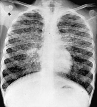
Figure 159-1 Chest radiograph of a 10 yr old girl with sarcoidosis showing widely disseminated peribronchial infiltrates, multiple small nodular densities, hyperaeration of the lungs, and hilar lymphadenopathy.
Extrathoracic lymphadenopathy and infiltration of the liver, spleen, and bone marrow also occur often. Infiltration of the liver and spleen typically leads to isolated hepatomegaly and splenomegaly, respectively, but actual organ dysfunction is rare. Cutaneous disease, such as plaques, nodules, erythema nodosum in acute disease, or lupus pernio in chronic sarcoidosis, appears in one quarter of cases and is usually present at onset. Red-brown to purple maculopapular lesions <1 cm on the face, neck, upper back, and extremities are the most common skin finding (Fig. 159-2). Ocular involvement is frequent and has variable manifestations, including anterior or posterior uveitis, conjunctival granulomas, eyelid inflammation, and orbital or lacrimal gland infiltration. The arthritis in sarcoidosis can be confused with juvenile rheumatoid arthritis. Central nervous system (CNS) involvement is rare in childhood but may manifest as seizures, cranial nerve involvement, intracranial mass lesions, and hypothalamic dysfunction. Kidney disease also occurs infrequently in children but typically manifests as renal insufficiency, proteinuria, transient pyuria, or microscopic hematuria as a result of either early monocellular infiltration or granuloma formation in kidney tissue. Only a small fraction of children have hypercalcemia or hypercalciuria, which is therefore an infrequent cause of kidney disease. Sarcoid granulomas can also infiltrate the heart and lead to cardiac arrhythmias and, rarely, sudden death. Other rare sites of disease involvement include blood vessels of any size, the gastrointestinal tract, muscles, bones, and testes.
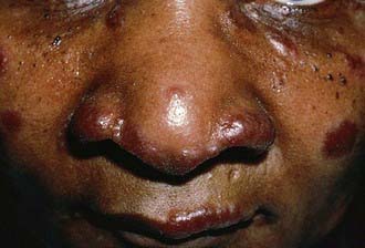
Figure 159-2 Sarcoidosis nodules on the face.
(From Shah BR, Laude TA: Atlas of pediatric clinical diagnosis, Philadelphia, 2000, WB Saunders.)
In contrast to the variable clinical presentation of sarcoidosis in older children, early-onset sarcoidosis classically manifests as the triad uveitis, arthritis, and rash. Pulmonary disease and lymphadenopathy are less common. The arthritis is polyarticular and symmetric, with large boggy effusions. The rash is diffuse, erythematous, papular, and somewhat scaly. Noncaseating granulomas are demonstrated with biopsy of the skin or joint synovium.
Laboratory Findings
There is no single laboratory test diagnostic of sarcoidosis. Anemia, leukopenia, and eosinophilia may be seen. Other nonspecific findings include hypergammaglobulinemia and elevations in acute-phase reactants, including erythrocyte sedimentation rate and C-reactive protein value. Hypercalcemia and/or hypercalciuria occur in only a small proportion of children with sarcoidosis. Angiotensin-converting enzyme (ACE) is produced by the epithelioid cells of the granuloma, and its serum value may be elevated, but this finding lacks diagnostic sensitivity and specificity. In addition, ACE values may be difficult to interpret because reference values for serum ACE are age dependent. Fluorodeoxyglucose F18 positron emission tomography (18FDG PET) can help identify nonpulmonary sites for a diagnostic biopsy.
Diagnosis
Definitive diagnosis ultimately requires demonstration of the characteristic noncaseating granulomatous lesions in a biopsy specimen (usually taken from the most readily available affected organ) and exclusion of other known causes of granulomatous inflammation. Skin and transbronchial lung biopsies have higher yield, greater specificity, and fewer associated adverse events than biopsy of mediastinal lymph nodes or liver. Additional diagnostic testing should include chest radiography, pulmonary function testing with measurement of diffusion capacity, hepatic enzyme measurements, and renal function asssessment. Ophthalmologic slit-lamp examination is essential, as ocular findings are frequent in sarcoidosis and vision loss is a sequela of untreated disease.
Bronchoalveolar lavage may be used to assess for disease activity, and the fluid typically reveals an excess of lymphocytes with an increased CD4+ : CD8+ ratio, 2 : 1- 13 : 1. The Kveim-Siltzbach test consists of an intradermal injection of homogenated human sarcoid tissue extract followed by observation for the formation of a granuloma several weeks later. This test is rarely used, owing to lack of available validated standardized test materials and safety concerns.
Differential Diagnosis
Because of its protean manifestations, the differential diagnosis of sarcoidosis is extremely broad and depends largely on the initial clinical manifestations. Granulomatous infections, including tuberculosis, cryptococcosis, pulmonary mycoses (histoplasmosis, blastomycosis, and coccidioidomycosis), brucellosis, tularemia, and toxoplasmosis, must be excluded. Other causes of granulomatous inflammation are Wegener granulomatosis, hypersensitivity pneumonia, chronic berylliosis, and other occupational exposures to metals. Combined variable immunodeficiency may also manifest as granulomatous lesions. Lymphoma should be ruled out in cases of hilar or other lymphadenopathy. Sarcoid arthritis may mimic juvenile rheumatoid arthritis. Evaluation for endocrine disorders is needed in the setting of hypercalcemia or hypercalciuria.
Treatment
There are no consensus guidelines for the treatment of childhood sarcoidosis. Treatment should be based on disease severity as well as the number and type of organs involved. Corticosteroids are the mainstay of treatment for most acute and chronic disease manifestations. The optimal dose and duration of corticosteroid therapy in children have not been established. Induction treatment typically begins with oral prednisone or prednisolone (1-2 mg/kg/day up to 40 mg daily) for 8-12 wk until manifestations improve. Corticosteroid dosage is then gradually decreased over 6-12 mo to the minimal effective dose that controls symptoms. Methotrexate may be effective as a corticosteroid-sparing agent. On the basis of the role of TNF-α in the formation of granulomas, there is rationale for use of TNF-α antagonists, and results of a small randomized trial in adults showed modest effect. Other therapeutics used for sarcoidosis manifestations include inhaled corticosteroids (lung), azathioprine (CNS), hydroxychloroquine (skin), thalidomide or its analogs (skin), topical corticosteroids (eye), and nonsteroidal anti-inflammatory drugs (NSAIDs).
Prognosis
The prognosis of childhood sarcoidosis is not well defined. The disease may be self-limited with complete recovery or may persist with a progressive or relapsing course. Outcome is worse in the setting of multiorgan or CNS involvement. Most children requiring treatment experience considerable improvement with corticosteroids, though a significant number have morbid sequelae, mainly involving the lungs and eyes. Children with early-onset sarcoidosis have a poorer prognosis and generally experience a more chronic disease course. The greatest morbidity is associated with ocular involvement, including cataract formation, development of synechiae, and loss of visual acuity or blindness. Progressive polyarthritis may result in joint destruction. The overall mortality rate in childhood sarcoidosis is low.
Serial pulmonary function tests and chest radiographs are useful in following the course of lung involvement. Monitoring for other organ involvement should also include electrocardiogram with consideration of an echocardiogram, urinalysis, renal function tests, and measurements of hepatic enzymes and serum calcium. Other potential indicators of disease activity include inflammatory markers and serum ACE, although changes in ACE level do not always correlate with other indicators of disease status. Given the frequency of asymptomatic eye disease and the ocular morbidity associated with pediatric sarcoidosis, all patients should have an ophthalmologic examination at presentation with monitoring at regular intervals, perhaps every 3-6 mo as recommended in children with juvenile rheumatoid arthritis.
Baumann RJ, Robertson WCJr. Neurosarcoid presents differently in children than in adults. Pediatrics. 2003;112:e480-e486.
Dempsey OJ, Paterson EW, Kerr KM, et al. Sarcoidosis. BMJ. 2009;339:620-625.
Ho LP, Urban BC, Thickett DR, et al. Deficiency of a subset of T cells with immunoregulatory properties in sarcoidosis. Lancet. 2005;365:1062-1072.
Hoffman AL, Milman N, Byg K-E. Childhood sarcoidosis in Denmark 1979–1994: incidence, clinical features and laboratory results at presentation in 48 children. Acta Paediatr. 2004;93:30-36.
Iannuzzi MC, Fontana JR. Sarcoidosis. JAMA. 2011;305(4):391-398.
Iannuzzi MC, Rybicki BA, Teirstein AS. Sarcoidosis. N Engl J Med. 2007;357:2153-2165.
Milman N, Hoffman AL. Childhood sarcoidosis: long-term follow-up. European Respir J. 2008;31:592-598.
Newman LS, Rose CS, Bresnitz EA, et al. A case control etiologic study of sarcoidosis: environmental and occupational risk factors. Am J Respir Crit Care Med. 2004;170:1324-1330.
Rosé CD, Wouters CH, Meiorin S, et al. Pediatric granulomatous arthritis. Arthritis Rheumatol. 2006;54:3337-3344.
Shetty AK, Gedalia A. Childhood sarcoidosis: a rare but fascinating disorder. Pediatr Rheumatol Online J. 2008;6:16.
Spagnolo P, du Bois RM. Genetics of sarcoidosis. Clinics Dermatol. 2007;25:242-249.
Sverrild A, Backer V, Kyvik KO, et al. Heredity in sarcoidosis: a registry-based twin study. Thorax. 2008;63:894-896.