Chapter 307 Common Lesions of the Oral Soft Tissues
Oropharyngeal Candidiasis
Oropharyngeal infection with Candida albicans (thrush, moniliasis) (Chapter 226.1) is common in neonates from contact with the organism in the birth canal or breast. The lesions of oropharyngeal candidiasis (OPC) appears as white plaques covering all or part of the oropharyngeal mucosa. These plaques are removable from the underlying surface, which is characteristically inflamed and has pinpoint hemorrhages. The diagnosis is confirmed by direct microscopic examination on potassium hydroxide smears and culture of scrapings from lesions. OPC is usually self-limited in the healthy newborn infant, but topical application of nystatin to the oral cavity of the baby and to the nipples of breast-feeding mothers will hasten recovery.
OPC is also a major problem during myelosuppressive therapy. Systemic candidiasis (SC), a major cause of morbidity and mortality during myelosuppressive therapy, develops almost exclusively in patients who have had prior oropharyngeal, esophageal, or intestinal candidiasis. This observation implies that prevention of OPC should reduce the incidence of SC. The use of oral rinses of 0.2% chlorhexidine solution, plus systemic antifungals may be effective in preventing OPC, SC, or candidal esophagitis.
Aphthous Ulcers
The aphthous ulcer (canker sore) is a distinct oral lesion, prone to recurrence. The differential diagnosis is noted in Table 307-1. Aphthous ulcers are reported to develop in 20% of the population. Their etiology is unclear, but allergic or immunologic reactions, emotional stress, genetics, and injury to the soft tissues in the mouth have been implicated. Aphthous-like lesions may be associated with inflammatory bowel disease, Behçet disease, gluten-sensitive enteropathy, periodic fever-aphthae-pharyngitis-adenitis syndrome, Sweet syndrome, HIV infection (especially if ulcers are large and slow to heal), and cyclic neutropenia. Clinically, these ulcers are characterized by well-circumscribed, ulcerative lesions with a white necrotic base surrounded by a red halo. The lesions last 10-14 days and heal without scarring. Over-the-counter palliative therapies, such as benzocaine and topical lidocaine, are effective, as well as topical steroids. Tetracycline has been shown to have benefit with severe outbreaks, but caution is necessary in pregnant women and young children to prevent tetracycline tooth staining during a child’s tooth development.
Table 307-1 DIFFERENTIAL DIAGNOSIS OF ORAL ULCERATION
| CONDITION | COMMENT |
|---|---|
| COMMON | |
| Aphthous (canker sore) | Painful, circumscribed lesions; recurrences |
| Traumatic | Accidents, chronic cheek biter, or after dental local anesthesia |
| Hand, foot, mouth disease | Painful; lesions on tongue, anterior oral cavity, hands, and feet |
| Herpangina | Painful; lesions confined to soft palate and oropharynx |
| Herpetic gingivostomatitis | Vesicles on mucocutaneous borders; painful, febrile |
| Recurrent herpes labialis | Vesicles on lips; painful |
| Chemical burns | Alkali, acid, aspirin; painful |
| Heat burns | Hot food, electrical |
| UNCOMMON | |
| Neutrophil defects | Agranulocytosis, leukemia, cyclic neutropenia; painful |
| Systemic lupus erythematosus | Recurrent, may be painless |
| Behçet’s syndrome | Resembles aphthous lesions; associated with genital ulcers, uveitis |
| Necrotizing ulcerative gingivostomatitis | Vincent stomatitis; painful |
| Syphilis | Chancre or gumma; painless |
| Oral Crohn disease | Aphthous-like; painful |
| Histoplasmosis | Lingual |
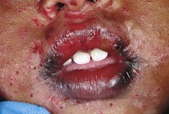
Herpetic Gingivostomatitis
After an initial incubation period of ∼1 wk, the initial infection with herpes simplex virus manifests as fever and malaise, usually in a child <5 yr (Chapter 244). The oral cavity can show various expressions, including the gingiva becoming erythematous, mucosal hemorrhages, and clusters of small vesicles erupting throughout the mouth. There is often involvement of the mucocutaneous margin and perioral skin (Fig. 307-1). The oral symptoms generally are accompanied by fever, lymphadenopathy, and difficulty eating and drinking. The symptoms usually regress within 2 wk without scarring. Fluids should be encouraged because the child may become dehydrated. Analgesics and anesthetic rinses can make the child more comfortable. Oral acyclovir if taken within the first 3 days of symptoms may be beneficial in shortening the duration of symptoms. Caution should be exercised to prevent autoinoculation or transmission of infection to the eyes.
Recurrent Herpes Labialis
Approximately 90% of the population develops antibodies to herpes simplex virus. In periods of quiescence, the virus is thought to remain latent in sensory neurons. Unlike primary herpetic gingivostomatitis, which manifests as multiple painful vesicles on the lips, tongue, palate, gingiva, and mucosa, recurrent herpes is generally limited to the lips. Other than the annoyance of causing pain and an unattractive appearance, there are generally no systemic symptoms. Reactivation of the virus is thought to be the result of exposure to ultraviolet light, tissue trauma, stress, or fevers. There is little advantage of antiviral therapy over palliative therapies in an otherwise healthy patient affected by recurrent herpes.
Bohn Nodules
Bohn nodules are small developmental anomalies located along the buccal and lingual aspects of the mandibular and maxillary ridges and in the hard palate of the neonate. These lesions arise from remnants of mucous gland tissue. Treatment is not necessary, because the nodules disappear within a few weeks.
Dental Lamina Cysts
Dental lamina cysts are small cystic lesions located along the crest of the mandibular and maxillary ridges of the neonate. These lesions arise from epithelial remnants of the dental lamina. Treatment is not necessary; they disappear within a few weeks.
Fordyce Granules
Almost 80% of adults have multiple yellow-white granules in clusters or plaquelike areas on the oral mucosa, most commonly on the buccal mucosa or lips. They are aberrant sebaceous glands. The glands are present at birth, but they can hypertrophy and 1st appear as discrete yellowish papules during the preadolescent period in ∼50% of children. No treatment is necessary.
Parulis
The parulis (gum boil) is a soft reddish papule located adjacent to the root of a chronically abscessed tooth. It occurs at the end-point of a draining dental sinus tract. Treatment consists of diagnosing which tooth is abscessed and extracting it or performing root canal on the offending tooth.
Cheilitis
This dryness of the lips followed by scaling and cracking and accompanied by a characteristic burning sensation is common in children. Cheilitis may be caused by sensitivity to contact substances, lip licking, vitamin deficiency, weakened immune system, or fungal or bacterial infections. Cheilitis often occurs in association with fever. Treatment may include antifungal or antibacterial agents and frequent application of petroleum jelly.
Ankyloglossia
Ankyloglossia or “tongue-tie” is characterized by an abnormally short lingual frenum that can hinder the tongue movement but rarely interferes with feeding or speech. The frenum might spontaneously lengthen as the child gets older. If the extent of the ankyloglossia is severe, speech may be affected and surgical correction may be indicated.
Geographic Tongue
Geographic tongue (migratory glossitis) is a benign and asymptomatic lesion and is characterized by one or more smooth, bright red patches, often showing a yellow, gray, or white membranous margin on the dorsum of an otherwise normally roughened tongue. The condition has no known cause, and no treatment is indicated (Chapter 656).
Fissured Tongue
The fissured tongue (scrotal tongue) is a malformation manifested clinically by numerous small furrows or grooves on the dorsal surface (Chapter 656). If the tongue is painful, brushing the tongue or irrigating with water can reduce the bacteria in the fissures.