Chapter 406 Pneumomediastinum
Pneumomediastinum is the presence or air or gas in the mediastinum.
Etiology
Pneumomediastinum is usually caused by alveolar rupture during acute or chronic pulmonary disease. A diverse group of nonrespiratory entities can also cause it, and the lung is not always the source of the air. Pneumomediastinum has been reported after dental extractions, adenotonsillectomy, normal menses, obstetric delivery, diabetes mellitus with ketoacidosis, acupuncture, anorexia nervosa, and acute gastroenteritis. It can also result from esophageal perforation, penetrating chest trauma, or inhaled foreign body. Occasionally, no underlying cause is found. Acute asthma is the most common cause of pneumomediastinum in older children and teenagers. Simultaneous pneumothorax is unusual in these patients.
Pathogenesis
After intrapulmonary alveolar rupture, air dissects through the perivascular sheaths and other soft tissue, planes toward the hilum, and enters the mediastinum.
Clinical Manifestations
Transient stabbing chest pain that may radiate to the neck is the principal feature of pneumomediastinum. Isolated abdominal pain and sore throat also occur. The patient may have dyspnea and cough. Pneumomediastinum is difficult to detect by physical examination alone. Subcutaneous emphysema, if present, is diagnostic. Cardiac dullness to percussion may be decreased, but the chests of many patients with pneumomediastinum are chronically overinflated, and it is unlikely that the clinician can be sure of this finding. A mediastinal “crunch” (Hamman sign) is occasionally heard but is easily confused with a friction rub.
Laboratory Findings
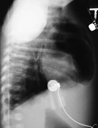
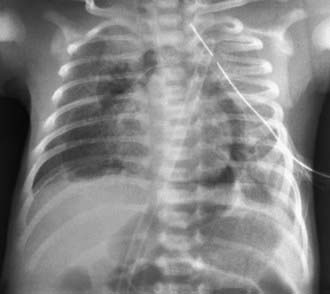
Chest radiography reveals mediastinal air, with a more distinct cardiac border than normal (Figs. 406-1 to 406-3). On the lateral projection, the posterior mediastinal structures are clearly defined, there may be a lucent ring around the right pulmonary artery, and retrosternal air can usually be seen (see Fig. 406-2). Vertical streaks of air in the mediastinum and subcutaneous air are often observed (see Fig. 406-1).
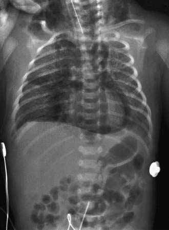
Figure 406-1 Large pneumomediastinum surrounding the heart and dissecting into the neck.
(From Clark DA: Atlas of neonatology, ed 7, Philadelphia, 2000, WB Saunders.)
Complications
Pneumomediastinum is rarely a major problem in older children because the mediastinum can be depressurized by escape of air into the neck or abdomen. In the newborn, however, the rate at which air can leave the mediastinum is limited, and pneumomediastinum can lead to dangerous cardiovascular compromise or pneumothorax (Chapters 95.12 and 405).
Treatment
Treatment is directed primarily at the underlying obstructive pulmonary disease or other precipitating condition. Analgesics are occasionally needed for chest pain. Rarely, subcutaneous emphysema can cause sufficient tracheal compression to justify tracheotomy; the tracheotomy also decompresses the mediastinum. Collar mediastinotomy and percutaneous drainage catheter placement are other treatment modalities.
Bullaro FM, Bartoletti SC. Spontaneous pneumomediastinum in children: a literature review. Pediatr Emerg Care. 2007;23:28-30.
Chiu CY, Wong KS, Yao TC, et al. Asthmatic versus non-asthmatic spontaneous pneumomediastinum in children. Asian Pac J Allergy Immunol. 2005;23:19-22.
Hauri-Hohl A, Baenziger O, Frey B. Pneumomediastinum in the neonatal and paediatric intensive care unit. Eur J Pediatr. 2008;167:415-418.
Shine NP, Sader C, Coates H. Cervicofacial emphysema and pneumomediastinum following pediatric adenotonsillectomy: a rare complication. Int J Pediatr Otorhinolaryngol. 2005;69:1579-1582.
Sundararaghavan S, Pitts TY, Suarez WA, et al. Chest pain among adolescents with anorexia nervosa. Pediatr Emerg Care. 2005;21:603-605.