Chapter 545 Breast Concerns
Girls with breast disorders commonly present with questions about the development and appearance of their breasts, breast pain, nipple discharge, and concerns about the presence of a mass. Although children and adolescents are unlikely to have malignant or life-threatening breast problems, this population of patients should be referred to practitioners who have experience and familiarity with the immature and developing breast to avoid overtreatment with unnecessary diagnostic or surgical procedures.
Breast Development
Development of the breast begins around wk 5 of gestation, when the ectoderm on the anterior body wall thickens into a ridge known as the milk line. This ridge of tissue extends from the area of the developing axilla to the area of the developing inguinal canal. The ridge above and below the area of the pectoralis muscle recedes in utero, leaving the mammary primordium, which is the origin of the lactiferous ducts. The initial lactiferous ducts form between wk 10 and 20 and become interspersed through the developing mesenchyme, which becomes the fibrous and fatty portions of the breast. The breast bud, under the stimulation of maternal estrogen, becomes palpable at wk 34 of gestation. This breast bud regresses within the 1st mo of life, because the estrogen stimulation is no longer present. The areola appears at 5 mo of gestation, and the nipple is seen shortly after birth. It is initially depressed and later becomes elevated.
Thelarche, or the onset of pubertal breast development, is hormonally mediated and normally occurs between the ages of 8 and 13 yr, with an average age of 10.3 years. The initiation of thelarche and progression in females is affected by race, with normal thelarche occurring earlier in African-American girls than in Caucasian or Asian girls.
Once thelarche is initiated, normal development of the breast occurs over 2-4 yr and is classified by the sexual maturity rating (SMR) system into 5 stages. Maturation can sometimes occur asymmetrically owing to fluctuation of the hormonal environments and various end organ sensitivities. Lack of development by age 13 yr is considered delayed and warrants endocrinology evaluation. Menarche usually occurs approximately 2 yr after initiation of breast development.
Breast Examination
A breast examination should be included in the annual examination of all children and adolescents. Examination of the newborn includes assessment of breast size, nipple position, presence of accessory nipples, and nipple discharge. Examination of the prepubertal girl includes inspection and palpation of the chest wall for masses, pain, nipple discharge, and signs of premature thelarche. Examination of the adolescent is performed with the patient in the supine position; the arm ipsilateral to the breast that is being examined should be placed next to the patient’s head. The breast tissue is examined with the flat pads of the middle fingers and the examiner can feel for abnormalities on the breast with a pattern similar to spokes on a wheel, in a circular clockwise pattern with concentric circles or by moving in a rotary fashion around the breast. Whatever the method used, the goal is to palpate all the breast tissue in a uniform fashion. The SMR should be noted and axillary, supraclavicular, and infraclavicular nodes evaluated for lymphadenopathy. The areola should be compressed to assess for nipple discharge.
Self Breast Examination
Controversy exists as to the utility of breast self-examination in the adolescent population. Experts believe that it might be ill advised to encourage breast self-examination in the adolescent due to a potential for unnecessary anxiety and possible unwarranted treatment in a population that is at low risk for malignant disease. The American College of Obstetricians and Gynecologists states that despite a lack of definitive data for or against breast self-examination, breast self-examination may be recommended beginning at age 19 yr. Women with previous exposure to therapeutic chest radiation therapy are advised to begin breast self-examination 10 yr after radiation therapy.
Abnormal Development
Neonatal Breast Abnormalities
The condition in which breasts enlarge in the newborn period is neonatal breast hypertrophy. This is quite common in term infants and can occur as a result of elevated circulating maternal endogenous steroid hormones in late gestation. As maternal estrogen levels fall, prolactin levels can increase and the breasts can produce a clear or cloudy (milklike) nipple discharge (“witch’s milk”). Repeated manipulation of the breast can exacerbate the condition. On occasion, the hypertrophy is associated with mastitis caused by a staphylococcal or streptococcal infection; antibiotics should be administered.
Precocious Puberty
Premature thelarche is usually an isolated condition but it may be the first symptom of precocious puberty. Precocious puberty occurs in 14-18% of girls with premature thelarche (Chapter 556). Serial examinations, with particular emphasis on growth velocity, secondary sex characters such as pubic hair, pigmentation of the labia or areola, or vaginal bleeding are imperative to identify precocious puberty. Unless there are associated signs of precocious puberty, the parents should be reassured and the child should be followed.
Amastia
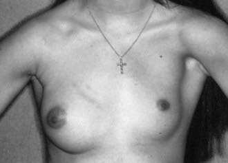
Complete absence of the breast, or amastia, is rare and is thought to occur from lack of formation or obliteration of the milk line. Amastia is usually unilateral and can be congenital or associated with systemic disorders (e.g., malnutrition, Crohn disease) or endocrine disorders (e.g., congenital adrenal hyperplasia, gonadal dysgenesis, hypogonadotropic hypogonadism). It can be associated with anomalies of the underlying mesoderm, such as abnormal pectoralis muscles seen in Poland syndrome (aplasia of the pectoralis muscles, rib deformities, webbed fingers, and radial nerve aplasia) (Fig. 545-1). Amastia or hypomastia can also be iatrogenic, resulting from injuries sustained during thoracotomy, chest tube placement, radiotherapy, severe burns, and inappropriate biopsy of the breast bud. Treatment is surgical correction.
Polymastia and Polythelia
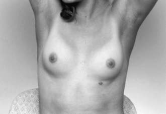
Supernumerary breast tissue (polymastia) and accessory nipples (polythelia) occur in approximately 1-2% of the population (Fig. 545-2). The abnormally placed tissue can be seen anywhere along the milk line but is usually noted on the chest, upper abdomen, or just inferior to the normally positioned breast. An association has been made between polythelia and anomalies of the urinary and cardiovascular system. Surgical excision of the accessory breasts or nipple is not usually needed. Resection of accessory tissue may be warranted if the patient has pain or for cosmetic reasons.
Breast Asymmetry and Hypomastia
Some degree of asymmetry is normal in women, and it may be more pronounced during puberty while the breasts are developing. Hypoplasia of the breasts varies in degree from a nearly total absence of breast tissue to well-formed breasts that are considered by the patient to be too small. There are several causes for poor or absent breast development. The onset of breast development may be delayed with normal secondary sex characters; the breasts develop slowly but are normal in all other respects; a patient’s family history might include late breast development. Other causes include ovarian dysfunction, hypothyroidism, and chest wall irradiation or surgery. Hypoplastic breast tissue can also be associated with a tuberous breast anomaly. Treatment depends on the underlying cause. Patients with mild asymmetry and with no other associated pathology should be reassured. Surgical correction is an option for women with marked asymmetry.
Juvenile or Virginal Hypertrophy
Spontaneous massive growth of the breasts during puberty and adolescence is thought to be the result of excessive end-organ sensitivity to gonadal hormones. The underlying cause, if any, should be determined and removed (Table 545-1). When growth is extreme it is termed macromastia or gigantomastia. It is more commonly bilateral, often occurs over a brief period, and most commonly affects adolescent girls. Physical and psychologic problems can affect posture and quality of life. Strong emotional support should be provided as this can affect an adolescent’s self-esteem at a vulnerable time in her psychologic development. Management should be individualized and may range from reassurance or the use of supportive brassieres to reduction mammoplasty. Surgery should be delayed until late adolescence to allow complete breast development. Medical therapy is available to slow down breast growth in extreme cases until surgery can be performed. Surgical intervention often necessitates relocation of the nipple, which can result in decreased sensation and altered lactation.
Infections
Mastitis is the most common infection of the breast. Though it is most common in lactating mothers, it can occur in young infants and adolescents. Neonatal mastitis is an infection that usually occurs in term or near-term infants. It should be treated aggressively to reduce the risk of forming abscesses. Adolescents can develop nonlactational mastitis or a breast abscess for unknown reasons, as a result of irritation of the skin (through shaving or nipple stimulation), trauma, a foreign body (e.g., piercing), ductal abnormality (such as ductal ectasia), or infection of an epidermal cyst. The initial therapy of all breast infections is antibiotics and analgesics. Staphylococcus aureus (Chapter 174.1) is the offending organism is almost all cases. Owing to the potential for breast abscess, the neonatal population should be treated with parenteral antibiotics guided by gram stain, when available. Adolescents may be initially treated with warm compresses and oral antibiotics. Abscesses should be surgically evaluated (with ultrasound guidance if necessary) and drained as necessary. If incision and drainage is performed, a small, periareolar incision is indicated.
Trauma and Inflammation
Breast trauma is common in adolescent girls participating in contact sports. The trauma usually takes the form of contusion or hematoma and can resolve spontaneously or may be associated with late cystic changes in the breast or fibrosis with retraction of the skin or the nipple over the injured area.
Nipple Discharge
Nipple discharge must be carefully evaluated and a distinction made among galactorrhea (milky white discharge), blood, or other discharge (Table 545-2). A careful history and physical examination directed at the possible etiologies of galactorrhea will help the practitioner determine the etiology. Examination of the discharge assists in diagnosis. Benign conditions are usually associated with a milky, sticky, thick discharge; infection is associated with a purulent discharge; intraductal papilloma and cancer are associated with a serous, serosanguineous, or bloody discharge.
Galactorrhea
To test for galactorrhea, obtain cytologic evaluation of the discharge fluid by Sudan (fat) stain. Serum pregnancy testing and prolactin and thyroid levels are obtained to rule out the presence of a thyroid abnormality, a pituitary prolactinoma, and pregnancy (in the postpubertal adolescent). If the prolactin level is elevated, visual field studies and a head MRI might reveal presence of a pituitary adenoma (Chapter 554). Treatment is directed by results of history, physical exam, and lab studies. Patients should be instructed to avoid nipple stimulation and stop any offending drugs. Hypothyroidism should be treated and prolactin tumors managed with appropriate medical or surgical care. Treatment of galactorrhea (not thyroid related) consists primarily of dopamine agonists such as bromocriptine or cabergoline. Surgical intervention, usually transsphenoidal hypophysectomy, is rarely required.
Bloody Discharge
In adolescent athletes, bloody discharge may be due to chronic nipple irritation (jogger’s nipple), discharge from the ducts of Montgomery (on the edge of the areola, not through the nipple) or duct ectasia. Cytologic assessment should be performed. Surgical consultation for a mass is indicated because intraductal breast papillomas have occurred in adolescents.
Mastalgia
Physiologic swelling and tenderness occur on a cyclic basis, most commonly during the premenstrual phase, and are secondary to hormonal stimulation and resulting proliferative changes. Hormonal imbalance can cause exaggerated responses in the breast tissue, especially in the upper and outer quadrants. Nodularity, poorly localized tenderness, and a soreness radiating to the axilla and arm are usual accompanying findings. The preferable term for these changes is benign breast changes rather than fibrocystic disease. Treatments recommended for this condition include a firm support bra, heat, and analgesics. Oral contraceptives often improve the breast pain. A course of nonsteroidal anti-inflammatory drugs has also been shown to be effective. Methylxanthines (caffeine in coffee, tea, carbonated drinks) and smoking should be eliminated. Evening primrose oil and vitamin E are popular but unproven treatments.
Breast Masses
A mass in the developing breast can be of concern to the adolescent and her family.
Peripubertal Masses
Initial breast development at the onset of thelarche can be asymmetric and thus mistaken for a “mass.” The breast bud is palpable in these cases and should be distinguishable. Such asynchronous thelarche should be recognized to avoid biopsy and potential injury to the maturing breast. If there is any question, ultrasound can be used to evaluate for a mass. Unilateral thelarche has also been reported as a side effect of cimetidine and is reversible with stopping the drug.
Common Adolescent Breast Masses
The differential diagnosis for breast masses in the adolescent patient is seen in Table 545-3. The patient should be questioned about the variation in symptoms with the menstrual cycle, associated symptoms such as nipple discharge, recent trauma to the breast, family history of breast masses or cancer, and history of chest radiation or malignancy. Because breast cancer in the adolescent is extremely rare, masses can be expectantly managed for extended periods with little concern for malignancy in this population.
Table 545-3 BREAST MASSES IN THE ADOLESCENT GIRL
BENIGN
MALIGNANT
Data from Dehner LP, Hill DA, Deschryver K: Pathology of the breast in children, adolescents, and young adults, Semin Diagn Pathol 16:235–247, 1999; Simmons PS: Diagnostic considerations in breast disorders of children and adolescents, Obstet Gynecol Clin North Am 19:91–102, 1992; and Laufer MR, Goldstein DP: The breast: examination and lesions. In Emans SJ, Laufer MR, Goldstein DP, editors: Pediatric and adolescent gynecology, ed 5, Philadelphia, Lippincott Williams & Wilkins, 2005, pp 729–759.
The most common solid mass seen in adolescent girls is the fibroadenoma. Fibroadenomas are most often located in the upper outer quadrant of the breast and are more common in African-American patients. The average size is 2-3 cm, and 10-25% of patients have multiple lesions. The physical examination is usually diagnostic because these lesions are well circumscribed, rubbery, mobile, and not tender. In equivocal cases, an ultrasound may be helpful in making the diagnosis. Mammography is not indicated in the adolescent patient.
Fibroadenomas can develop because of a local exaggerated response to estrogen stimulation and they can enlarge during the menstrual cycle. These lesions may be safely watched for at least 2 menstrual cycles, and some investigators suggest observation until adulthood. About 10% of fibroadenomas regress spontaneously. The option of expectantly managing the patient until adulthood should be considered because the risk of primary cancer is very low in this population. If expectant management is chosen, serial ultrasounds may be done to ensure that the mass does not have malignant characteristics on imaging and that it is not enlarging or changing in contour. About 4% of fibroadenomas grow, and so fine-needle aspiration or excision is recommended when a mass is enlarging, grows >5 cm (due to the risk of giant fibroadenoma or cystosarcoma phylloides), or the mass is causing anxiety to the patient or her family.
Cysts are very common masses seen in the pediatric breast. Cysts vary in size over the course of a menstrual cycle, so a patient with a possible cyst should be re-examined a few weeks after the initial evaluation to see if the mass is still present. If a mass persists, then it may be imaged by ultrasonography or aspirated with a needle to evaluate if it truly is a cyst. Aspirated fluid that is clear may be discarded. Bloody fluid and other aspirated material should be sent for cytology. Cystic lesions that resolve with aspiration should be reevaluated in 3 mo. If they recur they should be evaluated with sonography.
Malignant Masses
Primary breast cancer is extremely rare in adolescents. Surveillance Epidemiology and End Results (SEER) data from 1975-2006 lists no incident cases of breast cancer in situ in girls and women <20 yr and publishes an incidence of invasive breast cancer of 0.2/100,000 for females up to age 19 (http://seer.cancer.gov/csr/1975_2006/results_merged/sect_04_breast.pdf). Although malignancy is rare, lesions with suspicious imaging findings or progressive growth should undergo cytologic or histologic examination. A small study in Austria noted a 4.7% malignancy rate among palpable masses in teens. Additional research in this area is needed to assess the validity of the findings of the latter study.
Cystosarcoma phylloides can occur in adolescents. It is characterized by asymmetric breast enlargement in association with a firm, mobile, circumscribed mass. It can mimic a giant fibroadenoma. The tumor often grows rapidly and can become quite large. The majority of these tumors have a favorable prognosis, but malignant cystosarcoma phylloides has been reported to recur both locally and with metastases. Excision with 1 cm margins is the preferred initial therapy in adolescent patients, regardless of the histologic classification of the lesion. Fatal metastatic cystosarcoma phylloides in an adolescent has occurred.
Secondary cancers in adolescents with previous therapeutic radiation to the chest or with malignancies with the potential to metastasize to the breast should be monitored more closely for breast masses. Breast tumors also may be the first manifestation of relapse (extramedullary) in acute lymphoblastic leukemia.
Imaging of Breast Masses
Because the dense breast tissue of the adolescent obstructs the visualization of a palpable mass, mammography is not advised for this age group. Ultrasonography is the imaging modality of choice for breast abnormalities in the pediatric population. Color Doppler ultrasound can be useful in evaluating breast abnormalities such as fibroadenomas or abscesses.
Alterations to the Breast
A dramatic increase in adolescents desiring breast augmentation has occurred since the turn of the century. Breast augmentation in adolescents is discouraged owing to associated psychologic and physical immaturity. The American Society of Plastic Surgeons discourages breast augmentation in girls <18 yr for purely cosmetic reasons. The FDA also considers breast implants in adolescents <18 yr for solely cosmetic reasons to be an off-label use.
Breast reduction surgery might be considered when an adolescent is bothered by extremely large breasts that result in neck and back pain and prevent participation in sports. Breast reduction allows these girls to feel less self-conscious, have less pain, and be more active. Also girls with marked asymmetry in breasts due to the pathologies noted earlier can feel self-conscious and want breast surgery. Before performing breast-altering surgery, practitioners must ensure proper selection of teens and families who have an appropriate understanding of the risks and benefits of surgery and realistic expectations of the procedure.
American College of Obstetricians and Gynecologists. ACOG Committee opinion no. 350, November 2006: Breast concerns in the adolescent. Obstet Gynecol. 2006;108:1329-1336.
American College of Obstetricians and Gynecologists. Fact sheet: plastic surgery. Tool kit for teen care, ed 2. American College of Obstetricians and Gynecologists, Washington, DC, 2009.
American Society of Plastic Surgeons. Policy statement: breast augmentation in teenagers (PDF), http://www.plasticsurgery.org/d.xml?comp=x983. Accessed April 1, 2010
Centers for Disease Control and Prevention. Idiopathic granulomatous mastitis in Hispanic women—Indiana, 2006-2008. MMWR. 2009;58(47):1317-1321.
Chung EM, Cube R, Hall GJ, et al. From the archives of the AFIP: breast masses in children and adolescents: radiologic-pathologic correlation. Radiographics. 2009;29:907-931.
Dixon JM, Khan LR. Treatment of breast infection. BMJ. 2011;342:484-489.
Greydanus DE, Matytsina L, Gains M. Breast disorders in children and adolescents. Prim Care. 2006;33:455-502.
Henley DV, Lipson N, Korach KS, et al. Prepubertal gynecomastia linked to lavender and tea tree oils. N Engl J Med. 2007;365:479-485.
Laufer MR, Goldstein DP. The breast: examination and lesions. In: Emans SJ, Laufer MR, Goldstein DP, editors. Pediatric and adolescent gynecology. ed 5. Philadelphia: Lippincott Williams & Wilkins; 2005:729-759.
Ries LAG, Melbert D, Krapcho M, et al. SEER Cancer Statistics Review, 1975–2005 (website), http://seer.cancer.gov/csr/1975_2005/. Accessed April 1, 2010
Sadove AM, van Aalst JA. Congenital and acquired pediatric breast anomalies: a review of 20 years’ experience. Plast Reconstr Surg. 2005;115:1039-1050.
Stricker T. Mastitis in early infancy. Acta Paediatrica. 2005;94:166-169.
Tea MM, Asseryanis E, Kroiss R, et al. Surgical breast lesions in adolescent females. Pediatr Surg Int. 2009;25:73-75.
Tiryaki T, Senel E, Hucumenoglu S, et al. Breast fibroadenoma in female adolescents. Saudi Med J. 2007;28:137-138.