Chapter 639 Diseases of the Neonate
Minor evanescent lesions of newborn infants, particularly when florid, may cause undue concern. Most of the entities are relatively common, benign, and transient and do not require therapy.
Sebaceous Hyperplasia
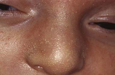
Minute, profuse, yellow-white papules are frequently found on the forehead, nose, upper lip, and cheeks of a term infant; they represent hyperplastic sebaceous glands (Fig. 639-1). These tiny papules diminish gradually in size and disappear entirely within the first few weeks of life.
Milia
Milia are superficial epidermal inclusion cysts that contain laminated keratinized material. The lesion is a firm cyst, 1-2 mm in diameter, and pearly, opalescent white. Milia may occur at any age but in neonates are most frequently scattered over the face and gingivae and on the midline of the palate, where they are called Epstein pearls. Milia exfoliate spontaneously in most infants and may be ignored; those that appear in scars or sites of trauma in older children may be gently unroofed and the contents extracted with a fine-gauge needle.
Sucking Blisters
Solitary or scattered superficial bullae present at birth on the upper limbs of infants at birth are presumably induced by vigorous sucking on the affected part in utero. Common sites are the radial aspect of the forearm, thumb, and index finger. These bullae resolve rapidly without sequelae and should be distinguished from sucking pads (calluses), which are found on the lips in the first few months and are due to combined intracellular edema and hyperkeratosis. The diagnosis can be confirmed by observing the neonate sucking the affected area.
Cutis Marmorata
When a newborn infant is exposed to low environmental temperatures, an evanescent, lacy, reticulated red and/or blue cutaneous vascular pattern appears over most of the body surface. This vascular change represents an accentuated physiologic vasomotor response that disappears with increasing age, although it is sometimes discernible even in older children. Cutis marmorata telangiectatica congenita is clinically similar, but the lesions are more intense, may be segmental, are persistent, and may be associated with loss of dermal tissue, epidermal atrophy, and ulceration.
Harlequin Color Change
A rare but dramatic vascular event, harlequin color change occurs in the immediate newborn period and is most common in low birthweight (LBW) infants. It probably reflects an imbalance in the autonomic vascular regulatory mechanism. When the infant is placed on one side, the body is bisected longitudinally into a pale upper half and a deep red dependent half. The color change lasts only for a few minutes and occasionally affects only a portion of the trunk or face. Changing the infant’s position may reverse the pattern. Muscular activity causes generalized flushing and obliterates the color differential. Repeated episodes may occur but do not indicate permanent autonomic imbalance.
Salmon Patch (Nevus Simplex)
Salmon patches are small, pale pink, ill-defined, vascular macules that occur most commonly on the glabella, eyelids, upper lip, and nuchal area of 30-40% of normal newborn infants. These lesions, which represent localized vascular ectasia, persist for several months and may become more visible during crying or changes in environmental temperature. Most lesions on the face eventually fade and disappear completely, although lesions occupying the entire central forehead often do not. Those on the posterior neck and occipital areas usually persist. The facial lesion should not be confused with a port-wine stain, which is a permanent lesion. The salmon patch is usually symmetric, with lesions on both eyelids or on both sides of midline. Port-wine stains are often larger and unilateral, and they usually end along the midline (Chapter 642).
Mongolian Spots
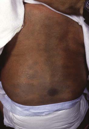
Mongolian spots, which are blue or slate-gray macular lesions, have variably defined margins; they occur most commonly in the presacral area but may be found over the posterior thighs, legs, back, and shoulders (Fig. 639-2). They may be solitary or numerous and often involve large areas. More than 80% of black, Asian, and East Indian infants have these lesions, whereas the incidence in white infants is <10%. The peculiar hue of these macules is due to the dermal location of melanin-containing melanocytes (mid-dermal melanocytosis) that are presumably arrested in their migration from neural crest to epidermis. Mongolian spots usually fade during the first few years of life as a result of darkening of the overlying skin. Malignant degeneration does not occur. The characteristic appearance and congenital onset distinguish these spots from the bruises of child abuse.
Erythema Toxicum
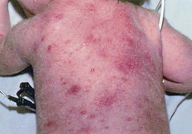
A benign, self-limited, evanescent eruption, erythema toxicum occurs in ≈ 50% of full-term infants; preterm infants are affected less commonly. The lesions are firm, yellow-white, 1- to 2-mm papules or pustules with a surrounding erythematous flare (Fig. 639-3). At times, splotchy erythema is the only manifestation. Lesions may be sparse or numerous and either clustered in several sites or widely dispersed over much of the body surface. The palms and soles are usually spared. Peak incidence occurs on the 2nd day of life, but new lesions may erupt during the 1st few days as the rash waxes and wanes. Onset may occasionally be delayed for a few days to weeks in premature infants. The pustules form below the stratum corneum or deeper in the epidermis and represent collections of eosinophils that also accumulate around the upper portion of the pilosebaceous follicle. The eosinophils can be demonstrated in Wright-stained smears of the intralesional contents. Cultures are sterile.
The cause of erythema toxicum is unknown. The lesions can mimic pyoderma, candidosis, herpes simplex, transient neonatal pustular melanosis, and miliaria but can be differentiated by the characteristic infiltrate of eosinophils and the absence of organisms on a stained smear. The course is brief, and no therapy is required. Incontinentia pigmenti and eosinophilic pustular folliculitis also have eosinophilic infiltration but can be distinguished by their distribution, histologic type, and chronicity.
Transient Neonatal Pustular Melanosis
Pustular melanosis, which is more common among black than among white infants, is a transient, benign, self-limited dermatosis of unknown cause that is characterized by 3 types of lesions: (1) evanescent superficial pustules, (2) ruptured pustules with a collarette of fine scale, at times with a central hyperpigmented macule, and (3) hyperpigmented macules (Fig. 639-4). Lesions are present at birth, and 1 or all types of lesions may be found in a profuse or sparse distribution. Pustules represent the early phase of the disorder, and macules, the late phase. The pustular phase rarely lasts more than 2-3 days; hyperpigmented macules may persist for as long as 3 mo. Sites of predilection are the anterior neck, forehead, and lower back, although the scalp, trunk, limbs, palms, and soles may be affected.
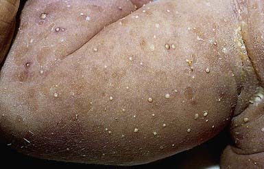
Figure 639-4 Transient neonatal pustular melanosis. Multiple papules present at birth on the arm of an infant.
(From Weston WL, Lane AT, Morelli JG, editors: Color textbook of pediatric dermatology, ed 3, Philadelphia, 2002, Mosby, p 331.)
The active phase shows an intracorneal or subcorneal pustule filled with polymorphonuclear leukocytes, debris, and an occasional eosinophil. The macules are characterized only by increased melanization of epidermal cells. Cultures and smears can be used to distinguish these pustules from those of erythema toxicum and pyoderma, because the lesions of pustular melanosis do not contain bacteria or dense aggregates of eosinophils. No therapy is required.
Infantile Acropustulosis
Onset of infantile acropustulosis generally occurs at 2-10 mo of age; lesions are occasionally noted at birth. Black males have a predisposition, but infants of both sexes and all races may be affected. The cause is unknown.
The lesions are initially discrete erythematous papules that become vesiculopustular within 24 hr and subsequently crust before healing. They are intensely pruritic. Preferred sites are the palms of the hands and the soles and sides of the feet, where the lesions may develop in profusion. A less dense eruption may be found on the dorsum of the hands and feet, ankles, and wrists. Pustules occasionally occur elsewhere on the body. Each episode lasts 7-14 days, during which time pustules continue to appear in crops. After a 2- to 4-wk remission, a new outbreak follows. This cyclic pattern continues for about 2 yr; permanent resolution is often preceded by longer intervals of remission between periods of activity. Infants with acropustulosis are otherwise well.
Wright-stained smears of intralesional contents show abundant neutrophils or, occasionally, a predominance of eosinophils. Histologically, well-circumscribed, subcorneal, neutrophilic pustules, with or without eosinophils, are noted.
The differential diagnosis in neonates includes transient neonatal pustular melanosis, erythema toxicum, milia, cutaneous candidosis, and staphylococcal pustulosis. In older infants and toddlers, additional diagnostic considerations include scabies; dyshidrotic eczema; pustular psoriasis; subcorneal pustular dermatosis; and hand-foot-and-mouth disease. A therapeutic trial of a scabicide is warranted in equivocal cases.
Therapy is directed at minimizing discomfort for infants. Topical corticosteroid preparations and/or oral antihistamines decrease the severity of the pruritus and an infant’s irritability. Dapsone 2 mg/kg/24 hr taken orally twice daily has been effective but has potentially serious side effects—notably, hemolytic anemia and methemoglobinemia—and should be used with caution.
Eosinophilic Pustular Folliculitis
Eosinophilic pustular folliculitis (EPF) is defined as recurrent crops of pruritic, coalescing, follicular papulopustules on the face, trunk, and extremities. Fifty percent of patients have peripheral eosinophilia with eosinophil counts exceeding 5%, and about 30% have leukocytosis (>10,000 leukocytes/mm3).
Infants account for <10% of all cases of EPF. The clinical and histologic appearances of this disorder in infants closely resemble those in immunocompetent adults, with minor exceptions. In infants, the lesions are most prominent on the scalp, although they also occur on the trunk and extremities and occasionally are found on the palms and soles. The classic annular and polycyclic appearance with centrifugal enlargement is not seen in infants. Adults have an eosinophilic infiltrate that invades sebaceous glands and the outer root sheath of hair follicles, often leading to spongiosis in the outer root sheath. The eosinophilic infiltrate in most infants, however, is perifollicular, without spongiosis in the outer root sheath. Because of the slight differences between clinical findings and course in immunocompetent adults and those in infants and patients with AIDS, it has been proposed that EPF) be subclassified into classic, HIV-related, and infantile forms. The differential diagnosis includes erythema toxicum neonatorum, infantile acropustulosis, localized pustular psoriasis, pustular folliculitis, and transient neonatal pustular melanosis.
High-potency or superpotent topical corticosteroids are the most effective treatment (see Table 638-1).