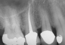
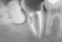
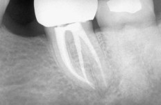
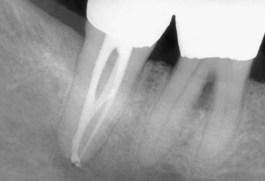
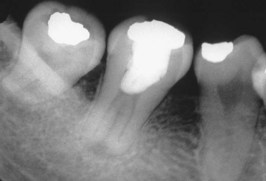
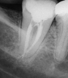
image

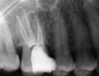
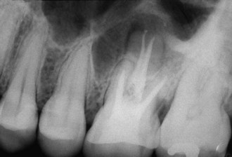
FIG. 7-74 A, Poor access placement and inadequate mesial extension leave both mesial orifices uncovered. Information about the position and location of pulp chambers can be obtained through evaluation of pretreatment radiographs, especially bite-wing radiographs, and assessment of the tooth anatomy at the cementoenamel junction (CEJ). B, Inadequate extension of the distal access cavity leaves the distobuccal canal orifice unexposed. All developmental grooves must be traced to their termination and must not be allowed to disappear into an axial wall. C, Gross overextension of the access cavity weakens the coronal tooth structure and compromises the final restoration. This mistake results from failure to determine correctly the position of the pulp chamber and the angulation of the bur. D, Allowing debris to fall into canal orifices results in an iatrogenic mishap. Amalgam fillings and dentin debris block canal orifices, preventing proper shaping and cleaning. Complete removal of the restoration and copious irrigation help prevent this problem. E, Failure to remove the roof of the pulp chamber is a serious underextension error; the pulp horns have been exposed. Bite-wing radiographs are excellent aids in determining vertical depth. F, Access preparation in which the roof of the pulp chamber remains and the pulp horns have been mistaken for canal orifices. The whitish color of the roof, the depth of the access cavity, and the lack of developmental grooves are clues to this underextension. Root canal orifices generally are positioned at or slightly apical to the CEJ. B, Buccal; D, distal; L, lingual; M, mesial.

image

FIG. 7-75 A, Overzealous tooth removal caused by improper bur angulation and failure to recognize the lingual inclination of the tooth. This results in weakening and mutilation of the coronal tooth structure, which often leads to coronal fractures. B, Inadequate opening; the access cavity is positioned too far to the gingival with no incisal extension. This can lead to bur and file breakage, coronal discoloration because the pulp horns remain, inadequate instrumentation and obturation, root perforation, canal ledging, and apical transportation. C, Labial perforation caused by failure to extend the preparation to the incisal before the bur shaft entered the access cavity. D, Furcation perforation caused by failure to measure the distance between the occlusal surface and the furcation. The bur bypasses the pulp chamber and creates an opening into the periodontal tissues. Perforations weaken the tooth and cause periodontal destruction. They must be repaired (see Fig. 7-72) as soon as they are made for a satisfactory result. E, Perforation of the mesial tooth surface caused by failure to recognize that the tooth is tipped and failure to align the bur with the long axis of the tooth. This is a common error in teeth with full crowns. Even when these perforations are repaired correctly, they usually cause a permanent periodontal problem because they occur in a difficult maintenance area.

image

FIG. 7-76 A, The most embarrassing error, with the greatest potential for medical and legal damage, is entering the wrong tooth because of incorrect dental dam placement. When the crowns of teeth appear identical, the clinician should mark the tooth with a felt-tip marker before the dental dam is placed. B, Burs and files can be broken if used with an improper motion, excessive pressure, or before the access cavity has been properly prepared. A broken instrument may lock into the canal walls, requiring excessive removal of tooth structure to retrieve it. On occasion, fragments may not be retrievable.

Morphology and Access Cavity Preparations for Individual Teeth

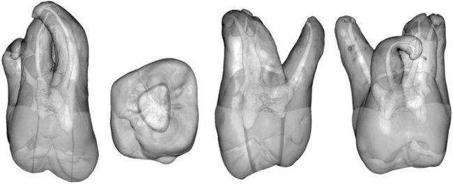
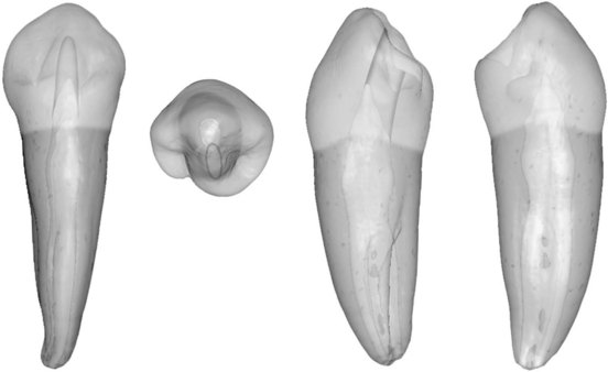
The anatomy shown in the following figures was obtained from human teeth through the use of recently developed 3-D imaging techniques. The teeth were scanned in a high-resolution, microcomputer-assisted tomographic scanner. The data were then manipulated with proprietary computer programs to produce the 3-D reconstructions and visualization. The following individuals made this project possible:

Tomographic scan: Courtesy Michael J. Flynn, PhD, Director, X-Ray Imaging Research Laboratory, Henry Ford Health Sciences, Detroit, MI; also professor (adjunct) of nuclear engineering and radiological science, University of Michigan, Ann Arbor, MI
3-D reconstructions and visualizations: Courtesy Kevin Montgomery, PhD, technical director, Stanford-NASA National Biocomputation Center, Palo Alto, CA
Facilitator: Dr. Paul Brown
Radiographs: Courtesy Dr. L. Stephen Buchanan, Santa Barbara, CA; Dr. John Khademi, Durango, CO; Dr. Raed S. Kasem, Clearwater, FL; Dr. Gary Manasse, Jacksonville, FL; Dr. Michael DeGrood, DeBary, FL; and Dr. Kevin Melker, Clearwater, FL
Access cavity illustrations: Designed and formatted by Dr. Richard Burns, San Mateo, CA; and Dr. Eric Herbranson, San Leandro, CA

Maxillary Central Incisor

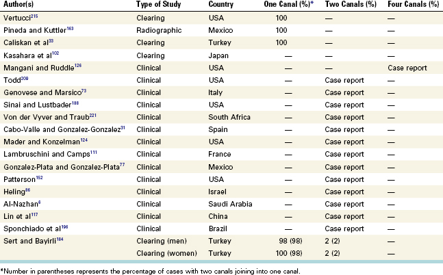
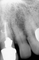
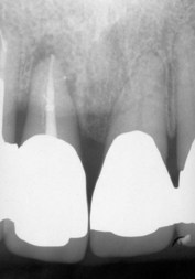
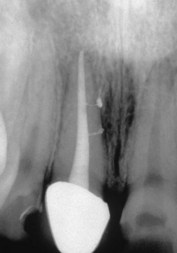
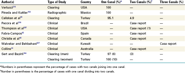
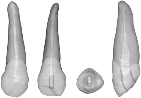
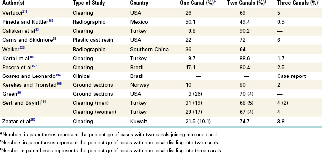
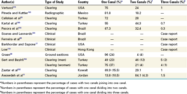
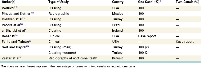
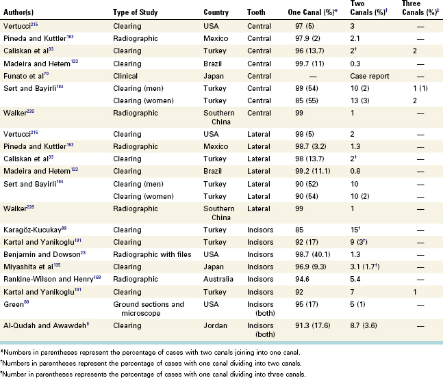
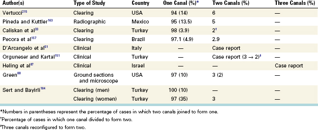
The root canal system outline of the maxillary central incisor reflects the external surface outline. A newly erupted central incisor has three pulp horns, and the pulp chamber is wider mesiodistally than buccolingually. A lingual shoulder usually is present, and it must be removed to gain access to the lingual wall of the root canal. The lingual shoulder prevents direct access to the root canal and deflects files labially, often resulting in a ledge or perforation. In cross-section, the root canal at the CEJ is triangular in young teeth and oval in older teeth. It gradually becomes round as it approaches the apical foramen. Multiple canals are rare (Table 7-8), but lateral canals are common.

TABLE 7-8 Studies of Apical Canal Configurations for the Maxillary Central Incisor

image

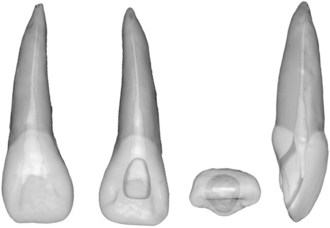
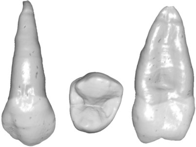
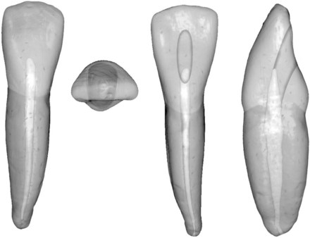
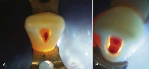
The external access outline form for the maxillary central incisor is a rounded triangle with its base toward the incisal aspect (Figs. 7-77 through 7-81). The width of the triangular base is determined by the distance between the mesial and distal pulp horns. The mesial and distal external walls should converge toward the cingulum. All internal walls should funnel to the root canal orifice. If the lingual shoulder has been removed properly, the entire orifice should be seen through the access opening. The incisal internal wall should approach the lingual surface of the tooth in a near butt joint to allow for a bulk of restorative material on this functional surface.

image

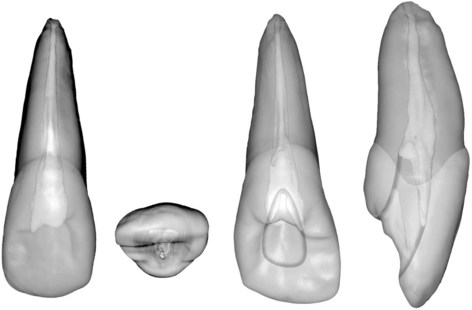
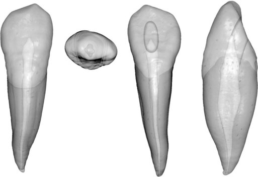
FIG. 7-77 Maxillary central incisor. Development and anatomic data: average time of eruption, 7 to 8 years; average age of calcification, 10 years; average length, 22.5 mm. Root curvature (most common to least common): straight, labial, distal.

image

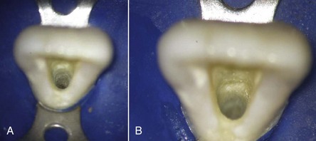
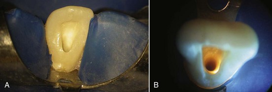
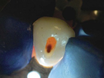
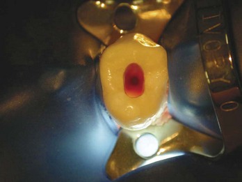
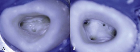
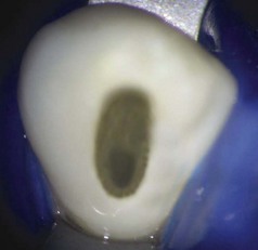
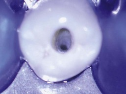
FIG. 7-78 Access cavity for a maxillary central incisor as viewed through the dental operating microscope. A, ×3.4 magnification. B, ×8.4 magnification.

image

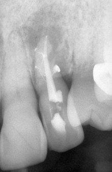
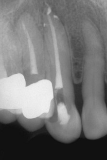
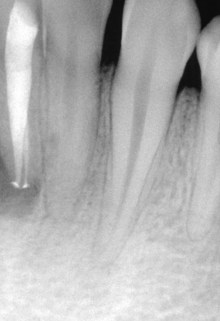
FIG. 7-79 Curved accessory canal with an intersecting straight lateral canal.

image

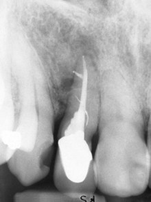
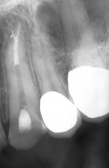
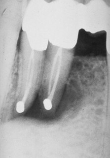
FIG. 7-80 Parallel accessory canal to main canal with a simple lateral canal.

image

FIG. 7-81 Double lateral canals.

Variation

The outline form of the access cavity changes to a more oval shape as the tooth matures and the pulp horns recede because the mesial and distal pulp horns are less prominent.

Maxillary Lateral Incisor

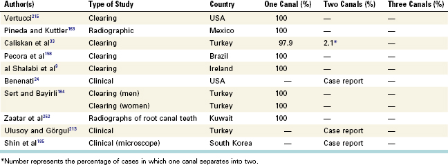
The pulp chamber outline of the maxillary lateral incisor is similar to that of the maxillary central incisor; however, it is smaller, and two or no pulp horns may be present. This tooth is wider mesiodistally than buccolingually. A cross-section at the CEJ shows a pulp chamber centered in the root, and its shape may be triangular, oval, or round. The clinician must know the anatomy of the pulp chamber before starting the access cavity. From the CEJ the pulp canal becomes round in cross-section in the midroot and apical areas. The lingual shoulder of dentin must be removed before instruments can be used to explore the canal. Normally only one root canal is present, but two and three canals have been reported (Table 7-9).

TABLE 7-9 Studies of Apical Canal Configurations for the Maxillary Lateral Incisor

image

The external access outline form for the maxillary lateral incisor may be a rounded triangle or an oval, depending on the prominence of the mesial and distal pulp horns (Figs. 7-82 through 7-85). When the horns are prominent, the rounded triangular shape is compressed mesiodistally relative to a central incisor, producing a more slender triangle. The outline form usually is oval if the mesial and distal pulp horns are not prominent. All other aspects of the access preparation are the same as those for the central incisor.

image

FIG. 7-82 Maxillary lateral incisor. Development and anatomic data: average time of eruption, 8 to 9 years; average age of calcification, 11 years; average length, 22 mm. Root curvature (most common to least common): distal, straight.

image

FIG. 7-83 Access cavity for a maxillary lateral incisor as viewed through the dental operating microscope. A, ×3.4 magnification. B, ×5.1 magnification with cervical fiberoptic transillumination.

image

FIG. 7-84 Lateral incisor with a canal loop and multiple lateral canals with associated lesions.

image

FIG. 7-85 Multiple accessory foramina.

The maxillary lateral incisor often has anomalies. One such variation in form is the presence of a lingual radicular or developmental groove.119,149-151,154-157 Although this groove may be present on the roots of all anterior teeth, it is more common in the maxillary lateral. The groove can lose its periodontal attachment as a result of both periodontal and endodontic etiologies. There is generally direct communication between the groove and the pulp cavity and this occurs primarily through dentinal tubules.

Dens invaginatus is another anomaly and this has been classified into three types based on severity,188 from simple to more complex. Type 1 is an invagination that is confined to the crown. Type 2 is an invagination that extends past the cementoenamel junction but does not involve the periradicular tissues. Type 3 is an invagination that extends beyond the cementoenamel junction and can have a second apical foramen. Often surgical and orthograde root canal therapy is necessary to treat this condition.*

* References 151, 160, 163, 170, 171, 173, 176, 182, 189, 200.

Maxillary Canine

The root canal system of the maxillary canine is similar in many ways to that of the maxillary incisors. A major difference is that it is wider labiolingually than mesiodistally. Another difference is that it has no pulp horns. Its smallest pointed incisal edge corresponds to the single cusp. The pulp chamber outline at the CEJ is oval. A lingual shoulder is present, which may prevent shaping and cleaning of the root canal in its lingual dimension. From this point, the root canal remains oval until it approaches the apical third of the root, where it becomes constricted. Because of this oval shape, the clinician must take care to circumferentially file labially and palatally to shape and clean the canal properly. Usually one root canal is present, although two canals have been reported (Table 7-10). The thin buccal bone over the canine eminence often disintegrates, and fenestration is an occasional finding. Accurate determination of the length is critical. Another effect of this fenestration is a slight, permanent apical pressure sensitivity that occasionally occurs after root canal therapy. This sensitivity can best be corrected by apical root surgery.

TABLE 7-10 Studies of Apical Canal Configurations for the Maxillary Canine

image

The external access outline form is oval or slot shaped because no mesial or distal pulp horns are present (Figs. 7-86 through 7-89). The mesiodistal width of the slot is determined by the mesiodistal width of the pulp chamber. The incisogingival dimension is determined by straight-line access factors and removal of the lingual shoulder. The incisal extension often approaches to within 2 to 3 mm of the incisal edge to allow for straight-line access. The incisal wall meets the lingual surface of the canine in a butt joint to provide adequate thickness for restorative material, because this tooth is heavily involved in excursive occlusal guidance and function. All internal walls funnel to the orifice.

image

FIG. 7-86 Maxillary canine. Development and anatomic data: average time of eruption, 10 to 12 years; average age of calcification, 13 to 15 years; average length, 26.5 mm. Root curvature (most common to least common): distal, straight, labial.

image

FIG. 7-87 Access cavity for a maxillary canine as viewed through the dental operating microscope. (×5.1 magnification with cervical fiberoptic transillumination.)

image

FIG. 7-88 Canine with multiple accessory foramina.

image

FIG. 7-89 Canine with lateral canal dividing into two canals.

Maxillary First Premolar

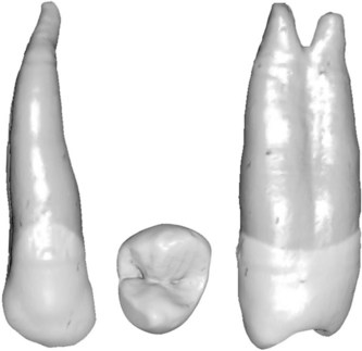
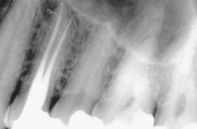
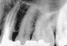
The majority of maxillary first premolars have two root canals regardless of the number of roots. Ethnicity plays a factor in that Asian people have a higher incidence of one canal than do other ethnic groups.118

A furcation groove or developmental depression on the palatal aspect of the buccal root is another anatomic feature. Its prevalence has been reported as between 62% and 100%.97,202 This groove may pose a risk to endodontic and prosthodontic treatment of this tooth. At the deepest part of the invagination, the average dentin thickness was found to be 0.81 mm.

The pulp chamber of the maxillary first premolar is considerably wider buccolingually than mesiodistally. In the buccolingual dimension the chamber outline shows a buccal and a palatal pulp horn. The buccal pulp horn usually is larger. From the occlusal level the chamber maintains a similar width to the floor, which is located just apical to the cervical line. The palatal orifice is slightly larger than the buccal orifice. In cross-section at the CEJ, the palatal orifice is wider buccolingually and kidney shaped because of its mesial concavity. From the floor, two root canals take on a round shape at midroot and rapidly taper to their apices, usually ending in extremely narrow, curved root canals. The palatal canal usually is slightly larger than the buccal canal. The maxillary first premolar may have one, two, or three roots and canals; it most often has two (Table 7-11). If two canals are present, they are labeled buccal and palatal; three root canals are designated mesiobuccal, distobuccal, and palatal. Directional positioning of endodontic pathfinder files can help identify the anatomy. The roots are considerably shorter and thinner than in the canines. In double-rooted teeth the roots most often are the same length. The buccal root can fenestrate through the bone, leading to the same problems that arise with canines (i.e., inaccurate apex location, chronic posttreatment sensitivity to palpation over the apex, and an increased risk of an irrigation accident).

TABLE 7-11 Studies of Apical Canal Configurations for the Maxillary First Premolar

image

The maxillary first premolar is prone to mesiodistal root fractures and fractures at the base of the cusps, particularly the buccal cusp. If a fracture is suspected, all restorations should be removed and the coronal anatomy inspected with fiberoptic light and magnification. Full occlusal coverage is required after root canal therapy to prevent cuspal and/or crown/root fracture.

The access preparation for the maxillary first premolar is oval or slot shaped (Figs. 7-90 through 7-94). It also is wide buccolingually, narrow mesiodistally, and centered mesiodistally between the cusp tips. In fact, the mesiodistal width should correspond to the mesiodistal width of the pulp chamber. The buccal extension typically is two thirds to three fourths up the buccal cusp incline. The palatal extension is approximately halfway up the palatal cusp incline. The buccal and palatal walls funnel directly into the orifices. Because of the mesial concavity of the root, the clinician must take care not to overextend the preparation in that direction, as this could result in perforation.

image

FIG. 7-90 Maxillary first premolar. Development and anatomic data: average time of eruption, 10 to 11 years; average age of calcification, 12 to 13 years; average length, 20.6 mm. Root curvature (most common to least common): buccal root—lingual, straight, buccal; palatal root—straight, buccal, distal; single root—straight, distal, buccal.

image

FIG. 7-91 Access cavity for a maxillary first premolar as viewed through the dental operating microscope. A, ×3.4 magnification. B, ×5.1 magnification. C, ×8.4 magnification with cervical fiberoptic transillumination. D, Schematic representation of a three-canal access preparation.

image

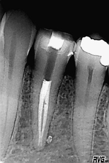
FIG. 7-92 Lateral bony lesion associated with a filled lateral canal.

image

FIG. 7-93 Two canals that have fused and then redivided.

image

FIG. 7-94 Three canals.

Variation

When three canals are present, the external outline form becomes triangular with the base on the buccal aspect. The mesiobuccal and distobuccal corners of the triangle should be positioned directly over the corresponding canal orifices (see Fig. 7-91, D).

Maxillary Second Premolar

The root canal system of the maxillary second premolar is wider buccolingually than mesiodistally. This tooth may have one, two, or three roots and canals (Table 7-12). Two or three canals can occur in a single root. Directional positioning of the endodontic pathfinder or a small file can help identify the anatomy. The mesiodistal and buccolingual aspects of the pulp chamber are similar to those of the first premolar. A buccal and a palatal pulp horn are present; the buccal pulp horn is larger. A single root is oval and wider buccolingually than mesiodistally. The canal(s) remain oval from the pulp chamber floor and taper rapidly to the apex.

TABLE 7-12 Studies of Apical Canal Configurations for the Maxillary Second Premolar

image

The roots of the maxillary second premolar are approximately as long as those of the first premolar, and apical curvature is common, particularly with large maxillary sinus cavities. The proximity of this tooth to the sinus can lead to drainage of a periradicular abscess into the sinus and exposure of the sinus during apical root surgery.

Like the maxillary first premolar, the second premolar is prone to mesiodistal root fractures and fractures at the base of the cusps, usually the buccal cusp. If a fracture is suspected, all restorations in the tooth should be removed and the coronal anatomy inspected with a fiberoptic light and magnification. Full occlusal coverage is required after root canal therapy to prevent cusp and/or crown/root fracture.

When two canals are present, the maxillary second premolar access preparation is nearly identical to that of the first premolar. Because this tooth usually has one root, if two canals are present, they are nearly parallel to each other and the external outline form must have a greater buccolingual extension to permit straight-line access to these canals than with the first premolar with two roots and diverging canals. If only one canal is present, the buccolingual extension is less and corresponds to the width between the buccal and palatal pulp horns (Figs. 7-95 through 7-99). If three canals are present, the external access outline form is the same triangular shape illustrated for the maxillary first premolar (see Fig. 7-91, D).

image

FIG. 7-95 Maxillary second premolar. Average time of eruption, 10 to 12 years; average age of calcification, 12 to 14 years; average length: 21.5 mm. Root curvature (most common to least common): distal, bayonet, buccal, straight.

image

FIG. 7-96 Access cavity for a maxillary second premolar as viewed through the dental operating microscope. (×5.1 magnification with cervical fiberoptic transillumination.)

image

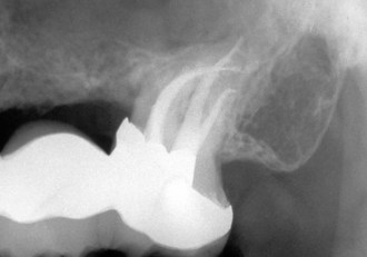
FIG. 7-97 Second premolar with three canals and a large lateral canal.

image

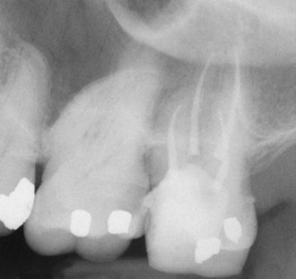
FIG. 7-98 Single canal that has divided into two canals.

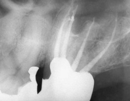
image

FIG. 7-99 Single canal that has split into three canals.

Maxillary First Molar

The maxillary first molar is the largest tooth in volume and one of the most complex in root and canal anatomy. The pulp chamber is widest in the buccolingual dimension, and four pulp horns are present (mesiobuccal, mesiopalatal, distobuccal, and distopalatal). The pulp chamber’s cervical outline form has a rhomboid shape, sometimes with rounded corners. The mesiobuccal angle is an acute angle; the distobuccal angle is an obtuse angle; and the palatal angles are basically right angles. The palatal canal orifice is centered palatally; the distobuccal orifice is near the obtuse angle of the pulp chamber floor; and the main mesiobuccal canal orifice (MB-1) is buccal and mesial to the distobuccal orifice and is positioned within the acute angle of the pulp chamber. The second mesiobuccal canal orifice (MB-2) is located palatal and mesial to the MB-1. A line drawn to connect the three main canal orifices—the mesiobuccal (MB) orifice, distobuccal (DB) orifice, and palatal (P) orifice—forms a triangle, known as the molar triangle.

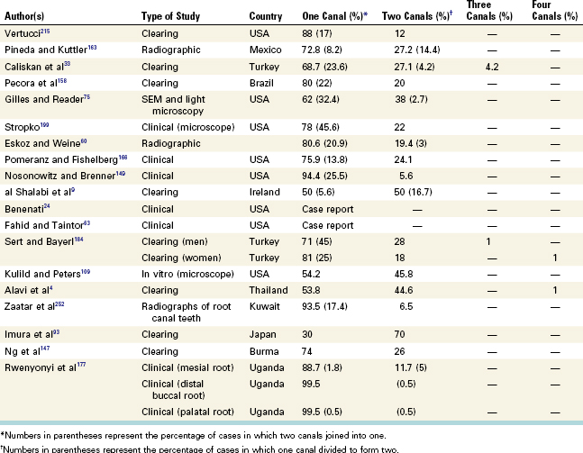
The three individual roots of the maxillary first molar (i.e., mesiobuccal root, distobuccal root, and palatal root) form a tripod. The palatal root is the longest, has the largest diameter, and generally offers the easiest access. It can contain one, two, or three root canals (Table 7-13). The palatal root often curves buccally at the apical one third, which may not be obvious on a standard periapical radiograph. From its orifice the palatal canal is flat, ribbonlike, and wider in a mesiodistal direction. The distobuccal root is conical and may have one or two canals (Table 7-14). From its orifice, the canal(s) first is oval and then becomes round as it approaches the apical third of the root. The mesiobuccal root has generated more research and clinical investigation than any other root in the mouth. It may have one, two, or three root canals (Table 7-15). A single mesiobuccal canal is oval and wider buccolingually; two or three canals are more circular. In general, a concavity exists on the distal aspect of the mesiobuccal root, which makes this wall thin. The clinician must take care not to instrument the wall excessively because a strip perforation may result.

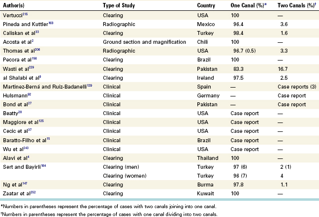
TABLE 7-13 Studies of Apical Canal Configurations for the Palatal Root of the Maxillary First Molar

image

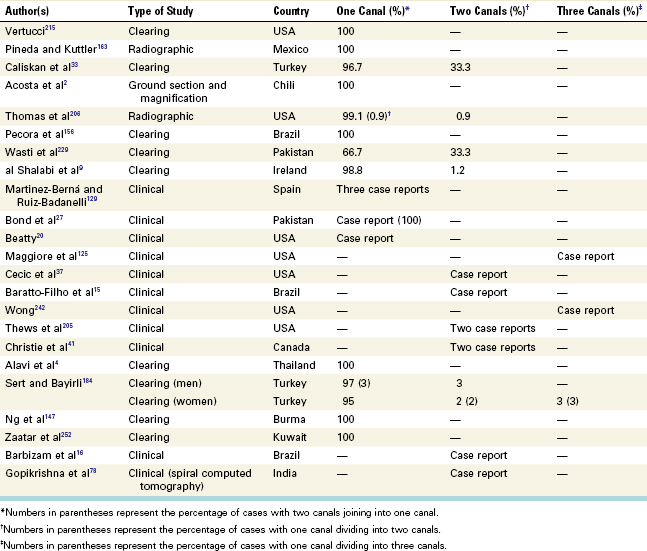
TABLE 7-14 Studies of Apical Canal Configurations for the Distobuccal Root of the Maxillary First Molar

image

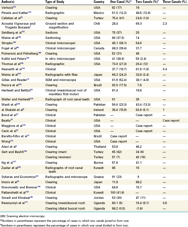
TABLE 7-15 Studies of Apical Canal Configurations for the Mesiobuccal Root of the Maxillary First Molar

image

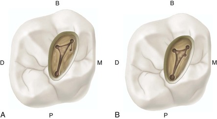
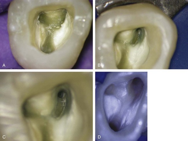
The DOM has been used to study the location and pathway of the MB-2 canal in maxillary first and second molars.79 The clinician must always keep in mind that the location of the MB-2 canal varies greatly; this canal generally is located mesial to or directly on a line between the MB-1 and palatal orifices, within 3.5 mm palatally and 2 mm mesially of the MB-1 orifice (Figs. 7-100 and 7-101). These authors79 found that not all MB-2 orifices lead to a true canal. A true MB-2 orifice was present in only 84% of molars in which a second orifice was identified (Fig. 7-102).197

image

FIG. 7-100 Maxillary first molar. Average time of eruption, 6 to 7 years; average age of calcification, 9 to 10 years; average length, 20.8 mm. Root curvature (most common to least common): mesiobuccal root—distal, straight; distobuccal root—straight, mesial, distal; palatal root—buccal, straight.

image

FIG. 7-101 The two locations of the second mesiobuccal (MB-2) canal orifices in a maxillary first molar. B, Buccal; D, distal; M, mesial; P, palatal.

image

FIG. 7-102 Access cavity for a maxillary first molar as viewed through the dental operating microscope. A, Four apparent orifices, with a projection of dentin covering the mesial groove. (×3.4.) B, Removal of the mesial projection and troughing of the mesial groove to locate the second mesiobuccal (MB-2) canal. (×5.1.) C, Despite deepening of the mesial groove, the MB-2 canal cannot be located. (×8.5.) D, The MB-2 canal cannot be found even after removal of the mesial groove. (×13.6.)

Negotiation of the MB-2 canal often is difficult; a ledge of dentin covers its orifice, the orifice has a mesiobuccal inclination on the pulp floor, and the canal’s pathway often takes one or two abrupt curves in the coronal part of the root. Most of these obstructions can be eliminated by troughing or countersinking with ultrasonic tips mesially and apically along the mesiobuccal pulpal groove (Figs. 7-103 through 7-107). This procedure causes the canal, when present, to shift mesially, meaning that the access wall must be moved farther mesially. Troughing may need to be 0.5 to 3 mm deep. Care must be taken to avoid furcal wall perforation of this root. Apical to the troughing level the canal may be straight or may curve sharply to the distobuccal, buccal, or palatal.

image

FIG. 7-103 Access preparation for a maxillary first molar as viewed through the dental operating microscope. A, Four apparent orifices located under ×3.4 magnification. B, The second mesiobuccal canal is located by deepening the mesial groove. (×5.1.) C, Four distinct canal orifices can be seen. (×5.1 magnification with cervical fiberoptic transillumination.)

image

FIG. 7-104 Four canals with loops and accessory canals.

image

FIG. 7-105 Two canals in both buccal roots with a common foramen in each root.

image

FIG. 7-106 Two separate canals in palatal root.

(Courtesy Dr. Paulo Nogueira, São Paulo, Brazil.)

image

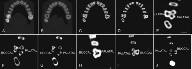
FIG. 7-107 A-D, Axial section spiral computerized tomography (SCT) image of the maxillary arch. E-G, Enlarged axial section SCT image of tooth #3 showing two palatal roots and two canals in the fused buccal root. H-J, Enlarged axial section SCT image of tooth #14, showing the two palatal roots and a single canal in the fused buccal root.

(From Gopikrishna V, Reuben J, Kandaswamy D: Oral Surg Oral Med Oral Pathol Oral Radiol Endod 105:e74, 2008.)

Because the maxillary first molar almost always has four canals, the access cavity has a rhomboid shape, with the corners corresponding to the four orifices (MB-1, MB-2, DB, and P) (see Fig. 7-103). One study demonstrated that the access cavity should not extend into the mesial marginal ridge.241 Distally, the preparation can invade the mesial portion of the oblique ridge, but it should not penetrate through the ridge. The buccal wall should be parallel to a line connecting the MB-1 and DB orifices and not to the buccal surface of the tooth.

Maxillary Second Molar

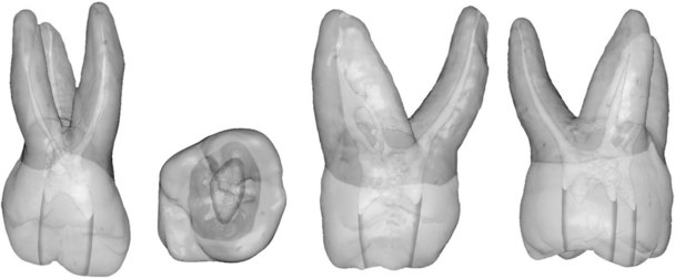
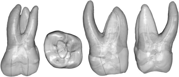
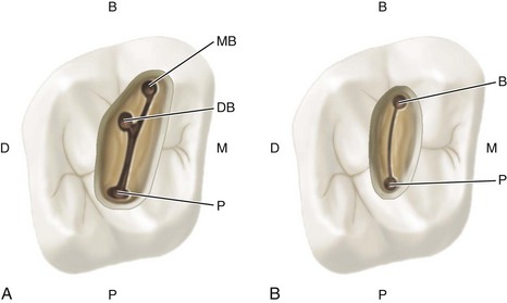
Coronally, the maxillary second molar closely resembles the maxillary first molar. The root and canal anatomy are similar to those of the first molar, although differences occur. The distinguishing morphologic feature of the maxillary second molar is that its three roots are grouped closer together and are sometimes fused. Also, they generally are shorter than the roots of the first molar and not as curved. The second molar usually has one canal in each root; however, it may have two or three mesiobuccal canals, one or two distobuccal canals, or two palatal canals (Tables 7-16 to 7-18). Four canals are less likely to be present in the second molar than in the first molar. The three main orifices (MB, DB, and P) usually form a flat triangle and sometimes almost a straight line (Figs. 7-108 through 7-113). The mesiobuccal canal orifice is located more to the buccal and mesial than in the first molar; the distobuccal orifice approaches the midpoint between the mesiobuccal and palatal orifices; and the palatal orifice usually is located at the most palatal aspect of the root. In general, the canal orifices in the maxillary second molar are closer mesially to each other than they are in the maxillary first molar.

TABLE 7-16 Studies of Apical Canal Configurations for the Mesiobuccal Root of the Maxillary Second Molar

image

TABLE 7-17 Studies of Apical Canal Configurations for the Distobuccal Root of the Maxillary Second Molar

image

TABLE 7-18 Studies of Apical Canal Configurations for the Palatal Root of the Maxillary Second Molar

image
image

FIG. 7-108 Maxillary second molar. Average time of eruption, 11 to 13 years; average age of calcification, 14 to 16 years; average length, 20 mm. Root curvature (most common to least common): mesiobuccal root—distal, straight; distobuccal root—straight, mesial, distal; palatal root—straight, buccal.

image

FIG. 7-109 A, Three canal orifices in a maxillary second molar. B, Two canal orifices in a maxillary second molar. B, Buccal; P, palatal; D, distal, DB, distobuccal; M, medial; MB, mesiobuccal.

image

FIG. 7-110 Access cavity for a maxillary second molar as viewed through the dental operating microscope. A, Dentin projection covering the mesial aspect of the floor of the pulp chamber. (×8.4.) B, Fourth canal orifice (second mesiobuccal, MB-2), which was identified after removal of the dentin projection and troughing of the groove connecting the MB-1 orifice and the palatal (P) canal orifice. (×8.4.)

image

FIG. 7-111 Severely curved mesiobuccal root with a right-angle curve in the distobuccal (DB) root.

image

FIG. 7-112 Four canals (MB-1, MB-2, DB, and P) can be seen.

image

FIG. 7-113 Four canals with two distinct palatal roots and canals.

The floor of the pulp chamber is markedly convex, which gives the canal orifices a slight funnel shape. On occasion the canals curve into the chamber at a more horizontal angle, requiring removal of a lip of dentin so that a canal can be entered more in a direct line with the axis. Teeth with fused roots occasionally have only two canals; in rare cases, they have only one. Teeth with two canals usually have a buccal and a palatal canal of equal length and diameter (see Fig. 7-109, B). These parallel root canals are frequently superimposed radiographically, but they can be imaged by exposing the radiograph from a distal angle. To enhance radiographic visibility, especially when interference arises from the malar process, a more perpendicular and distoangular radiograph may be exposed. When two roots are present, each root may have one canal or the buccal root may have two canals that join before reaching a single foramen. One study found that two palatal roots and two palatal canals occur in 1.47% of these teeth.159

When four canals are present, the access cavity preparation of the maxillary second molar has a rhomboid shape and is a smaller version of the access cavity for the maxillary first molar (see Fig. 7-110). If only three canals are present, the access cavity is a rounded triangle with the base to the buccal. As with the maxillary first molar, the mesial marginal ridge need not be invaded. Because the tendency in maxillary second molars is for the distobuccal orifice to move closer to a line connecting the MB and P orifices, the triangle becomes more obtuse and the oblique ridge usually is not invaded.

If only two canals are present, the access outline form is oval and widest in the buccolingual dimension. Its width corresponds to the mesiodistal width of the pulp chamber, and the oval usually is centered between the mesial pit and the mesial edge of the oblique ridge.

Maxillary Third Molar

Loss of the maxillary first and second molars often is the reason the third molar must be considered as a strategic abutment. Another indication for root canal therapy and full coverage is a fully functioning mandibular third molar in an arch that has sufficient room for full eruption and oral hygiene.

Careful examination of the root morphology is important before treatment is determined. The radicular anatomy of the third molar is completely unpredictable, and it may be advisable to explore the root canal morphology to evaluate the likelihood and degree of success. Even so, many third molars have adequate root formation; given reasonable accessibility, they can serve well as functioning dentition after root canal therapy.

The root anatomy of the maxillary third molar varies greatly. This tooth can have one to four roots and one to six canals, and C-shaped canals also can occur. The third molar usually has three roots and three root canals (Table 7-19; Figs. 7-114 through 7-116). The tooth may be tipped significantly to the distal, the buccal, or both, which creates an even greater access problem than with the second molar.

TABLE 7-19 Studies of Apical Canal Configurations for the Maxillary Third Molar

image
image

FIG. 7-114 Maxillary third molar. Average time of eruption, 17 to 22 years; average age of calcification, 18 to 25 years; average length: 17 mm.

image

FIG. 7-115 Canals that have fused into a single canal. Multiple accessory canals can be seen in the second molar.

image

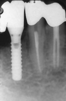
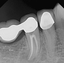
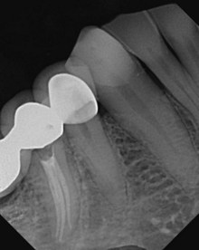
FIG. 7-116 Distal bridge abutment with major accessory canal.

The access cavity form for the third molar can vary greatly. Because the tooth typically has one to three canals, the access preparation can be anything from an oval that is widest in the buccolingual dimension to a rounded triangle similar to that used for the maxillary second molar. The MB, DB, and P orifices often lie nearly in a straight line as the DB orifice moves even closer to the line connecting the MB and P orifices. The resultant access cavity is an oval or highly obtuse triangle.

Mandibular Central and Lateral Incisors

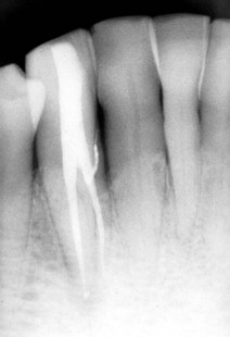
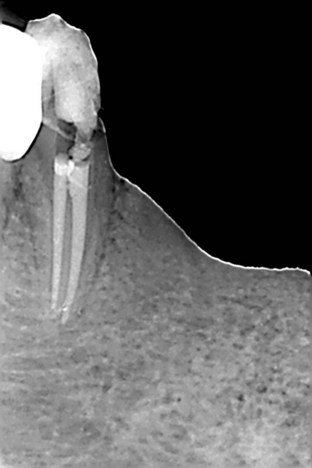
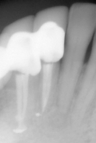
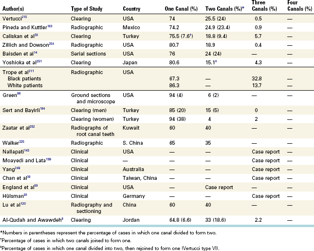
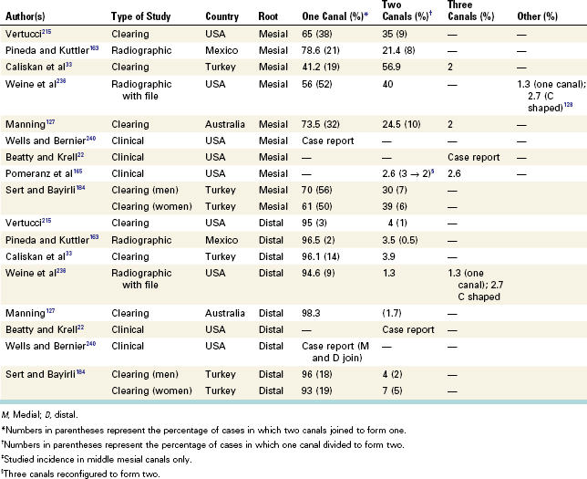
The root canal systems and access cavities for the two mandibular incisors are so similar they are discussed together. As with the maxillary incisors, a lingual shoulder must be eliminated to allow direct-line access. The shoulder conceals the orifice to a second canal that, if present, is found immediately beneath it. Unlike the maxillary incisors, the pulp outline of the mandibular incisors is wider labiolingually. At the CEJ the pulp outline is oval, wider labiolingually than mesiodistally. At midroot the canal outline is still oval, but the canal is more constricted and narrower labiolingually. Most mandibular incisors have a single root with what radiographically appears to be a long, narrow canal. However, it is a broad canal labiolingually. Often a dentinal bridge is present in the pulp chamber that divides the root into two canals. The two canals usually join and exit through a single apical foramen, but they may persist as two separate canals. On occasion one canal branches into two canals, which subsequently rejoin into a single canal before reaching the apex (Table 7-20). One study determined that a relationship existed between crown size (expressed as the M-D/F-L index) and the incidence of bifid root canals in these teeth.228 Double root canals occur more often in teeth with a smaller index.

TABLE 7-20 Studies of Apical Canal Configurations for the Mandibular Incisors

image

The mandibular incisors, because of their small size and internal anatomy, may be the most difficult access cavities to prepare. The external outline form may be triangular or oval, depending on the prominence of the mesial and distal pulp horns (Figs. 7-117 through 7-120). When the form is triangular, the incisal base is short and the mesial and distal legs are long incisogingivally, creating a long, compressed triangle. Without prominent mesial and distal pulp horns, the oval external outline form also is narrow mesiodistally and long incisogingivally. One study148 determined that by age 40 years the mandibular incisor pulp chamber has decreased in size sufficiently to routinely justify an oval access cavity. Complete removal of the lingual shoulder is critical, because this tooth often has two canals that are buccolingually oriented, and the lingual canal most often is missed. To avoid missing this canal, the clinician should extend the access preparation well into the cingulum gingivally. Because the lingual surface of this tooth is not involved with occlusal function, butt joint junctions between the internal walls and the lingual surface are not required.

image

FIG. 7-117 Mandibular central/lateral incisors. Average time of eruption, 6 to 8 years; average age of calcification, 9 to 10 years; average length: 20.7 mm. Root curvature (most common to least common): straight, distal, labial.

image

FIG. 7-118 Access cavity for the mandibular incisors as viewed through the dental operating microscope. A, One canal orifice. (×8.5 magnification with cervical fiberoptic transillumination.) B, Two canal orifices. (×8.5 magnification with cervical fiberoptic transillumination.)

image

FIG. 7-119 Double-rooted mandibular lateral incisor.

image

FIG. 7-120 Two canals in the mandibular lateral and central incisors.

Mandibular Canine

The root canal system of the mandibular canine is very similar to that of the maxillary canine, except that the dimensions are smaller, the root and root canal outlines are narrower in the mesiodistal dimension, and the mandibular canine occasionally has two roots and two root canals located labially and lingually (Table 7-21). The root canal of the mandibular cuspid is narrow mesiodistally but usually very broad buccolingually. A lingual shoulder must be removed to gain access to the lingual wall of the root canal or to the entrance of a second canal. The lingual wall is almost slitlike compared with the larger buccal wall, which makes the canal a challenge to shape and clean.

TABLE 7-21 Studies of Apical Canal Configurations for the Mandibular Canine

image

The access cavity for the mandibular canine is oval or slot shaped (Figs. 7-121 through 7-126). The mesiodistal width corresponds to the mesiodistal width of the pulp chamber. The incisal extension can approach the incisal edge of the tooth for straight-line access, and the gingival extension must penetrate the cingulum to allow a search for a possible lingual canal. As with the mandibular incisors, butt joint relationships between internal walls and the lingual surface are not necessary.

image

FIG. 7-121 Mandibular canine. Average time of eruption, 9 to 10 years; average age of calcification, 13 years; average length, 25.6 mm. Root curvature (most common to least common): straight, distal, labial.

image

FIG. 7-122 Access preparation for a mandibular canine as viewed through the dental operating microscope. (×5.1.)

image

FIG. 7-123 One canal with a sharp mesial curvature at the apex.

image

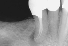
FIG. 7-124 Two canals with the lateral canal above the crest of bone; the lateral canal is probably responsible for the pocket depth.

image

FIG. 7-125 Two separate root canals.

image

FIG. 7-126 Mandibular canine and lateral incisor with two canals.

Mandibular First Premolar

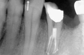
As a group, the mandibular premolars are difficult to treat. They have a high flare-up and failure rate. A possible explanation may be the extreme variations in root canal morphology in these teeth. The root canal system of the mandibular first premolar is wider buccolingually than mesiodistally. Two pulp horns are present: a large, pointed buccal horn and a small, rounded lingual horn. At the cervical line the root and canal are oval; this shape tends to become round as the canal approaches the middle of the root. If two canals are present, they tend to be round from the pulp chamber to their foramen. In another anatomic variation, a single, broad root canal may bifurcate into two separate root canals. Direct access to the buccal canal usually is possible, whereas the lingual canal may be quite difficult to find. The lingual canal tends to diverge from the main canal at a sharp angle. In addition, the lingual inclination of the crown tends to direct files buccally, making location of a lingual canal orifice more difficult. To counter this situation, the clinician may need to extend the lingual wall of the access cavity farther lingually; this makes the lingual canal easier to locate. The mandibular first premolar sometimes may have three roots and three canals (Table 7-22). One study reported a C-shaped canal anatomy in this tooth.14

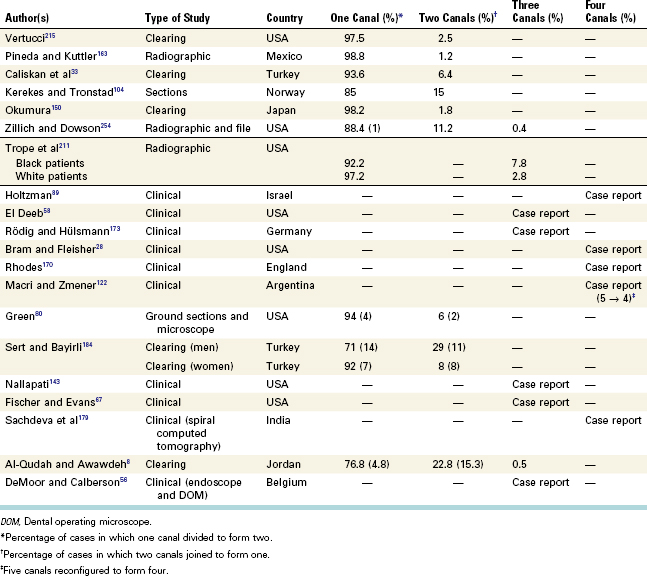
TABLE 7-22 Studies of Apical Canal Configurations for the Mandibular First Premolar

image

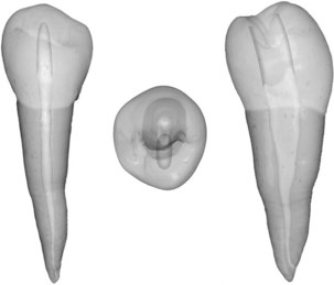
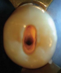
The oval external outline form of the mandibular first premolar typically is wider mesiodistally than its maxillary counterpart, making it more oval and less slot shaped (Figs. 7-127 through 7-131). Because of the lingual inclination of the crown, buccal extension can nearly approach the tip of the buccal cusp to achieve straight-line access. Lingual extension barely invades the poorly developed lingual cusp incline. Mesiodistally the access preparation is centered between the cusp tips. Often the preparation must be modified to allow access to the complex root canal anatomy frequently seen in the apical half of the tooth root.

image

FIG. 7-127 Mandibular first premolar. Average time of eruption, 10 to 12 years; average age of calcification, 12 to 13 years; average length: 21.6 mm. Root curvature (most common to least common): straight, distal, buccal.

image

FIG. 7-128 Access cavity for a mandibular first premolar as viewed through the dental operating microscope: one orifice. (×5.1.)

image

FIG. 7-129 Two canals.

(Courtesy Dr. Raed Kasem, Clearwater, FL.)

image

FIG. 7-130 Single canal that has divided into two.

image

FIG. 7-131 Three canals.

Mandibular Second Premolar

The mandibular second premolar is similar to the first premolar, with the following differences: the lingual pulp horn usually is larger; the root and root canal are more often oval than round; the pulp chamber is wider buccolingually; and the separation of the pulp chamber and root canal normally is distinguishable compared with the more regular taper in the first premolar. The canal morphology of the mandibular second premolar is similar to that of the first premolar with its many variations: two, three, and four canals and a lingually tipped crown. Fortunately, these variations are found less often in the second premolar (Table 7-23).

TABLE 7-23 Studies of Apical Canal Configurations for the Mandibular Second Premolar

image

The access cavity form for the mandibular second premolar varies in at least two ways in its external anatomy. First, because the crown typically has a smaller lingual inclination, less extension up the buccal cusp incline is required to achieve straight-line access. Second, the lingual half of the tooth is more fully developed, and therefore the lingual access extension typically is halfway up the lingual cusp incline.56 The mandibular second premolar can have two lingual cusps, sometimes of equal size. When this occurs, the access preparation is centered mesiodistally on a line connecting the buccal cusp and the lingual groove between the lingual cusp tips. When the mesiolingual cusp is larger than the distolingual cusp, the lingual extension of the oval outline form is just distal to the tip of the mesiolingual cusp (Figs. 7-132 through 7-138).

image

FIG. 7-132 Mandibular second premolar. Average time of eruption, 11 to 12 years; average age of calcification, 13 to 14 years; average length: 22.3 mm. Root curvature (most common to least common): straight, distal, buccal.

image

FIG. 7-133 Access cavity for a mandibular second premolar as viewed through the dental operating microscope: one canal orifice. (×5.1 magnification with cervical fiberoptic transillumination.)

image

FIG. 7-134 Two canals.

(Courtesy Dr. Haider AlZubaidi, Ocala, FL.)

image

FIG. 7-135 Single canal that has divided at the apex.

(Courtesy Dr. Haider AlZubaidi, Ocala, FL.)

image

FIG. 7-136 Single canal that has divided and crossed over at the apex.

image

FIG. 7-137 Three separate canals.

(Courtesy Dr. Haider AlZubaidi, Ocala, FL.)

image

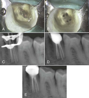
FIG. 7-138 A, Initial access opening. B, Access opening showing orifices of the four root canals. C, Working length determination. D, Immediate posttreatment radiograph. E, Recall radiograph 1 year posttreatment.

(From Sachdeva GS, Ballal S, Gopikrishna V, Kandaswamy D: J Endod 34:104, 2008.)

Mandibular First Molar

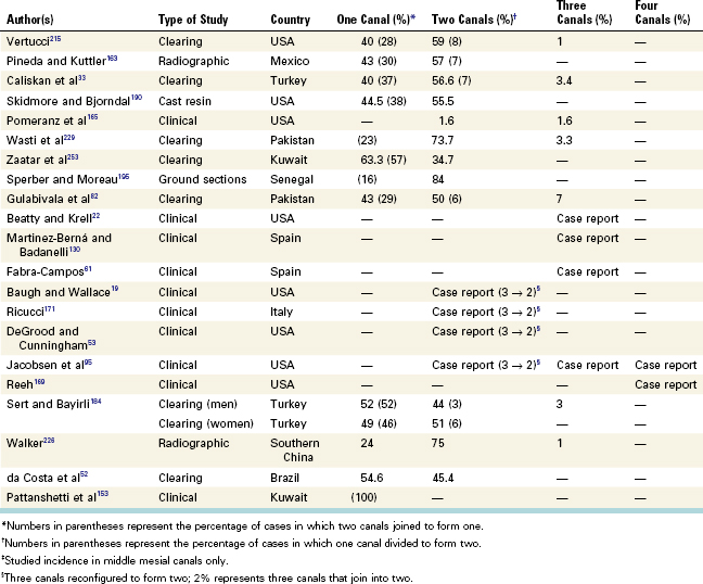
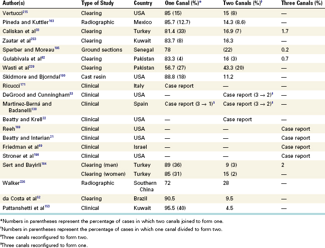
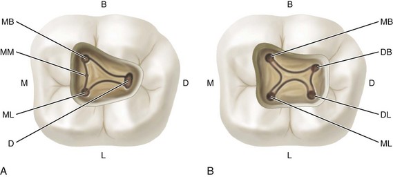
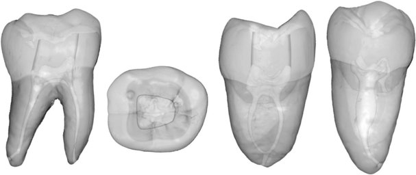
The earliest permanent posterior tooth to erupt, the mandibular first molar seems to be the tooth that most often requires root canal treatment. It often is extensively restored, and it is subjected to heavy occlusal stress. Therefore the pulp chamber frequently has receded or is calcified. The tooth usually has two roots, but occasionally it has three, with two or three canals in the mesial root and one, two, or three canals in the distal root (Tables 7-24 and 7-25). The canals in the mesial root are the MB and ML canals; a middle mesial (MM) canal sometimes is present in the developmental groove between the MB and ML canals. The incidence of an MM canal ranges from 1%215 to 15%.76 The canals in the distal root include the distal canal (if only one canal is present) and the DB, DL, and middle distal (MD) canals (if more than one is present).74 The orifices to these canals are connected by a developmental groove. Orifices to all canals usually are located in the mesial two thirds of the crown, and the pulp chamber floor is roughly trapezoid or rhomboid. Usually four pulp horns (MB, ML, DB, and DL) are present.

TABLE 7-24 Studies of Apical Canal Configurations for the Mesial Root of the Mandibular First Molar

image

TABLE 7-25 Studies of Apical Canal Configurations for the Distal Root for the Mandibular First Molar

image

The presence of two separate distal roots is rare but does occur. In such cases the DL root is smaller than the DB root and usually more curved. Also, the DL root often has a sharp apical hook toward the buccal side that is not obvious on radiographs. The mesial root, the wider of the two roots, curves mesially from the cervical line to the middle third of the root and then angles distally to the apex. The buccal and lingual surfaces are convex throughout their length, whereas the distal surface of the mesial root and the mesial surface of the distal root have a root concavity, which makes the dentin wall very thin. Care must be taken to minimize instrumentation against these walls, because overzealous cutting of the concavity can lead to a strip perforation of the root.

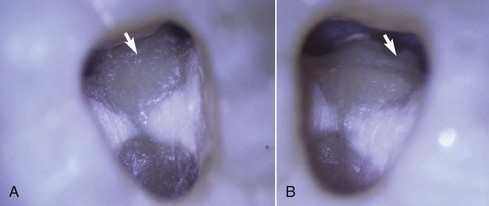
The mesial canal orifices usually are well separated within the main pulp chamber and connected by a developmental groove. The MB orifice commonly is under the mesiobuccal cusp, whereas the ML orifice generally is found just lingual to the central groove. On occasion an MM canal orifice is present in the groove between the MB and ML orifices (Figs. 7-139 through 7-147). The clinician must always check for such an orifice after shaping and cleaning the main root canals. A bur is used to remove any protuberance from the mesial axial wall that would prevent direct access to the developmental groove between the MB and ML orifices. The clinician should use magnification to explore this developmental groove carefully with the sharp tip of an endodontic explorer. If a depression or orifice is located, the groove can be troughed with ultrasonic tips, at the expense of the mesial aspect, until a small file can negotiate the canal. This is best accomplished with the DOM (see Fig. 7-141).

image

FIG. 7-139 Mandibular first molar. Average time of eruption, 6 years; average age of calcification, 9 to 10 years; average length, 21 mm. Root curvature (most common to least common): mesial root—distal, straight; distal root—straight, distal.

image

FIG. 7-140 Access cavities for the mandibular first molar. A, Three mesial canal orifices and one distal canal orifice. B, Two mesial and two distal canal orifices. B, Buccal; D, distal, distal orifice; DB, distobuccal orifice; DL, distolingual orifice; L, labial; M, mesial; MB, mesiobuccal orifice; ML, mesiolingual orifice; MM, middle mesial orifice.

image

FIG. 7-141 Access cavity for a mandibular first molar as viewed through the dental operating microscope. A, Three canal orifices (MB, ML, and D) and a dentinal projection (arrow) can be seen. (×5.1.) A possible middle mesial (MM) orifice may be present along the mesial groove. B, Troughing of the mesial groove allows identification of the MM canal orifice (arrow). (×5.1.)

image

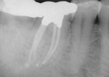
FIG. 7-142 Two mesial and two distal canals.

image

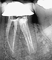
FIG. 7-143 Three canals in distal root.

(Courtesy Dr. Raed Kasem, Clearwater, FL.)

image

FIG. 7-144 Three mesial canals.

image

FIG. 7-145 Three orifices in the mesial root and three orifices in the distal root.

(Courtesy Dr. Haider AlZubaidi, Ocala, FL.)

image

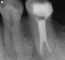
FIG. 7-146 Radix entomolaris. Notice lingual position of its orifice in relation to the two canals in the distal root.

(Courtesy Dr. William J. Aippersbach, Venice, FL.)

image

FIG. 7-147 Pretreatment radiograph. Notice position and appearance of the distolingual root.

(Courtesy Dr. William J. Aippersbach, Venice, FL.)

When only one distal canal is present, the orifice is oval buccolingually and the opening generally is located distal to the buccal groove. This orifice usually can be explored from the mesial with either an endodontic explorer or a small K-file. If the file tip takes a sharp turn in a distobuccal or distolingual direction, the clinician should search for yet another orifice; in rare cases an MD canal orifice is present.

If three root canals (MB, ML, and D) are present in this tooth, each is oval in the cervical and middle thirds of the root and round in the apical third. If two canals (DB and DL) are present in the distal root, they usually are more round than oval for their entire length. The mesial root canals usually are curved, with the greatest curvature in the MB canal. This canal can have a significant curvature in the buccolingual plane that may not be apparent on radiographs. Such a curvature usually can be detected with precurved pathfinder instruments.

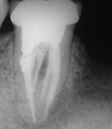
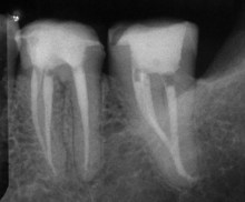
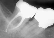
Multiple accessory foramina are located in the furcation of the mandibular molars. These foramina usually are impossible to clean and shape directly; they are rarely seen, except occasionally on a posttreatment radiograph if they have been filled with root canal sealer or thermoplastic filling material. Because sodium hypochlorite solutions can dissolve organic debris, the pulp chamber should be thoroughly exposed to allow the solution to reach the tiny openings. Fractures occasionally occur on proximal marginal ridges and extend down the root or under the lingual cusps.

The access cavity for the mandibular first molar typically is trapezoid or rhomboid regardless of the number of canals present. When four or more canals are present, the corners of the trapezoid or rhombus should correspond to the positions of the main orifices. Mesially the access need not invade the marginal ridge. Distal extension must allow straight-line access to the distal canal(s). The buccal wall forms a straight connection between the MB and DB orifices, and the lingual wall connects the ML and DL orifices without bowing.

A variation in root morphology is the presence of an extra distolingual root.195 Usually this root has a type I canal configuration. It was found that two thirds of the first mandibular molars of a Chinese population possessed this variation.250 Similarly, this distolingual root occurred in 4% of mandibular first molars of a Kuwait population.154 These results confirm the observation that East Asian populations have more three-rooted mandibular first molars than other racial groups.7

Mandibular molars, mainly first molars, may also have an additional root located lingually or buccally. Although this is a rare occurrence in white populations it is more common in Asian populations.212 The radix entomolaris (RE) is a supernumerary root located distolingually in mandibular molars, whereas the radix paramolaris (RP) is an extra root located mesiobuccally. Each root usually contains a single root canal. The orifice of the RE is located disto- to mesiolingually from the main canal or canals of the distal root; the orifice of the RP is located mesio- to distobuccally from the main mesial root canals. A dark line or groove from the main root canal on the pulp chamber floor leads to these orifices.32 These anatomic variations present definite challenges to therapy because of their orifice inclination and root canal curvature. The canal can be straight, have a coronal curvature, or can have separate coronal and apical curvatures.55

Mandibular Second Molar

The mandibular second molar is somewhat smaller coronally than the first molar and tends to be more symmetrical. This tooth is identified by the proximity of its roots. The two roots often sweep distally in a gradual curve, with the apices close together. In some cases only one root is present. The degree of canal curvature and the configuration were studied in the mesial roots of 100 randomly selected mandibular first and second molars; 100% of the specimens showed curvature in both buccolingual and mesiodistal views.48

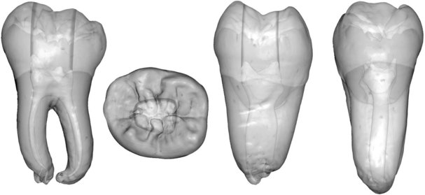
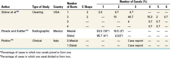
The pulp chamber and canal orifices of the mandibular second molar generally are not as large as those of the first molar. This tooth may have one, two, three, or four root canals (Table 7-26; Figs. 7-148 through 7-153). The two mesial orifices are located closer together. In some mandibular second molars with single or fused roots, a file placed in the mesiobuccal canal may appear to be in the distal canal. This happens because the two canals sometimes are connected by a semicircular slit, a variation of the C-shaped canal134 (see Fig. 7-144, A) that often occurs in this tooth. The distal aspect of the mesial root and the mesial aspect of the distal root have concavities, which must be evaluated during cleaning and shaping procedures. The apices of this tooth often are close to the mandibular canal; therefore the clinician must take care not to allow instruments or filling material to invade this space, because paresthesia may result.

TABLE 7-26 Studies of Apical Canal Configurations of the Mandibular Second Molar

image
image

FIG. 7-148 Mandibular second molar. Average time of eruption, 11 to 13 years; average age of calcification, 14 to 15 years; average length, 19.8 mm. Root curvature (most common to least common): mesial root—distal, straight; distal root—straight, distal, mesial, buccal; single root—straight, distal, bayonet, lingual.

image

FIG. 7-149 Access cavity for a mandibular second molar as viewed through the dental operating microscope. A, Two canal orifices (M and D). (×5.1.) B, Three canal orifices (MB, ML, and D). (×3.4 magnification with cervical fiberoptic transillumination.) C, Four canal orifices identified (MB, ML, DB, and DL). (×5.1 magnification with cervical fiberoptic transillumination.)

image

FIG. 7-150 Classification of C-shaped canal configuration.

(From Fan B, Cheung G, Fan M, Gutmann J: J Endod 30:899, 2004.)

image

FIG. 7-151 Anastomosis of all canals into one.

image

FIG. 7-152 Two canals with an accessory canal at the distal root apex.

image

FIG. 7-153 Fusion of mesial canals at the apex.

Mandibular second molars may have one to six canals, although the most prevalent configurations are two, three, and four canals (see Fig. 7-149). When three canals are present, the access cavity is similar to that for the mandibular first molar, although perhaps a bit more triangular and less rhomboid. The distal orifice is less often ribbon shaped buccolingually; therefore the buccal and lingual walls converge more aggressively distally to form a triangle. The second molar may have only two canals, one mesial and one distal, in which case the orifices are nearly equal in size and line up in the buccolingual center of the tooth. The access cavity for a two-canal second molar is rectangular, wide mesiodistally and narrow buccolingually. The access cavity for a single-canal mandibular second molar is oval and is lined up in the center of the occlusal surface.

Mandibular Third Molar

The mandibular third molar is anatomically unpredictable and must be evaluated on the basis of its root formation. Fused short, severely curved, or malformed roots often support well-formed crowns. This tooth may have one to four roots and one to six canals (Table 7-27; Figs. 7-154 through 7-157). C-shaped canals also can occur. Most of these teeth can be successfully treated endodontically regardless of anatomic irregularities; however, the long-term prognosis is determined by the root surface volume in contact with bone. The clinician must weigh the benefit of treatment against the prognosis.

TABLE 7-27 Studies of Apical Canal Configurations for the Mandibular Third Molar

image
image

FIG. 7-154 Mandibular third molar. Average time of eruption, 17 to 21 years; average age of calcification, 18 to 25 years; average length: 18.5 mm.

image

FIG. 7-155 Five canals.

(Courtesy Dr. Paulo Nogueira, São Paulo, Brazil.)

image

FIG. 7-156 Complex root anatomy.

(Courtesy Dr. Paulo Nogueira, São Paulo, Brazil.)

image

FIG. 7-157 Complex apical anatomy.

The anatomy of the mandibular third molar is unpredictable; therefore the access cavity can take any of several shapes. When three or more canals are present, a traditional rounded triangle or rhomboid shape is typical. When two canals are present, a rectangular shape is used. For single-canal molars, an oval shape is customary.

Teeth With C-Shaped Root Canal Systems

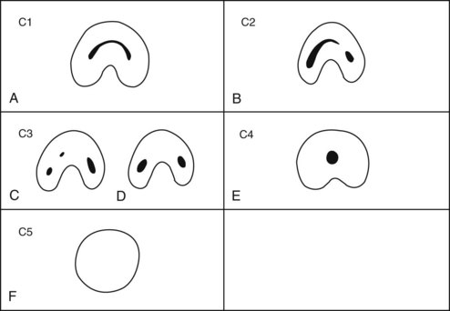
The main cause for C-shaped roots and canals is the failure of Hertwig’s epithelial root sheath to fuse on either the buccal or lingual root surface. The C-shaped canal system can assume many variations in its morphology. The original classification133 has been modified and has produced a more detailed description of C-shaped root and canal morphology. The “C” shape canal configuration can vary along the root depth so that the appearance of the orifices may not be good predictors of the actual canal anatomy.64

Category I (C1): The shape is an uninterrupted “C” with no separation or division (see Fig. 7-150, A).
Category II (C2): The canal shape resembles a semicolon resulting from a discontinuation of the “C” outline (see Fig. 7-150, B), but either angle α or β should be no less than 60 degrees.
Category III (C3): Two or three separate canals (see Fig. 7-150, C and D) and both angles, α and β, are less than 60 degrees.
Category IV (C4): Only one round or oval canal is in the cross-section (see Fig. 7-150, E).
Category V (C5): No canal lumen can be observed (is usually seen near the apex only) (see Fig. 7-150, F).

The C-shaped root canal system was first reported in 1979.46 Most C-shaped canals occur in the mandibular second molar (Figs. 7-158 through 7-166), but they also have been reported in the mandibular first molar, the maxillary first and second molars, and the mandibular first premolar. Two investigators reported the first case of a C-shaped canal in a maxillary molar.146 One study reported the incidence of C-shaped canal anatomy in maxillary first molars as 2 in 2175 (0.092%); this study also determined that the DB and palatal orifices were connected by a common groove (see Fig. 7-159, B).54 Investigators who examined 309 Chinese maxillary second molars found C-shaped root canals in 4.9%.248

image

FIG. 7-158 C-shaped canal anatomy: one continuous canal from pulp chamber floor to apex.

image

FIG. 7-159 C-shaped canal anatomy. A, Mandibular second molar. B, Maxillary first molar.

image

FIG. 7-160 Types of pulpal floor. M, Mesial side; D, distal side.

(From Min Y, Fan B, Cheung G, Gutmann J, Fan M: J Endod 32, 1155, 2006.)

image

FIG. 7-161 Three-dimensional classification of C-shaped canal configuration. A, Merging type; B, symmetrical type; C, asymmetrical type.

(From Gao Y, Fan B, Cheung G, Gutmann J, Fan M: J Endod 32, 1062, 2006.)

image

FIG. 7-162 Microcomputed tomographic image of the minimal wall thickness measurement. a, The outer root surface; b, the inner canal wall; c, the thinnest wall thickness.

(From Gao Y, Fan B, Cheung G, Gutmann J, Fan M: J Endod 32, 1062, 2006.)

image

FIG. 7-163 Mandibular second molar with multiple foramina.

image

FIG. 7-164 Mandibular second molar with interconnecting canal anatomy.

image

FIG. 7-165 Pretreatment appearance of a mandibular first molar with a C-shaped canal.

image

FIG. 7-166 Root canal obturation showing the ribbonlike canal space.

(Courtesy Dr. Paulo Nogueira, São Paulo, Brazil.)

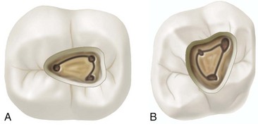
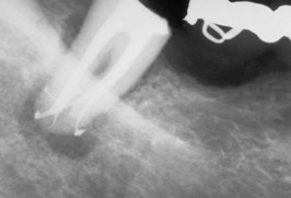
C-shaped mandibular molars are so named because of the cross-sectional morphology of their fused roots and their root canals. Instead of having several discrete orifices, the pulp chamber of a molar with a C-shaped root canal system is a single, ribbon-shaped orifice with an arc of 180 degrees or more. It starts at the mesiolingual line angle and sweeps around either to the buccal or the lingual to end at the distal aspect of the pulp chamber (see Fig. 7-159, A). Below the orifice, the root structure can show a wide range of anatomic variations. These can be classified into two basic types: those with a single, ribbonlike, C-shaped canal from orifice to apex, and those with three or more distinct canals below the usual C-shaped orifice. Fortunately, molars with a single swath of canal are the exception rather than the rule. More common is the second type, with discrete canals that take unusual forms.46 Other investigators determined that C-shaped canals in mandibular second molars can vary in shape and number along the root length.133 This makes cleaning, shaping, obturation, and restoration of these teeth difficult.

One study reported a case of a mandibular first molar with a normal mesiolingual orifice and a C-shaped groove that ran continuously from the mesiobuccal orifice along the buccal wall to the distal canal orifice.18 The groove ran continuously down the root to the apical third, where it divided into two canals. Other researchers reported a C-shaped groove in a mandibular first molar that extended from the DL to the DB orifice and across the buccal surface to the MB orifice.26 The ML orifice remained separate. Four separate apical foramina were noted. One investigator evaluated 811 endodontically treated mandibular second molars and found that 7.6% had C-shaped canals.233 Several variants of the C-shaped canal morphology were noted, the most common being two or three canals that merged and exited as one canal.233

Significant ethnic variation can be seen in the incidence of C-shaped root canal systems. This anatomy is much more common in Asians than in the white population. Investigators in Japan107 and China247 found a 32% incidence of C-shaped canals. Others found the occurrence of C-shaped canals in a Chinese population to be 23% in mandibular first molars and 32% in mandibular second molars.247 Another study found a 19% rate in Lebanese subjects83 and 11% of mandibular second molars from a Saudi Arabian population.5 Mandibular molars in a Burmese population were found to have C-shaped canals in 22% of teeth.82 Another investigation found that 33% of Koreans had a C-shaped canal morphology in mandibular second molars.183

Four types of pulpal floors were found in mandibular second molars:

Type I: A peninsula-like floor with continuous C-shaped orifice (see Fig. 7-160)
Type II: A buccal, striplike dentin connection between the peninsula-like floor and the buccal wall of the pulp chamber that separates the C-shaped groove into mesial (M) and distal (D) orifices. Sometimes the mesial orifice is separated into a mesiobuccal (MB) orifice and a mesiolingual (ML) orifice by another striplike dentin connection between the peninsula-like floor and the mesial wall of the pulp chamber (most common) (see Fig. 7-160)
Type III: Only one mesial, striplike dentin connection exists between the peninsula-like floor and the M wall, which separates the C-shaped groove into a small ML orifice and a large MB-D orifice. The MB-D orifice was formed by the merging of the MB orifice and the D orifice (second most common) (see Fig. 7-160)
Type IV: Non–C-shaped floors. One distal canal orifice and one oval or two round mesial canal orifices are present (least common) (see Fig. 7-160)

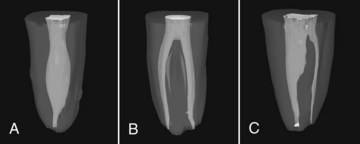
Not all C-shaped mandibular second molars with C-shaped canal systems have a C-shaped pulpal floor. This makes diagnosis difficult. However, it was determined radiographically that a tooth with a C-shaped canal system always has a fused root with a longitudinal groove in the middle of the root.65 It was also found that there are three types of C-shaped canal systems in mandibular second molars (see Fig. 7-161).72

Type I (merging type): Canals merge to one main canal before exiting at the apical foramen
Type II (symmetrical type): Separated mesial and distal canals in each root, which exit as separate canals
Type III (asymmetrical type): Separated mesial and distal canals, with the distal canal having a long isthmus across the furcation area

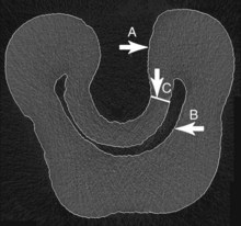
The minimal wall thickness in the middle and apical parts of type III and in the apical part of type II makes these regions danger zones for canal enlargement procedures (see Fig. 7-162).72

Another study on mandibular molars found that there is a higher risk of root perforation at the thinner lingual walls of C-shaped canals during shaping and after canal preparation procedures. Both buccal and lingual canal walls were frequently narrower at the mesial locations.38

The access cavity for teeth with a C-shaped root canal system varies considerably and depends on the pulp morphology of the specific tooth. Teeth with C-shaped anatomy pose a considerable technical challenge; however, use of the DOM, sonic and ultrasonic instrumentation, and plasticized obturation techniques have made treatment successful.

References

1. Abbott PV. Assessing restored teeth with pulp and periapical diseases for the presence of cracks, caries, and marginal breakdown. Aust Dent J. 2004;49:33.

2. Acosta Vigouraux SA, Trugeda Bosaans SA. Anatomy of the pulp chamber floor of the permanent maxillary first molar. J Endod. 1978;4(7):214.

3. Alapati S, Zaatar EI, Shyama M, Al-Zuhair N. Maxillary canine with two root canals. Med Princ Pract. 2006;15:74.

4. Alavi AM, Opasanon A, Ng Y-L, Gulabivala K. Root and canal morphology of Thai maxillary molars. Int Endod J. 2002;35:478.

5. Al-Fougan KS. C-shaped root canals in mandibular second molars in a Saudi Arabian population. Int Endod J. 2002;34:499.

6. Al-Nazhan S. Two root canals in a maxillary central incisor with enamel hypoplasia. J Endod. 1991;17:469.

7. Al Nazhan S. Incidence of fourth canal in root canal treated mandibular first molars in a Saudi Arabian sub-population. Int Endod J. 1999;32:49.

8. Al-Qudah AA, Awawdeh LA. Root canal morphology of mandibular incisors in a Jordanian population. Int Endod J. 2006;39:873.

9. al Shalabi RM, Omer OE, Glennon J, Jennings M, Claffey NM. Root canal anatomy of maxillary first and second permanent molars. Int Endod J. 2000;33(5):405.

10. Awawheh L, Abdullah H, Al-Qudah A. Root forms and canal morphology of Jordanian maxillary first premolars. J Endod. 2008;34:956.

11. Awawdeh LA, Al-Qudah AA. Root form and canal morphology of mandibular premolars in a Jordanian population. Int Endod J. 2008;41:240.

12. Bahcall JK, Barss JT. Fiberoptic endoscope usage for intracanal visualization. J Endod. 2001;27(2):128.

13. Baldassari-Cruz LA, Lilly JP, Rivera EM. The influence of dental operating microscopes in locating the mesiolingual canal orifice. Oral Surg Oral Med Oral Pathol Oral Radiol Endod. 2002;93(2):190.

14. Baisden MK, Kulilid JC, Weller RN. Root canal configuration of the mandibular first premolar. J Endod. 1992;18(10):505.

15. Baratto-Filho F, Fariniuk LF, Ferreira EF, Pecora JD, Cruz-Filho AM, Sousa-Neto MD. Clinical and macroscopic study of maxillary molars with two palatal roots. Int Endod J. 2002;35(9):796.

16. Barbizam JVB, Ribeiro RG, Filho MT. Unusual anatomy of permanent maxillary molars. J Endod. 2004;30:668.

17. Barkhordar RA, Sapone J. Surgical treatment of a three-rooted maxillary second premolar: report of a case. Oral Surg Oral Med Oral Pathol. 1987;63(5):614.

18. Barnett F. Mandibular molar with C-shaped canal. Dent Traumatol. 1986;2:79.

19. Baugh D, Wallace J. Middle mesial canal of the mandibular first molar: a case report and literature review. J Endod. 2004;30(3):185.

20. Beatty RG. A five-canal maxillary first molar. J Endod. 1984;10(4):156.

21. Beatty RG, Interian CM. A mandibular first molar with five canals: report of case. J Am Dent Assoc. 1985;111(5):769.

22. Beatty RG, Krell K. Mandibular molars with five canals: report of two cases. J Am Dent Assoc. 1987;114(6):802.

23. Benjamin KA, Dowson J. Incidence of two root canals in human mandibular incisor teeth. Oral Surg Oral Med Oral Pathol. 1974;38(1):122.

24. Benenati FW. Maxillary second molars with two palatal canals and a palatogingival groove. J Endod. 1985;11(7):308.

25. Bjørndal L, Carlsen O, Thuesen G, Darvann T, Keriborg S. External and internal macromorphology in 3D reconstructed maxillary molars using computerized x-ray microtomography. Int Endod J. 1999;32(1):3.

26. Bolger WL, Schindler WG. A mandibular first molar with a C-shaped root configuration. J Endod. 1998;14(10):515.

27. Bond JL, Hartwell G, Portell FR. Maxillary first molar with six canals. J Endod. 1988;14(5):258.

28. Bram SM, Fleisher R. Endodontic therapy in a mandibular second bicuspid with four canals. J Endod. 1991;17(10):513.

29. Buhrley LJ, Barrows MJ, BeGole EA, Wenckus CS. Effect of magnification on locating the MB-2 canal in maxillary molars. J Endod. 2002;28(4):324.

30. Burch JG, Hulen S. The relationship of the apical foramen to the anatomic apex of the tooth root. Oral Surg Oral Med Oral Pathol. 1972;34(2):262.

31. Cabo-Valle M, Gonzalez-Gonzalez JM. Maxillary central incisor with two root canals: an unusual presentation. J Oral Rehabil. 2001;28(8):797.

32. Calberson FL, DeMoor RJ, Deroose CA. The radix entomolaris and paramolaris: a clinical approach in endodontics. J Endod. 2007;33:58.

33. Caliskan MK, Pehlivan Y, Sepetcioglu F, Turkun M, Tuncer SS. Root canal morphology of human permanent teeth in a Turkish population. J Endod. 1995;21(4):200.

34. Cambruzzi JV, Marshall FJ. Molar endodontic surgery. J Can Dent Assoc. 1983;1:61.

35. Card SJ, Sigurdsson A, Orstavik D, Trope M. The effectiveness of increased apical enlargement in reducing intracanal bacteria. J Endod. 2002;28(11):779.

36. Carnes EJ, Skidmore AE. Configurations and deviation of root canals of maxillary first premolars. Oral Surg Oral Med Oral Pathol. 1973;36(6):880.

37. Cecic P, Hartwell G, Bellizzi R. The multiple root canal system in the maxillary first molar: a case report. J Endod. 1982;8(3):113.

38. Chai WL, Thong YL. Cross-sectional morphology and minimum canal wall widths in C-shaped roots of mandibular molars. J Endod. 2004;30:509.

39. Chan K, Yew SC, Chao SY. Mandibular premolars with three root canals: two case reports. Int Endod J. 1992;25:261.

40. Christie WH, Peikoff MD, Acheson DW. Endodontic treatment of two maxillary lateral incisors with anomalous root formation. J Endod. 1981;7(11):528.

41. Christie WH, Peikoff MD, Fogel HM. Maxillary molars with two palatal roots: a retrospective clinical study. J Endod. 1991;17:80.

42. Cimilli H, Mumcu G, Cimilli T, Kartal N, Wesselink P. Correlation between root canal patterns. Oral Surg Oral Med Oral Pathol Oral Radiol Endod. 2006;102(2):e16.

43. Coelho de Carvalho MC, Zuolo ML. Orifice locating with a microscope. J Endod. 2000;26(9):532.

44. Collins IJ. Maxillary lateral incisor with two roots. Aust Endod J. 2001;27(1):37.

45. Contreras MA, Zinman EH, Kaplan SK. Comparison of the first file that fits at the apex before and after early flaring. J Endod. 2001;27(2):113.

46. Cooke HG, Cox FL. C-shaped canal configurations in mandibular molars. J Am Dent Assoc. 1979;99(5):836.

47. Coolidge ED. Anatomy of the root apex in relation to treatment problems. J Am Dent Assoc. 1929;16:1456.

48. Cunningham CJ, Senia ES. A three-dimensional study of canal curvature in the mesial roots of mandibular molars. J Endod. 1992;18:294.

49. Cutright DE, Bhaskar SN. Pulpal vasculature as demonstrated by a new method. Oral Surg Oral Med Oral Pathol. 1969;27:678.

50. Dankner E, Friedman S, Stabholz A. Bilateral C-shape configuration in maxillary first molars. J Endod. 1990;16(12):601.

51. D’Arcangelo C, Varvara G, De Fazio P. Root canal treatment in mandibular canines with two roots: a report of two cases. Int Endod J. 2001;34(4):331.

52. da Costa LF, Sousa Neto MD, Fidel SR. External and internal anatomy of mandibular molars. Braz Dent J. 1996;7:33.

53. DeGrood ME, Cunningham CJ. Mandibular molar with five canals: report of a case. J Endod. 1997;23(1):60.

54. DeMoor RJG. C-shaped root canal configuration in maxillary first molars. Int Endod J. 2002;35(2):200.

55. DeMoor RJ, Calberson FL. The radix entomolaris in mandibular first molars: an endodontic challenge. Int Endod J. 2004;37:789.

56. DeMoor RJG, Calberson FLG. Root canal treatment in a mandibular second premolar with three root canals. J Endod. 2005;31:310.

57. Dummer PMH, McGinn JH, Rees DG. The position and topography of the apical canal constriction and apical foramen. Int Endod J. 1984;17:192.

58. El Deeb ME. Three root canals in mandibular second premolars: literature review and a case report. J Endod. 1982;8(8):376.

59. England MCJr, Hartwell GR, Lance JR. Detection and treatment of multiple canals in mandibular premolars. J Endod. 1991;17:174.

60. Eskoz N, Weine FS. Canal configuration of the mesiobuccal root of the maxillary second molar. J Endod. 1995;21(1):38.

61. Fabra-Campos H. Unusual root anatomy of mandibular first molars. J Endod. 1985;11(12):568.

62. Fabra-Campos H. [Upper lateral incisor with two canals.]. Endodoncia. 1991;9(2):104.

63. Fahid A, Taintor JF. Maxillary second molar with three buccal roots. J Endod. 1988;14(4):181.

64. Fan B, Cheung GSP, Fan M, Gutmann JL, Bian Z. C-shaped canal system in mandibular second molars. I. Anatomical fractures. J Endod. 2004;30:899.

65. Fan B, Cheung GSP, Fan M, Gutmann JL, Fan W. C-shaped canal system in mandibular second molars. II. Radiographic. J Endod. 2004;30:904.

66. Ferreira CM, de Moraes IG, Bernardineli N. Three-rooted maxillary second premolar. J Endod. 2000;26(2):105.

67. Fischer GM, Evans CE. A three rooted mandibular second premolar. Gen Dent. 1992;40:139.

68. Fogel HM, Peikoff MD, Christie WH. Canal configuration in the mesiobuccal root of the maxillary first molar: a clinical study. J Endod. 1994;20(3):135.

69. Friedman S, Moshonov J, Stabholz A. Five root canals in a mandibular first molar. Dent Traumatol. 1986;2:226.

70. Funato A, Funato H, Matsumoto K. Mandibular central incisor with two root canals. Dent Traumatol. 1998;14(6):285.

71. Gani O, Visvisian C. Apical canal diameter in the first upper molar at various ages. J Endod. 1999;25(10):689.

72. Gao Y, Fan B, Cheung GSP, Gutmann JL, Fan M. C-shaped canal system in mandibular second molars. IV. Morphological analysis and transverse measurement. J Endod. 2006;32:1062.

73. Genovese FR, Marsico EM. Maxillary central incisor with two roots: a case report. J Endod. 2003;29(3):220.

74. Ghoddusi J, Naghavi N, Zarei M, Rohani E. Mandibular first molar with four distal canals. J Endod. 2007;33:1481.

75. Gilles J, Reader A. An SEM investigation of the mesiolingual canal in human maxillary first and second molars. Oral Surg Oral Med Oral Pathol. 1990;70(5):638.

76. Goel NK, Gill KS, Taneja JR. Study of root canal configuration in mandibular first permanent molars. J Indian Soc Pedod Prev Dent. 1991;8:12.

77. Gonzalez-Plata RR, Gonzalez-Plata EW. Conventional and surgical treatment of a two-rooted maxillary central incisor. J Endod. 2003;29:422.

78. Gopikrishna V, Reuben J, Kandaswamy D. Endodontic management of a maxillary first molar with two palatal roots and a fused buccal root diagnosed with spiral computed tomography: a case report. Oral Surg Oral Med Oral Pathol Oral Radiol Endod. 2008;105:e74.

79. Görduysus MO, Görduysus M, Friedman S. Operating microscope improves negotiation of second mesiobuccal canals in maxillary molars. J Endod. 2001;27(11):683.

80. Green D. Double canals in single roots. Oral Surg Oral Med Oral Pathol. 1973;35:689.

81. Gutierrez JH, Aguayo P. Apical foraminal openings in human teeth: number and location. Oral Surg Oral Med Oral Pathol Oral Radiol Endod. 1995;79:769.

82. Gulabivala K, Aung TH, Alavi A, Mg Y-L. Root and canal morphology of Burmese mandibular molars. Int Endod J. 2001;34:359.

83. Haddad GY, Nehma WB, Ounsi HF. Diagnosis, classification and frequency of C-shaped canals in mandibular second molars in the Lebanese population. J Endod. 1999;25:268.

84. Hartwell G, Bellizzi R. Clinical investigation of in vivo endodontically treated mandibular and maxillary molars. J Endod. 1982;8:555.

85. Haznedaroglu F, Ersev H, Odaba H, Yetkin G, Batur B, Asci S, et al. Incidence of patent furcal accessory canals in permanent molars of a Turkish population. Int Endod J. 2003;36(8):515.

86. Heling B. A two-rooted maxillary central incisor. Oral Surg Oral Med Oral Pathol. 1977;43:649.

87. Heling I, Gottlieb-Dadon I, Chandler NP. Mandibular canine with two roots and three root canals. Dent Traumatol. 1995;11(6):301.

88. Hess W, Zurcher E. The Anatomy of Root Canals of the Teeth of the Permanent and Deciduous Dentitions. New York: William Wood; 1925.

89. Holtzman L. Root canal treatment of mandibular second premolar with four root canals: a case report. Int Endod J. 1998;31(5):364.

90. Hsu Y, Kim S. The resected root surface: the issue of canal isthmuses. Dent Clin North Am. 1997;3:529.

91. Hülsman M. Mandibular first premolars with three root canals. Endod Dent Traumatol. 1990;6:189.

92. Hulsmann M. A maxillary first molar with two distobuccal root canals. J Endod. 1997;23(11):707.

93. Imura N, Hata GI, Toda T. Two canals in mesiobuccal roots of maxillary molars. Int Endod J. 1998;31:410.

94. Iqbal M, Fillmore I. Preoperative predictors of number of root canals clinically detected in maxillary molar: a PennEndo database study. J Endod. 2008;34:413-416.

95. Jacobsen EL, Dick K, Bodell R. Mandibular first molars with multiple mesial canals. J Endod. 1994;20(12):610.

96. Jenkins S, Kulild J, Williams K, Lyons W, Lee C. Sealing ability of three materials in the orifice of root canal systems obturated with gutta-percha. J Endod. 2006;32(3):225-227.

97. Joseph I, Varma BR, Bhat KM. Clinical significance of furcation anatomy of the maxillary first premolar: a biometric study on extracted teeth. J Periodontol. 1996;67:386.

98. Jurecko M, Vertucci FJ, Britto L. Effect of Sonic Oscillation on Canal Cleanliness: a Preliminary Study [Master’s Thesis]. Gainesville, FL: University of Florida; 2004.

99. Karagöz-Kucukay I. Root canal ramifications in mandibular incisors and efficacy of low-temperature injection thermoplasticized gutta-percha filling. J Endod. 1994;20(5):236.

100. Kartal N, Ozcelik B, Cimilli H. Root canal morphology of maxillary premolars. J Endod. 1998;24:417.

101. Kartal N, Yanikoglu FC. Root canal morphology of mandibular incisors. J Endod. 1992;18(11):562.

102. Kasahara E, Yasuda E, Yamamoto A, Anzai M. Root canal systems of the maxillary central incisor. J Endod. 1990;16(4):158.

103. Kerekes K, Tronstad L. Long-term results of endodontic treatment performed with a standardized technique. J Endod. 1979;5:83.

104. Kerekes K, Tronstad L. Morphometric observations in root canals of human premolars. J Endod. 1997;3:74.

105. Kerekes K, Tronstad L. Morphometric observations on root canals of human premolars. J Endod. 1998;3:417.

106. Kim S, Pecora G, Rubinstein R, Dorscher-Kim J. Color Atlas of Microsurgery in Endodontics. Philadelphia: WB Saunders; 2001.

107. Kotoku K. Morphological studies on the roots of the Japanese mandibular second molars. Shika Gakuho. 1985;85:43.

108. Krasner P, Rankow HJ. Anatomy of the pulp chamber floor. J Endod. 2004;30(1):5.

109. Kulild JC, Peters DD. Incidence and configuration of canal systems in the mesiobuccal root of maxillary first and second molars. J Endod. 1990;16(7):311.

110. Kuttler Y. Microscopic investigation of root apexes. J Am Dent Assoc. 1955;50:544.

111. Lambruschini GM, Camps J. A two-rooted maxillary central incisor with a normal clinical crown. J Endod. 1995;19(2):95.

112. Langeland K. Tissue Changes in the Dental Pulp: an Experimental Histologic Study. Oslo: Oslo University Press; 1957.

113. Langeland K. The histopathologic basis in endodontic treatment. Dent Clin North Am. 491, Nov, 1967.

114. Langeland K. Tissue response to dental caries. Dent Traumatol. 1987;3:149.

115. Langeland K. Reacción tisular a los materiales de obturación del conducto. In: Guldener PHA, Langeland K, editors. Endodoncia. Barcelona: Springer-Verlag Ibérica, 1995.

116. Leeb J. Canal orifice enlargement as related to biomechanical preparation. J Endod. 1983;9:463.

117. Lin W-C, Yang S-F, Pai S-F. Nonsurgical endodontic treatment of a two-rooted maxillary central incisor. J Endod. 2006;32:478.

118. Loh HS. Root morphology of the maxillary first premolar in Singaporeans. Aust Endod J. 1998;43:399.

119. Low D. Unusual maxillary second premolar morphology: a case report. Quintessence Int. 2001;32(8):626.

120. Lu T-Y, Yang S-F, Pai S-F. Complicated root canal morphology of mandibular first premolars in a Chinese population using cross section method. J Endod. 2006;32:932.

121. Lumley PJ, Walmsley AD, Walton RE, Rippin JW. Cleaning of oval canals using ultrasonics and sonic instrumentation. J Endod. 1993;19:453.

122. Macri E, Zmener O. Five canals in a mandibular second premolar. J Endod. 2000;26(5):304.

123. Madeira MC, Hetem S. Incidence of bifurcations in mandibular incisors. Oral Surg Oral Med Oral Pathol. 1973;36(4):589.

124. Mader CL, Konzelman JL. Double-rooted maxillary central incisors. Oral Surg Oral Med Oral Pathol. 1980;50(1):99.

125. Maggiore F, Jou YT, Kim S. A six canal maxillary first molar: case report. Int Endod J. 2002;35(5):486.

126. Mangani F, Ruddle CJ. Endodontic treatment of a “very particular” maxillary central incisor. J Endod. 1994;20(11):560.

127. Manning SA. Root canal anatomy of mandibular second molars. I. Int Endod J. 1990;23(1):34.

128. Manning SA. Root canal anatomy of mandibular second molars. II. C-shaped canals. Int Endod J. 1990;23(1):40.

129. Martinez-Berná A, Ruiz-Badanelli P. Maxillary first molars with six canals. J Endod. 1983;9(9):375.

130. Martinez-Berná A, Badanelli P. Mandibular first molars with six root canals. J Endod. 1985;11(8):348.

131. Martinez-Lozano MA, Forner-Navarro L, Sanchez-Cortes JL. Analysis of radiologic factors in determining premolar root canal systems. Oral Surg Oral Med Oral Pathol Oral Radiol Endod. 1999;88(6):719.

132. Mauger MJ, Waite RM, Alexander JB, Schindler WG. Ideal endodontic access in mandibular incisors. J Endod. 1999;25(3):206.

133. Melton DC, Krell KV, Fuller MW. Anatomical and histological features of C-shaped canals in mandibular second molars. J Endod. 1991;17:384.

134. Min Y, Fan B, Cheung GSP, Gutmann JL, Fan M. C-shaped canal system in mandibular second molars. III. The morphology of the pulp chamber floor. J Endod. 2006;32:1155.

135. Miyashita M, Kasahara E, Yasuda E, Yamamoto A, Sekizawa T. Root canal system of the mandibular incisor. J Endod. 1997;23(8):479.

136. Mizutani T, Ohno N, Nakamura H. Anatomical study of the root apex in the maxillary anterior teeth. J Endod. 1992;18(7):344.

137. Mjör IA, Nordahl I. The density and branching of dentinal tubules in human teeth. Arch Oral Biol. 1996;41:401.

138. Mjör IA, Smith MR, Ferrari M, Mannocei F. The structure of dentin in the apical region of human teeth. Int Endod J. 2001;34(5):346.

139. Moayedi S, Lata D. Mandibular first premolar with three canals. Endodontology. 2004;16:26.

140. Monnan G, Smallwood ER, Gulabivala K. Effects of access cavity location and design on degree and distribution of instrumented root canal surface in maxillary anterior teeth. Int Endod J. 2001;34:176.

141. Moreinis SA. Avoiding perforation during endodontic access. J Am Dent Assoc. 1979;98:707.

142. Morfis A, Sylaras SN, Georgopoulou M, Kernani M, Prountzos F. Study of the apices of human permanent teeth with the use of a scanning electron microscope. Oral Surg Oral Med Oral Pathol. 1994;77(2):172.

143. Nallapati S. Three canal mandibular first and second premolars: a treatment approach: a case report. J Endod. 2005;31:474.

144. Nattress BR, Martin DM. Predictability of radiographic diagnosis of variations in root canal anatomy in mandibular incisor and premolar teeth. Int Endod J. 1991;24(2):58.

145. Neaverth EJ, Kotler LM, Kaltenbach RF. Clinical investigation (in vivo) of endodontically treated maxillary first molars. J Endod. 1987;13(10):506.

146. Newton CW, McDonald S. A C-shaped canal configuration in a maxillary first molar. J Endod. 1984;10:397.

147. Ng YL, Aung TH, Alavi A, Gulabivala K. Root and canal morphology of Burmese maxillary molars. Int Endod J. 2001;34:620.

148. Nielsen CJ, Shahmohammadi K. The effect of mesio-distal chamber dimension on access preparation in mandibular incisors. J Endod. 2005;31:88-90.

149. Nosonowitz DM, Brenner MR. The major canals of the mesiobuccal root of the maxillary first and second molars. NY J Dent. 1973;43:12.

150. Okumura T. Anatomy of the root canals. Trans Seventh Int Dent Cong. 1926;1:170.

151. Orguneser A, Kartal N. Three canals and two foramina in a mandibular canine. J Endod. 1998;24(6):444.

152. Patterson JM. Bifurcated root of upper central incisor. Oral Surg Oral Med Oral Pathol. 1970;29:222.

153. Pattanshetti N, Gaidhane M, Al Kandari AM. Root and canal morphology of the mesiobuccal and distal roots of permanent first molars in a Kuwait population: a clinical study. Int Endod J. 2008;41(9):755.

154. Pecora JD, Santana SVS. Maxillary lateral incisor with two roots: case report. Braz Dent J. 1991;2(2):151.

155. Pecora JD, Saquy PC, Sousa Neto MD, Woelfel JB. Root form and canal anatomy of maxillary first premolars. Braz Dent J. 1991;2(2):87.

156. Pecora JD, Sousa Neto MD, Saquy PC, Woelfel JB. In vitro study of root canal anatomy of maxillary second premolars. Braz Dent J. 1992;3(2):81.

157. Pecora JD, Sousa Neto MD, Saquy PC. Internal anatomy, direction and number of roots and size of mandibular canines. Braz Dent J. 1993;4:53.

158. Pecora JD, Woelfel JB, Sousa Neto MD, Issa EP. Morphologic study of the maxillary molars. II. Internal anatomy. Braz Dent J. 1992;3(1):53.

159. Peikoff MD, Christie WH, Fogel HM. The maxillary second molar: variations in the number of roots and canals. Int Endod J. 1996;29(6):365.

160. Peters OA, Laib A, Gohring TN, Barbakow F. Changes in root canal geometry after preparation assessed by high-resolution computed tomography. J Endod. 2001;27(1):1.

161. Peters OA, Laib A, Rüegsegger P, Barbakow F. Three-dimensional analysis of root canal geometry by high resolution computed tomography. J Dent Res. 2000;79(6):1405.

162. Peters OA, Peters CI, Schonenberger K, Barbakaw F. ProTaper rotary root canal preparation: assessment of torque force in relation to canal anatomy. Int Endod J. 2003;36(2):93.

163. Pineda F, Kuttler Y. Mesiodistal and buccolingual roentgenographic investigation of 7275 root canals. Oral Surg Oral Med Oral Pathol. 1972;33:101.

164. Plotino G. A mandibular third molar with three mesial roots: a case report. J Endod. 2008;34:224.

165. Pomeranz H, Eidelman DL, Goldberg MG. Treatment considerations of the middle mesial canal of mandibular first and second molars. J Endod. 1981;7(12):565.

166. Pomeranz H, Fishelberg G. The secondary mesiobuccal canal of maxillary molars. J Am Dent Assoc. 1974;88:119.

167. Ponce EH, Vilar Fernandez JA. The cemento-dentino-canal junction, the apical foramen, and the apical constriction: evaluation by optical microscopy. J Endod. 2003;29(3):214.

168. Rankine-Wilson RW, Henry P. The bifurcated root canal in lower anterior teeth. J Am Dent Assoc. 1965;70(5):1162-1165.

169. Reeh ES. Seven canals in a lower first molar. J Endod. 1998;24(7):497.

170. Rhodes JS. A case of an unusual anatomy of a mandibular second premolar with four canals. Int Endod J. 2001;34(8):645.

171. Ricucci D. Three independent canals in the mesial root of a mandibular first molar. Dent Traumatol. 1997;13(1):47.

172. Ricucci D, Langeland K. Apical limit of root canal instrumentation and obturation. 2. A histologic study. Int Endod J. 1998;31(6):394.

173. Rödig T, Hülsmann M. Diagnosis and root canal treatment of a mandibular second premolar with three root canals. Int Endod J. 2003;36(12):912.

174. Rödig T, Hülsmann M, Mühge M, Schäfers F. Quality of preparation of oval distal root canals in mandibular molars using nickel–titanium instruments. Int Endod J. 2002;35:919.

175. Rubinstein R, Kim S. Short-term observation of the results of endodontic surgery with the use of a surgical operating microscope and Super EBA as root-end filling material. J Endod. 1999;25:43.

176. Rubinstein R, Kim S. Long-term follow-up of cases considered healing 1 year after apical microsurgery. J Endod. 2002;28:378.

177. Rwenyonyi CM, Kutesa AM, Muwazi LM, Buwembo W. Root and canal morphology of maxillary first and second permanent molar teeth in a Ugandan population. Int Endod J. 2007;40:679.

178. Saad AY, Al-Yahya AS. The location of the cementodentinal junction in single-rooted mandibular first premolars from Egyptian and Saudi patients: a histologic study. Int Endod J. 2003;36:541.

179. Sachdeva GS, Ballal S, GopiKrishna V, Kandas Wamy D. Endodontic management of a mandibular second premolar with four roots and four root canals with the aid of spiral computed tomography: a case report. J Endod. 2008;34:104.

180. Schilder H. Filling root canals in three dimensions. Dent Clin North Am. 723, Nov 1967.

181. Schwarze T, Baethge C, Stecher T, Geurtsen W. Identification of second canals in the mesiobuccal root of maxillary first and second molars using magnifying loupes or an operating microscope. Aust Endod J. 2002;28(2):57.

182. Seidberg BH, Altman M, Guttuso J, Suson M. Frequency of two mesiobuccal root canals in maxillary first molars. J Am Dent Assoc. 1973;87:852.

183. Seo MS, Park DS. C-shaped root canals of mandibular second molars in a Korean population: clinical observation and in vitro analysis. Int Endod J. 2004;37(2):139.

184. Sert S, Bayirli GS. Evaluation of the root canal configurations of the mandibular and maxillary permanent teeth by gender in the Turkish population. J Endod. 2004;30(6):391.

185. Shin SJ, Park JW, Lee JK, Hwang SW. Unusual root canal anatomy in maxillary second molars: two case reports. Oral Surg Oral Med Oral Pathol Oral Radiol Endod. 2007;104:e61.

186. Sidow SJ, West LA, Liewehr FR, Loushine RJ. Root canal morphology of human maxillary and mandibular third molars. J Endod. 2000;26(11):675.

187. Simon JHS. The apex: how critical is it? Gen Dent. 1994;42:330.

188. Sinai IH, Lustbader S. A dual-rooted maxillary central incisor. J Endod. 1984;10(3):105.

189. Sjögren U, Hägglund B, Sundqvist G, Wing K. Factors affecting the long-term results of endodontic treatment. J Endod. 1990;16:498.

190. Skidmore AE, Bjorndal AM. Root canal morphology of the human mandibular first molar. Oral Surg Oral Med Oral Pathol. 1971;32(5):778.

191. Slowey RE. Root canal anatomy: road map to successful endodontics. Dent Clin North Am. 1979;23(4):555.

192. Smadi L, Khraisat A. Detection of a second mesiobuccal canal in the mesiobuccal roots of maxillary first molar teeth. Oral Surg Oral Med Oral Pathol Oral Radiol Endod. 2007;103:e77.

193. Smulson MH, Hagen JC, Ellenz SJ. Pulpoperiapical pathology and immunologic considerations. In Weine FS, editor: Endodontic Therapy, ed 5, St. Louis: Mosby, 1996.

194. Soares JA, Leonardo RT. Root canal treatment of three-rooted maxillary first and second premolars: a case report. Int Endod J. 2003;36(10):705.

195. Sperber GH, Moreau JL. Study of the number of roots and canals in Senegalese first permanent mandibular molars. Int Endod J. 1998;31(2):117.

196. Sponchiado EC, Ismail HAA, Braga MRL, de Carvalho FK, Simoes CACG. Maxillary central incisor with two-root canals: a case report. J Endod. 2006;32:1002.

197. Stabholz A, Goultschin J, Friedman S, Korenhouser S. Crown-to-root ratio as a possible indicator of the presence of a fourth root canal in maxillary first molars. Israel J Dent Sci. 1984;1:85.

198. Stroner WF, Remeikis NA, Carr GB. Mandibular first molar with three distal canals. Oral Surg Oral Med Oral Pathol. 1984;57(5):554.

199. Stropko JJ. Canal morphology of maxillary molars: clinical observations of canal configurations. J Endod. 1990;25:446.

200. Subay RK, Kayatas M. Dens invaginatus in an immature lateral incisor: a case report of complex endodontic treatment. Oral Surg Oral Med Oral Pathol Oral Radiol Endod. 2006;102:e37.

201. Sykaras S, Economou P. [Root canal morphology of the mesio-buccal root of the maxillary first molar.]. Odontostomatol Proodos. 1970;24:99.

202. Tamse A, Katz A, Pilo R. Furcation groove of buccal root of maxillary first premolars: a morphometric study. J Endod. 2000;26:359.

203. Taylor GN. Techniiche per la preparazione e l’otturazione intracanalare. La Clinica Odontoiatrica del Nord America. 1988;20(3):566.

204. Teixeira FB, Sano CL, Gomes BP, Zara AA, Ferraz CC, Souza-Filho FJ. A preliminary in vitro study of the incidence and position of the root canal isthmus in maxillary and mandibular first molars. Int Endod J. 2003;36(4):276.

205. Thews ME, Kemp WB, Jones CR. Aberrations in palatal root and root canal morphology of two maxillary first molars. J Endod. 1979;5(3):94.

206. Thomas RP, Moule AJ, Bryant R. Root canal morphology of maxillary permanent first molar teeth at various ages. Int Endod J. 1993;26(5):257.

207. Thompson BH, Portell FR, Hartwell GR. Two root canals in a maxillary lateral incisor. J Endod. 1985;11(8):353.

208. Tidmarsh BG, Arrowsmith MG. Dentinal tubules at the root ends of apicected teeth: a scanning electron microscope study. Int Endod J. 1989;22:184.

209. Todd HW. Maxillary right central incisor with two root canals. J Endod. 1976;2(8):227.

210. Torabinejad M, Walton R. Endodontics: Principles and Practice, ed 4. St. Louis: Saunders; 2009.

211. Trope M, Elfenbein L, Tronstad L. Mandibular premolars with more than one root canal in different race groups. J Endod. 1986;12(8):343.

212. Tu M-G, Tsai C-C, Jou M-J, Chen W-L, Chang Y-F, Chen S-Y, et al. Prevalence of three rooted mandibular first molars among Taiwanese individuals. J Endod. 2007;33:1163.

213. Ulusoy OI, Görgül G. Endodontic treatment of a maxillary second molar with two palatal roots: a case report. Oral Surg Oral Med Oral Pathol Oral Radiol Endod. 2007;104(4):e95.

214. Usman N, Baumgartner JC, Marshall JG. Influence of instrument size on root canal debridement. J Endod. 2004;30(2):110.

215. Vertucci FJ. Root canal anatomy of the human permanent teeth. Oral Surg Oral Med Oral Pathol. 1984;58:589.

216. Vertucci FJ, Anthony RL. A scanning electron microscopic investigation of accessory foramina in the furcation and pulp chamber floor of molar teeth. Oral Surg Oral Med Oral Pathol. 1986;62:319.

217. Vertucci FJ, Beatty RG. Apical leakage associated with retrofilling techniques: a dye study. J Endod. 1986;12:331.

218. Vertucci FJ, Seelig A, Gillis R. Root canal morphology of the human maxillary second premolar. Oral Surg Oral Med Oral Pathol. 1974;38:456.

219. Vertucci FJ, Williams RG. Furcation canals in the human mandibular first molar. Oral Surg Oral Med Oral Pathol. 1974;38:308.

220. Von der Lehr WN, Marsh RA. A radiographic study of the point of endodontic egress. Oral Surg Oral Med Oral Pathol Oral Radiol Endod. 1973;35:705.

221. Von der Vyver PJ, Traub AJ. Maxillary central incisor with two root canals: a case report. J Dent Assoc South Afr. 1995;50(3):132.

222. Wada M, Takase T, Nakanuma K, Arisue K, Nagahama F, Yamazaki M. Clinical study of refractory apical periodontitis treated by apicoectomy. 1. Root canal morphology of resected apex. Int Endod J. 1998;31:53.

223. Walker RT. Root form and canal anatomy of maxillary first premolars in a southern Chinese population. Dent Traumatol. 1987;3:130.

224. Walker RT. Root form and canal anatomy of mandibular first molars in a southern Chinese population. Dent Traumatol. 1988;4:19.

225. Walker RT. Root form and canal anatomy of mandibular first premolars in a southern Chinese population. Dent Traumatol. 1988;4:226.

226. Walker RT. The root canal anatomy of mandibular incisors in a southern Chinese population. Int Endod J. 1988;21:218.

227. Walvekar SV, Behbehani JM. Three root canals and dens formation in a maxillary lateral incisor: a case report. J Endod. 1997;23(3):185.

228. Warren EM, Laws AJ. The relationship between crown size and the incidence of bifid root canals in mandibular incisor teeth. Oral Surg Oral Med Oral Pathol. 1981;52(4):425.

229. Wasti F, Shearer AC, Wilson NH. Root canal systems of the mandibular and maxillary first permanent molar teeth of South Asian Pakistanis. Int Endod J. 2001;34(4):263.

230. Webber RT, del Rio CE, Brady JM, Segall RO. Sealing quality of a temporary filling material. Oral Surg Oral Med Oral Pathol. 1978;46(1):123.

231. Weine FS. Case report: three canals in the mesial root of a mandibular first molar. J Endod. 1982;8(11):517.

232. Weine FS, editor Endodontic Therapy ed 5 1996 Mosby St. Louis 243

233. Weine FS. members of the Arizona Endodontic Association: The C-shaped mandibular second molar: incidence and other considerations. J Endod. 1998;24(5):372.

234. Weine FS, Hayami S, Hata G, Toda T. Canal configuration of the mesiobuccal root of the maxillary first molar of a Japanese subpopulation. Int Endod J. 1999;32(2):79.

235. Weine FS, Healy HJ, Gerstein H, Evanson L. Canal configuration in the mesiobuccal root of the maxillary first molar and its endodontic significance. Oral Surg Oral Med Oral Pathol. 1969;28:419.

236. Weine FS, Pasiewicz RA, Rice RT. Canal configuration of the mandibular second molar using a clinically oriented in vitro method. J Endod. 1988;14(5):207.

237. Weisman MI. A rare occurrence: a bi-rooted upper canine. Aust Endod J. 2000;26:119.

238. Weller RN, Hartwell G. The impact of improved access and searching techniques on detection of the mesiolingual canal in maxillary molars. J Endod. 1989;15:82.

239. Weller NR, Niemczyk SP, Kim S. Incidence and position of the canal isthmus. 1. Mesiobuccal root of the maxillary first molar. J Endod. 1995;21:380.

240. Wells DW, Bernier WE. A single mesial canal and two distal canals in a mandibular second molar. J Endod. 1984;10(8):400.

241. Wilcox LR, Walton RE, Case WB. Molar access: shape and outline according to orifice location. J Endod. 1989;15(7):315.

242. Wong M. Maxillary first molar with three palatal canals. J Endod. 1991;17(6):298.

243. Wu M-K, Barkis D, R’oris A, Wesselink PR. Does the first file to bind correspond to the diameter of the canal in the apical region? Int Endod J. 2002;35:264.

244. Wu M-K, R’oris A, Barkis D, Wesselink PR. Prevalence and extent of long oval canals in the apical third. Oral Surg Oral Med Oral Pathol Oral Radiol Endod. 2000;89:739.

245. Wu M-K, Wesselink PR. A primary observation on the preparation and obturation of oval canals. Int Endod J. 2001;34:137.

246. Wu M-K, Wesselink P, Walton R. Apical terminus location of root canal treatment procedures. Oral Surg Oral Med Oral Pathol Oral Radiol Endod. 2000;89:99.

247. Yang Z-P, Yang S-F, Lee G. The root and root canal anatomy of maxillary molars in a Chinese population. Dent Traumatol. 1998;4:215.

248. Yang Z-P, Yang S-F, Lin YL. C-shaped root canals in mandibular second molars in a Chinese population. Dent Traumatol. 1988;4:160.

249. Yang ZP. Multiple canals in a mandibular first premolar: case report. Aust Dent J. 1994;39:18.

250. Yew SC, Chan K. A retrospective study of endodontically treated mandibular first molars in a Chinese population. J Endod. 1993;19:471.

251. Yoshioka T, Villegas JC, Kobayashi C, Suda H. Radiographic evaluation of root canal multiplicity in mandibular first premolars. J Endod. 2004;30(2):73.

252. Zaatar EI, Al-Kandari AM, Alhomaidah S, Al Yasin IM. Frequency of endodontic treatment in Kuwait: radiographic evaluation of 846 endodontically treated teeth. J Endod. 1997;23:453.

253. Zaatar EI, al Anizi SA, al Duwairi Y. A study of the dental pulp cavity of mandibular first permanent molars in the Kuwait population. J Endod. 1998;24(2):125.

254. Zillich R, Dowson J. Root canal morphology of mandibular first and second premolars. Oral Surg Oral Med Oral Pathol. 1973;36(5):738.
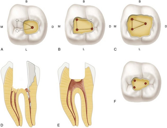
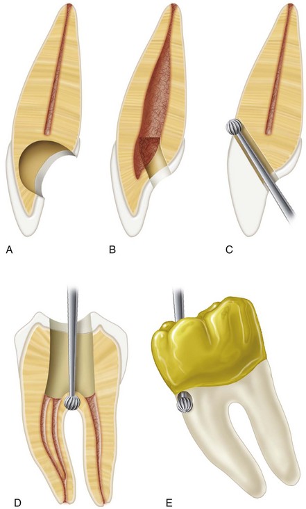
* References 30, 80, 81, 163, 176, 200, 215, and 220.