Derivatives of the skin
Hair
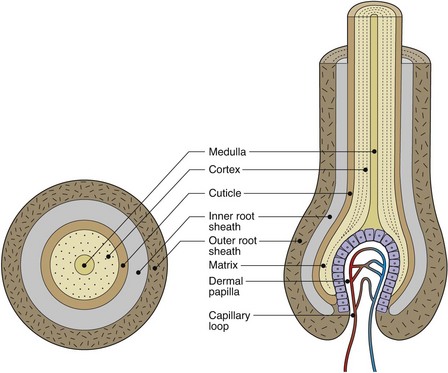
Hairs are found over the entire surface of the skin, with the exception of the glabrous skin of the palms, soles, glans penis and vulval introitus. The density of follicles is greatest on the face. Embryologically, the hair follicle has an input from the epidermis, which is responsible for the matrix cells and the hair shaft, and the dermis, which contributes to the papilla, with its blood vessels and nerves.
There are three types of hair:
1. Lanugo hairs are fine and long, and are formed in the fetus at 20 weeks’ gestation. They are normally shed before birth, but may be seen in premature babies.
2. Vellus hairs are the short, fine, light-coloured hairs that cover most body surfaces.
3. Terminal hairs are longer, thicker and darker, and are found on the scalp, eyebrows, eyelashes and also on the pubic, axillary and beard areas. They originate as vellus hair; differentiation is stimulated at puberty by androgens.
Structure
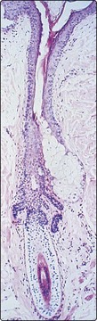
The hair follicle is an invagination of the epidermis containing a hair. The portion above the site of entry of the sebaceous duct is the infundibulum. The hair shaft consists of an outer cuticle that encloses a cortex of packed keratinocytes with (in terminal hairs) an inner medulla (Fig. 1). The germinative cells are in the hair bulb; associated with these cells are melanocytes, which synthesize pigment. The arrector pili muscle is vestigial in humans; it contracts with cold, fear and emotion to erect the hair, producing ‘goose pimples’.
Nails
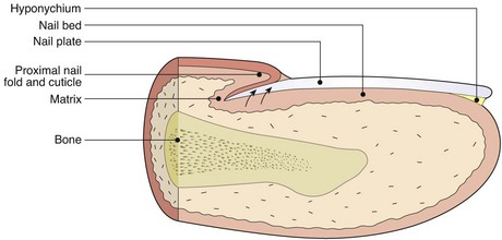
The nail is a phylogenetic remnant of the mammalian claw and consists of a plate of hardened and densely packed keratin. It protects the fingertip and facilitates grasping and tactile sensitivity in the finger pulp.
Structure
The nail matrix contains dividing cells which mature, keratinize and move forward to form the nail plate (Fig. 2). The nail plate has a thickness of 0.3–0.5 mm and grows at a rate of 0.1 mm/24 h for the fingernail. Toenails grow more slowly. The nail bed, which produces small amounts of keratin, is adherent to the nail plate. The adjacent dermal capillaries produce the pink colour of the nail; the white lunula is the visible distal part of the matrix. The hyponychium is the thickened epidermis that underlies the free margin of the nail.
Sebaceous glands
Sebaceous glands are found associated with hair follicles (Fig. 3), especially those of the scalp, face, chest and back, and are not found on non-hairy skin. They are formed from epidermis-derived cells and produce an oily sebum, the function of which is uncertain. The glands are small in the child, but become large and active at puberty, being sensitive to androgens. Sebum is produced by holocrine secretion in which the cells disintegrate to release their lipid cytoplasm.
Sweat glands
Sweat glands (Fig. 4) are tube-like and coiled glands, located within the dermis, which produce a watery secretion. There are two separate types: eccrine and apocrine.
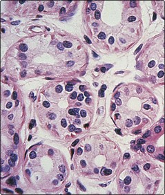
A cross-section through the coiled secretory portion of an eccrine sweat gland, situated deep in the dermis.
Eccrine
Eccrine sweat glands develop from downbudding of the epidermis. The secretory portion is a coiled structure in the deep reticular dermis; the excretory duct spirals upwards to open onto the skin surface. An estimated 2.5 million sweat ducts are present on the skin surface. They are universally distributed, but are most profuse on the palms, soles, axillae and forehead where the glands are under both psychological and thermal control (those elsewhere being under thermal control only). Eccrine sweat glands are innervated by sympathetic (cholinergic) nerve fibres.
Apocrine
Also derived from the epidermis, apocrine sweat glands open into hair follicles and are larger than eccrine glands. They are most numerous around the axillae, perineum and areolae. Their sweat is generated by ‘decapitation’ secretion of the gland’s cells and is odourless when produced; an odour develops after skin bacteria have acted upon it. Sweating is controlled by sympathetic (adrenergic) innervation. The apocrine glands represent a phylogenetic remnant of the mammalian sexual scent gland.
Other structures in skin
Nerve supply
The skin is richly innervated (Fig. 5), with the highest density of nerves being found in areas such as the hands, face and genitalia. All nerves supplying the skin have their cell bodies in the dorsal root ganglia. Both myelinated and non-myelinated fibres are found. The nerves contain neuropeptides, e.g. substance P.
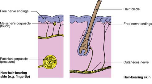
Free sensory nerve endings are seen in the dermis and also encroaching into the epidermis where they may abut onto Merkel cells. These nerve endings detect pain, itch and temperature. Specialized corpuscular receptors are distributed in the dermis, such as the Pacinian corpuscle (detecting pressure and vibration) and touch-sensitive Meissner’s corpuscles, which are mainly seen in the dermal papillae of the feet and hands.
Autonomic nerves supply the blood vessels, sweat glands and arrector pili muscles. The nerve supply is dermatomal with some overlap.
Blood and lymphatic vessels
The skin also has a rich and adaptive blood supply. Arteries in the subcutis branch upwards, forming a superficial plexus at the papillary/reticular dermal boundary. Branches extend to the dermal papillae (Fig. 6), each of which has a single loop of capillary vessels, one arterial and one venous. Veins drain from the venous side of this loop to form the mid-dermal and subcutaneous venous networks. In the reticular and papillary dermis, there are arteriovenous anastomoses that are well innervated and concerned with thermoregulation (see p. 7).
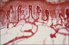
Fig. 6 Superficial dermal blood vessels.
Capillary loops branch off the superficial vascular plexus and extend into each dermal papilla.
The lymphatic drainage of the skin is important, and abundant meshes of lymphatics originate in the papillae and assemble into larger vessels that ultimately drain into the regional lymph nodes.
Derivatives
 Sebaceous glands, associated with hair follicles, are androgen sensitive.
Vellus hairs cover most body surfaces; terminal hairs occur on the scalp, beard, axillary and pubic areas.
Skin has extensive nerve networks with specialized nerve endings.
Skin has a rich and adaptive blood supply; lymphatics drain to regional lymph nodes.
Eccrine sweat glands, with sympathetic innervation, are under thermal/psychological control; apocrine glands are largely vestigial in humans.