Terminology of skin lesions
Dermatology has a vocabulary that is quite distinct from that of other medical specialties and without which it is impossible to describe skin disorders. A lesion is a general term for an area of disease, usually small. An eruption (or rash) is a more widespread skin involvement, normally composed of several lesions, which may be the primary pathology (e.g. papules, vesicles or pustules) or due to secondary factors such as scratching or infection (e.g. crusting, lichenification or ulceration). Below is a selection of other commonly encountered dermatological terms.
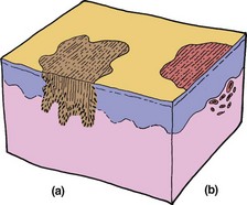
Macule
A macule is a localized area of colour or textural change in the skin. Macules can be hypopigmented, as in vitiligo; pigmented, as in a freckle (a); or erythematous, as in a capillary haemangioma (b).
Papule
A papule is a small solid elevation of the skin, generally defined as less than 5 mm in diameter. Papules may be flat topped, as in lichen planus; dome shaped, as in xanthomas; or spicular if related to hair follicles.
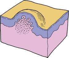
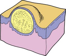
Nodule
Similar to a papule but larger (i.e. greater than 5 mm in diameter), nodules can involve any layer of the skin and can be oedematous or solid. Examples include a dermatofibroma (below) and secondary deposits.
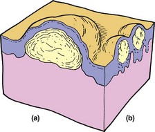
Bulla
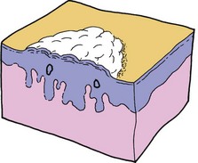
A bulla is similar to a vesicle but larger: greater than 5 mm in diameter. The blisters of bullous pemphigoid (see figure; a) and pemphigus vulgaris (p. 78) are examples.
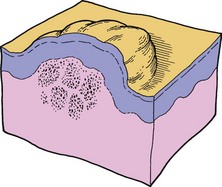
Vesicle
A vesicle is a small blister (less than 5 mm in diameter) consisting of clear fluid accumulated within or below the epidermis. Vesicles may be grouped as in dermatitis herpetiformis (subepidermal). Intraepidermal vesicles are shown in the figure (b).
Glossary of other dermatological terms
 Abscess: A localized collection of pus formed by necrosis of tissue.
Alopecia: Absence of hair from a normally hairy area.
Atrophy: Loss of epidermis, dermis or both. Atrophic skin is thin, translucent and wrinkled with easily visible blood vessels.
Burrow: A tunnel in the skin caused by a parasite, particularly the acarus of scabies.
Callus: Local hyperplasia of the horny layer, often of the palm or sole, due to pressure.
Carbuncle: A collection of boils (furuncles) causing necrosis in the skin and subcutaneous tissues.
Crust: Dried exudate (normally serum, blood or pus) on the skin surface.
Ecchymosis: A macular red or purple haemorrhage, more than 2 mm in diameter, in the skin or mucous membrane.
Erosion: A superficial break in the epidermis, not extending into the dermis, which heals without scarring.
Erythema: Redness of the skin due to vascular dilatation.
Excoriation: A superficial abrasion, often linear, which results from scratching.
Fissure: A linear split in the epidermis, often just extending into the dermis.
Cellulitis: A purulent inflammation of the skin and subcutaneous tissue.
Folliculitis: An inflammation of the hair follicles.
Comedo: A plug of sebum and keratin in the dilated orifice of a pilosebaceous gland.
Freckle: A macular area in which there is increased pigment formation by melanocytes.
Pustule
A pustule is a visible collection of free pus in a blister. Pustules may indicate infection (e.g. a furuncle), but not always, as pustules seen in psoriasis, for example, are not infected.
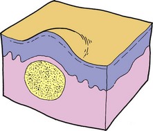
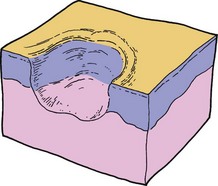
Cyst
A cyst is a nodule consisting of an epithelial-lined cavity filled with fluid or semisolid material. An epidermal (‘sebaceous’) cyst is shown below.
Wheal
A wheal is a transitory, compressible papule or plaque of dermal oedema, red or white in colour and usually signifying urticaria.
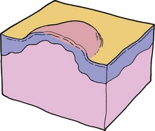
Plaque
A plaque is a palpable, plateau-like elevation of skin, usually more than 2 cm in diameter. Plaques are rarely more than 5 mm in height and can be considered as extended papules. Certain lesions of psoriasis (below) and mycosis fungoides are good examples.
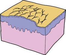
Scale
A scale is an accumulation of thickened, horny layer keratin in the form of readily detached fragments. Scales usually indicate inflammatory change and thickening of the epidermis. They may be fine, as in ‘pityriasis’; white and silvery, as in psoriasis (below); or large and fish-like, as seen in ichthyosis.
Ulcer
An ulcer is a circumscribed area of skin loss extending through the epidermis into the dermis. Ulcers are usually the result of impairment of the vascular or nutrient supply to the skin, e.g. as a result of peripheral arterial disease.
Glossary of other dermatological terms
Furuncle: A pyogenic infection localized in a hair follicle.
Hirsuties: Excessive male pattern hair growth.
Hypertrichosis: Excessive hair growth in a non-androgenic pattern.
Keloid: An elevated and progressive scar not showing regression.
Keratosis: A horn-like thickening of the skin.
Lichenification: Chronic thickening of the skin with increased skin markings, as a result of rubbing or scratching.
Milium: A small white cyst containing keratin.
Papilloma: A nipple-like projection from the skin surface.
Petechia: A haemorrhagic punctuate spot measuring 1–2 mm in diameter.
Poikiloderma: A combination of hyperpigmentation, telangiectasia and atrophy seen together in a dermatosis.
Purpura: Extravasation of blood resulting in red discoloration of the skin or mucous membranes.
Scar: The replacement of normal tissue by fibrous connective tissue at the site of an injury.
Stria: An atrophic linear band in the skin – white, pink or purple in colour. The result of connective tissue changes.
Telangiectasia: Dilated dermal blood vessels giving rise to a visible lesion.