Connective tissue diseases
The inflammatory disorders of connective tissue often affect several organs, as in systemic lupus erythematosus (LE), but they may also involve the skin alone (e.g. discoid LE). Autoantibodies are a feature of these diseases, which can thus be regarded as ‘autoimmune’.
Lupus erythematosus
Lupus erythematosus represnts a spectrum of cutaneous disease from scarring (discoid) to multisystem (systemic).
Aetiopathogenesis
There is increasing evidence that the induction of autoantibodies against self-proteins derives from an impaired ability to clear dying (apoptotic) cells. Both genetic (human leucocyte antigen (HLA)) and environmental (toxins, drugs, UV radiation, viruses) factors have been implicated in disease pathogenesis.
Pathology
Discoid lesions show epidermal atrophy, hyperkeratosis and basal layer degeneration. Subacute lesions show greater atrophy but the other features are less evident. Systemic lesions have similar changes with dermal oedema and fibrinoid change, inflammatory infiltrate and sometimes vasculitis. Direct immunofluorescence shows a ‘lupus band’ at the dermoepidermal junction in lesional systemic and discoid LE, but this is also found in approximately 20% of healthy individuals on sun-exposed skin.
Clinical presentation
Systemic lupus erythematosus (99% ANA, 50% Ro, 60% dsDNA positive)
Skin signs are found in 80%. The facial butterfly eruption (Fig. 1) is characteristic, but photosensitivity, discoid lesions, diffuse alopecia, mouth lesions and vasculitis also occur. Multisystem involvement with serological or haematological abnormalities must be demonstrated to diagnose systemic LE (Table 1). The female : male ratio is 8 : 1.
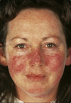
Table 1 Organ involvement in systemic LE
| Organ | Involvement |
|---|---|
| Skin | Photosensitivity, facial rash, vasculitis, hair loss, Raynaud’s phenomenon |
| Blood | Anaemia, thrombocytopenia |
| Joints | Arthritis, tenosynovitis, calcification |
| Kidney | Glomerulonephritis, nephrotic syndrome |
| Heart | Pericarditis, endocarditis, hypertension |
| Central nervous system | Psychosis, infarction, neuropathy |
| Lungs | Pneumonitis, effusion |
Subacute lupus erythematosus (60% ANA, 80% Ro, <5% dsDNA positive)
The skin involvement is usually on the neck, trunk and arms. The erythema is non-scarring and may be papulosquamous or annular and resolves with hypopigmentation and telangiectasia. Mouth ulceration, livedo reticularis, periungal telangiectasia and Raynaud’s phenomenon may also be noted. Multisystem involvement can be seen, but is usually mild.
Discoid lupus erythematosus (35% ANA, 2% Ro, <5% dsDNA positive)
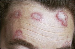
One or more round or oval plaques appear on the face, scalp or hands (Fig. 2). The lesions are well demarcated, red, atrophic, scaly and show keratin plugs in dilated follicles. Scarring leaves alopecia on the scalp and hypopigmentation in those with a pigmented skin. Remission occurs in over 50%. Internal involvement is not a feature, and only 6% develop systemic LE. Women outnumber men by 2 : 1.
Differential diagnosis
Discoid LE can usually be differentiated from other facial rashes such as rosacea, seborrhoeic dermatitis, lupus vulgaris or psoriasis. A biopsy should be performed. The photosensitive eruption of systemic LE may resemble polymorphic light eruption, dermatomyositis or drug reaction.
Management
Immunological screening is important for diagnosis and to predict complications (e.g. anti-Ro and congenital heart block; anticardiolipin and thromboses and spontaneous abortions; antihistone and drug-induced lupus). Discoid LE usually responds to potent or very potent topical steroids which, in this instance, can be applied to the face. Sunblock creams are essential. Widespread disease may need systemic therapy with hydroxychloroquine; the small risk of retinopathy demands monitoring of visual acuity. The treatment of systemic LE depends on the type of involvement. Sunscreens reduce photosensitivity but, if there is internal disease, antimalarials and systemic steroids are required, often with immunosuppressive agents.
Systemic sclerosis
Systemic sclerosis is an uncommon, progressive multisystem disease in which collagen deposition and fibrosis occur in several organs.
Aetiopathogenesis
Dysregulated transforming growth factor (TGF)-β-induced fibroblast overproduction of collagen is central to tissue fibrosis. However, endothelial cell damage by T cells and macrophages may also be important.
Clinical presentation
Raynaud’s phenomenon (p. 70) is frequently the presenting sign. The skin of the fingers, forearms and lower legs becomes tight, waxy and stiff, and the finger pulps are resorbed (Fig. 3). Facial signs include perioral furrowing, telangiectasia (p. 115) and restricted mouth opening. Internal organ involvement, e.g. renal failure, may prove fatal (Table 2). Women are affected more than men (F : M ratio 4 : 1). Some 90–95% of cases are ANA positive. The diagnosis is rarely in doubt in diffuse disease, although chronic graft-versus-host disease shows similar changes. Limited cutaneous systemic sclerosis has a better prognosis and usually affects only the neck, forearms and lower legs. A subset described as CREST syndrome, is confined to Calcinosis, Raynaud’s phenomenon, oEsophageal dysmotility, Sclerodactyly and Telangiectasia.
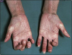
Table 2 Organ involvement in systemic sclerosis
| Organ | Involvement |
|---|---|
| Skin | Raynaud’s phenomenon, calcinosis, sclerodactyly, telangiectasia |
| Gut | Oesophageal dysmotility, malabsorption, dilated bowel |
| Lung | Fibrosis, pulmonary hypertension |
| Heart | Pericarditis, myocardial fibrosis |
| Kidney | Renal failure, hypertension |
| Muscle | Myositis, tendon involvement |
Management
Treatment is mainly supportive. Nifedipine can help Raynaud’s phenomenon. Hypertension is controlled. Systemic steroids, penicillamine and immune-suppressives have been used with little benefit. Photophoresis may be tried (p. 115). Renal crises (associated with antiRNA polymerase I and III antibodies) should be managed aggressively with angiotensin converting enzyme (ACE) inhibitors.
Morphoea (localized scleroderma)
Morphoea consists of localized indurated plaques or bands of sclerosis on the skin. Internal disease is not found. The cause is unknown, although it may follow trauma. Histology shows bands of collagen with loss of appendages.
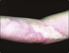
Morphoea presents with round or oval plaques of induration and erythema, often with a purplish edge (Fig. 4). These become shiny and white, eventually leaving atrophic hairless pigmented patches. The trunk or proximal limbs are affected. Morphoea is more common in women (F : M ratio 3 : 1). Linear morphoea may involve the face or a limb and, when seen in a child, can retard growth of the underlying tissues, including bone.
There is no well established treatment (p. 115), although topical steroids are often given. The disease usually resolves spontaneously within months to years.
Dermatomyositis
Dermatomyositis is an uncommon disorder in which inflammation of skin, muscle and blood vessels gives a distinctive eruption, with muscle weakness of varying severity. The cause is unknown but an underlying malignancy is found in a subgroup.
Clinical presentation
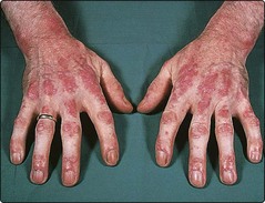
In dermatomyositis/polymyositis, skin changes or muscle weakness may predominate. The typical eruption is a lilac–blue discoloration around the eyelids, cheeks and forehead, often with oedema. Bluish–red papules or streaks on the dorsal aspects of the hands (Fig. 5), elbows and knees are seen, sometimes with pigmentation and nail fold telangiectasia. Photosensitivity is common. There is no strong association with autoantibodies, although anti-Jo-1 antibodies predict lung involvement. An association with malignancy exists in patients over 40 years, 40% of whom have an underlying tumour, usually of the lung, breast or stomach. A childhood variant mainly affects the muscles and causes calcinosis and contractures.
Management
Investigations must define the degree of myositis and, in adults, exclude the possibility of underlying neoplasia. Treatment is with systemic steroids in moderate to high dosage, often with an immunosuppressive such as azathioprine or methotrexate. Immunoglobulin infusion and possibly photophoresis (p. 115) may help.
Connective tissue diseases
 Systemic LE is an autoimmune, multisystem disease in which a butterfly rash, photosensitivity, vasculitis and alopecia may be seen. Treatment depends on the type and degree of involvement and often includes systemic steroids and immunosuppressive agents.
Subacute LE is a less aggressive form of LE with predominantly cutaneous features.
Discoid LE is confined to the skin. Scaly atrophic plaques and scarring alopecia are found. Topical steroids and sunscreens are helpful. Sometimes, systemic therapy, e.g. with hydroxychloroquine, is used.
Systemic sclerosis is a serious multisystem disorder. Sclerodactyly, Raynaud’s phenomenon, telangiectasia and calcinosis are seen.
Morphoea is characterized by white indurated plaques, usually on the trunk and proximal limbs. In children, it may retard growth of underlying tissues, producing atrophy. In adults, the condition is generally self-limiting.
Dermatomyositis is an autoimmune inflammation of skin and muscle. The skin signs are a lilac–blue discoloration around the eyelids and red streaks on the dorsa of the hands. Exclude underlying malignancy in those over 40 years of age.