Vascular and lymphatic diseases
Blood vessel disorders
Erythema
Erythema is redness of the skin, usually due to vasodilatation (Table 1). It may be localized, e.g. with pregnancy or liver disease (on palms), fixed drug eruption and infection (e.g. Lyme disease), or generalized, as with drug eruption, toxic erythema (e.g. viral exanthem) and connective tissue disease.
Table 1 Classification of blood vessel and lymphatic disorders
| Vessel | Process | Resulting lesion |
|---|---|---|
| Small blood vessels |
Erythema, flushing, telangiectasia Urticaria (p. 76), oedema |
|
| Arteries |
Vasculitis (p. 82) |
|
| Veins |
Thrombosis, skin changes, ulceration (p. 72) |
|
| Lymphatics |
Flushing
Flushing is erythema due to vasodilatation. The causes are:
 physiological (autonomic response to emotion, heat or exercise)
menopause (hormonal; often with associated sweating)
foods (e.g. spices – gustatory; alcohol – aldehyde related)
drugs (angiotensin-converting enzyme (ACE) inhibitors, 5-hydroxytryptamine (5-HT3) antagonists, nifedipine)
Flushing is common and affects the face, neck and upper trunk. It is usually benign. A sudden onset and systemic symptoms (e.g. diarrhoea or fainting) mean that carcinoid syndrome or phaeochromocytoma must be excluded. In treatment, first remove the cause, e.g. spices or alcohol. Embarrassing physiological flushing may improve with a small dose of propranolol.
Telangiectasia
Telangiectasia is a visible dilatation of dermal venules or, in spider naevi (Fig. 1), an arteriole. It results from:
congenital (e.g. hereditary haemorrhagic telangiectasia)
skin atrophy (topical steroids, ageing skin, radiation dermatitis)
excess oestrogen (e.g. liver disease, pregnancy, ‘the pill’)
connective tissue disease (systemic sclerosis, lupus erythematosus, dermatomyositis)
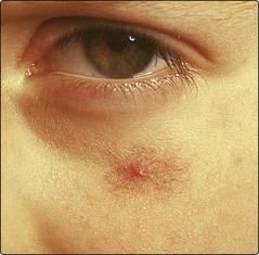
Isolated spider naevi are common and of little significance, but their number may increase with pregnancy and liver disease. A venous lake – acquired venous ectasia – is often seen on the lower lip of the elderly. Telangiectasia is treated by fine-needle cautery, hyfrecation or laser (p. 113).
Purpura
Purpura is a blue–brown discoloration of the skin due to the extravasation of erythrocytes (Fig. 2). It results from a variety of mechanisms:
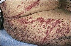
Petechiae are small dot-like purpura, whereas ecchymoses are more extensive. Purpura is often seen in the elderly or those on steroids, and develops spontaneously or after minor trauma. Idiopathic pigmented purpura is seen as brownish punctate lesions (capillaritis) on the legs.
Mostly, there is no specific therapy. Underlying causes, e.g. blood disorders or vasculitis, are treated as necessary.
Raynaud’s phenomenon
Raynaud’s phenomenon is characterized by a paroxysmal vasoconstriction of the digital arteries, usually provoked by cold, in which the fingers turn white (due to ischaemia), cyanotic blue (due to capillary dilatation with a stagnant blood flow) and then red (due to reactive hyperaemia). When no cause is found, it is known as ‘Raynaud’s disease’. Causes include:
arterial occlusion: atherosclerosis, Buerger’s disease
connective tissue disease: systemic sclerosis (including CREST syndrome), systemic lupus erythematosus (p. 81)
hyperviscosity syndrome: polycythaemia, cryoglobulinaemia
neurological defects: syringomyelia, peripheral neuropathy
reflux vasoconstriction: with use of vibration tools (p. 124)
Raynaud’s phenomenon mostly affects women. It may be the forerunner of a connective tissue disease. The hands should be kept warm and protected from the cold. Smoking must be stopped. A calcium channel blocker (e.g. nifedipine) or naftidrofuryl may help. In resistant cases, prostacyclin infusions are given.
Livedo reticularis
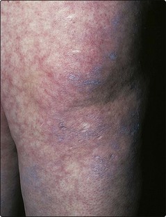
Livedo reticularis is a marble-patterned cyanosis of the skin, due to reduced arteriole blood flow, usually in women. The condition has the following causes:
Physiological, i.e. cold induced.
Vasculitis due to connective tissue disease, e.g. systemic lupus erythematosus and polyarteritis nodosa.
Hyperviscosity due to cryoglobulinaemia, polycythaemia.
Sneddon syndrome, which consists of livedo vasculitis with cerebrovascular disease and circulating antiphospholipid antibodies.
Cold-induced livedo reticularis gives a mottled meshwork pattern on the outer thighs of children and is reversible. Fixed livedo (Fig. 3) is due to vasculitis and requires investigation. Treatment is aimed at the underlying disease.
Erythema ab igne
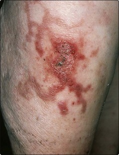
Erythema ab igne is a reticulate pigmented erythema (Fig. 4) due to heat-induced damage. It is seen on the shins of the elderly who sit before a fire, or with the use of heat pads or laptop computers.
Chilblains
Chilblains are inflamed and painful purple–pink swellings on the fingers, toes or ears that appear in response to cold. Chilblains result from an overcompensatory cold-induced vasoconstriction of cutaneous arterioles and venules. They occur in the winter and usually affect women. Warm housing and clothing are advised. Oral nifedipine may help.
Lymphatic disorders
Lymphoedema
Lymphoedema is oedema, often of a limb, due to inadequate lymphatic drainage. The condition may be primary or secondary. Primary lymphoedema is the result of a congenital developmental defect. Secondary causes include:
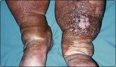
Primary lymphoedema presents in adolescence and may follow infection. The lower legs are commonly affected. In chronic lymphoedema, the oedema is non-pitting and fibrotic, and the overlying epidermis hyperkeratotic (Fig. 5). Radiolabelled lymphoscintigraphy (or X-ray or magnetic resonance imaging (MRI) lymphangiography) shows the defect.
A lymphoedematous limb is at risk of repeated infection (particularly erysipelas), and long-term prophylaxis with oral phenoxymethylpenicillin is recommended. Exercise, compression support and massage can help. Surgical reconstruction is rarely possible.
Lymphangitis
Lymphangitis is defined as infection of the lymphatic vessels, usually due to streptococci. It presents as a tender red line extending proximally up a limb, usually from a focus of infection. Hospital admission is usually necessary. Therapy is with a suitable intravenous antibiotic (e.g. benzylpenicillin).
Vascular and lymphatic diseases
Erythema: can be localized, e.g. liver palms, or generalized, e.g. toxic erythema.
Flushing: usually emotional; rarely carcinoid syndrome or phaeochromocytoma.
Telangiectasia: commonly seen with skin atrophy, but lesions can occur with oestrogen excess or connective tissue disease. Treatment is by hyfrecation or laser.
Purpura: caused by defects of the vessel wall, supporting dermis or clotting mechanism, or ‘idiopathic’. Treat the underlying disorder.
Livedo reticularis: physiological or due to underlying hyperviscosity or connective tissue disorder.
Chilblains: describes cold-induced perniosis of the fingers, toes and ears.
Raynaud’s phenomenon: vasoconstriction of the digital arteries with colour changes.
Lymphoedema: results from absence of or damage to lymphatics. Long-term prophylaxis with antibiotics prevents recurrent infection in chronic cases.