Patient Education and the Dental Radiographer
After completion of this chapter, the student will be able to do the following:
• Summarize the importance of educating patients about dental radiographs
• List the three methods that can be used by the dental radiographer to educate patients about dental radiographs
• Answer common patient questions about the need for dental radiographs, x-ray exposure, the safety of dental x-rays, digital imaging, and other miscellaneous concerns
The dental radiographer must be able to educate patients about the importance of dental radiographs and also be prepared to answer common questions asked by patients about the need for dental radiographs, x-ray exposure, the safety of dental x-rays, and miscellaneous concerns. The purpose of this chapter is to discuss the importance of patient education, to describe different methods of patient education, and to review common patient questions and answers about dental radiography.
Importance of Patient Education
Educating dental patients about the importance of dental radiographs is critical, yet patient education is often overlooked by dental professionals. Many patients do not understand the value of dental radiographs. Often, the patient is simply told that “dental x-rays are required by the dentist,” and little additional information is provided. As a result, many patients fear the use of x-radiation. Others believe that dental radiographs are a way for the dentist to make extra money. To address such fears and misconceptions, the dental radiographer must be prepared to educate the patient about the value of dental radiographs.
Many patients have heard or read about the damaging effects of x-radiation. Newspaper articles, magazine articles, and television magazine shows often highlight the damaging effects of radiation and cast doubt on the necessity and benefit of radiographic examinations. Such reports are often misleading and are not well researched. As a result, these reports cause the patient to fear the use of x-radiation and to avoid all radiation exposure.
Because of the presence of such misinformation, the dental radiographer must take the time to educate the patient. In some instances, the patient may have to be completely re-educated. The dental radiographer must be prepared to explain exactly why dental radiographs are important, how dental radiographs are used, and how they are beneficial. In addition, the dental professional must be able to discuss common conditions and lesions that can be detected only through the use of dental radiographs (see Chapters 4 and 11).
Comprehensive dental health education is one of the greatest services that a dental professional can provide to the patient. Education enhances understanding. A patient who is knowledgeable about the importance of dental radiographs is more likely to realize the benefit of dental radiographs, accept the prescribed treatment, and follow prevention plans. Patient education is also likely to decrease fears of x-ray exposure, increase cooperation, and increase motivation for regular dental visits.
Methods of Patient Education
Patient education about dental radiographs can be accomplished in a number of ways. The dental radiographer can use an oral presentation, printed literature, or a combination of both to educate the dental patient.
An oral presentation, in conjunction with sample dental images, can be used to communicate the importance of dental radiographs. For example, the dental radiographer can show the patient a prepared series of radiographs illustrating typical normal and abnormal conditions. Through the use of such radiographs, the dental radiographer includes a visual component in the educational process; visual aids enhance patient comprehension. A prepared oral presentation with visual aids allows the patient to develop greater confidence in the expertise of the dental radiographer. A prepared presentation also communicates to the patient that the dental radiographer is organized and competent.
The use of digital imaging may further aid in patient education. This helps patients view their own periapical, bite-wing, or extraoral images on a computer monitor or television screen instead of looking at detailed information on mounted radiographs. The use of digital imaging helps explain concepts such as caries, periodontal changes, or oral diseases (Figure 13-1).
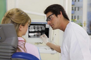
FIGURE 13-1 Dentist and patient reviewing digital images on a large computer moitor helps to facilitate patient education. (Courtesy of DEXIS, Des Plaines, III.)
Printed information about dental radiographs is useful to educate the dental patient as well. Brochures can be placed in the reception area of the dental office or handed to patients before the radiographic examination. The pamphlet Dental X-rays: Your Dentist’s Advice, for example, discusses dental x-rays and how dental radiographs benefit the patient. This and other brochures about dental radiographs and x-ray exposure can be obtained from the American Dental Association and the Bureau of Radiologic Health. Printed literature about dental radiographs can also be custom designed by the dental professional and then printed for use in the dental office.
A combination of an oral presentation and printed literature is probably the most effective method of educating the dental patient about dental radiographs. The use of both approaches can stimulate a question-and-answer type of discussion about dental radiographs.
Common Questions and Answers
The dental radiographer must be prepared to answer common questions about the need for dental radiographs, x-ray exposure, safety of dental x-rays, digital imaging, and other concerns. Many patients ask the dental auxiliary, rather than the dentist, questions about x-radiation. The dental radiographer can answer many of the patient’s questions. However, some questions must be answered only by the dentist; such questions must be established by the dentist and understood by all members of the dental team. For example, questions about diagnosis must be answered only by the dentist.
Necessity Questions
Patients often ask questions about the need for dental x-rays, the frequency of dental x-rays for adults and children, the refusal of dental x-rays, and the use of dental x-rays from a previous dentist. Examples of questions and answers follow:
Question: Are dental x-rays really necessary?
Answer: Yes. Many diseases and conditions such as tooth decay, gum disease, cysts, and tumors cannot be detected simply by looking into your mouth. Many diseases and conditions produce no signs or symptoms. Without dental x-rays, these conditions may go unnoticed for a long time. As these conditions progress, extensive damage and pain may occur; these, in turn, may result in more extensive and costly treatment. Some oral diseases can even affect your general health or become life threatening.
Dental radiographs are always taken to benefit you, the patient; the primary benefit is disease detection. Through the use of dental radiographs, conditions and diseases that cannot be detected in any other way can be identified early. Early identification and treatment minimize and prevent problems, such as pain and the need for surgical procedures.
Question: How often should I have dental x-rays?
Answer: The first step to limiting the amount of radiation that you receive is the proper prescribing, or ordering, of dental radiographs. Decisions about the number, type, and frequency of dental x-rays are determined by the dentist based on your individual needs. Guidelines published by the American Dental Association are used by the dentist to aid in prescribing the number, type, and frequency of dental radiographs.
Because every patient’s dental condition is different, the frequency of radiographic examinations is also different. The frequency of your dental x-ray examinations is based on your individual needs. No set interval exists between x-ray examinations. For example, a patient with tooth decay or gum disease needs more frequent radiographic examinations than a patient without such diseases.
Question: How often should children have dental x-rays?
Answer: The interval between radiographic examinations should be based on the individual needs of the child. Because every child’s dental condition is different, the frequency of radiographic examinations is different as well. There is no set interval between x-ray examinations. For example, a child with tooth decay needs more frequent radiographic examinations than a child without tooth decay.
Question: Can I refuse x-rays and be treated without them?
Answer: No. When you refuse dental x-rays, the dentist cannot treat you. The standard of care requires that the dentist refuse treatment when a patient refuses x-rays that are necessary. Treatment without necessary radiographs is considered negligent. No document can be signed to release the dentist from liability. For example, if you were to sign a paper stating that you refused dental x-rays but released the dentist from any and all liability, you would be consenting to negligent care. Legally, you cannot consent to negligent care.
Question: Instead of taking x-rays now, can you use the x-rays from my previous dentist?
Answer: Yes. Previous dental radiographs can be used, provided they are recent and of acceptable diagnostic quality. Additional dental radiographs may be necessary, however, based on your individual needs. If your previous dental radiographs, even if recent, are not of diagnostic quality, you will have to have another radiograph taken.
Exposure Questions
Patients often ask questions about x-ray measurement, amounts of x-ray exposure, the use of the lead apron during exposure, dental x-radiation during pregnancy, and the reason for the dental radiographer leaving the room during exposure. Examples of questions and answers follow:
Question: How are x-rays measured?
Answer: Special units are used to measure x-ray exposure and absorption. The radiation that reaches the surface of the skin is measured in roentgen units. The unit for dose, or the amount of energy absorbed by a tissue, is termed radiation absorbed dose (rad). Because of the small quantities of radiation used during radiographic procedures, very small multiples of these radiation units are used. The prefix milli-, meaning “one one-thousandth,” is used to express the small quantities of exposure in milliroentgens and the dose in millirads.
Question: How much radiation will I receive from dental x-rays?
Answer: Because no amount of radiation is considered safe, strict guidelines are followed to limit the amount of x-radiation. For example, the dentist custom-orders your x-rays on the basis of your individual needs. During exposure, a thyroid collar, a lead apron, fast film, digital imaging, and a beam alignment device will be used to protect you from excess radiation. Good exposure technique and careful processing are also used to limit your exposure to x-radiation.
The actual amount of x-radiation received will vary depending on the film speed, the technique used, and exposure factors. For example, when a single intraoral D-speed film is exposed, the x-rays expose a small area of skin, and the exposure to the skin of the face is about 250 milliroentgens. With faster F-speed film, a single intraoral film results in a surface skin exposure of 125 milliroentgens. The use of digital imaging reduces the radiation dose even further. For dental x-rays to produce permanent skin damage, such as skin cancer, exposures in the range of thousands of roentgens are needed. Such exposures are inconceivable in dental radiography and are not possible with dental x-ray equipment.
Question: Why do you use a lead apron?
Answer: A lead apron and a thyroid collar are used to protect reproductive, blood-forming, and thyroid tissues from scatter radiation. The lead acts as a shield and actually prevents the radiation from reaching these radiosensitive organs. The lead apron protects you from unnecessary radiation exposure.
Question: Is it safe to take dental x-rays during pregnancy?
Answer: When a lead apron is used during dental radiographic procedures, the amount of radiation received in the gonadal region is nearly zero. No detectable exposure to the embryo or fetus occurs with the use of the lead apron. The American Dental Association, together with the Food and Drug Administration, has stated in the Guidelines for Prescribing Dental Radiographs that the recommended guidelines “do not need to be altered because of pregnancy.” Although scientific evidence indicates that dental x-ray procedures can be performed during pregnancy, many dentists elect to postpone such x-ray procedures because of patient concerns.
Question: Why do you leave the room when x-rays are used?
Answer: When you are exposed to x-rays, you receive the diagnostic benefit of the dental radiographs; I do not receive any benefit. An individual should only be exposed to x-radiation when the benefit of disease detection outweighs the risk of exposure. Since I do not benefit from your x-ray exposure, I must use proper protection measures. One of the most effective ways for me to limit my x-ray exposure is to maintain adequate distance and shielding, which is why I step out of the room during your x-ray exposure.
Safety Questions
Patients often ask questions about the safety of dental x-rays and wonder whether dental x-rays cause cancer. Examples of questions and answers follow:
Question: Are dental x-rays safe?
Answer: All x-rays are harmful to living tissue. The amount of x-radiation used in dental radiography is small, but biologic damage does occur. No amount of radiation is considered safe. As a result, dental x-rays must be prescribed only when the benefit of disease detection outweighs this risk of harm.
Question: Will dental x-rays cause cancer?
Answer: Not a single recorded case of a patient developing cancer from diagnostic x-rays exists. The radiation exposure that occurs during a dental x-ray examination is very small, and the chance that it will contribute to or cause cancer is extremely low. For example, the potential risk of dental radiography inducing a fatal cancer has been estimated to be 3 in one million. The risk of a person developing cancer spontaneously is much higher, or 3300 in one million. When these two numbers are compared, it is evident that when cancer occurs, it is much more likely to be unrelated to radiation exposure.
Digital Imaging Questions
Question: What are the advantages of digital imaging?
Answer: Digital imaging requires less exposure to radiation which benefits you, the patient. Digital information can be stored, transmitted, and manipulated electronically. Digital imaging also gives us instant images that are environmentally friendly, as no processing chemicals are used.
Question: Are there any risks associated with digital imaging?
Answer: Because radiation is involved, a certain amount of risk does exist. With digital radiography, your exposure is less than with traditional x-rays. Your radiation exposure time may be reduced by 50% to 80%.
Miscellaneous Questions
Question: Can a panoramic x-ray be taken instead of a complete series?
Answer: No. A panoramic radiograph cannot be substituted for a complete series of dental radiographs. A complete series of dental radiographs is required when information about the details of the teeth and surrounding bone are needed. A panoramic radiograph does not clearly reveal changes in teeth, as in tooth decay, or the details of the supporting bone. The panoramic radiograph is useful for showing the general condition of a patient’s teeth and bone.
Question: Who owns my dental radiographs?
Answer: All your dental records, including the dental radiographs, are the property of the dentist. As a patient, however, you have the privilege of reasonable access to your dental records. For example, you can request a copy of your dental radiographs or request that a copy be sent to a dentist of your choice. Digital images may also be electronically sent to a referring doctor. The dentist usually retains the original dental radiographs as part of the patient record.
Summary
• The dental radiographer must be able to educate patients about dental radiographs. A patient who is knowledgeable about the importance of dental radiographs is more likely to have reduced fears about x-ray exposure, realize the benefits of dental radiographs, accept prescribed treatment, and follow prevention plans.
• The dental radiographer can use an oral presentation, printed literature, or a combination (probably the most effective method) to educate the dental patient about radiographs.
• The dental radiographer must be prepared to answer common patient questions about the need for dental radiographs, x-ray exposure, safety of dental x-rays, digital imaging, and miscellaneous concerns.
• Some patient questions such as those about diagnosis must be answered only by the dentist, and these questions must be established by the dentist and understood by the dental team.
Frommer, HH, Savage-Stabulas, JJ, Operator protection. Radiology for the dental professional, ed 9, St. Louis, Mosby, 2011.
Frommer, HH, Savage-Stabulas, JJ, Patient protection. Radiology for the dental professional, ed 9, St. Louis, Mosby, 2011.
Haring, JI, Lind, LJ. The importance of dental radiographs and interpretation. In: Radiographic interpretation for the dental hygienist. Philadelphia: Saunders; 1993.
Johnson, ON, Thomson, EM, Patient relations and education. Essentials of dental radiography for dental assistants and hygienists, ed 8, Upper Saddle River, NJ, Pearson, 2007.
Thunthy, KH. X-rays: Detailed answers to frequently asked questions. Compend Contin Educ Dentistry. 1993;14(3):394–398.
Short Answer
3. Are dental x-rays really necessary?
4. How often should adults have dental x-rays?
5. How often should children have dental x-rays?
6. Can a patient refuse dental x-rays and be treated without them?
7. Can radiographs from a previous dentist be used instead of taking dental x-rays?
9. How much radiation is received from dental x-rays?
10. Why is a lead apron used during x-ray exposure?
11. Can dental x-rays be taken during pregnancy?
12. Why does the dental radiographer leave the room during x-ray exposure of the patient?
14. Will dental x-rays cause cancer?
15. Can a panoramic x-ray be taken instead of a complete series?
16. Who owns the dental radiographs—the dentist or the patient?