Identification of Restorations, Dental Materials, and Foreign Objects
After completion of this chapter, the student will be able to do the following:
• Define the key terms associated with identifying restorations, materials, and foreign objects on dental images
• Discuss the importance of interpreting dental images while the patient is present
• On dental images, identify and describe the appearance of the following restorations: amalgam, gold, stainless steel and chrome, post and core, porcelain, porcelain-fused-to-metal, composite, and acrylic
• On dental images, identify and describe the appearance of the following dental materials and devices: base materials, metallic pins, gutta percha, silver points, removable partial dentures, complete dentures, orthodontic bands, brackets and wires, fixed retainers, implants, suture wires, splints, and stabilizing arches and wires
• On dental images, identify and describe the appearance of the following miscellaneous objects: jewelry, eyeglasses, and patient napkin chains
Dental images are an important diagnostic tool that enables the practitioner to view dental restorations and dental materials. Dental images are also useful in identifying and locating foreign objects. Some restorations, materials, and foreign objects are easily identified on dental images; others are not. On dental images, the appearances of restorations, materials, and foreign objects vary, depending on the thickness of the material, density, and atomic number. Some restorations, materials, and foreign objects may be identified by the degree of radiopacity present, outline, contour, or size; others require additional clinical information.
The dental professional should interpret all dental images while the patient is present. If questions arise as to what is seen on a dental image concerning dental restorations, dental materials, or foreign objects, clinical examination of the patient can be done to obtain additional information or to verify what is seen. When dental images are interpreted without the patient present, some important clinical information is not available.
The purpose of this chapter is to review common dental restorations, dental materials, and foreign objects that may be seen on dental images.
Identification of Restorations
A variety of common restorative materials, including amalgam, gold, stainless steel, porcelain, composite, and acrylic, can be identified on dental images.
Metallic restorations (e.g., amalgam, gold) absorb x-rays, and as a result, very little (if any) radiation comes in contact with the receptor. Consequently, that area of the receptor remains unexposed, and the metallic restorations appear completely radiopaque (light or white) on a dental image. To illustrate this concept, place a processed film with an amalgam restoration on a printed page. The underlying print can be easily read through the radiopaque area on the film (Figure 32-1).
FIGURE 32-1 When a radiograph with a metallic restoration is placed on a printed page, the print can be easily seen.
Nonmetallic restorations (e.g., porcelain, composite, acrylic) may vary in appearance from radiolucent (dark or black) to slightly radiopaque, depending on the density of the material. Of the nonmetallic restorations, porcelain is the most dense and least radiolucent, and acrylic is the least dense and most radiolucent.
Amalgam Restorations
Amalgam is the most common restorative material used in dentistry. Amalgam absorbs the x-ray beam and prevents x-rays from reaching the receptor; consequently, amalgam appears completely radiopaque on a dental image. Amalgam may be seen in a variety of shapes, sizes, and locations on a dental image.
One-Surface Amalgam Restorations
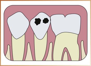
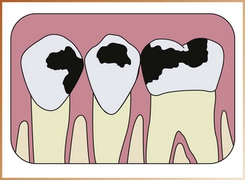
One-surface amalgam restorations (“pit amalgams”) appear as distinct, small, round or ovoid radiopacities (Figures 32-2 and 32-3). One-surface amalgams may be seen on the buccal, lingual, or occlusal surfaces of teeth. Larger two-surface and multi-surface amalgam restorations also appear radiopaque and are characterized by their irregular outlines or borders (Figures 32-4 and 32-5). Multi-surface amalgam restorations may involve any tooth surface.
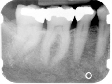
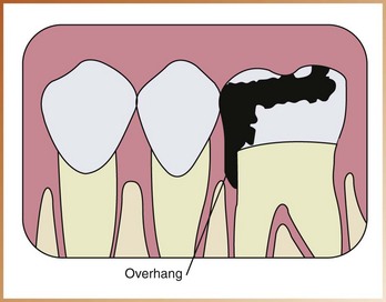
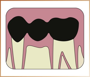
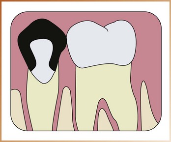
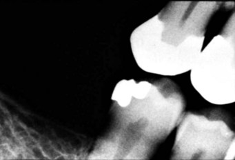
Amalgam Overhangs
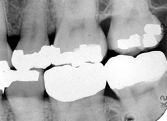
Amalgam overhangs can be described as extensions of amalgam seen beyond the crown portion of a tooth in the interproximal region. An amalgam overhang results from improper band placement around a tooth before condensing the amalgam restoration. Amalgam overhangs can be easily visualized on a dental image and appear radiopaque (Figures 32-6 and 32-7). An amalgam overhang disrupts the natural cleansing contours of the tooth, traps food and plaque, and contributes to bone loss. To prevent destruction of interproximal bone, amalgam overhangs must be removed.
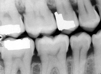
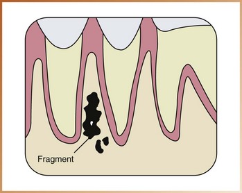
Amalgam Fragments
Fragments of amalgam may be inadvertently embedded in adjacent soft tissue during restoration of a tooth. Amalgam fragments or scraps vary in size and shape and appear radiographically as dense radiopacities with irregular borders on a dental image (Figures 32-8 and 32-9). Amalgam fragments may be seen in any location where soft tissue is present. If amalgam fragments are displaced into soft tissue during the placement or removal of an amalgam restoration or during the extraction of a tooth with an amalgam restoration present, a permanent area of pigmentation, known as an amalgam tattoo, may occur (Figure 32-10). Amalgam tattoos may be seen clinically and radiographically.
Gold Restorations
It is not always possible to differentiate one metallic restoration from another on a dental image; however, an educated guess is often possible if the shape and size of the restoration are considered. Both gold and amalgam appear equally radiopaque on a dental image. A large radiopaque restoration with smooth borders is most likely gold. Gold restorations appear completely radiopaque and, unlike amalgam restorations, exhibit a smooth marginal outline (Figures 32-11 and 32-12). If dental images are interpreted with the patient present, examination of the patient can help obtain additional information about the material—gold versus amalgam—or to verify what is seen.
Gold Crowns and Bridges
Gold crowns and bridges appear as large radiopaque restorations with smooth contours and regular borders (Figure 32-13). Similarly, gold inlays and onlays exhibit marginal outlines that appear smooth and regular (Figures 32-14 and 32-15).
Gold Foil Restorations
One-surface gold foil restorations appear as small round radiopacities on a dental image and are indistinguishable from one-surface amalgam restorations. A two-surface gold foil restoration may appear similar to a gold inlay, with smooth, regular marginal outlines, or may exhibit slightly irregular margins and resemble a two-surface amalgam.
Stainless Steel and Chrome Crowns
Stainless steel and chrome crowns are prefabricated restorations that are usually used as interim or temporary restorations. These crowns are thin and do not absorb dental x-rays to the extent that amalgam, gold, and other cast metals do. As a result, both stainless steel and chrome crowns appear radiopaque, but not as densely radiopaque as amalgam or gold.
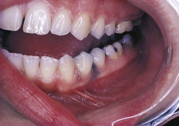
Stainless steel and chrome crowns appear radiopaque on a dental image. Because these crowns are prefabricated, their outlines and margins appear very smooth and regular. Often these crowns are not contoured properly to the cervical portion of the tooth and thus do not appear to fit the tooth well (Figure 32-16). Restorations that are not contoured with the shape of the tooth may cause periodontal problems ranging from food impaction to gingival bleeding or possible bone loss. Because stainless steel and chrome crowns are thin, some areas may appear “see-through” on the dental image (Figures 32-17 and 32-18).
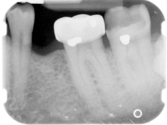
Post and Core Restorations
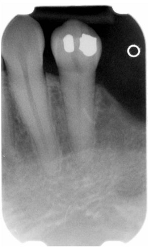
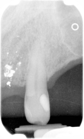
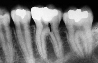
Post and core restorations can be seen in teeth treated with endodontic therapy. The post and core restoration is cast metal and appears as radiodense as amalgam or gold. Post and core restorations appear radiopaque on a dental image. The core portion of the restoration resembles the prepared portion of a tooth crown, and the post portion extends into the pulp canal (Figure 32-19).
Porcelain Restorations
A porcelain restoration appears radiopaque on a dental image. Unlike metallic restorations, which appear totally radiopaque, porcelain restorations are slightly radiopaque and resemble the radiodensity of dentin.
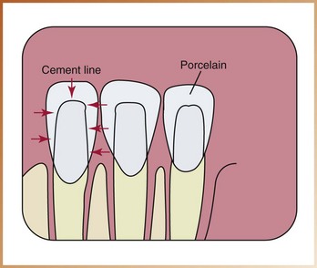
All-Porcelain Crowns
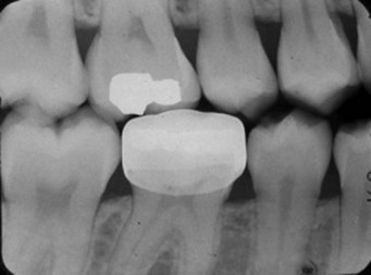
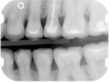
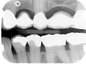
All-porcelain crowns appear slightly radiopaque on a dental image (Figure 32-20). A thin radiopaque line outlining the prepared tooth may be evident through the slightly radiopaque porcelain crown (Figures 32-21 and 32-22). This thin line represents cement. The radiodensity of an all-porcelain bridge appears identical to that of the all-porcelain crown.
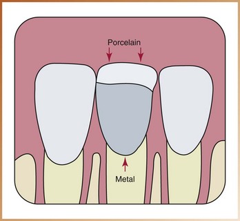
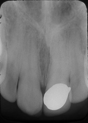
Porcelain-Fused-to-Metal Crown
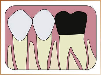
When viewed on a dental image, a porcelain-fused-to-metal crown has two components. The metal component appears completely radiopaque, and the porcelain component appears slightly radiopaque (Figures 32-23 and 32-24). The radiodensities of a porcelain-fused-to-metal bridge appear identical to that seen in the porcelain-fused-to-metal crown (Figure 32-25).
Composite Restorations
When viewed on a dental image, a composite restoration may vary in appearance from radiolucent to slightly radiopaque, depending on the composition of the composite material (Figures 32-26 and 32-27). Some manufacturers of composite materials add radiopaque particles to their products to help the viewer differentiate a composite restoration from dental caries (which appears radiolucent) on a dental image.
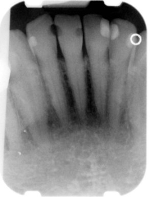
FIGURE 32-27 Composite restorations seen on mandibular anterior teeth. Note the radiolucent and radiopaque appearance of these restorations.
Should there be any confusion between a composite restoration and dental caries, a careful visual and digital examination of the tooth in question enables the clinician to distinguish between the two.
Identification of Materials used in Dentistry
A number of materials are used in dentistry for a variety of reasons, each specific to the specialty that requires the material. Practitioners in restorative dentistry, endodontics, prosthodontics, orthodontics, and oral surgery all use materials that can be identified on dental images.
Materials Used in Restorative Dentistry
Base materials, which include zinc phosphate cement and zinc oxide–eugenol paste, are used as cavity liners to protect the pulp of the tooth. Base materials are placed on the floor of a cavity preparation. A restorative material, such as amalgam, is then placed over the base material. A base material appears radiopaque. Compared with amalgam, the base material appears less radiodense (Figure 32-28).
Metallic Pins
Metallic pins, used to enhance the retention of amalgam or composite, appear as cylindrical or screw-shaped radiopacities on a dental image (Figures 32-29 and 32-30).
Materials Used in Endodontics
Gutta percha is a claylike material used in endodontic therapy to fill the canals of the pulp. Gutta percha appears radiopaque, similar in density to that of base materials (Figure 32-31). When compared with metallic restorations, gutta percha appears less radiodense.
Silver Points
Silver points are also used in endodontic therapy to fill the canals of the pulp. Silver points appear highly radiopaque, similar to other metallic materials. Silver points appear more radiodense than does gutta percha (Figure 32-32).
Materials Used in Prosthodontics
Complete and removable partial dentures are common and may be occasionally observed on dental images. The appearances of complete and removable partial dentures vary, depending on the base materials and type of denture teeth used.
Patients should be instructed to remove all complete and partial dentures before dental images are taken. If not removed, complete and partial dentures may obscure important information concerning adjacent teeth and underlying bone.
Complete Dentures
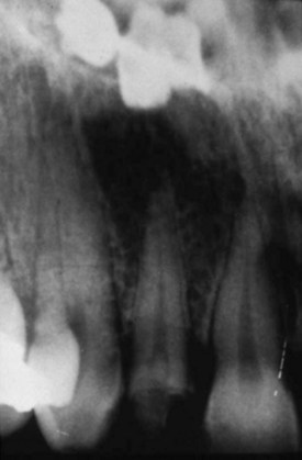
A complete denture consists of two component parts: (1) a base material and (2) denture teeth. The typical denture base material is composed of acrylic and appears as a very faint radiopacity on a dental image or, in some cases, may not be seen at all. Denture teeth may be composed of porcelain or acrylic and vary in appearance. Porcelain denture teeth appear radiopaque and resemble the radiodensity of dentin. Anterior porcelain denture teeth include one or two metal retention pins, or diatorics. On a dental image, diatorics appear as tiny, dense radiopacities superimposed over the radiopaque porcelain denture teeth (Figure 32-33). Posterior porcelain denture teeth also appear radiopaque but do not contain diatorics. Acrylic (plastic) denture teeth lack density and appear faintly radiopaque or radiolucent on a dental image.
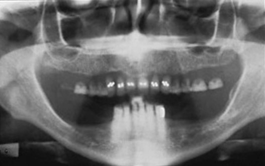
A complete denture that is not removed before the exposure of a dental image gives the illusion of rootless, or “floating,” teeth (Figure 32-34).
Removable Partial Dentures
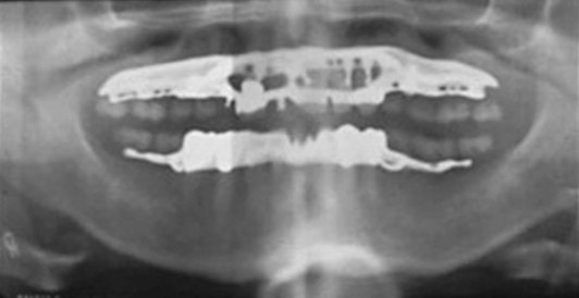
A removable partial denture can be constructed from a variety of base materials, including cast metal, a combination of cast metal and acrylic, and all acrylic. The removable partial denture constructed of cast metal appears densely radiopaque on a dental image. The size and shape of the radiopacity depend on the design of the metal framework of the partial denture. A removable partial denture constructed of a metal base with acrylic saddles appears densely radiopaque where metal is present and slightly radiopaque in the areas of acrylic. A removable partial denture base constructed totally of acrylic is usually seen with wrought-metal clasps. The acrylic base appears slightly radiopaque or radiolucent on a dental image. The metal clasps appear radiopaque and are seen resting on abutment teeth.
Teeth in a removable partial denture may be composed of acrylic or porcelain. Porcelain teeth appear radiopaque and resemble the radiodensity of dentin. Acrylic teeth appear faintly radiopaque or radiolucent (Figure 32-35).
Materials Used in Orthodontics
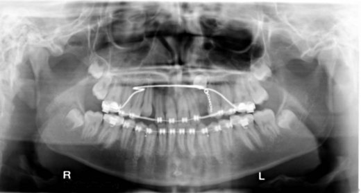
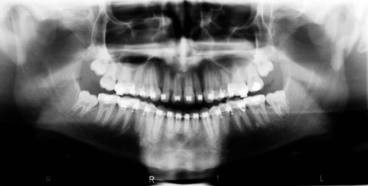
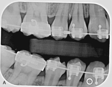
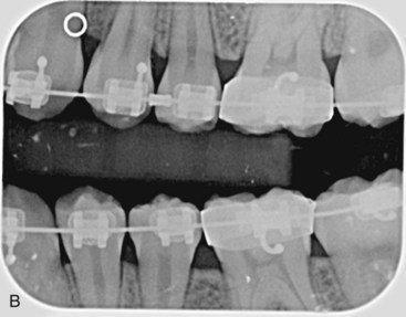
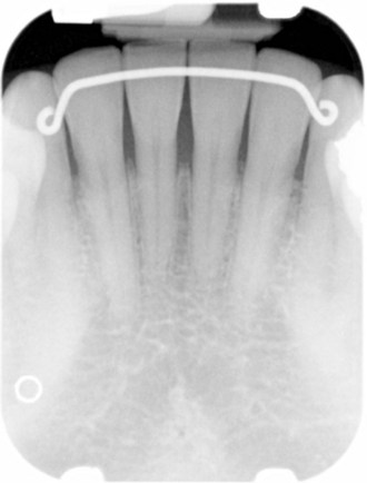
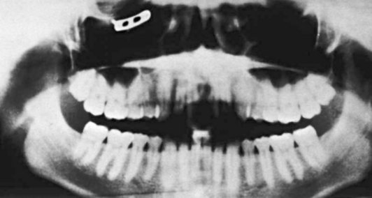
Orthodontic bands, brackets, and wires may be observed on dental images. Each of these orthodontic materials has a characteristic radiopaque appearance (Figures 32-36, 32-37 and 32-38). Fixed orthodontic retainers may also be observed on dental images and have an equally characteristic appearance (Figure 32-39).
Materials Used in Oral Surgery
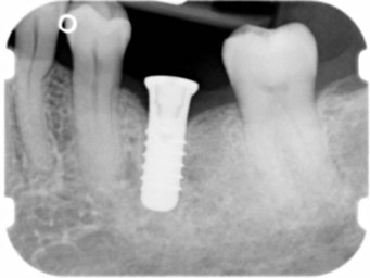
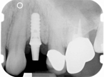
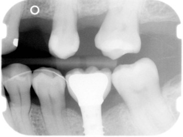
Implants are being used in oral surgery with increased frequency. The appearances of the numerous endosteal implants that are currently used vary, depending on their shapes and designs. The endosteal implant is made of a metallic material and appears radiopaque on a dental image (Figures 32-40, 32-41, and 32-42).
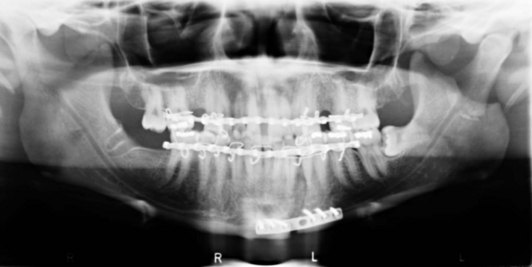
Suture wires, metallic splints and plates, bone screws, and stabilizing arches are used in oral surgery to stabilize fractures of the maxilla and the mandible. Suture wires appear as thin radiopaque lines. Metallic splints, plates, fixation screws, and stabilizing arches also appear radiopaque; their characteristic shapes and sizes may vary (Figures 32-43 and 32-44).
Identification of Miscellaneous Objects
A number of miscellaneous objects, including jewelry, eyeglasses, and napkin chains, can be viewed on dental images. Such objects may obscure important diagnostic information. Some miscellaneous objects may be seen on intraoral images; others may be noted on extraoral images.
To avoid nondiagnostic images, patients should be instructed to remove earrings, necklaces, nose jewelry, eyeglasses, and napkin chains before exposure of extraoral images. In the case of intraoral images, patients should be instructed to remove eyeglasses and nose jewelry, if necessary.
Jewelry
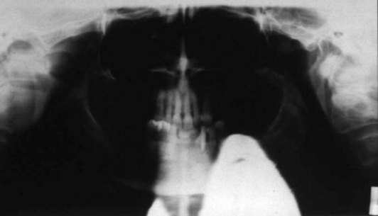
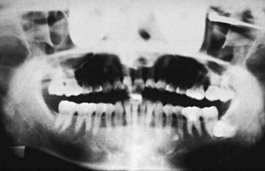
Earrings most often appear on extraoral images. Metal earrings appear as dense radiopacities on a dental image; the size and shape of the radiopacity correspond to the size and shape of the earring (Figures 32-45 and 32-46). Plastic earrings with metallic posts and backings or metal clips can also be seen on dental images; the metallic portions appear as dense radiopacities. A radiodense object, such as a metal earring, causes an artifact, known as a ghost image, on panoramic images (see Chapter 22). Ghost images can obscure important information about teeth and bones and render the image nondiagnostic (Figure 32-47).
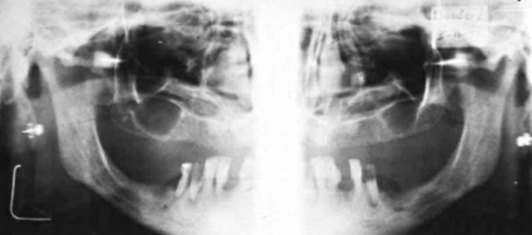
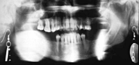
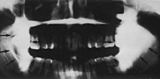
FIGURE 32-47 Note the loss of diagnostic information that results from the ghost images of the large metallic earrings.
A necklace may also appear on an extraoral image as a radiopacity that corresponds in shape and size to the jewelry (Figure 32-48). Nose jewelry (small studs or hoop earrings worn on the nose) may be seen on extraoral as well as intraoral images (e.g., maxillary anterior periapical images). This type of jewelry appears as a radiopacity on a dental image and corresponds in size and shape to the object it represents (Figure 32-49).
Eyeglasses and Napkin Chain
Eyeglasses may be seen on extraoral and intraoral images. Most eyeglasses have some metal components in their frames. The metal portion of the frames appears as a radiopacity on a dental image (Figure 32-50).
The napkin chain, similar to a necklace, may be seen on an extraoral image. If the napkin chain is in the path of the x-ray beam, a radiopacity resembling the napkin chain will be seen on the image.
Summary
• Dental restorations, dental materials, and foreign objects can be seen and evaluated on extraoral and intraoral images.
• The appearances of restorations, materials, and foreign objects vary, depending on material composition, density, and thickness. The appearances of restorations and materials can range from completely radiopaque to totally radiolucent.
• Some restorations and objects are easily identified on dental images; others may require additional clinical information. Dental images play an important role in the evaluation of dental restorations, materials, and objects.
• It is important that the dental professional interpret dental images with the patient present. Without the patient present, important clinical information is not available.
• With the patient present, if a question arises about what is seen on a dental image, a clinical examination can provide additional information or verify what is seen.
Frommer, HH, Savage-Stabulas, JJ, Film mounting and radiographic anatomy. Radiology for the dental professional, ed 9, St. Louis, Mosby, 2011.
Langlais, RP. Exercises in oral radiology and interpretation, ed 4. St. Louis: Saunders; 2004.
White, SC, Pharoah, MJ, Normal radiographic anatomy. Oral radiology: principles and interpretation, ed 6, St. Louis, Mosby, 2009.
Identification
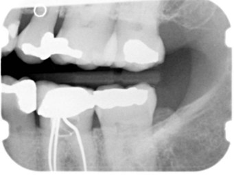
___ 1. Identify in the image the restorative material used in the pulp canal of the maxillary first molar (Figure 32-51).
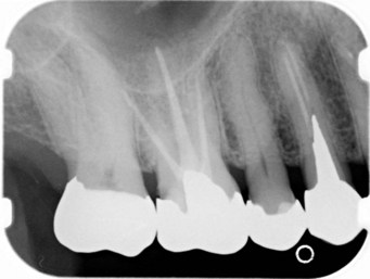
FIGURE 32-51
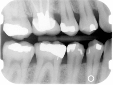
___ 2. Identify the restorative material seen in each tooth of this dental image (Figure 32-52).
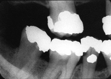
FIGURE 32-52
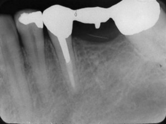
___ 3. Identify the restorative material used to fabricate this bridge (Figure 32-53).
FIGURE 32-53
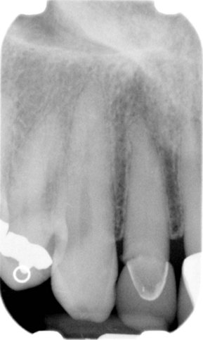
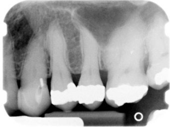
___ 4. Identify in the image the restorative material used in the maxillary anterior region (Figure 32-54).
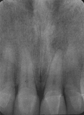
FIGURE 32-54
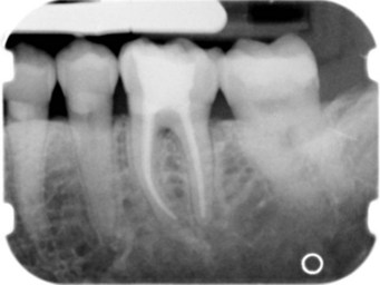
___ 5. Identify in the image the restoration present in the area of the mandibular first molar (Figure 32-55).
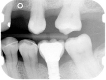
FIGURE 32-55
___ 6. Identify in the image the large radiopacity seen in the posterior mandible (Figure 32-56).
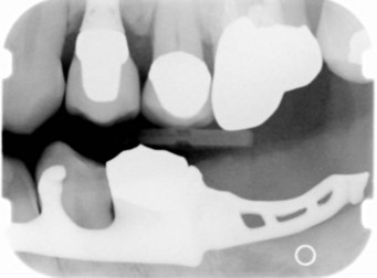
FIGURE 32-56
___ 7. Identify the radiopacity, which is seen in the middle of this panoramic image, obscuring the border of the mandible (Figure 32-57).
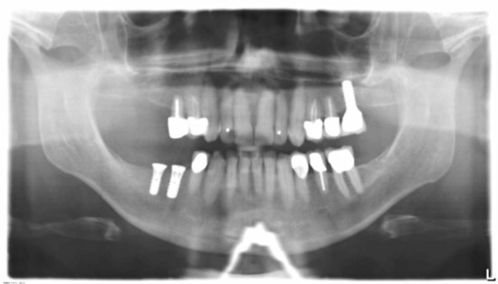
FIGURE 32-57
___ 8. Explain why the maxillary teeth in this dental image appear to be “floating” (Figure 32-58).

FIGURE 32-58
Multiple Choice
___ 9. Rank the following restorative materials from most radiopaque (1) to least radiopaque or radiolucent (4).
___ 10. Which of the following appear equally radiopaque on a dental image?
___ 11. Which of the following is most radiopaque?
___ 12. Which of the following is least radiopaque?
___ 13. Which of the following describes how gold can be distinguished from amalgam on a dental image?