Chapter 102The Bucked-Shin Complex
 Etiology, Pathogenesis, and Conservative Management
Conditions of fatigue failure of bone and inadequacy of bone modeling and remodeling of the third metacarpal bone (McIII) in the racehorse are part of a condition known as bucked shins or dorsal metacarpal disease.1,2 Bucked shins start in young healthy racehorses, usually Thoroughbreds (TBs) and Quarter Horses, but occasionally Standardbreds (STBs), that undergo intense training for racing, usually as 2-year-olds, while the skeleton is still immature and in the growth phase (Figure 102-1, A). The true incidence of bucked shins is unknown and may vary geographically, but reports range from 30% to 90%. A North American questionnaire cited an incidence of 70%.1 Stress fractures (dorsal cortical or saucer fractures) usually occur some months after initial signs of bucked shins and may be a potentially life-threatening injury if a horse is raced or exercised at speed (see Figure 102-1, B). The diagnosis of bucked shins is easy and often made by the trainer or owner. The history of sudden tenderness or soreness of the left McIII (in North America) or both the McIIIs after high-speed work or the first race, or soreness developing the day after, are cardinal signs of early bucked shins. Horses with severe disease manifest acute lameness and extreme sensitivity to palpation of the dorsal cortex of the McIII and are unwilling to train or race. All gradations of pain or disability may be seen. Swelling and tenderness may suggest new bone proliferation. Radiology is helpful to determine the amount of periosteal new bone formation, which determines prognosis. Large accumulations of periosteal new bone on the dorsal or dorsomedial surface of the McIII suggest a serious imbalance between exercise and bone fatigue and may portend actual stress fractures that will be seen on the dorsolateral aspect of the McIII some months later.
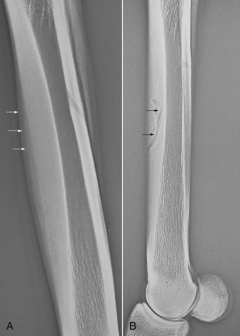
Fig. 102-1 A, Dorsolateral-palmaromedial oblique radiographic image of a third metacarpal bone (McIII). Periosteal new bone formation (arrows) over the dorsomedial aspect of the McIII is evidence of bucked shins. B, Lateromedial radiographic image of the metacarpal region. This dorsal cortical fracture of the dorsolateral aspect of the McIII represents a common type of fatigue (stress) fracture (arrows) that usually occurs months after an episode of bucked shins.
Research Findings
An understanding of the etiology, pathomechanics, and pathogenesis of bucked shins in the TB is helpful in determining prevention and/or treatment modalities and training regimens. It was formerly suggested that bucked shins resulted from microfractures on the dorsal aspect of the McIII, caused by high-speed exercise. However, microfractures should heal without periosteal callus, which does not fit with the clinical observation of extensive periosteal new bone on the dorsomedial aspect of the McIII. Work in our laboratory led us to propose a different cause of bucked shins3 and an exercise program that significantly reduces the incidence of bucked shins and may help to eliminate catastrophic stress fractures. The following summarizes our investigations.
Geometric Properties of the Third Metacarpal Bone: Comparison of Thoroughbreds and Standardbreds
The McIIIs of TBs and STBs of known age were examined, and comparisons were made between breeds of a particular age group and among the age groups of a particular breed. The second moments of area relate to bending stiffness in dorsopalmar and mediolateral directions and were used to determine the minimum and maximum principal moments of inertia (Imin and Imax). The most significant changes in the bone occurred at the midsections between the ages of 1 and 2 years, but continued change occurred until age 3 or 4 years. Imin was smaller in the yearling TBs but larger in the adult TBs compared with STBs. During the first 2 to 4 years of life, Imin changed to a greater extent in TBs.4
In Vitro Comparison of Local Fatigue Failure of the Third Metacarpal Bone
Dumbbell-shaped specimens machined from adult McIIIs from TBs and STBs were tested in fully reversed cyclic bending experiments using a constant-strain rotating cantilever model that measured load decrement. All tests were performed at 40 Hz and continued until the specimen broke or had a 30% loss of stiffness. Three different offsets were used to establish nominal strains of 7500, 6000, or 4500 microstrain in the specimens.
Data were analyzed using a power regression model for each horse and for each breed. Statistical differences were not found among the curves for individual horses of the same breed or for the curves between breeds. Pooled data then were used to describe fatigue characteristics of cortical specimens of the McIIIs from TBs and STBs of various ages, subjected fully to reversed cyclic loading (Figure 102-2).5 The bone from young horses was much more susceptible to fatigue failure.
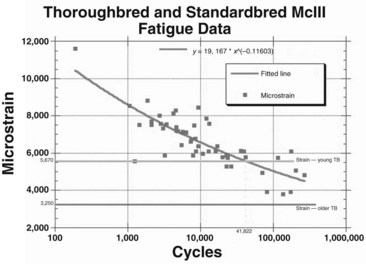
Fig. 102-2 In vitro fatigue data are plotted for the adult Thoroughbred (TB) and Standardbred. The regression line shows where the third metacarpal bone (McIII) will fail from repeated cycles. Superimposition of the strain levels of young TBs shows that 41,822 cycles would result in fatigue failure. The superimposition of an older TB shows that more than a million cycles would be needed before bone failure.
Because the in vitro fatigue was similar in TBs and STBs, other factors appeared to be important in the pathogenesis of fatigue failure of bone in the TB. This, together with the different inertial properties noted in STBs and TBs, led to the hypothesis that differences in training regimens or racing speeds between breeds might influence the incidence of disease.
Third Metacarpal Bone Stiffness Measurements
Whole bone stiffness measurements were made from horses of 2 months to 28 years of age using an Instron testing machine (Instron, Canton, Massachusetts, United States) and a nondestructive three-point dorsopalmar bending test. The bones showed general increases in stiffness until they reached a plateau at about 6 years of age.
The material included the McIIIs from 12 2-year-old TBs, three of which had bucked shins. These three horses had differences in stiffness between the left and right McIIIs of 16% to 27%, respectively, whereas other trained or control 2-year-olds had considerably smaller left-right differences.6
In Vivo Strain Measurements: Relationship to Exercise
Bone strain in the McIII was measured in horses of varying ages, training at or near racing speed, by placing a rosette strain gauge on the dorsolateral aspect of the McIII and recording using telemetry.7 The mean peak compressive strain in four horses 2 years of age was −4841 ± 572 microstrain, compared with −3317 in a horse 12 years of age. One 2-year-old developed bucked shins, and its strain measurements were about 6 standard deviations greater than in the other three.
After acquiring in vivo strain data, we correlated these data with in vitro fatigue data previously generated by determining the average number of cycles that a young TB would gallop in training before the onset of bucked shins. The training records of six 2-year-old TBs that developed bucked shins were analyzed to determine the total distance worked before the onset of bucked shins. Stride length at canter, gallop, and racing speed was measured in a group of TBs to determine the number of strides (cycles) per mile. The total number of gait cycles was estimated based on the distances covered at a canter, at a gallop, and at work. The six horses were trained in these gaits for 10,000 to 12,000 cycles per month and developed bucked shins at 35,284 to 53,299 training cycles. These data were compared with the in vitro data described previously and showed good correlation (see Figure 102-2).
Changing from the trot to the gallop changed the principal strain direction by more than 40 degrees on the dorsolateral surface of the McIII. Although trotting horses showed tensile strains in the long axis of the bone on the dorsal or dorsolateral surface, at racing speeds this same surface of the bone showed compressive strains.8
Relationship of Exercise to Bone Fatigue
With the understanding that slow-speed gaits produced tensile strains on the dorsal surface of the McIII, whereas high-speed exercise induced compressive strains, a study was undertaken to determine the effects of different training regimens and track surfaces on the modeling and remodeling of the McIII in TBs.
Eight untrained 2-year-old TBs were divided into four groups of two horses each. Classical training methods were used for the horses in groups I and II. Group I horses trained on a dirt track. Group II horses trained on a wood chip track. Group III horses (control group) were not trained, but they were allowed free exercise in a large pasture. Group IV horses were trained using a modified classical training program on a dirt track.
The classical training program consisted of daily gallops (approximately 18-second furlongs or 11.2 m/sec) of 1 to 2 miles per day (1.6 to 3.2 km), followed by shorter workouts or breezes at racing speed (approximately 14-second furlongs or 14.4 m/sec) once every 7 to 10 days that increased in distance from 2 to 6 furlongs (0.4 to 1.2 km) progressively over the course of the study. The modified classical training method used similar daily gallops, but the frequency of the high-speed workouts increased to three per week, and distances increased progressively from 1 to 4 furlongs (0.2 to 0.8 km). After 5 months the McIIIs were harvested from all horses. Microradiographs of bone sections were made to determine the extent of the remodeling activity (Figure 102-3). Bone modeling on the periosteal and endosteal surfaces of the McIII changed the cross-sectional geometry differently among the experimental groups. Classically trained horses (groups I and II) responded with appositional new bone formation on the dorsomedial periosteal surface, giving the impression that the medullary cavity, although reduced in diameter, was displaced laterally. Horses in the modified training group (group IV) had bone deposition dorsally and a slightly larger medullary cavity that was not displaced laterally. Control horses (group III) had new bone formation on the medial, lateral, and dorsal surfaces. The medullary cavity remained large and centrally placed. Examination of the McIII inertial properties showed that Imin in groups I, II, and III was similar, but the Imin of group IV horses was greater and was similar to the Imin previously reported for mature racehorses.
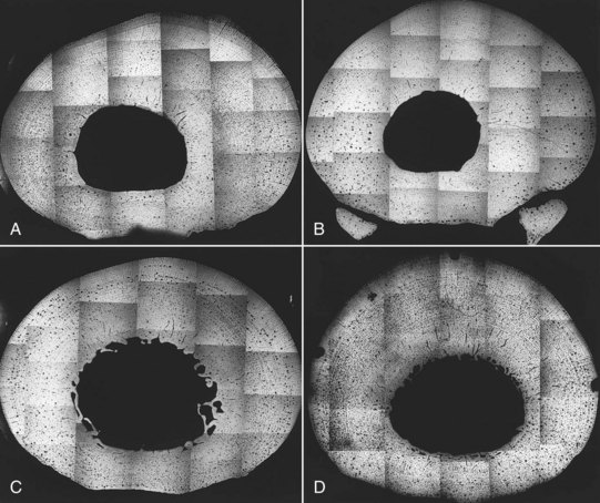
Fig. 102-3 Fifty-percent length cross-sections of the third metacarpal bone were used to make these microradiographs. Individual photographs were taken with cross-sections magnified four times, and giant montages (approximately 50 × 70 cm) were constructed to be able to evaluate individual haversian systems of each individual bone of each horse. Lateral is to the left. A, Group I horses: classically trained on a dirt track. B, Group II horses: classically trained on a wood chip track. C, Group III horses: controls. D, Group IV horses: modified training on a dirt track. Changes in modeling, remodeling, and shape can be seen easily by comparing different groups. Group II horses appear to be earlier in the remodeling cycle than are group I horses, which already have remodeled in the medial and lateral cortices. All specimens are from the left forelimb, and increased new bone formation is seen on the medial surface of the classically trained horses (groups I and II). Modified training (group IV) and the control horses (group III) do not show this change. The lack of haversian remodeling in the dorsal-dorsolateral cortex of the classically trained horses in groups I and II is notable. In this area catastrophic stress fractures occur in racehorses.
Microradiographic sections from the middiaphysis of the McIII showed that bone remodeling occurred only medially and laterally in groups I and II. Filling of secondary haversian systems with new bone was most complete in group I specimens, indicating that the remodeling process was advanced further in horses exercised on a harder surface. A distinct lack of remodeling activity was apparent in the dorsal and dorsolateral regions in groups I and II. Horses in groups III and IV showed extensive remodeling throughout the cortex, including remodeling in the dorsal and dorsolateral aspects.
These data suggested that in horses training on a hard surface, bone remodeling occurred at a faster rate than in horses training on a compliant (wood chip) surface. Previous studies showed that classically trained horses on a hard surface have a higher incidence of bucked shins than do horses trained on a more compliant surface. One horse in group I developed bucked shins during the training period. Inertial properties (Imin) of the McIII in the dorsopalmar direction were no different in horses trained on a hard or soft track. Horses in group IV had a significant change in Imin of the dorsal cortex of the McIII, similar to that seen in adult racehorses evaluated previously that were no longer at risk for developing bucked shins.3 This supported the concept that exercise could be designed to optimize the shape and modeling or remodeling status of the McIII and thus reduce the incidence of bucked shins.9
Exercise Programs Designed to Decrease the Incidence of Fatigue Failure
To determine the efficacy of an adapted training program in decreasing the incidence of bucked shins, a prospective and retrospective study was performed using work training data from five commercial training stables. Two of the stables (2 and 5) were already using our modified classical training program, and the others (1, 3, and 4) were training in a classical manner. Horses in stables 1, 2, and 4 were trained on a commercial racetrack, whereas those in stables 3 and 5 were trained on private farm training tracks. All horses in the study were TBs, 2 years of age, and starting training for the first time and were followed for 1 year. Data collection stopped if horses developed bucked shins, were sold, or stopped training because of another event not related to bucked shins. The study included 11 years of training data from 226 2-year-olds. Fifty-six of the 226 horses developed bucked shins, and 170 horses completed the observation period or were sold.
Regression analysis and survival analysis techniques were used to explore the data. Horses in stable 2 had the best survival, and those in stables 1 and 4 the worst, and evaluation of data suggested that relationships between galloping and breezing were important. Horses in stable 2 had the highest breezing rate and the lowest incidence of bucked shins, whereas those in stables 1 and 4 had the highest galloping rates and the highest incidence of bucked shins.
Galloping increased the likelihood of bucked shins by 36.4%, whereas breezing short distances was protective, reducing the likelihood of bucked shins by 98.6%.10 It is important to note that long-distance breeze rates are detrimental.11 The winter of 1994 brought severe ice storms to the northeast. Horses in stable 2 could not be trained using the modified program and instead were trained using the standard classical program. An unintentional crossover design was created, and 62% of the horses trained that year developed bucked shins. When 1994 data are not used, only 9.3% of horses developed bucked shins.
In Vitro Bone Testing
In vitro compression testing of cylindrical specimens obtained from the McIIIs of 16 horses (139 specimens) in four different training regimens that included no training were tested to failure to determine differences in modulus and strength. These specimens included three horses that developed bucked shins. Because these in vitro studies showed no statistical differences in the material properties of bones with different training regimens, a change in material properties of bone is not a factor in the etiology or pathogenesis of bucked shins. From this study and previous ones carried out in our laboratory, it can be hypothesized that early in a TB’s racing career the McIII experiences extreme strain conditions. These strains cause bone apposition on the dorsal periosteal surface of the McIII in an attempt to lessen the strains. This causes a change in the geometric properties (section properties) but not the material properties of the existing bone.
Discussion
Two decades of evolutionary experiments and clinical observations based on an initial observation of a significant difference in the incidence of fatigue fractures between TB and STB racehorses have led to a natural model for fatigue failure of bone.11,12 We can now compare in vitro and in vivo fatigue behavior and observe bone adaptation with different exercise regimens. Adaptive exercise was shown to change the geometric properties of the McIII without changing the material properties, to influence bone modeling and remodeling, and to reduce the incidence of bucked shins and catastrophic fatigue failure of the McIII in the TB.13
Comparisons of TBs and STBs show major changes in inertial properties of the McIII resulting from growth and superimposed training. Comparisons of young TBs that are susceptible to fatigue failure, with older, resistant horses suggest that changes in bone inertial properties are an important factor affecting the incidence of bucked shins. Large McIII Imin values reflect probable increases in the McIII stiffness in the dorsopalmar direction and thus reduced peak strain during high-speed exercise. The inertial properties of the proximal aspect of the tibia were shown to predict development of fatigue fractures in military recruits,14,15 just as the inertial property measurement of the McIIIs of 5-year-old TBs shows that these horses are no longer at risk for bucked shins.4
In vivo strain measurements of the McIII demonstrated higher peak strains under physiological loading than ever had been reported previously in any species. Although in vitro test conditions differed from the in vivo loading, both involved significant bending components that can be expected to produce accumulated fatigue failure in bone. Superimposition of the in vivo strains reported for the young and older horses at racing speed produced a striking predictive relationship for risk of developing bucked shins (see Figure 102-2).
Large surface strains, measured in vivo at high speeds on the dorsolateral aspect of the McIII in young TBs in training, contrast dramatically with the smaller strains measured in adults that have raced successfully. Strains, under a given load regimen, measured on the surface of bone relate to the modulus and inertial properties (section modulus) of the bone. Because inertial properties were shown to increase with age, and bone strains during high-speed exercise were shown to decrease in older horses, the hypothesis was that changes in bone inertial properties and modulus serve to lower the peak bone strains as a young racehorse matures. However, training regimens possibly can outpace adaptive response. In fact, we observed that a certain percentage of young horses actually increased the McIII surface strains after several months of training. Whole bone stiffness measurements showed right-to-left differences of up to 27% in horses in training, whereas no right-to-left difference was found in the nontrained control horses. Because bucked shins occur bilaterally but sequentially in TBs, usually on the left side before the right, the developmental stiffness changes in limbs possibly are not synchronized but may respond to the predominance of the left lead used by the horse in its racing gait as the horse works in a counterclockwise direction. Maximal strains at exercise and bone stiffness parameters probably change with time and may be declining on one side, while increasing on the other. Increasing bone strain measured at high speed during training, as seen in four of seven TBs, suggested rapid bone stiffness changes in vivo from exercise. Because we now believe that material properties of the bone do not change dramatically during this phase of training, this means that the horses must be providing larger forces on the bone (running faster or applying increased muscular input into the bones).
If Wolff’s law is applied strictly, it follows that a bone that adapts to a particular peak tensile strain may not be prepared adequately to resist far larger peak compressive strains in the same location. Hence training adapts bone to training, and training that mimics racing adapts bone to racing.
An in vitro fatigue study of equine McIIIs showed a difference in fatigue resistance to bending loads in different anatomical quadrants. Bone that was loaded in bending around the physiological bending axis had greater fatigue resistance than bone bent at 90 degrees to this axis.16
We hypothesized that to adapt adequately for racing, the McIII should be exposed during training to strains of the actual magnitude and direction experienced during racing. Furthermore, the high incidence of bucked shins in TBs suggested that loading to produce such peak strains and concomitant adaptive remodeling did not occur in a large number of TBs in classical training programs before racing.
Previous in vivo studies using a functionally isolated rooster ulna, have shown that low numbers of loading cycles (four per day) were adequate to maintain bone mass. Thirty-six cycles were enough to stimulate bone formation.17 The resulting periosteal new bone formation is the same type of bone reaction observed in the TB McIII with fatigue injury.
With these observations taken into account, an exercise (training) regimen was developed that modestly increased the small numbers of high-load cycles using peak load magnitudes and directions that were seen during racing. Increasing the number of short-distance workouts (breezes) from once every 7 to 10 days, as occurs with classical training programs, to three per week produced large changes in modeling, remodeling, and inertial property measurements of the McIII. Classical training produced little progressive change in the inertial properties of the McIII, whereas the new modified training program showed inertial property development that equaled or surpassed that observed in established older successful TBs, those horses apparently no longer susceptible to bucked shins.3
The idea of using exercise to produce adaptive bony remodeling is not new. Woo and colleagues18 showed that 12 months of exercise in young pigs produced dramatic changes in the femur, increasing cross-sectional area by 23% and inertial properties (Imin) by 27%, without intrinsic bone property changes.18 Milgrom and co-workers19 looked for exercise that can adapt bone and showed that playing basketball for 2 years or more was protective in reducing the incidence of bone fatigue failure in military recruits.
In the 11-year longitudinal study, we proved that adaptive exercise could be used to reduce the incidence of bucked shins. The relative incidence of bucked shins in classically trained TBs is probably higher than we found. The mean time taken for horses to develop bucked shins was about 25 days shorter than the mean time to loss for other reasons. Bucked shins developed in most horses by 200 days in training. We would not expect horses in the modified training program to experience additional fatigue fractures after the observation period, because the McIII should resemble more closely that of adult, nonsusceptible racehorses.
Adaptive exercise changed the geometric parameters of a specific bone in a way that would be expected to reduce fatigue damage while significantly reducing the incidence of bucked shins. This correlation, although not explicit proof of the interrelationship between the factors measured, is convincing.
In addition, approximately 10% of the catastrophic breakdown injuries involving the skeleton of TB racehorses are complete fractures of the McIII. These injuries are direct sequelae to fatigue fractures of the dorsolateral aspect of the McIII that are often seen several months after horses develop bucked shins. Theoretically, if bucked shins could be prevented there would be an immediate 10% decrease in the incidence of these catastrophic fractures and thereby a decrease in racing fatalities.
Training to Prevent Bucked Shins
A training program was developed to limit the incidence of bucked shins and can be modified by trainers to suit their situations.12,20
The principles of this training program are the following:
These principles were developed based on experimental studies that showed that at slow gaits the dorsal surface of the McIII is under tension and at high speed is bending to produce compression. Structurally, materials are designed differently to support tension or compression. Think how strong a rope is in tension and how useless it is in supporting loads in compression. Short, high-speed exercise exposes the bone to compression on its dorsal surface and introduces the bone to the loads expected during a race. Experimental studies showed that this sort of training regimen changed the cross-section of the McIII to the adult racing shape within 6 months.
Long, low-speed jogging adapts the bone only to jogging, creating tension on the dorsal surface of the McIII. In stable 2 in the icy winter of 1994 the horses were jogged for 30 days, without any high-speed exercise, and then resumed the exercise program. The incidence of bucked shins increased fivefold compared with previous years and returned to normal the following year. Although training is not a cookbook recipe for success, the fundamentals of the program are described here with an actual schedule.12
The schedule assumes that the young horse is broken to ride in the fall and is able to gallop 1 mile (18- to 20-second furlongs) by the end of December. The training program starts in January and can be broken into three stages. The principle involved is that the horse’s bones need to see the strain environment of racing as soon as possible so that bone modeling and remodeling can begin in a timely manner. The training program is 6 days a week with Sundays off. The horses walk to and from the racetrack. On the track the horse walks  mile and jogs
  mile to warm up. The horse then gallops 1 mile.
 -mile open gallop in 15 seconds. This speed work is done on Tuesdays and Saturdays. This speed and distance of the open gallop is repeated 10 times (5 weeks).
 mile in 30 seconds (15-second furlongs). This speed work is repeated 10 times, which takes up the next 5 weeks. All open gallops in stage 1 and stage 2 are at the end of the gallop and are included in the 1-mile gallop.
 mile in approximately 26 seconds (13-second furlongs). This is repeated four times (4 weeks). In stage 3 the daily gallops are extended to
  miles twice a week. After the fourth week the
  -mile breeze is continued with a strong “gallop out” for an additional furlong. This makes the 3-furlong total about 40 seconds. This is done for an additional 3 weeks, giving stage 3 a total time of 7 weeks.
After stage 3, the McIII shape and architecture are effectively established for the longer high-speed workouts necessary for racing. The horse can now go on to 4- to 6-furlong workouts as needed to further develop other body systems to complete fitness for racing. The total time of this initial training program is 119 to 147 days, depending on the availability of a race for the horse. This does not include any downtime for sickness or injury. Gate work is started early and often in this program. The horses are introduced to the gate in January as the program starts. All young horses are turned out in a small paddock for 1 to  hours of exercise before daily training. This training program has shown no increase in the injury rate of young horses. An excellent byproduct of this training program is the mental development of these 2-year-olds. Because of the very relaxed atmosphere of walking to and from the racetrack, these horses exhibit no anxiety about work. For this training program to work, the rider cannot be in a hurry to get back to the barn and on the next horse. The 2-year-olds are not anxious about speed work because it has been in the weekly schedule since the beginning of training. All the horses walk back to the barn. Walking is a great exercise that does not seem to negatively influence bone modeling or remodeling. The schedule described here for training to negate bucked shins is just that—a schedule. An understanding of the principles behind this training regimen is important. Training programs that take advantage of this information can be developed that will meet an individual horse’s needs.
Horses that develop respiratory disease during training and are off for 10 days or more are backed up about 10 days in the training program. Shin sore horses are treated for soreness with phenylbutazone and ice water and are walked until soreness is gone. These horses then get put in an abbreviated program in which the breezing distances and galloping distances are cut in half. These horses are usually coming from a classical training program, in which bucked shins is a common occurrence. When presented with these horses (often from a 2-year-old in training sale), one is far better off putting them into this bone-conditioning program initially rather than going on with them and hoping that they do not develop bucked shins, as they invariably do. These horses take far longer to get to the first race. Sore horses should be individually evaluated based on clinical and radiological signs. In most horses that develop early shin soreness the training program can be modified and the horse can continue training. A short rest period with handwalking, the administration of phenylbutazone, and the application of ice water can get most horses back into training rather quickly.
Although bucked shins are commonly accepted by veterinarians, trainers, and owners as a normal training event in young TB racehorses, with estimated losses to the industry of $10,000,000 per year in lost training and racing days, they are far more important than that! Horses with bucked shins are at increased risk to develop dorsal cortical fractures of the McIII and then may have catastrophic failure of the McIII during racing. Horses without bucked shins are at very low risk to develop these fractures.
Horses with bucked shins can be trained using this program, provided that the horse is sound and periosteal new bone formation is not substantial. The secret is to decrease the horse’s galloping distance (cut it in half) and add short high-speed workouts. For example, if a horse usually gallops  miles/day, reduce the distance to
  mile/day and introduce short workouts twice weekly. We usually back the horse up about 1 month in its exercise program and get the high-speed workouts started over short distances. If horses are sore or lame, training is stopped and cold therapy and administration of nonsteroidal antiinflammatory drugs (NSAIDs) are begun. Once sound, horses are returned to training. In some horses NSAIDs may need to be administered for chronic soreness, but horses with frank lameness should be allowed to rest until sound.
If a horse’s training schedule is interrupted by an unrelated lameness or upper respiratory tract disease, remodeling that starts with bone resorption is sudden. When the primary problem resolves, if horses are returned to training at a level similar to that before rest but the McIII porosity is increased, the horse is at risk for bucked shins. Therefore if a horse is out of the exercise program for 10 days or more, the horse should be backed up in its training schedule to minimize the risk of bucked shins when it restarts training.
With proper training the incidence of bucked shins can be reduced dramatically (almost eliminated). This can be accomplished without increasing the training time or risking added injury. The added benefit of this program is the concurrent absence of dorsal cortical fractures of the McIII in horses without previous bucked shins.
Stress Fractures of the Third Metacarpal Bone: Surgical Management
Most stress fractures of the McIII occur on the dorsolateral aspect of the left forelimb but may be bilateral (Figure 102-4). Treatment options include controlled exercise (see page 958), screw placement in the dorsal cortex, and cortical drilling around the fracture line. Recently extracorporeal shock wave therapy has been used with apparent early success (Editors, see Chapter 96). The placement of a screw does not result in interfragmentary compression, but it may have a local effect on bone modeling and remodeling. Cortical drilling is thought to improve vascularization and new bone formation at the fracture line.1 Advantages of cortical drilling alone are that a second surgery is not necessary to remove the screw, and in some horses this procedure can be performed with the horse in a standing position, obviating the need for general anesthesia. However, drill breakage in a standing horse is a risk and, if the bit piece is not retrieved, may lead to chronic lameness. Clinical reports indicate that the use of cortical drilling and screw placement has a lower rate of fracture recurrence than cortical drilling alone. Success rates were 97% and 85%, respectively.2,3 I use the combination technique.
Fig. 102-4 Lateromedial radiographic image of a third metacarpal bone (McIII) of a 4-year-old Thoroughbred. Dorsal is to the left. Typical dorsal cortical stress fracture of the third metacarpal bone, cortical thickening of the McIII, and numerous oblique fracture lines are present. This horse is a candidate for drilling or screw placement.
Horses with radiological evidence of substantial endosteal or periosteal healing often heal satisfactorily without surgical intervention. Horses with severe fractures that are in multiple locations, or spiral around the bone, have a risk of catastrophic failure on recovery from general anesthesia and usually are treated conservatively (Figure 102-5). To my knowledge no objective reports of conservative management of dorsal cortical stress fractures exist. In my experience conservative management is less reliable than surgical treatment and does not result in a reduction in training days missed. In addition, I have experienced a higher rate of recurrent fracture, or failure of fracture to heal, with conservative compared with surgical management.
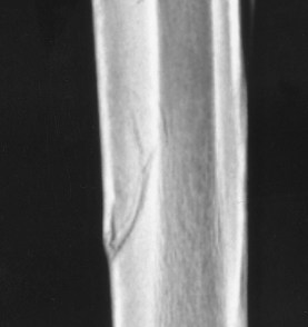
Fig. 102-5 Lateromedial radiographic image of the third metacarpal bone (McIII) of a 2-year-old Thoroughbred. Dorsal is to the left. There is a severe, dorsal cortical fracture of the McIII. Conservative management in horses with severe fractures is recommended. This fracture is saucer-shaped in appearance and could propagate during recovery from general anesthesia.
Surgical Procedure
The horse is positioned in lateral recumbency under general anesthesia with the affected limb uppermost. Both limbs can be operated on from this position. The periosteal new bone usually can be felt through the skin, and a 6- to 8-cm vertical incision is made over the fracture, usually between the common and lateral digital extensor tendons. Radiographic examination is used if necessary. The periosteum should not be elevated. A sharp 2.5-mm drill bit is used to drill a thread hole for a 3.5-mm screw perpendicular to the fracture line. Screws are placed as positional screws rather than in lag fashion. Only the dorsal cortex is drilled. Overdrilling may result in impact on the palmar cortex and instrument breakage. During or after screw placement, radiological examination is necessary to determine proper screw placement. The screw is placed after countersinking, tapping, and measuring to determine appropriate screw length. Usually a single screw is used, but occasionally several screws are used for a long fracture. Five or six unicortical drill holes approximately 1 cm apart are made using a 2.5-mm drill bit in the region of the fracture, before routine closure and bandaging of the limb.
If dorsal cortical drilling alone is performed with a horse in the standing position, under sedation and perineural analgesia, a 3.2-mm drill bit is used to decrease the risk of drill breakage if the horse moves during the procedure.
Postoperative Treatment
Four weeks of stall rest with handwalking followed by 4 weeks of small paddock turnout, continued handwalking, or swimming are recommended. Screw removal is performed with the horse standing at 8 weeks, based on fracture healing. Return to light jogging can occur 2 weeks after screw removal, but more intense training should not start until at least 16 weeks after surgery and should be based on lack of clinical signs of lameness and radiological signs of healing.
Prognosis
Prognosis is considered good, with 85% to 97% of horses returning to racing.2,3 Recurrence of fracture is more common after cortical drilling alone, compared with the combination of cortical drilling and screw placement. Catastrophic failure has occurred after cortical drilling alone but not with the combined technique.