36 Non-sporing anaerobes
Wound infection; periodontal disease; abscess; normal flora
Key points
• Non-sporing anaerobes are found as part of normal flora in health.
• Most infections with anaerobes are of endogenous origin and are often polymicrobial. They act as opportunistic pathogens at damaged and necrotic tissue sites.
• Production of putrid odour is a common feature of infection.
• Fusobacterium nucleatum is often recovered from head and neck infections.
• Anaerobic Gram-negative rods, especially Bacteroides fragilis and anaerobic Gram-positive cocci, are the most common cause of non-clostridial anaerobic infections.
• Black-pigmented Porphyromonas and Prevotella species occur in abscesses and soft tissue infections in various parts of the body.
• Penicillins and nitroimidazoles, especially metronidazole, are the main agents used for treatment.
The significance of obligate anaerobes in general and of non-sporing anaerobes in particular is now well recognized. This heightened awareness of the important role that such organisms play, both as part of the normal microbial flora of the body and in a wide variety of infections, has come about largely through the application of greatly improved laboratory techniques for the isolation and cultivation of anaerobic bacteria, and the pioneering efforts of ‘anaerobe enthusiasts’ in various parts of the world.
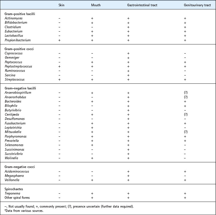
A bewildering range of anaerobes is found in the mouth and oropharynx, gastrointestinal tract and female genital tract of healthy individuals as part of the commensal flora. These include Gram-positive and Gram-negative cocci, rods and filaments, as well as a number of spiral forms (Table 36.1). Most infections with these organisms are of endogenous origin, except in the case of animal and human bite wounds, where the infecting organisms, usually mixed, are derived from the mouth of the aggressor.
The flora of the lower intestinal tract, in particular, harbours vast numbers of anaerobes; quantitative studies on the bacterial flora of human faeces (Table 36.2) reveal a total content of over 1010 anaerobes per gram of faeces.
Table 36.2 The bacterial flora of faeces of English subjects
| Bacterial group | Mean bacterial counta |
|---|---|
| Gram-negative anaerobic rods | 9.8 |
| Bifidobacterium spp. | 9.8 |
| Clostridium spp. | 5.0 |
| Veillonella spp. | 4.2 |
| Lactobacillus spp. | 6.5 |
| Bacillus spp. | 3.7 |
| Enterobacteria | 7.9 |
| Streptococcus spp. | 7.1 |
| Enterococcus spp. | 5.8 |
| Total anaerobes | 10.1 |
| Total aerobes | 8.0 |
a Log10 viable organisms per gram of faeces.
From Hill M J, Drasar B S, Hawksworth G et al 1971 Bacteria and aetiology of cancer of large bowel. Lancet 1: 95–100.
Many of the bacteria isolated from anaerobic infections are opportunist pathogens. Such organisms are particularly likely to set up infections in damaged and necrotic tissue, when they are translocated to sites other than their normal habitat, or in a host that is compromised or debilitated in a way that leads to impairment of immunological or other defence mechanisms. Anaerobic infections of the head, neck and respiratory tract are often associated with organisms found in the mouth, whereas infections in the abdominal and pelvic regions are more commonly associated with gut bacteria.
Features of anaerobic infections
Clinical signs
A common, but not invariable, feature is the production of a foul or putrid odour. Foul-smelling pus or discharge should always alert the clinician to the likelihood that anaerobes are present, as no other organisms produce this effect, but the absence of this sign does not necessarily exclude the involvement of anaerobic bacteria. Other clues to the clinical diagnosis are listed in Box 36.1.
Box 36.1
Some clinical signs and indicators of non-clostridial anaerobic infections
• Presence of foul-smelling pus, discharge or lesion
• Production of a large amount of pus (abscess formation)
• Proximity of lesion to mucosal surface or portal of entry
• Failure to isolate organisms from pus (’sterile’ pus)
• Infection associated with necrotic tissue
• Gas formation in tissues (crepitus)
• Failure to respond to conventional antimicrobial therapy
• Pus that shows red fluorescence under ultraviolet light (Porphyromonas spp.)
• Detection of ‘sulphur granules’ in pus (actinomycosis)
Adapted from Finegold and George (1989).
Polymicrobial flora
Infections involving non-clostridial anaerobes are often polymicrobial. The composition of these mixed infections varies according to the site affected. The complexity may vary from two or three species up to a dozen or more, and may include strict anaerobes, facultatively anaerobic and micro-aerophilic organisms. Such combinations frequently comprise mixtures of Gram-negative rods (such as Bacteroides, Prevotella and Fusobacterium species) and Gram-positive cocci (such as streptococci). In most cases, with the occasional exception of actinomycosis, it is not possible to accurately predict which organisms are present from the clinical presentation, although the detection of red fluorescing pus under ultraviolet light usually indicates the involvement of one of the black-pigmented Porphyromonas species.
Laboratory diagnosis
When anaerobic infection is suspected, it is important that adequate clinical specimens are collected and transported as soon as possible to the bacteriology laboratory, preferably under reducing conditions. After direct microscopical examination of the material, appropriate culture media should be inoculated for incubation in an anaerobic cabinet or in anaerobic jars. As many anaerobes are relatively slow growing, it is essential that cultures are incubated for several days before being discarded. In mixed infections, fast-growing aerobic or facultatively anaerobic organisms are often detected within 24 h, whereas some anaerobes may require incubation for 7–10 days before their colonies can be recognized.
In some laboratories, gas–liquid chromatography is carried out directly on pus and other clinical specimens in order to detect metabolic products, such as butyric and propionic acids, that are characteristic of certain anaerobes. Molecular based techniques are also increasingly used for the rapid identification of anaerobes.
Gram-negative bacilli
Fusobacterium spp
Fusobacteria colonize the mucous membranes of human beings and animals, and are generally regarded as commensals of the upper respiratory and gastrointestinal tracts. They tend to form long filamentous rods, often with pointed ends, sometimes described as fusiform or spindle shaped. Species such as F. nucleatum, F. periodonticum and F. naviforme are generally isolated from the oral cavity and are often associated with infections of this and related sites. F. nucleatum, the most studied species, is frequently recovered from mixed infections of the head and neck region, including dental abscesses and the central nervous system, and is also quite commonly isolated from transtracheal aspirates and pleural fluid. Five subspecies are recognized; animalis, fusiforme, nucleatum, polymorphum and vincentii. F. nucleatum subspecies nucleatum is an important periodontal pathogen, particularly during the period when quiescent periodontitis becomes active.
F. necrophorum is an important animal pathogen. It is associated with human necrobacillosis and occasionally infections similar to those caused by F. nucleatum.
F. mortiferum, F. necrogenes, F. gonidiaformans and F. varium are generally isolated from the gastrointestinal and urogenital tracts of man and animals. These species, together with F. nucleatum, are often associated with mixed intra-abdominal infections, perirectal abscesses, osteomyelitis, decubitus, and other ulcers and various soft tissue infections. F. ulcerans is associated with tropical ulcers but may be found in other sites.
Leptotrichia buccalis
This species shares a number of properties with the fusobacteria. It is normally considered to be an oral species, but also occurs outside the oral cavity. It has been reported in acute necrotizing ulcerative gingivitis (Vincent’s gingivitis), together with Treponema, Porphyromonas and Fusobacterium species. Some isolates described as L. buccalis probably represent separate species within the genus.
Bacteroides, Porphyromonas and Prevotella species
Bacteria once thought of as typical members of the genus Bacteroides, especially those isolated from human beings, form three broad groups according to whether they are asaccharolytic, moderately saccharolytic or strongly saccharolytic (Table 36.3):
1. The asaccharolytic, pigmented species are classified in the genus Porphyromonas, which includes the important periodontal pathogen P. gingivalis.
2. The moderately saccharolytic species that are inhibited by 20% bile and are largely indigenous to the oral cavity are assigned to the genus Prevotella.
3. The genus Bacteroides is now restricted to B. fragilis and related species that are saccharolytic and grow in 20% bile.
Table 36.3 Current taxonomic status of Bacteroides, Porphyromonas and Prevotella species
| Group | Species |
|---|---|
| Saccharolytic (B. fragilis and related species) | B. fragilis, B. caccae, B. eggerthii, B. ovatus, B. stercoris, B. thetaiotaomicron, B. uniformis, B. vulgatus, Parabacteroides distasonis, Para. merdae |
| Moderately saccharolytic (Prevotella spp.) | Prev. melaninogenica, Prev. bivia, Prev. buccae, Prev. buccalis, Prev. corporis, Prev. denticola, Prev. disiens, Prev. enoeca, Prev. heparinolytica, Prev. intermedia, Prev. loescheii, Prev. nigrescens, Prev. pollens, Prev. oralis, Prev. oris, Prev. oulorum, Prev. tannerae, Prev. veroralis, Prev. zoogleoformans, Prev. salivae, Prev. shahii, Prev. multiformis, Prev. marshii, Prev. baroniae |
| Asaccharolytic (Porphyromonas spp.) | P. asaccharolytica, P. catoniae, P. gingivalis, P. endodontalis, P. uenonis |
| Other (uncertain taxonomic status) | B. splanchnicus |
To add to the taxonomic complexity, many other former Bacteroides species that are usually isolated from nonhuman sources have undergone reclassification. B. gracilis and B. ureolyticus now belong to the genus Campylobacter, and the former B. ochraceus now belongs to the genus Capnocytophaga.
Infections with Bacteroides, Porphyromonas and Prevotella species
Bacteroides species and related Gram-negative rods are, together with anaerobic cocci, the most common cause of non-clostridial anaerobic infections in man. Organisms of the B. fragilis group are particularly significant, as they are the most commonly isolated and tend to be more resistant to antimicrobial agents than most anaerobes.
B. fragilis itself is substantially outnumbered by other Bacteroides species in the normal bowel microflora, but is often associated with intra-abdominal and soft tissue infections below the waist. B. fragilis is also the most common anaerobe found in bacteraemia, and has even occasionally been reported from head and neck infections, despite its apparent absence from the normal flora of the mouth. Species of the B. fragilis group account for about a quarter of all anaerobes isolated from clinical specimens.
Black-pigmented species, including those from the genera Porphyromonas and Prevotella, occur in abscesses and soft tissue infections in various parts of the body. They are rarely isolated in pure culture. P. gingivalis is associated with chronic adult periodontitis and P. endodontalis with dental root canal (endodontic) infections.
Gram-positive anaerobic cocci
Gram-positive anaerobic cocci comprise part of the normal microbial flora of the mouth, gastrointestinal tract, genitourinary tract and skin (see Table 36.1). Most are found as part of the flora of the bowel and are not usually considered to be significant in infections.
However, several genera of clinically significant strictly anaerobic Gram-positive cocci, formerly regarded as belonging to the genus Peptostreptococcus, are now recognized (Box 36.2). They are not easy to identify precisely, and are often described simply as ‘anaerobic cocci’.
Box 36.2
Currently recognized species (human) of Gram-positive anaerobic cocci
Infections with anaerobic cocci
Anaerobic cocci are isolated from infections in various parts of the body, particularly from abscesses (Box 36.3). They are often found in association with other anaerobic, facultatively anaerobic or aerobic organisms. As with all mixed infections, it is difficult to assess the contribution of each individual organism to the pathogenic process. However, there is sufficient evidence from both clinical and experimental studies to confirm their pathogenic potential.
Box 36.3
Types of infection and clinical specimens from which anaerobic Gram–positive cocci are isolated
Gram-negative anaerobic cocci
Among genera recorded as part of the normal flora of the gastrointestinal tract (see Table 36.1), only Veillonella is found regularly at other sites. In the mouth, for example, this genus is a regular component of supragingival dental plaque and the tongue microflora. Veillonellae are able to use some of the lactic acid produced by bacteria such as streptococci and lactobacilli that potentially induce dental caries.
The role of Veillonella species and other anaerobic Gram-negative cocci in disease, if any, has not been clearly established, although they may be isolated from a variety of clinical conditions. In general, they are regarded as a minor component of mixed anaerobic infections, and antimicrobial chemotherapy is not generally directed specifically against them.
Non-sporing Gram-positive rods
The spore-forming genus Clostridium is well known for its involvement in serious infections (see Ch. 22). The role of anaerobic non-sporing Gram-positive rods, on the other hand, is less well understood, although they are present in significant numbers in the normal flora of the mouth, skin, gastrointestinal and female genitourinary tracts, and are isolated from a variety of types of infection. The main genera and some of their characteristics are listed in Table 36.4.
Table 36.4 Some characteristics of anaerobic non-sporing Gram-positive rods
| Genus | Common sites | Acid end-products |
|---|---|---|
| Propionibacterium | Skin, mouth, gut, vagina | Propionic acid |
| Bifidobacterium | Gut, mouth, vagina | Acetic and lactic acids |
| Lactobacillus | Mouth, gut, vagina | Lactic acid (major end-product) |
| Actinomyces | Mouth, gut, vagina | Succinic, lactic and acetic acids |
| Eubacteriuma | Mouth, gut, vagina | Butyric and other acids |
a Taxonomy currently undergoing revision; includes several different genera.
Infections with Gram-positive rods
Any of these bacteria can occur as components of mixed anaerobic infections, and Actinomyces species can undoubtedly adopt a pathogenic role. Most cases of actinomycosis are caused by Actinomyces israelii and are cervicofacial, although the disease can also occur in the thorax, abdomen and female genital tract (see Ch. 20). Actinomyces species are not themselves strict anaerobes, but A. israelii requires good anaerobic conditions for primary isolation, and plates should be incubated for 7–10 days.
Propionibacterium propionicum is morphologically and biochemically very similar to A. israelii. It is particularly associated with infection of the tear duct in the condition called lachrymal canaliculitis. The significance of other genera in infections is not clear. Some species are found in acne; they are also isolated occasionally in infective endocarditis and in infections associated with implanted prostheses. Eubacterium species are a large group (possibly mistaken for Actinomyces species in some reports), many of which are being reclassified into new genera, including Slackia and Eggerthella. These bacteria may play a role in infections around intra-uterine devices; others, for example Slackia exigua, may be involved in human periodontal disease. There is only limited evidence for the pathogenicity of Bifidobacterium species, although Bif. dentium has been isolated occasionally from pulmonary infections; bifidobacteria can also be isolated from dental caries lesions by use of appropriate cultural methods.
Spiral-shaped motile organisms
Several Treponema species are found in the mouth and elsewhere in the body (see Table 36.1). They are thought to be an important component of the mixed anaerobic infection associated with acute necrotizing ulcerative gingivitis along with fusobacteria and Prev. intermedia, and may also contribute to other forms of periodontal disease. The proportion of motile spiral organisms seen by dark-ground microscopy in samples from the gingival pocket increases markedly when there is evidence of periodontal destruction.
Motile, spiral-shaped, Gram-negative anaerobes of the genus Anaerobiospirillum have been isolated from patients with diarrhoea and from bacteraemia. Although comparatively rarely isolated from man, they can cause serious infections. The distribution and normal habitat of this and other morphologically similar organisms are not well understood. In some cases the source of infection may be domestic animals and pets.
Treatment
In many infections caused by anaerobes the most important aspect of treatment is surgical. This often involves drainage of pus from abscesses, but may also include debridement, curettage and removal of necrotic tissue. For minor infections surgical drainage alone may be sufficient, but in many cases antimicrobial chemotherapy is also indicated. The main groups of agents used are the penicillins and the nitroimidazoles, particularly metronidazole. Other agents with good anti-anaerobe activity include chloramphenicol, clindamycin and cefoxitin, but resistant strains occur.
Metronidazole is effective against virtually all obligate anaerobes, including Bacteroides, Porphyromonas, Prevotella and Fusobacterium species, but not against facultatively anaerobic or micro-aerophilic bacteria such as actinomyces and streptococci. Resistance to metronidazole is still relatively uncommon.
Most anaerobic species are sensitive to benzylpenicillin, but members of the B. fragilis group are usually resistant. Such resistance is associated with β-lactamase production and these organisms are usually susceptible to combinations of penicillins with β-lactamase inhibitors (e.g. co-amoxiclav) and to carbapenems such as imipenem.
Allaker RP, Young KA, Langlois T, et al. Dental plaque flora of the dog with reference to fastidious and anaerobic bacteria associated with bites. Journal of Veterinary Dentistry. 1997;14:127–130.
Borriello SP, Murray PR, Funke G, eds. Topley and Wilson’s Microbiology and Microbial Infections, Vol. 2. Oxford: Wiley-Blackwell, 2005. Bacteriology
Duerden BI, Drasar BS. Anaerobes in Human Disease. London: Edward Arnold, 1991.
Finegold SM, George WL. Anaerobic Infections in Humans. San Diego: Academic Press, 1989.
Fuller R, Perdigon G. Gut Flora, Nutrition, Immunity, and Health. Oxford: Blackwell Publishing, 2003.
Marsh P, Martin MV. Oral Microbiology. Edinburgh: Churchill Livingstone; 2009.
Wilson M. Bacteriology of Humans. Oxford: Blackwell Publishing; 2008.
