Page 753

Chapter 54 Bleeding in pregnancy

Amanda Hutcherson

Learning Outcomes

After reading this chapter, you will be able to:

identify the causes of vaginal bleeding in pregnancy
discuss the midwife’s role in bleeding in pregnancy both before and after the 24th week
describe the possible implications for the health and wellbeing of the mother and the fetus
discuss therapeutic termination of a pregnancy.

Introduction

Vaginal bleeding during pregnancy is always considered to be abnormal and should always be investigated. It may be extremely frightening for the woman, so must be managed with sensitivity, ensuring that the woman is fully informed and involved in her plan of care. An important part of the management lies within the diagnosis of the cause and in the accurate assessment and reporting of the woman’s previous and present history. It is also important to recognize that medical definitions and terms, such as ‘abortion’, will need to be explained. This term may have a different meaning for women and families.

Bleeding from the genital tract can be divided into two categories, depending on whether it occurs before or after the 24th week of pregnancy (Drife 2002).

Bleeding before the 24th week of pregnancy

Bleeding from the genital tract in early pregnancy – that is, before the 24th week – may be caused by:

implantation bleeding
abortion
hydatidiform mole
ectopic pregnancy
cervical lesions
vaginitis.

Implantation bleeding

There may be a little bleeding when the trophoblast embeds into the endometrial lining of the uterus. The bleeding is usually bright red and of short duration. As implantation takes place 8–12 days after fertilization, the bleeding usually occurs just before the menstrual period is due. If mistakenly thought to be a menstrual period, this may confuse the expected date of delivery. A careful menstrual history is essential to detect probable implantation bleeding, thereby avoiding miscalculation of dates.
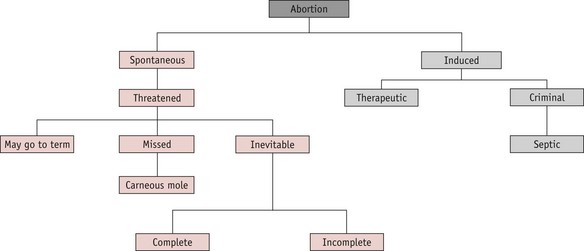
Abortion

A pregnancy that ends before 24 completed weeks of gestation, and where the fetus is not alive, is termed an abortion. The classification is shown in Figure 54.1.

image

Figure 54.1 The classification of abortion.

Page 754

Spontaneous abortion

Approximately 15–20% of confirmed pregnancies end in spontaneous abortion, most of these occurring before the 12th week of pregnancy. Midwives should be aware that the term ‘abortion’ may cause confusion. Many women who have lost a wanted pregnancy find the word offensive and it should not, therefore, be used when talking to women about a pregnancy ending from natural causes. In these circumstances, the use of the word ‘miscarriage’ is more appropriate.

Causes

Maldevelopment of the conceptus: The most common cause of spontaneous abortion is a defective conceptus. Chromosomal abnormalities account for approximately 70% of defective conceptions, although spontaneous mutations may arise (Baker 2006, Beischer et al 1997).
Defective implantation (Beischer et al 1997).
Page 755
Hydatidiform mole (Baker 2006).
Maternal infection: Any acute illness, particularly a high temperature, may cause abortion. This may be due to the general metabolic effect of a high fever or the result of transplacental passage of viruses. Infections known to be associated include influenza, rubella, pneumonia, toxoplasmosis, cytomegalovirus, listeriosis, syphilis and brucellosis. Appendicitis in pregnancy may also be a cause (Silver & Branch 2007).
Genital tract infection: Examples include bacterial vaginosis and vaginal mycoplasma infection (Donders et al 2000, Silver & Branch 2007).
Medical disorders: These include diabetes, thyroid disease, renal disease and hypertensive disorders (Baker 2006).
Endocrine abnormalities: These include poor development of the corpus luteum, inadequate secretory endometrium and low serum progesterone levels (Beischer et al 1997).
Uterine abnormalities: The majority of the female genital tract arises from the two müllerian ducts, which form during embryonic life. Failure of development may cause structural abnormalities, such as double uterus, unicornuate, bicornuate, septate or subseptate uterus. Such structural uterine abnormalities are implicated in approximately 15% of early pregnancy losses. Sometimes the uterus fails to develop to the full adult size, remaining infantile. Pregnancy in such a uterus may also end in abortion (Baker 2006, Silver & Branch 2007).
Fibroids (Baker 2006).
Retroversion of the uterus: This does not itself cause abortion. As the uterus enlarges, it will usually rise into the abdomen. If it fails to do so, vaginal and abdominal manipulation to correct the retroversion may cause an abortion (Baker 2006).
Cervical weakness (‘incompetent’ cervix): Laceration of the cervix or undue stretching of the internal cervical os, produced by a previous abortion or childbirth, may allow the membranes to bulge through the cervical canal and rupture. This condition is often a cause of repeated late pregnancy losses. Cervical cerclage (a nylon tape or suture inserted and tied around the cervix) at about the 14th week may prevent this. The tape must be removed before the onset of labour (Baker 2006, Beischer et al 1997).
Environmental factors: External influences may be a cause. These include environmental teratogens, such as lead and radiation, and ingested teratogenic substances, such as drugs (especially cocaine) and alcohol (Carr & Coustan 2007; Silver & Branch 2007).
Smoking: Exposure to tobacco smoke has been linked with spontaneous abortion but research findings remain inconclusive (Carr & Coustan 2007).
Maternal age: Women in their late 30s and older have higher rates of pregnancy loss irrespective of obstetric history (Andersen et al 2000, Silver & Branch 2007).
Stress and anxiety: Severe emotional upset may cause abortion by disrupting hypothalamic and pituitary functions. However, other factors may be implicated, as women experiencing adverse life events often have higher rates of smoking and alcohol use (Boyles et al 2000).
Paternal causes: Poor sperm quality may be a factor. The father may also be the source of chromosomal abnormalities, particularly in cases of recurrent abortion (Beischer et al 1997, Gopalkrishnan et al 2000).
Immunological: Maternal lymphocytes with natural killer cell activity may affect trophoblast development, disrupting implantation and embryonic growth. Autoimmune diseases, such as antiphospholipid syndrome, may also cause abortion (Beischer et al 1997, Silver & Branch 2007).

Despite detailed investigations, no cause can be found in the majority of cases.

Inevitable abortion

The key feature of inevitable abortion is cervical dilatation with an outcome of unavoidable pregnancy loss. The gestation sac separates from the uterine wall and the uterus contracts to expel the conceptus. This uterine activity causes discomfort similar to that of labour contractions. Speculum examination reveals a dilating cervix, possibly with products of conception protruding through. The gestation sac may be expelled complete (complete abortion), or in part, usually with placental tissue retained (incomplete abortion).

The midwife who is called by a woman with signs of inevitable abortion should arrange immediate care. The woman’s vital signs should be recorded and an estimate of blood loss made. If the fetus has been expelled and the woman is bleeding, local policies for the management of the third stage of labour and control of postpartum bleeding should be followed. Any products of conception passed should be saved for inspection. The midwife should refer the woman for medical care either by her GP or by a gynaecologist at her local hospital. If the bleeding is severe or the woman is showing signs of shock, a paramedic team from the local ambulance service should be requested. They will resuscitate the woman and stabilize her condition before transfer to hospital. In hospital, evacuation of retained products of conception (ERPC) from the uterus may be carried out and a blood transfusion may be given if blood loss has been severe.

Medical management of inevitable or incomplete abortion is possible, using prostaglandin analogues such as misoprostol or gemeprost. Once the uterus is empty, vulval hygiene is important for comfort and to reduce the likelihood of infection: the woman should be advised to change her sanitary towels frequently and keep the vulva clean, using a bidet or shower if possible. All women who have required surgical evacuation should be screened for chlamydial infection (RCOG 2006).

If the breasts begin to secrete, the woman should be advised to wear a well-fitting brassiere in order to minimize discomfort. Cabergoline 1 mg may be prescribed by a medical practitioner or qualified midwife prescriber to suppress lactation. If the woman is rhesus negative, anti-D gammaglobulin is given within 60 hours of abortion to prevent isoimmunization and potential rhesus problems in subsequent pregnancies. Women who are non-immune to rubella may be given rubella vaccination at this time and advised to avoid the risk of pregnancy for the next 3 months.

Missed abortion (delayed abortion, silent abortion)

In this condition, bleeding occurs between the gestation sac and the uterine wall and the embryo dies. The uterus ceases to increase in size and as the presence of the retained fetus appears to inhibit menstruation, the woman may think that her pregnancy is continuing, although other signs of pregnancy have disappeared. The bleeding from the vagina varies from nothing, to a trickle of brownish discharge. As the signs of pregnancy gradually disappear, some women become aware that all is not well.

The diagnosis is confirmed by ultrasound. The uterus would eventually expel the fetus spontaneously, but this may not occur for some time. Treatment is usually to evacuate the uterus, either surgically or with misoprostol, either alone or in combination with methotrexate (Creinin et al 2003, Neilson et al 2006). ‘Expectant’ management may be offered: the woman is given the option of returning home for a few days to await spontaneous expulsion of the fetus.

If a well-formed fetus is retained in the uterus, it can become flattened and mummified as a fetus papyraceous (Fig. 54.2), rather than being reabsorbed. This is more commonly associated with a multiple pregnancy.

image

Figure 54.2 Fetus papyraceous.

(From Beischer et al 1997.)

Recurrent abortion

This is a term used when three or more consecutive spontaneous abortions have occurred. Careful investigation should be undertaken to find the cause. Occasionally, the causative factors are different for each, with no clear single factor associated. However, some conditions may be implicated in recurrent pregnancy loss (Backos & Regan 2006).

Structural abnormalities of the uterus: These appear in up to 50% of women with recurrent abortion (for example, bicornuate uterus).
Page 756
Weak (‘incompetent’) cervix.
Maternal systemic disease: Diabetes and antiphospholipid antibodies.
Genetic causes: The incidence of chromosome disorders is approximately 50% in first trimester abortions. The majority are balanced translocations. In cases of consanguineous or cousin marriage, a lethal recessive gene may cause recurrent losses.
Uterine infection: Especially toxoplasmosis, Mycoplasma hominis, Ureaplasma urealyticum and chlamydia.
Hormonal deficiency: Luteal phase deficits may be associated, although this theory is not universally accepted.
Immunological factors.

Psychological effects

Many women experience a marked grief reaction following abortion and may require considerable counselling and support. Psychological distress may be severe and some women become clinically depressed. The grief experienced by the partner may be as intense as that of the woman, though he is less likely to receive support (Conway & Russell 2000). Staff should treat the parents with sensitivity. The couple may wish to see their baby and staff should take account of their wishes. The guidelines written by the Stillbirth and Neonatal Death Society are useful (SANDS 2009).

After the end of the 24th week of pregnancy, the infant must be registered as a stillbirth (Home Office 2008). Many maternity hospitals offer a funeral or memorial service for pre-viable fetuses and all must offer respectful disposal. In this situation, the hospital chaplain may be a valuable source of support and advice. Antenatal Results and Choices (ARC) can provide non-directive support and counselling for parents who have received high-risk antenatal screening results or diagnosis of a fetal abnormality (ARC 2009).

Reflective activity 54.1

Talk to the manager of your early pregnancy unit. What are the referral criteria?

If there is no local early pregnancy unit, identify what services are available for women with early pregnancy problems and early fetal loss.

Induced abortion

This term refers to the deliberate termination of a pregnancy. Induced abortions are classified as therapeutic or criminal.

Therapeutic abortion

Therapeutic abortion has been legal in the UK since 1967, when the Abortion Act became law. This Act allows termination of a pregnancy if two registered medical practitioners are of the opinion that continuance of the pregnancy:

1 involves risk to the life of the pregnant woman
2 involves risk of injury to her physical or mental health
3 involves risk to any existing children of her family, greater than if the pregnancy were terminated; or
4 carries a substantial risk that if the child were born it would suffer from such physical or mental abnormalities as to be seriously handicapped.
Page 757

In current legislation, the upper gestation limit for legal termination is defined as the end of the 24th week (Dimond 2001, Human Fertilization and Embryology Act 1990). The only circumstances in which therapeutic abortion may be carried out after the 24th week are:

1 if there is a risk to the woman’s life
2 if there is a risk of grave permanent damage to the woman’s physical or mental health; or
3 if there is a substantial risk that the child would be seriously handicapped.

The law also allows for selective fetal reduction in multifetal pregnancy (see website),

Abortions after 24 weeks may only be carried out in NHS hospitals.

Methods of surgical abortion (Fig. 54.3)

image

Figure 54.3 Dilatation and vacuum aspiration.

Criminal abortion

This is the termination of a pregnancy outside the terms of the Abortion Act, possibly by unauthorized and untrained persons, and is an offence punishable by law. The incidence has fallen sharply since the introduction of the 1967 Abortion Act. However, cases still occur: four such offences were detected in the year 2000–2001 (Home Department 2001) and seven in 2004–2005 (Home Office 2009). The abortion may be induced either by the woman herself or by some other person, by use of drugs or instruments. Whether successful or not, the action is illegal. The methods used may cause sudden death from haemorrhage, air embolus or vagal inhibition. Because of lack of asepsis, infection readily occurs and may lead to chronic ill-health or salpingitis and sterility (see website).

Septic abortion

Uterine infection may occur after spontaneous or induced abortion. It is more likely to occur following criminal abortion or spontaneous abortion where there are retained products of conception. The incidence of septic abortion has declined in countries that allow legal termination of pregnancy but it is still a cause of maternal death: five maternal deaths from sepsis following spontaneous abortion were recorded in the UK between 1997 and 1999 (Lewis 2001), and five in the triennium 2003–2005 (Lewis 2007) (see website).

Reflective activity 54.2

Find out what services midwives offer locally to women who have had a spontaneous abortion or second trimester termination of pregnancy.

Gestational trophoblastic disease (hydatidiform mole and choriocarcinoma)

Hydatidiform mole

This condition occurs as a result of degeneration of the chorionic villi at an early stage of pregnancy (Fig. 54.4). Usually, the embryo is absent; occasionally, a hydatidiform mole may be found in a twin pregnancy alongside a viable fetus (Kauffman et al 1999). Molar pregnancy may be complete, with an intrauterine multivesicular mass composed of hydropic chorionic villi, or partial, where vesicular tissue is present, but less well developed, along with a fetus. Vesicle formation may occur within the placenta of an apparently normal pregnancy.

image

Figure 54.4 Hydatidiform mole.

Page 758

Signs and symptoms

Often, the minor disorders of pregnancy, such as nausea and breast tenderness, are more severe. The woman may complain of intermittent bleeding per vaginam from around the 12th week of pregnancy. When the mole begins to abort, there may be profuse haemorrhage. Pre-eclampsia may develop even in the early weeks of pregnancy. Severe nausea and vomiting may occur. On abdominal examination, the uterus is usually large for the period of gestation and may feel soft and doughy to the fingers. No fetal parts are palpable and the fetal heart is absent. There may be signs of mild thyrotoxicosis due to the thyroid-stimulating hormone (TSH)-like activity of human chorionic gonadotrophin (hCG) which is secreted in large amounts by the molar vesicles. The diagnosis is suggested by the clinical findings and is confirmed by an ultrasound scan which will reveal no fetal parts but only a speckled or snowstorm appearance (Oats & Abraham 2004). Urinary or serum hCG levels wiil be high.
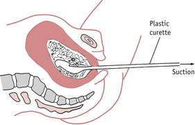
Treatment

Once the diagnosis of molar pregnancy is confirmed, the uterus must be completely evacuated at once. This is achieved by careful suction curettage (see Fig. 54.3). Uterine contractions may cause molar tissue to enter the circulation via the sinuses of the placental bed. These emboli may set up metastatic disease in other sites, commonly the lungs. Medical termination should therefore be avoided. Unless the woman is haemorrhaging, oxytocic drugs are withheld until the uterus has been surgically emptied. A Syntocinon infusion may then be used to maintain uterine contraction and haemostasis. The woman should be registered at a specialist follow-up centre in London, Sheffield or Dundee (RCOG 2004a).

After treatment for hydatidiform mole, careful observation is required as approximately 3% of these women will develop malignant trophoblastic disease (choriocarcinoma). Partial moles are less likely to become malignant but still require follow-up (Seckl et al 2000). Serum beta-hCG levels are monitored fortnightly until the values fall to within the normal range. Urine samples are then normally tested every 4 weeks until 1 year after evacuation. In the second year of follow-up, urinary hCG testing is carried out every 3 months.

If any molar tissue remains in the uterus, it will continue to grow and may invade the myometrium. Perforation of the uterine wall is then likely and this will cause major internal haemorrhage. Signs that the mole is continuing to grow are indicated by the persistence of high hCG levels 24 hours after uterine evacuation and high levels 1 month after treatment. If the serum or urinary hCG fails to return to normal levels within 6 months or begins to rise again, the woman is at risk of malignant trophoblastic disease.

The woman must avoid another pregnancy until she has been discharged from the follow-up programme. Use of the oral contraceptive pill increases the risk of the development of invasive disease and should therefore be avoided until hCG levels have been normal for three successive months.

Choriocarcinoma

Choriocarcinoma is a malignant disease of trophoblastic tissue. It occurs following approximately 3% of complete moles (Seckl et al 2000). hCG levels will rise and the pregnancy test will become strongly positive again. Choriocarcinoma may occur in the next normal pregnancy following an evacuation of a mole.

As the growth infiltrates the uterus and vagina, the affected woman will experience increasingly severe pain. The condition will be rapidly fatal unless treated. The disease spreads by local invasion and via the bloodstream; metastases may occur in the lungs, liver and brain. Transplacental fetal metastases may occur during a pregnancy but this is very rare.

Choriocarcinoma responds extremely well to chemotherapy. Cytotoxic drugs, such as methotrexate, etoposide and actinomycin-D, are used singly or as combination therapy, and are nearly always completely successful. The woman should avoid another pregnancy for at least 1 year after the completion of treatment and will require hCG monitoring after any future pregnancy, as there is a risk of disease recurrence (Oats & Abraham 2004).

Ectopic or extrauterine gestation

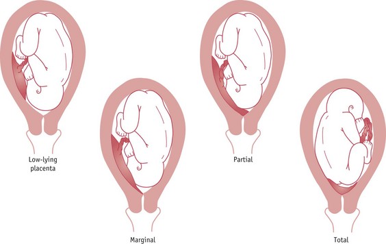
Ectopic pregnancy occurs when the fertilized ovum implants outside the uterine cavity. In 95% of cases the site of implantation is the uterine tube and these are known as tubal pregnancies. Occasionally, the site may be the ovary, the abdominal cavity or the cervical canal, but these are rare. The incidence of ectopic pregnancy is 1 : 150 pregnancies (Baker 2006). Ectopic pregnancy is the major cause of maternal death before 20 weeks’ gestation in the industrialized world (Benrubi 2005, Lewis 2007).

Page 759

Tubal pregnancy

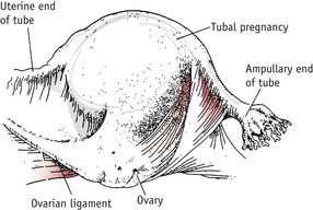
This is the commonest type of ectopic pregnancy and the incidence has increased two- to threefold in the last 30 years (Wiznitzer & Shener 2007). Tubal pregnancy occurs when there is a delay in the transport of the zygote along the fallopian tube. This may be due to a congenital malformation of the uterine tubes or more commonly to tubal scarring following pelvic infection. The ovum implants and begins to develop in the lining of the tube. The ampulla is the most common site (Fig. 54.5).

image

Figure 54.5 Tubal pregnancy.

Although tubal pregnancy may occur in the absence of any significant history, there are certain risk factors (Lemus 2000, Wiznitzer & Shener 2007):

History of previous tubal pregnancy.
Tubal surgery.
Hormonal ovulation induction – drugs such as clomifene may interfere with tubal motility.
Progesterone-releasing intrauterine contraceptive devices (this is related to higher-dose devices).
Tubal endometriosis.
Pelvic inflammatory disease.
Appendicectomy, pelvic or abdominal surgery which may cause adhesion formation.
Postcoital contraception using diethylstilbestrol.
Contraceptive methods – the intrauterine contraceptive device and the progesterone-only pill may increase the relative risk because they protect against intrauterine pregnancy but do not prevent ovulation and fertilization. This may make tubal pregnancy more likely in a woman who conceives while using these methods.
IVF pregnancy.

Diagnosis

Diagnosis based on clinical signs alone may be difficult because the clinical picture may appear similar to pelvic inflammatory disease or threatened abortion. Delay in diagnosis and treatment may contribute to maternal mortality and morbidity. The most accurate method currently available is a combination of serum hCG levels and transvaginal ultrasound scanning. hCG levels rise steadily in early pregnancy. Levels lower than normal or falling below the doubling time (the time in which serum levels can be expected to double) are indicative of ectopic gestation (Tin-Chiu et al 1999). The ultrasound scan may reveal a tubal mass or a fluid collection in the pelvis but is most useful for confirming the absence of an intrauterine sac. As the conceptus develops and grows, the tube distends to accommodate it.

Initially, the woman will experience the usual signs of pregnancy, such as nausea and breast changes, although amenorrhoea is not always present. The uterus will soften and enlarge under the influence of the pregnancy hormones. As the tube becomes further distended, the woman will experience abdominal pain and some vaginal bleeding. The blood loss is uterine in origin and signifies endometrial degeneration. Gastrointestinal disturbance such as diarrhoea and pain on defecation are also common signs (Baker 2006, Lewis 2007).

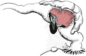
If the site of implantation is the narrower proximal end of the tube, tubal rupture is likely to occur between the 5th and 7th weeks of pregnancy (Fig. 54.6).

image

Figure 54.6 Rupture of the uterine tube.

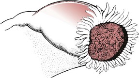
If the pregnancy is located in the wider ampullary section, the gestation may continue until the 10th week. Occasionally, the gestation sac is expelled from the fimbriated end (Fig. 54.7).

image

Figure 54.7 Tubal abortion.

Page 760

As the ovum separates from its attachment to the ampullary part of the tube, layers of blood clot may be deposited around the dead ovum to form a mass of blood clot which may remain in the uterine tube or be expelled from the fimbriated end of the tube. When the tube ruptures, there will be severe intraperitoneal haemorrhage and the woman will experience intense abdominal pain. There may also be referred shoulder-tip pain on lying down as blood tracks up towards the diaphragm. The woman will appear pale, shocked and nauseated and may collapse. The abdomen is tender and may be distended. Pelvic examination is usually exquisitely tender, especially on movement of the cervix. Ruptured ectopic pregnancy is an acute surgical emergency requiring immediate treatment.

Management

If ectopic pregnancy is suspected, a large-bore intravenous cannula (size 16 gauge) should be inserted and blood taken for cross-matching. The woman must be transferred to theatre as soon as possible. Laparoscopic salpingotomy may be performed unless the woman is suffering from haemorrhagic shock, when laparotomy is preferable.

If the condition is detected in the early stages, non-surgical management may be attempted with injections of prostaglandin F2a or systemic prostaglandin E2. Methotrexate may also be given, either intramuscularly or directly into the gestation sac (RCOG 2004b, Reis et al 2008).

Heterotopic or combined pregnancy

Heterotopic pregnancy occurs when a blastocyst from a multiple gestation implants outside the uterine cavity. It is associated with dizygotic twinning, and the extrauterine pregnancy is nearly always tubal. It may follow in vitro fertilization and embryo transfer. The incidence is thought to be approximately 1 : 15000 and is likely to rise as the incidence of both multiple gestation and ectopic pregnancy rises. Diagnosis can be difficult and management options are limited by the presence of the intrauterine pregnancy. The ectopic sac must be removed but the uterus should be disturbed as little as possible and methotrexate must be avoided if the intrauterine pregnancy is to survive (Wiznitzer & Shener 2007).

Secondary abdominal pregnancy

Very rarely, when rupture of a tubal pregnancy occurs, there may be partial extrusion of the ovum into the peritoneal cavity but with enough chorionic villi remaining attached to the tube to ensure that the embryo does not die. Chorionic villi on the surface of the ovum then become attached to the neighbouring abdominal organs and the pregnancy continues with the fetus developing free within the abdominal cavity (see website). The fetus is at risk of severe growth restriction because of the relatively poor placentation and may also suffer pressure deformities as there is no protective uterine wall.

This condition is usually detected by ultrasound scanning but may be suggested by a persistently abnormal fetal lie and the fact that fetal parts are unusually easy to palpate. Delivery is by laparotomy. The placenta is usually left in situ to be absorbed, as an attempt to detach it may cause uncontrollable haemorrhage.

Ectopic pregnancy is a significant cause of maternal death and the incidence is rising. There were 10 reported deaths in the UK from this cause between 2003 and 2005 (Lewis 2007). The midwife must be aware of the associated risk factors and seek an obstetric opinion for any woman with signs or symptoms suggestive of extrauterine gestation without delay.

Bleeding from associated conditions

The following conditions may cause bleeding, although, strictly speaking, they are not bleeding of early pregnancy since the bleeding is not from the site of the pregnancy.

Cervical polyp

This is a small red gelatinous growth attached by a pedicle to the cervix, close to the external os. It may give rise to slight irregular bleeding.

Ectropion of the cervix

A cervical erosion is formed when the columnar epithelium lining the cervical canal proliferates owing to the action of the pregnancy hormones. The ectropion forms a reddish area on the cervix, extending outwards from the external os. It may give rise to a blood-stained discharge from the vagina. No treatment is necessary and the ectropion will recede during the puerperium.

Carcinoma of the cervix

Invasive cervical carcinoma is rarely seen in pregnancy, although cervical intraepithelial neoplasia (CIN) may occasionally be discovered if a cervical smear is taken. If the cervical cytology report suggests precancerous changes, colposcopy is performed to identify the affected areas and a small cervical biopsy may be carried out. Treatment is deferred until after delivery if the condition is not invasive.

Page 761

Invasive cervical cancer is very serious as the disease may progress quickly. On vaginal examination, the cervix is hard and irregular and bleeds when touched. There may also be a purulent vaginal discharge. If the condition is discovered in the first trimester, the pregnancy may be terminated and treatment initiated. In the third trimester, the fetus is viable and may be delivered by caesarean section. Once the infant is born, the obstetrician may carry out a radical hysterectomy (Wertheim’s hysterectomy). Vaginal delivery is associated with a poorer prognosis for the mother as cervical dilatation may cause dissemination of tumour cells and metastases have been reported in episiotomy sites (Sood et al 2000).

A dilemma arises if the condition is discovered in the second trimester, because the fetus is unlikely to survive if delivered. The woman may choose to postpone treatment for a time to allow further fetal growth; however, the delay should be no longer than 4 weeks.

Bleeding in early pregnancy may occur for a variety of reasons. It is a serious sign and the underlying condition may be life-threatening. Any woman who reports vaginal bleeding during pregnancy must be referred to an obstetrician without delay.

Bleeding after the 24th week – antepartum haemorrhage

Antepartum haemorrhage is defined as bleeding from the genital tract after the 24th week of pregnancy and before the birth of the baby. Bleeding that occurs during labour is sometimes referred to as intrapartum haemorrhage.

Antepartum haemorrhage is a serious complication which may result in the death of the mother or the baby.

There are two main varieties of haemorrhage:

Placenta praevia (unavoidable or inevitable haemorrhage) is bleeding from separation of an abnormally situated placenta. (The placenta lies partly or wholly in the lower uterine segment and bleeding is inevitable when labour begins.)
Abruptio placentae (placental abruption) is bleeding from separation of a normally situated placenta.

However, extraplacental bleeding may sometimes occur. This is vaginal bleeding from some other part of the birth canal, for example, a cervical polyp, as described above.

Placenta praevia

The incidence of placenta praevia at term ranges from 0.5% to 1%. It is usually detected on ultrasound scanning in early pregnancy and may be seen in as many as one-quarter of all pregnancies in the second trimester. As the lower segment grows and stretches, the placental site appears to rise up the uterine wall, away from the internal os uteri, until at term in the majority of cases the placenta no longer occupies the lower segment. Those cases where the placenta overlies the internal os in early pregnancy, are at highest risk of haemorrhage. The classification of placenta praevia is shown in Table 54.1. The types are illustrated in Figure 54.8.

Table 54.1 Classification of placenta praevia

Low-lying placenta Placenta mainly in the upper segment but encroaching on the lower segment
Marginal Placenta reaches to, but does not cover, the internal os
Partial Placenta covers the internal os when closed but not completely when it is dilated
Total Placenta completely covers the internal os
image

Figure 54.8 Placenta praevia.

Causes

The cause of placenta praevia is unknown but the following factors are known to be associated (Baker 2006, Benrubi 2005, Handler et al 1994):

Multiparity: The increased size of the uterine cavity following repeated childbearing may predispose to placenta praevia.
Multiple pregnancy: The larger placental site is more likely to encroach on the lower segment of the uterus.
Age: Risk rises with maternal age.
Scarred uterus: One previous caesarean section doubles the risk of placenta praevia.
Previous myomectomy or hysterotomy.
Smoking: The exact mechanism is unclear but the relative hypoxia induced by smoking may cause enlargement of the placenta in order to compensate for the reduced oxygen supply.
Placental abnormality: Bipartite and succenturiate placentae may cause placenta praevia. Placenta membranacea (placenta diffusa) may also be a cause. This is a rare developmental abnormality of the placenta where all the chorion is covered with functioning villi. The placenta develops as a thin membranous structure, covering an unusually large surface of the uterus. The condition may be diagnosed on ultrasound. It may cause severe haemorrhage possibly requiring hysterectomy. It may not separate readily in the third stage of labour. Fetal nutrition appears to be relatively undisturbed in cases of placenta membranacea.
Fetal sex: There may be an association between male fetal sex and placenta praevia (Wen et al 2000).
Page 762

Associated conditions

A low-lying placenta puts the woman and her fetus at risk of other complications. The most serious of these is placenta accreta. This usually occurs where the previous delivery was by caesarean section (Yang et al 2007). The combination of the relatively thin decidua in the lower segment and the presence of scar tissue increases the likelihood of trophoblastic invasion of the myometrium.

Intrauterine growth restriction may occur, possibly as a result of repeated small haemorrhages (Drife 2002).

Signs and symptoms

As placenta praevia is normally diagnosed on ultrasound scanning in early pregnancy (Baulies et al 2007), midwives will usually be aware of any woman in their care who has a low-lying placenta. However, there are women who will not have had an ultrasound scan during pregnancy, including women who choose not to do so, women who have concealed their pregnancy, women who have not accessed care, or women who may have spent their antenatal period in a country where ultrasound scans are not readily available. Therefore the midwife must be aware of the signs that indicate a possible placenta praevia:

Malpresentation of the fetus: Although the presentation may be cephalic, often it is not. The placenta occupies space in the pelvis and the midwife may find that the breech presents, as there is more room for the head in the fundus, or that the lie is oblique and the fetal shoulder presents.
Non-engagement of the presenting part: This is especially likely with a partial or total placenta praevia.
Difficulty in identifying fetal parts on palpation: An anterior placenta praevia lies between the fetus and the midwife’s hand like a cushion. This makes the fetal parts relatively difficult to identify.
Loud maternal pulse below the umbilicus: An anterior placenta praevia may often be detected by the presence of loud maternal arterial sounds from the placental bed. This is more easily heard with an electronic fetal heart monitor (Doppler). The fetal heart sounds may be difficult to detect as they are muffled by the placenta, especially in a cephalic presentation.
An anterior placenta praevia will cushion some of the fetal movements and the woman may mention that she only feels fetal movement above the umbilicus.
Bleeding after sexual intercourse: Stimulation of the cervix during intercourse may provoke bleeding.
Page 763

When bleeding occurs, it usually begins after the 24th–28th week of pregnancy, although it may occur earlier. In the third trimester of pregnancy, the lower segment is completing its development, Braxton Hicks contractions are increasing, and towards the end of pregnancy, the cervix is becoming effaced. Bleeding is caused by detachment of the placenta which cannot stretch to adapt to these changes in uterine structure. As the placenta is in the lower pole of the uterus, the blood escapes easily, thus giving rise to the classical unprovoked, fresh, painless bleeding of placenta praevia. ‘Warning haemorrhages’ are associated with placenta praevia. These are small, recurrent, fresh and painless haemorrhages occurring during the third trimester. Each episode of bleeding indicates further placental detachment. If the placenta is torn, some fetal bleeding will occur and this will further compromise the condition of the fetus. Torrential maternal haemorrhage may occur at any time but is more likely once labour begins as the cervix begins to dilate.

It is impossible to predict the course of events in a case of placenta praevia and even in the absence of bleeding the condition is regarded as a major and life-threatening complication of pregnancy.

Management

If no serious haemorrhage has made it imperative to act, the woman will be delivered at about 38 weeks’ gestation, which should avoid problems of prematurity for the infant.

If the placental location is unclear, an examination may be carried out in theatre, by a senior obstetrician, with the woman suitably anaesthetized and the theatre set up in readiness for a caesarean section. An intravenous infusion is commenced and four units of cross-matched blood is immediately available. With the woman in the lithotomy position, the obstetrician makes a very gentle and cautious vaginal examination, passing a finger through the cervix into the lower pole of the uterus. If the placenta is palpable, the obstetrician will immediately perform a caesarean section. If the placenta is not palpable in the lower segment, the membranes may be ruptured and the woman allowed to labour. This procedure avoids unnecessary caesarean section for women in whom placenta praevia is not confirmed.

However, nearly all women have access to ultrasound scanning and the diagnosis is usually clear. Vaginal delivery is usually possible in cases of a low lying placenta, if the fetal head is engaged. However, the presence of placental tissue within 2 cm of the internal os is a contraindication to vaginal birth (RCOG 2005).

Active treatment

In cases where bleeding first occurs at 38 weeks or later, conservative treatment is not appropriate, as the fetus is mature. Active treatment is also necessary in cases where labour has started, if bleeding is severe or there are signs of fetal distress. An intravenous infusion is commenced and the woman’s condition is stabilized if necessary. A senior obstetrician performs an emergency caesarean section under general anaesthesia. A paediatrician should be present to attend to the baby, who may be asphyxiated at birth. Preterm infants of mothers with placenta praevia have an increased risk of developing respiratory distress syndrome (Lin et al 2001).

Third stage

Postpartum haemorrhage may complicate the third stage of labour since there are few oblique muscle fibres to control bleeding from the placental site in the lower uterine segment.

Placenta accreta may occur in women who have had a previous caesarean section and torrential haemorrhage may result from attempts to separate the placenta. Surgical treatments, such as ligation of the internal iliac arteries and interventional radiology, may be required in order to control the haemorrhage (RCOG 2007). Hysterectomy is undertaken as a last resort to save the woman’s life. The midwife must be familiar with local guidelines for management of massive obstetric haemorrhage (see Box 54.1) and adequate supplies of cross-matched blood should be available before surgery commences.

Box 54.1

Key points from guidelines for the management of massive obstetric haemorrhage (DH 1994, Lewis 2007)

Dealing with ill, bleeding women requires skilled teamwork between obstetric and anaesthetic teams, with appropriate help from other specialists, including haematologists, vascular surgeons and radiologists
Immediate involvement of all key staff, including senior obstetrician, anaesthetist, haematologist, blood transfusion service and portering staff
Minimum 20 mL sample of blood for cross-matching and coagulation studies
Minimum 6 units of cross-matched blood, with use of plasma expanders as necessary (not dextrans)
Blood of the patient’s own group to be used for transfusion; uncross-matched O-negative blood to be used only if immediate transfusion is required
Minimum of two peripheral intravenous lines, using 16-gauge cannulae
Immediate commencement of CVP monitoring
Facilities for monitoring central venous and intra-arterial pressure, ECG, blood gases and acid–base status should be available in consultant units
Rapid administration of blood and fluids (blood filtration is not necessary)
Use of blood-warming equipment
Repeated estimation of haemoglobin and coagulation studies
Use of an early warning scoring system such as the Modified Early Warning Scoring System (MEWSS)
Page 764

It cannot be emphasized too strongly that vaginal examination in placenta praevia is an extremely dangerous procedure and should not be attempted, except with the precautions described above.

Current interventions for suspected placenta praevia are reviewed by Neilson et al (2003), a summary of which can be found on the website.

Abruptio placentae

Abruptio placentae is bleeding due to the separation of a normally situated placenta (Fig. 54.9). It is sometimes referred to as placental abruption or ‘accidental’ bleeding. Placental abruption may occur at any stage of pregnancy, or during labour, and may complicate approximately 1% of pregnancies.

image

Figure 54.9 Abruptio placentae.

Causes

The cause of the placental separation cannot always be satisfactorily explained and in 40% of cases no cause can be found (Rana et al 1999). The following risk factors have been associated with the condition (Carr & Coustan 2007, Ray & Laskin 1999, Reis et al 2000)

Hypertensive disease: essential hypertension, pregnancy-induced hypertension (PIH) or pre-eclampsia.
Sudden decompression of the uterus: such as may follow spontaneous rupture of the membranes in cases of polyhydramnios.
Preterm prelabour: rupture of the membranes.
Previous history: placental abruption.
Trauma: for example, following external cephalic version, road traffic accident, a fall or a blow.
Smoking.
Drug abuse: for example, cocaine, crack or marijuana.
Folate and vitamin B12 deficiency: although the evidence for this association is not conclusive.

Maternal hypertension is the most consistent finding in cases of placental abruption.

Types

The bleeding may be revealed, concealed or partially revealed (Fig. 54.10).

image

Figure 54.10 Types of abruptio placentae.

Revealed bleeding

This occurs when the site of detachment is at the placental margin. The blood thus dissects between the membranes and the decidua and escapes through the os uteri. With revealed placental abruption the degree of shock is in proportion to the visible vaginal blood loss.

Concealed bleeding

This occurs when the site of detachment is close to the centre of the placenta. The blood cannot escape and a large retroplacental clot forms. The blood may infiltrate the myometrium, sometimes as far as the peritoneal covering, causing a marbled, petechial pattern of bleeding. This is called a Couvelaire uterus. There is no visible blood loss but the pain and shock may be severe as the intrauterine tension rises. Increasing abdominal girth or rising fundal height are suspicious signs of concealed haemorrhage. Backache may accompany abruption in a posteriorly sited placenta.

Partially revealed bleeding

This occurs when some of the blood trickles between the membranes and the decidua to become visible as vaginal bleeding. Not all the blood escapes and a variable amount remains concealed. In this situation the bleeding and thus the degree of shock will be much more severe than the visible loss suggests.

The severity of placental abruption may be classified as mild, moderate or severe.

Mild abruptio placentae

The loss is usually slight and the bleeding may be entirely concealed, although often there is a slight trickle per vaginam. The woman may experience no more than mild abdominal pain, the uterus is not tender and the fetus is alive. There is no sign of maternal shock.

Moderate abruptio placentae

The blood loss is heavier, the abdominal pain more severe and, on palpation, the uterus may be tender and firm. The mother may be hypotensive and have a tachycardia and usually there are signs of fetal distress.

Severe abruptio placentae

This is an obstetric emergency. More than half the placenta will have separated, the blood loss will exceed 1 litre and the mother will be very shocked. Abdominal pain will be severe. On palpation, the uterus may be hard and tender, and on auscultation, fetal heart sounds will not be heard. There is an increased risk of coagulation disorders. It is essential to remember that the amount of bleeding per vaginam is no guide to the degree of placental separation.

Outcome

If there is only minor detachment of the placenta and the mother and fetus are in good condition, the woman will be advised to stay in hospital for observation. If the bleeding ceases and all appears well, she may be discharged. The pregnancy will be closely monitored with ultrasound scans and regular cardiotocography to assess fetal growth and wellbeing. There is an increased risk of poor fetal growth and preterm birth following an episode of placental abruption (Rasmussen et al 2000).

In a case of moderate or severe abruptio placentae, the most important treatment is to empty the uterus. If fetal condition permits, labour is induced by rupturing the membranes and an oxytocic infusion is commenced. Vaginal delivery may be possible.

If the fetus is in poor condition, delivery will be by caesarean section unless the woman is already in the second stage of labour, when a forceps delivery will be performed. Ergometrine 500 mcg is given intravenously at delivery to control haemorrhage in the third stage of labour. It is usual to continue the Syntocinon infusion for some hours after delivery in order to maintain uterine contraction.

The most effective treatment for severe haemorrhage and defective coagulation is the transfusion of fresh blood. If fresh blood is not available, fresh frozen plasma should be given as this contains fibrinogen, platelets and clotting factors III, V and VIII.

Reflective activity 54.3

Locate and read your Unit policy for management of bleeding in later pregnancy.

Management of antepartum haemorrhage and the midwife’s role

At home

If called by a woman with bleeding, the midwife must ascertain the amount of bleeding and organize immediate care. If the reported bleeding is heavy, the midwife should ask a paramedic team from the local ambulance service to attend in order to avoid delay in transfer to hospital.

Page 766

The woman should lie on her side or with a pillow or towel wedged under the right hip to achieve a slight pelvic tilt and avoid supine hypotensive syndrome. Blood pressure, pulse rate and temperature should be recorded, blood loss noted and a sanitary pad applied. The presence of pain is suggestive of abruptio placentae, while painless bright bleeding may indicate placenta praevia. Soiled pads and clothing should be saved to allow accurate estimation of blood lost. An obstetrician from the local hospital must be contacted, as the woman will need to be admitted. The midwife should inform the obstetrician of the severity of the bleeding, colour of the blood (fresh or dark), nature and location of pain, if present, and the woman’s general condition. If the initial loss is small, the woman’s blood pressure and pulse rate will be normal and she will appear well. The assessment of blood pressure should be treated with caution, with reference to the woman’s medical record if possible. A normal blood pressure may falsely represent a low blood pressure in women with a history of hypertension. In these cases, the pulse and respiration rate may be more indicative of bleeding. Should the loss be severe, she will present the typical picture of a woman who has had a haemorrhage: pale, sweating, restless, thirsty, with a rising pulse rate, rising respiration rate and falling blood pressure. In this case, emergency assistance is needed and a paramedic team from the ambulance service should be called. An intravenous infusion is started and group O rhesus-negative blood may be given if necessary. Plasma expanders, such as Haemaccel, Gelofusine or hetastarch (Hespan) preparations, may be used.

When attending to a woman who is bleeding, no vaginal examination should be made. If this is a case of placenta praevia, a vaginal examination could precipitate a disastrous haemorrhage. Rectal examinations are similarly dangerous. Abdominal examination should be avoided if possible as this may provoke Braxton Hicks contractions, which may accelerate bleeding.

In hospital

The woman is admitted to the delivery suite and the attendance of a senior obstetrician is requested. If there are signs of severe haemorrhage, a senior anaesthetist and haematologist should also be in attendance.

Until the diagnosis is clear, she must be treated as having a potential placenta praevia, although there may be some features which help in making a diagnosis (see Table 54.2). Often the distinction is not clear; it is particularly difficult if the mother has had a small revealed abruption but no pain, no apparent cause for the bleeding and no signs of pre-eclampsia.

Table 54.2 Differential diagnosis with placenta praevia

Clinical sign Placenta praevia Abruptio placentae
Pain No pain Uterine pain, may be severe; backache if placenta is posterior
Colour of blood loss Bright, fresh May be darker
‘Warning’ haemorrhages Yes No
Onset of bleeding Possibly following coitus, otherwise unexpected May follow trauma, exertion
Degree of shock In proportion to visible loss May be more severe than visible loss suggests
Consistency of uterus Soft, non-tender Increased uterine tone, may be tense, rigid, ‘woody’
Palpation Fetus usually fairly easy to palpate Tense uterus makes palpation difficult
Presentation May be a malpresentation Probably cephalic
Engagement Not engaged May be engaged
Fetal heartbeat Probably present May be absent
Abdominal girth Equivalent to gestation May increase due to concealed haemorrhage

If bleeding has been severe, the treatment must be swift, as the woman’s condition may deteriorate rapidly. The aim is to restore normal blood volume and thus improve the mother’s general condition, deliver the baby if necessary, and avoid the dangerous complications of renal failure and blood coagulation disorders. Two intravenous cannulae (16 guage) are inserted, blood is taken for grouping and cross-matching, and the duty haematologist and blood bank must be informed if large quantities of blood are likely to be needed. Other blood tests include full blood count (essential for haemoglobin estimation and platelet count), urea and electrolytes, clotting studies and fibrin degradation products.

Page 767

Temperature, pulse, blood pressure, fetal heart and vaginal loss should be observed. The pulse and blood pressure are recorded as frequently as the woman’s condition dictates: quarter-hourly if the bleeding is continuing. Maternal oxygen saturation should be observed using a pulse oximeter. Oxygen may be given by facemask if required. The fetal heart should be continuously monitored by external cardiotocography whilst bleeding persists. Gentle pressure should be used so as not to stimulate further bleeding or uterine activity. Placing a mark on the abdomen durng the first assessment of the fetal heart may reduce excessive palpation. A Foley urinary catheter is inserted and the urinary output is closely monitored, a marked decrease being a grave sign. The urine is tested for protein. The midwife records an estimate of blood loss, observing for the appearance of blood clotting: blood with normal clotting factors will clot in room air; if this fails to happen, deranged clotting should be suspected. Analgesia, such as morphine, may be required if the woman is in pain. An intravenous infusion of normal saline 0.9% or Hartmann’s solution (Ringer’s lactate) will be commenced. Blood transfusion may be required and several units of blood or packed cells may be needed if the haemorrhage has been substantial. In such cases, central venous pressure should be monitored to avoid the dangers of over- or undertransfusion.

The abdominal girth is sometimes measured and recorded. Meticulous observation and recording of vital signs, blood loss and fluid balance are essential in order to assess the woman’s condition and plan her care. Ongoing management of the situation is governed by the condition of the mother and fetus. The midwife may need to involve the social worker if there are other children at home for whom care arrangements need to be made. The needs of the partner should also be addressed, which include support and information.

If the haemorrhage is not severe and urgent delivery is not indicated, the woman may be transferred to the antenatal ward once the bleeding settles and her condition is stable. In the absence of pain or active bleeding, the woman should not be confined to her bed and can be encouraged to wear her usual clothes during the day. In cases of placenta praevia, at least two units of cross-matched blood should always be available; local policy will dictate the regularity of sample update, usually weekly. This enables cross-match of fresh blood and ensures ready availability of blood should haemorrhage occur. If the bleeding is due to placenta praevia, the woman may be advised to remain in hospital until delivery. The midwife must ensure that the woman and her partner are fully informed about her condition and the likely management.

Following antepartum haemorrhage, tests to assess fetal wellbeing will be carried out because premature separation of the placenta may result in impaired placental function. Fetal growth will be assessed by ultrasound, and periodic continuous fetal heart monitoring will be performed. Women who have a rhesus-negative blood group should be given anti-D gammaglobulin in line with local policy, after each episode of bleeding.

Complications

Blood coagulation disorders (see Ch. 68)

When tissue damage occurs, there is a release of thromboplastin from the local cells. Thromboplastin activates the clotting mechanism and this results in the conversion of fibrinogen into fibrin. The sticky web of fibrin traps the cellular components of the blood and a clot forms, sealing off the bleeding point. The clot is later dispersed by plasmin, which is the active product of the fibrinolytic system. When a clot is broken down, fibrin degradation products (FDPs) are formed. Clot dispersal is a protective mechanism to prevent capillary blockage.

The system of initial clot formation followed by fibrinolysis is normally delicately balanced. If the coagulation system fails, bleeding will persist, while if the fibrinolytic system fails, clotting will persist.

Occasionally, tissue damage is so severe or widespread that there is a massive release of thromboplastin into the general circulation. Widespread clotting will then occur throughout the body. This condition is known as disseminated intravascular coagulation (DIC). This is extremely dangerous as the microthrombi generated by the thromboplastin will occlude small blood vessels. This results in ischaemic tissue damage within the body organs: the damaged tissue releases thromboplastin, which stimulates further clotting. Thus a vicious circle of tissue damage and uncontrolled clotting occurs. Any body organ may be affected: renal damage will result in oliguria or anuria; liver damage will lead to jaundice. If the lungs are affected, dyspnoea and cyanosis will occur; convulsions or coma indicate cerebral involvement. Microthrombi in the retina may cause blindness; if the pituitary gland is affected, Sheehan’s syndrome, a condition that may have serious long-term effects on the mother’s health and her future fertility, may occur (see website). Eventually, the available circulating platelets are depleted. Clotting factors, such as prothrombin (factor II), thromboplastin (factor III), proaccelerin (factor V), antihaemophilic factor (factor VIII) and fibrinogen (factor I), are exhausted. No further coagulation can take place: bleeding becomes apparent. This may take the form of oozing from venepuncture sites, mucous membrane bleeding, petechiae and uncontrollable uterine haemorrhage.

DIC is always a secondary event, occurring as a result of massive tissue damage and thromboplastin release. It may complicate conditions such as severe pre-eclampsia, septicaemia or amniotic fluid embolism. It may also occur following abruptio placentae when thromboplastin is released from the damaged placental, decidual and myometrial tissue. Unless DIC is recognized and treated promptly, the condition may become uncontrollable with death an inevitable outcome. The midwife must be aware of any woman who is at risk of DIC and be alert for the signs of coagulation failure. All maternity units should have an emergency protocol for dealing with such cases. Any woman with an abruptio placentae should have screening tests for coagulation defects. These tests include:

partial thromboplastin time (normally 35–45 seconds)
prothrombin time (normally 10–14 seconds)
thrombin time (normally 10–15 seconds)
fibrinogen levels (2.5–4 g/L)
fibrin degradation products
whole blood film and platelet count.

Fresh frozen plasma, packed cells and platelets are used in the treatment of DIC. Heparin is rarely used as it may exacerbate the haemorrhage, especially if the uterus is not empty.

Acute renal failure

This may occur following severe shock in cases of antepartum haemorrhage.

Postpartum haemorrhage

Following severe abruptio placentae, postpartum haemorrhage is most likely to be caused by a blood coagulation disorder, whereas following placenta praevia it is due to the inability of the lower uterine segment to contract effectively. Aortic compression may be necessary to control cases of intractable haemorrhage.

Infection

Sepsis is likely owing to the woman’s lowered resistance following a state of severe shock, a large blood transfusion, increased intervention in labour and anaemia.

Anaemia

The haemoglobin must be checked and anaemia corrected in the puerperium.

Psychological disturbances/psychoses

Psychological disorders after childbirth are more likely following complications of pregnancy and labour, and may be due to the ensuing anaemia or post-traumatic stress (PTS) syndrome (Ch. 69).

Vasa praevia

This unusual condition may result in vaginal bleeding. It is associated with velamentous insertion of the cord. One of the fetal vessels traverses the membranes in the region of the internal os, in front of the presenting part. Occlusion of the vessel may occur as the presenting part compresses the membranes. When the membranes rupture, the vessel can be torn and severe fetal bleeding occurs. The perinatal mortality associated with this condition is high (Suzuki & Igarash 2008). Diagnosis is difficult but a pulsating vessel may be felt on vaginal examination. Velamentous insertion can often be detected on routine ultrasound examination from the second trimester of pregnancy and vasa praevia can be confirmed by transvaginal colour Doppler scanning (Nomiyama et al 1998).

If vasa praevia is suspected, the midwife should leave the membranes intact and inform the obstetrician.

The midwife should be aware that there is a higher risk of vasa praevia in placenta praevia, where there is a succenturiate lobe and in IVF pregnancies (Olayinka et al 1999).

Conclusion

Bleeding in pregnancy remains a major cause of maternal morbidity and mortality. The midwife must be familiar with local policy for management of bleeding in pregnancy and regular emergency drills should be held in maternity units to ensure that the obstetric team can respond to haemorrhage quickly and appropriately (Lewis 2007). Midwives should be familiar with the recommended guidelines for the management of massive obstetric haemorrhage, to be found in Lewis (2007).

Midwives also need to be able to identify women who might be at risk, and refer appropriately, also ensuring that the woman and her family are informed and supported throughout. Often the midwife will be the person providing continuity during the woman’s pregnancy, facilitating holistic, sensitive and appropriate care to the woman, her baby and family.

Key Points

Vaginal bleeding at any stage of pregnancy is always abnormal and may be indicative of a serious complication.
Midwives need to educate the woman and her family regarding deviations from normal, and ensure that they are aware of what action should be taken and with whom they should communicate.
Midwives must be aware of the possible causes of bleeding in pregnancy.
A prompt and appropriate response may prevent the loss of the fetus and may save the mother’s life.
Midwives must be aware of the possible emotional, social and psychological impact of bleeding or pregnancy loss for the woman and her partner.

References

1967 Abortion Act. London: HMSO, 1967.

Andersen N, Wohlfahrt J, Christens P, et al. Maternal age and fetal loss: population based register linkage study. British Medical Journal. 2000;320(7251):1708-1712.

ARC (Antenatal Results and Choices). (website) www.arc-org.uk, 2009. Accessed January 27, 2009

Backos M, Regan L. High risk pregnancy: management options. Edinburgh: Saunders; 2006.

Baker PN. Obstetrics by Ten Teachers. London: Arnold; 2006.

Baulies S, Maize N, Munoz A, et al. Prenatal ultrasound diagnosis of placenta previa and analysis of risk factors. Prenatal Diagnosis. 2007;27(7):595-599.

Page 769

Beischer N, Mackay E, Colditz P. Obstetrics and the newborn. London: WB Saunders; 1997.

Benrubi GI. Handbook of obstetric emergencies. Philadelphia: Lippincott Williams & Wilkins; 2005.

Boyles S, Ness R, Grisso J, et al. Life event stress and the association with spontaneous abortion in gravid women at an urban emergency department. Health Psychology. 2000;19(6):510-514.

Carr R, Coustan D. Nonprescription drugs and alcohol: abuse and effects in pregnancy. In Reece EA, Hobbins JC, editors: Clinical obstetrics: the fetus & mother, ed 3, Oxford: Blackwell, 2007.

Conway K, Russell G. Couples’ grief and experience of support in the aftermath of miscarriage. British Journal of Medical Psychology. 2000;73(4):531-545.

Creinin MD, Potter C, Holovanisin M, et al. Mifepristone and misoprostol and methotrexate/misoprostol in clinical practice for abortion. American Journal of Obstetrics and Gynecology. 2003;188(3):664-669.

Department of Health (DH). Report on Confidential Enquiries into Maternal Deaths in the United Kingdom 1988–1990. London: HMSO; 1994.

Dimond B. Termination of pregnancy. British Journal of Midwifery. 2001;9(2):108.

Donders G, Van Bulck B, Caudron J, et al. Relationship of bacterial vaginosis and mycoplasmas to the risk of spontaneous abortion. American Journal of Obstetrics and Gynecology. 2000;183(2):431-437.

Drife J. Bleeding in pregnancy. In: Chamberlain G, editor. Turnbull’s obstetrics. Edinburgh: Churchill Livingstone, 2002.

Gopalkrishnan K, Padwal V, Meherji P, et al. Poor quality sperm as it affects repeated early pregnancy loss. Archives of Andrology. 2000;45(2):111-117.

Handler A, Mason E, Rosenberg D, et al. The relationship between exposure during pregnancy to cigarette smoking and cocaine use and placenta praevia. American Journal of Obstetrics and Gynecology. 1994;170(3):884-889.

Home Department. Criminal statistics England and Wales 2000. London: The Stationery Office; 2001.

Home Office. Identity and Passport Service, General Registrar Office, official information on births, deaths and marriages. (website) www.gro.gov.uk/gro/content, 2008. Accessed January 26, 2009

Home Office. Crime statistics. (website) www.homeoffice.gov.uk/rds/recordedcrime1.html, 2009. Accessed February 12, 2009

1990 Human Fertilization and Embryology Act. London: Office of Public Sector Information, 1990.

Kauffman D, Sutkin G, Heine R, et al. Metastatic complete hydatidiform mole with a surviving co-existent twin. A case report. Journal of Reproductive Medicine. 1999;44(2):131-134.

Lemus J. Ectopic pregnancy: an update. Current Opinion in Obstetrics and Gynecology. 2000;12(5):369-375.

Lewis G, editor. Why mothers die 1997–1999: the fifth report of the Confidential Enquiries into Maternal Deaths in the United Kingdom. London: RCOG, 2001.

Lewis G, editor. Saving mothers’ lives: reviewing maternal deaths to make motherhood safer – 2003–2005. The Seventh Report on Confidential Enquiries into Maternal Deaths in the United Kingdom. London: CEMACH, 2007.

Lin C, Wang S, Hsu Y, et al. Risk for respiratory distress syndrome in preterm infants born to mothers complicated by placenta previa. Early Human Development. 2001;60(3):215-224.

Neilson JP: Interventions for suspected placenta praevia, Cochrane Database of Systematic Reviews (Online) (2):CD001998, 2003.

Neilson JP, Hickey M, Vazquez J: Medical treatment for early fetal death (less than 24 weeks) Cochrane Database of Systematic Reviews (Online) (3):CD002253, 2006.

Nomiyama M, Toyota Y, Kawano H. Antenatal diagnosis of velamentous umbilical cord insertion and vasa previa with color Doppler imaging. Ultrasound in Obstetrics and Gynecology. 1998;12(6):426-429.

Oats JN, Abraham S. Llewellyn-Jones fundamentals of obstetrics and gynaecology, ed 8. London: Elsevier; 2004.

Olayinka K, Turner M, Lees C, et al. Vasa previa: an avoidable obstetric tragedy. Obstetrical and Gynecological Survey. 1999;54(2):138-145.

Rana A, Sawhney H, Gopolan S, et al. Abruptio placentae and chorioamnionitis – microbiological and histological correlation. Acta Obstetricia et Gynecologica Scandinavica. 1999;78(5):363-366.

Rasmussen S, Irgens L, Dalaker K. Outcome of pregnancies subsequent to placental abruption: a risk assessment. Acta Obstetricia et Gynecologica Scandinavica. 2000;79(6):496-501.

Ray J, Laskin C. Folic acid and homocystine metabolic defects and the risk of placental abruption, pre-eclampsia and spontaneous pregnancy loss: a systematic review. Placenta. 1999;20(7):519-529.

Reis P, Sander C, Pearlman M. Abruptio placentae after auto accidents. A case control study. Journal of Reproductive Medicine. 2000;45(1):6-10.

Reis JL, Condeco P, Ventura P, et al. Conservative management of a cervical pregnancy with methotrexate. Female Patient. 2008;33(3):36-38.

Royal College of Obstetricians and Gynaecologists (RCOG). The management of gestational trophoblastic neoplasia. Green top guideline 38. London: RCOG; 2004.

Royal College of Obstetricians and Gynaecologists. The management of tubal pregnancies. Green top guideline 21. London: RCOG; 2004.

Royal College of Obstetricians and Gynaecologists (RCOG). Placenta praevia and placenta praevia acreta: diagnosis and management. Green top guideline 27. London: RCOG; 2005.

Royal College of Obstetricians and Gynaecologists (RCOG). Management of early pregnancy loss. Green top guideline 25. (2006). London: RCOG, 2006.

Royal College of Obstetricians and Gynaecologists (RCOG). The role of emergency and elective interventional radiology in postpartum haemorrhage. Practice guideline 6. London: RCOG; 2007.

Seckl M, Fisher R, Salwerno G, et al. Choriocarcinoma and partial hydatidiform moles. Lancet. 2000;356(9223):36-39.

Silver R, Branch D. Sporadic and recurrent pregnancy loss. In Reece EA, Hobbins JC, editors: Clinical obstetrics: the fetus & mother, ed 3, Oxford: Blackwell, 2007.

Page 770

Sood A, Sorosky J, Mayr N, et al. Cervical cancer diagnosed shortly after pregnancy: prognostic variables and delivery routes. Obstetrics and Gynecology. 2000;95(6 Pt 1):832-838.

Stillbirth and Neonatal Death Society (SANDS). www.uk-sands.org, 2009. Stillbirth and neonatal death charity supporting anyone affected by the death of a baby and promoting research to reduce the loss of babies’ lives. Accessed July 13, 2010

Suzuki S, Igarash M. Clinical significance of pregnancies with succenturiate lobes of the placenta. Archives of Gynecology and Obstetrics. 2008;227(4):299-301.

Tin-Chiu L, Bates S, Pearce M. Biochemical tests in complications in early pregnancy. In: O’Brien P, editor. The yearbook of obstetrics and gynaecology, vol 7. London: RCOG; 1999.

Wen S, Demissie K, Liu S, et al. Placenta praevia and male sex at birth: results from a population-based study. Paediatric and Perinatal Epidemiology. 2000;14(4):300-304.

Wiznitzer A, Shener E. Ectopic and heterotopic pregnancies. In: Reece EA, Hobbins JC, editors. Clinical obstetrics: the fetus & mother. Oxford: Blackwell, 2007.

Yang Q, Wen SW, Oppeheimer I, et al. Association of caesarean delivery for first birth with placenta previa and placental abruption in second pregnancy. British Journal of Obstetrics and Gynaecology. 2007;114(5):609-613.