External Parasites
If a parasite resides on the surface of its host, it is termed an ectoparasite. The majority of ectoparasites of domesticated animals are either insects (e.g., fleas, lice, flies) or arachnids (e.g., ticks, mites). However, a few nematodes have been included in this chapter because they demonstrate either immature or adult stages that are found within the animal’s skin or subcutaneous tissues.
This chapter is designed to aid the veterinary technician in the diagnosis of ectoparasitism in dogs, cats, cattle, sheep, goats, horses, and swine. To diagnose an ectoparasitic infestation, the technician must be able to collect the ectoparasite or its life cycle stages and then identify the organism involved. This chapter explains procedures most commonly used to collect ectoparasites from the host and describes those parasites so that a correct diagnosis can be made.
Although a complete review of the biology of ectoparasites is beyond the scope of this chapter, a brief review of the life cycles of the major groups of ectoparasites is included.
COLLECTION OF SAMPLES
Skin scraping is one of the most common diagnostic procedures used in evaluating animals with dermatologic problems. Equipment needed includes an electric clipper with a #40 blade, a scalpel or spatula, mineral oil in a small dropper bottle, and a compound microscope. Typical lesions or sites most likely to harbor the particular parasite should be scraped (e.g., ear margins for Sarcoptes scabei var. canis).
The scraping is performed with a #10 scalpel blade, with or without a handle. A 165-mm stainless steel spatula (Sargent-Welsh Scientific, Detroit, MI) is preferred by some clinicians. The scalpel blade should be held between the thumb and forefinger (Fig. 7-1). Before the skin is scraped, the blade is dipped in a drop of mineral oil on the slide, or a drop of mineral oil may be placed on the skin.
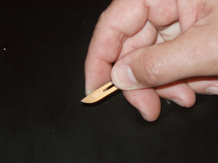
Figure 7-1 A scalpel blade may be held safely between the thumb and forefinger for skin scrapings. (From Hendrix CM, Robinson E: Diagnostic parasitology for veterinary technicians, ed 3, St Louis, 2006, Mosby.)
During the scraping process, the blade must be held perpendicular to the skin. Holding it at another angle may result in an accidental incision. The average area scraped should be approximately 3 to 4 cm2. Multiple sites should be scraped to increase the chances of collecting the parasite.
The depth of the scraping varies with the typical location of the parasite in question. When scraping for mites that live in tunnels (e.g., Sarcoptes species) or in hair follicles (e.g., Demodex species), the veterinary technician should scrape the skin until a small amount of capillary blood oozes from the scraped area. Clipping the area with a #40 blade before scraping enables better visualization of the lesion and removes excess hair that impedes proper scraping and interferes with collection of epidermal debris. Scraping at the interface between affected and unaffected sites is important. For surface-dwelling mites (e.g., Cheyletiella, Psoroptes, or Chorioptes species), the skin is scraped superficially to collect any loose scales or crusts. Clipping before scraping is not necessary when infestation with surface-dwelling mites is suspected.
All scraped debris on the forward surface of the blade is then spread in a drop of mineral oil on the glass slide. A glass cover slip is placed on the material, and the slide is ready for microscopic examination using the 4× (scanning) objective. The slide should be examined systematically in rows so that the entire area under the cover slip is evaluated. Low light intensity and high contrast increase visualization of translucent mites and eggs. If necessary, the slide may be evaluated with the 10× (low power) objective.
Demonstration of the characteristic mite or egg is frequently diagnostic for most diseases. In certain circumstances, however, more than just identification of the parasite is necessary. For example, determination of live/dead ratios and observation of immature larval and nymphal stages of demodectic mites are important in determining a patient’s prognosis. A decrease in the number of live mites and eggs during treatment is an excellent prognostic indicator.
Cellophane Tape Preparation
When attempting to demonstrate lice or mites that live primarily on the surface of the skin (e.g., Cheyletiella, Psoroptes, or Chorioptes species), the veterinary technician may use a cellophane tape preparation instead of a skin scraping. Clear cellophane tape (e.g., Scotch Transparent Tape, 3M, Minneapolis, MN) is applied to the skin to pick up epidermal debris. A ribbon of mineral oil is placed on a glass slide, and the adhesive surface of the tape is then placed on the mineral oil. Additional mineral oil and a cover slip may be placed on the tape to prevent the tape from wrinkling, but this is not necessary. The slide is then examined microscopically for parasites (see Procedure 6-9).
Parasite Identification
When arthropods or helminths cannot be grossly identified, the intact specimen should be collected in a sealed container containing 10% formalin or ethyl alcohol and submitted to an arthropodologist or parasitologist for identification. A complete history should accompany the specimen. The specimen should not be fragmented or squashed, which could distort morphologic features necessary for proper identification. Forensic identification of parasites is difficult.
TERMINOLOGY
Ectoparasites usually live on or in skin surfaces or feed on them. Ectoparasites infest the skin or external surfaces of animals and produce an infestation on the animal. By contrast, internal parasites, or endoparasites, infect the internal organs of domestic animals and produce an infection within that animal. In diagnostic parasitology, these terms always should be used in the proper context.
Life cycle describes the development of a parasite through its various life stages. Every parasite’s life cycle has a definitive host and may have one or more intermediate hosts. The definitive host harbors the adult, sexual, or mature stages of a parasite. The intermediate host harbors the juvenile, asexual, or immature stages of a parasite.
Many of the ectoparasites in this chapter belong to the phylum Arthropoda or, more simply, are considered arthropods. All adult arthropods have jointed legs. In addition, a few larval arthropods may be associated with dermal lesions. Later in this chapter is a discussion of ectoparasites that reside within the skin; these ectoparasites are not arthropods but rather belong to the phylum Nematoda, or more simply, the roundworms.
CLASSIFICATION SYSTEM
In beginning biology, students must learn the classification scheme perfected by Linnaeus, an early biologist. Every living organism on this planet may be classified with the following classification scheme: Kingdom, Phylum, Class, Order, Family, Genus, and species. Students often remember this scheme with this simple mnemonic device, “King Philip came over for good spaghetti.”
Every living thing is known by a scientific name that is made up of two components: the genus and the specific epithet. The dog’s scientific name is Canis familiaris. Canis is the Genus and familiaris is the specific epithet or species. Similar species are grouped together into the same Genus. Similar Genera (plural of genus) are grouped together into the same Family. Similar Families are grouped together into the same Order. Similar Orders are grouped together into the same Class. Similar Classes are grouped together into the same Phylum. Similar Phyla are grouped together into the same Kingdom. Therefore the dog’s classification scheme is as follows:
Every living creature on Earth has a distinct classification scheme.
PHYLUM: ARTHROPODA
The first part of this chapter discusses the Orders belonging to the Class Insecta and the Families belonging to the Class Acarina and how these ectoparasites are relevant to veterinary practice.
Order: Hymenoptera
Hymenoptera includes ants, bees, and wasps. Fire ants are indigenous to the southeastern United States and can bite and sting almost any domestic animal. “Downer cows” and young newborn animals are particularly susceptible to the bites and stings of fire ants.
Bees, wasps, and hornets can sting domestic animals, particularly curious dogs and cats. When stung, the animals may show an extremely swollen nose or face.
Africanized honeybees, or “killer bees,” have now crossed the Mexico/United States border. Almost any domestic animal (or person) could stumble on a hive of these bees, angering the inhabitants. Death often results from thousands of bee stings. Animals are particularly at risk because these bees often nest at or near the surface of the ground.
Order: Hemiptera
Members of Hemiptera are true bugs. Although some adult Hemipterans are wingless, most adult Hemipterans have two pairs of wings. The posterior pair of wings is membranous in appearance. The anterior pair of wings has a leathery basal portion with a membranous apical portion, which gives the appearance of being a “half wing,” hence, the origin of the ordinal name: hemi-, meaning half, and -ptera, meaning wing. Two groups of Hemipterans are of veterinary importance: reduviid bugs (“kissing bugs”) and bed bugs. Reduviid bugs are periodic parasites, making frequent visits to the host to obtain a blood meal. These bugs serve as intermediate hosts for Trypanosoma cruzi, a protozoan parasite that can produce a rare disease in people and dogs called Chagas disease. This disease also is called South American trypanosomiasis and rarely occurs in dogs and other animals in the United States. Reduviid bugs take blood meals from an infected host and transmit the parasite as they defecate.
Bed bugs are dorsoventrally flattened, wingless Hemipterans that often infest homes. They are periodic parasites, making frequent visits to the host to obtain a blood meal. Although bed bugs are human parasites, they also may be found in rabbit colonies, poultry houses, and pigeon colonies. Bed bugs do not naturally transmit any human or animal pathogen.
Orders: Mallophaga and Anoplura
Lice are some of the most prolific ectoparasites of domestic animals. Two orders of lice exist: the Mallophaga (chewing or biting lice) and the Anoplura (sucking lice). Lice are dorsoventrally flattened, wingless insects. They have three body divisions: the head, with its mouthparts and antennae; the thorax, with its three pairs of legs and its lack of wings; and the abdomen, the portion that bears the reproductive organs. These body divisions and their relations to each other are important in diagnostic veterinary parasitology.
Members of the order Mallophaga, or chewing/biting lice, are smaller than sucking lice. They are usually yellow and have a large, round head. The mouthparts are mandibulate and are adapted for chewing or biting the host. Characteristically, the head of every chewing louse is wider than the widest portion of the thorax. On the thorax are three pairs of legs, which may be adapted for clasping or moving rapidly among feathers or hairs. Chewing/biting lice may parasitize birds, dogs, cats, cattle, sheep, goats, and horses (Fig. 7-2).
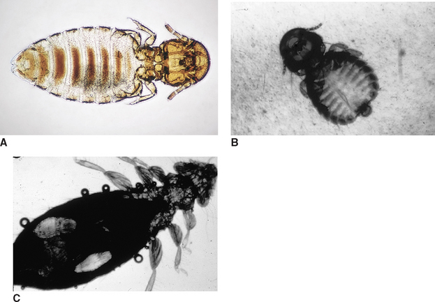
Figure 7-2 Damalinia caprae (A), Goniodes dissimilis (B), and Goniodes gallinae (C), assorted chewing lice of goats and fowl. (From Hendrix CM, Robinson E: Diagnostic parasitology for veterinary technicians, ed 3, St Louis, 2006, Mosby.)
Members of the order Anoplura (sucking lice) are larger than members of the order Mallophaga (chewing lice). These lice are red to gray; their color usually depends on the amount of blood ingested from the host. In contrast to the wide head of the Mallophagans, Anoplurans heads are narrower than the widest part of the thorax. Their mouthparts are piercing and are adapted for sucking blood. Their pincerlike claws are adapted for clinging to the host’s hairs. Although they are found on many species of domestic animals, sucking lice do not parasitize birds or cats (Fig. 7-3).
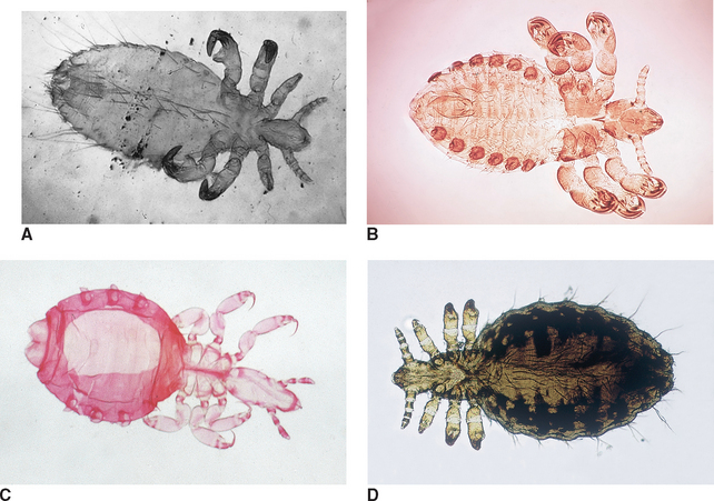
Figure 7-3 Solenopotes capillatus (A), Pedicinus obtusus (B), Pedicinus obtusus (C), and Linognathus setosus (D), assorted sucking lice of sheep, monkeys, and dogs. (Reprinted from Hendrix CM, Robinson E: Diagnostic parasitology for veterinary technicians, ed 3, St Louis, 2006, Mosby.)
Anoplurans and Mallophagans have the same type of life cycle. This life cycle has only three developmental stages: the egg, nymphal, and adult stages.
The egg stage is also called a nit. The nit is tiny, approximately 0.5 to 1.0 mm in length. It is oval, white, and usually found cemented to the hair or feather shaft of the host (Fig. 7-4). Figure 7-5 shows a gravid female sucking louse and an associated nit collected from a dog. Nits hatch approximately 5 to 14 days after being laid by the adult female louse.
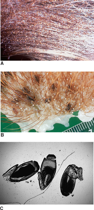
Figure 7-4 A, Thousands of nits can be cemented by female lice to the haircoat of domesticated animals. This calf’s tail contains thousands of nits. B, Pediculosis can be defined as infestation by either chewing or sucking lice, in this case, Haematopinus suis infestation in a pig. C, Appearance of operculated nits viewed by compound microscope. (From Hendrix CM, Robinson E: Diagnostic parasitology for veterinary technicians, ed 3, St Louis, 2006, Mosby.)
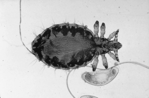
Figure 7-5 Linognathus setosus, a gravid female sucking louse, and associated nit collected from a dog. (From Hendrix CM, Robinson E: Diagnostic parasitology for veterinary technicians, ed 3, St Louis, 2006, Mosby.)
The nymphal stage is similar in appearance to the adult. However, it is smaller and lacks functioning reproductive organs and genital openings. The three nymphal stages are each progressively larger than its predecessor. The nymphal stage lasts from 2 to 3 weeks.
The adult stage is similar in appearance to the nymphal stage but larger. It has functional reproductive organs. Male and female lice copulate, the female lays eggs, cementing them to the hair or feather, and the life cycle begins again. It takes 3 to 4 weeks to complete the cycle. Nymphal and adult stages live no longer than 7 days if removed from the host. Eggs hatch within 2 to 3 weeks during warm weather but seldom hatch off the host.
Lice usually are transmitted by direct contact, but all life stages may be transmitted by fomites (inanimate objects such as blankets, brushes, and other grooming equipment). Lice are easily transmitted among young, old, and malnourished animals. Veterinarians often cannot understand why certain animals in a flock or herd are heavily infested, whereas others have only a few lice. Lice are species specific; that is, they will parasitize only their specific hosts. For example, dog lice parasitize dogs. They may temporarily reside on another species of animal, but they will not set up housekeeping on that animal.
Infestation by lice (whether Mallophagan or Anopluran) is referred to as pediculosis. Sucking lice can ingest blood to such a degree that they produce severe anemia; fatalities can occur, especially in young animals. The packed cell volume can drop as much as 10% to 20%. Severely infested animals may harbor many thousands of lice. Infested animals become more susceptible to other diseases and parasites and may succumb to stresses not ordinarily pathologic to uninfested animals. When animals are poorly fed and kept in overcrowded conditions, they often become severely infested with lice and quickly become anemic and unthrifty.
Diagnosis: Careful examination of the haircoat or feathers of infested animals easily reveals lice and their accompanying nits. Hair clippings also serve as a good source for lice. Infestation of animals with a thick haircoat may be easily overlooked. A handheld magnifying lens or a binocular headband magnifier may aid observation of adult and nymphal lice crawling through or clinging to hair or feathers or tiny nits cemented to individual hairs.
Any lice and/or their nits observed may be collected with thumb forceps and placed in a drop of mineral oil on a glass microscope slide. A cover slip should be placed over the specimen and the slide examined with the 4× or 10× objective.
Identification of louse to Genus and specific epithet is difficult and best left to a trained specialist. Veterinary technicians should be able to identify the specimen as being Anopluran (sucking) or Mallophagan (chewing or biting) by visual examination of head size in relation to the thorax.
Order: Diptera
Diptera are a large, complex order of insects. As adults, most members have one pair of wings, which is the origin of the ordinal name: di-, meaning two, and -ptera, meaning wing. Its members vary in size, food source preference, and developmental stage that parasitizes the animal or produces lesions.
Dipterans produce two contrasting pathologic scenarios. As adults, they may feed intermittently on vertebrate blood, saliva, tears, or mucus. As larvae, they may develop in the subcutaneous tissues or within internal organs. Adult Dipterans that make frequent visits to the vertebrate host to intermittently feed on blood are referred to as periodic parasites. When Dipteran larvae develop in the tissue or organs of vertebrate hosts, they produce a condition known as myiasis.
As periodic parasites, blood-feeding Dipterans may be classified with regard to which sex feeds on vertebrate blood, as well as food preference. In certain Dipteran groups only the females feed on vertebrate blood; these female flies require vertebrate blood for laying their eggs. In this group are the biting gnats (e.g., Simulium, Lutzomyia, and Culicoides species), the mosquitoes (e.g., Anopheles, Aedes, and Culex species), the horse flies (e.g., Tabanus species), and the deer flies (e.g., Chrysops species).
In the second group of blood-feeding Dipterans, both male and female flies require a vertebrate blood meal. These species include the stable fly Stomoxys calcitrans, the horn fly Haematobia irritans, and the sheep ked Melophagus ovinus. Musca autumnalis (the face fly) feeds on mucus, tears, and saliva of large animals, particularly cattle.
Simulium species: Members of the genus Simulium are commonly called “black flies,” although their coloration may vary from gray to yellow. They also are called “buffalo gnats” because their thorax is humped over the head, giving the appearance of a buffalo’s hump (Fig. 7-6). These are tiny flies, ranging from 1 to 6 mm in length. They have broad, unspotted wings with prominent veins along the cranial margins of the wings. These tiny flies have serrated, scissorlike mouthparts that inflict painful bites.
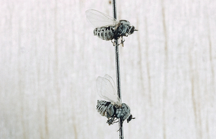
Figure 7-6 Simulium species, black flies. These tiny flies range in size from 1 to 6 mm in length. (From Hendrix CM, Robinson E: Diagnostic parasitology for veterinary technicians, ed 3, St Louis, 2006, Mosby.)
Because the females lay their eggs in well-aerated water, these flies often are found in the vicinity of swiftly flowing streams. They are swift fliers and move in great swarms, inflicting painful bites and sucking the host’s blood. These flies may keep cattle from grazing or cause them to stampede. The animal’s ears, neck, head, and abdomen are favorite feeding sites. These flies also feed on poultry and can serve as an intermediate host for the protozoan parasite Leukocytozoon.
Diagnosis.: Adult black flies are most often collected in the field and are not found on animals presented to a veterinary clinic. They are identified by their small size, humped back, and strong venation in the cranial region of the wings. Identification of black flies is probably best left to an entomologist.
Lutzomyia (New World Sand Fly): Members of the genus Lutzomyia are commonly referred to as “New World sand flies.” They are tiny, mothlike flies, rarely more than 5 mm long. A key feature for identification is that their bodies and wings are covered with fine hairs. They tend to be active only at night and are weak fliers. These tiny flies transmit the protozoan parasite Leishmania species.
Culicoides species: Culicoides gnats are also commonly known as “no see-ums,” “punkies,” or “sand flies.” They are tiny gnats (1 to 3 mm in length), similar in appearance to black flies. They inflict painful bites, suck the blood of their hosts, and are active at dusk and dawn, especially during the winter months. These gnats tend to feed on the dorsal or ventral areas of the host; the feeding site depends on the species of biting gnat.
Horses often become allergic to the bites of Culicoides gnats, scratching and rubbing bitten areas, causing alopecia, excoriations, and thickening of the skin. This condition has several names, including “Queensland itch,” “sweat itch,” “sweet itch,” and “summer dermatitis” (the latter name because it is often seen during the warmer months of the year). These flies also serve as the intermediate host for Onchocerca cervicalis, a nematode whose microfilariae are found in the skin of horses. These flies also transmit the bluetongue virus of sheep.
Anopheles, Aedes, and Culex Species (Mosquitoes): Although they are tiny, fragile Dipterans, mosquitoes are some of the most voracious blood feeders on domestic animals and human beings (Fig. 7-7). Mosquitoes can plague livestock and in swarms have been known to keep cattle from grazing in certain areas or cause them to stampede. The feeding of large numbers of swarming mosquitoes may cause significant anemia in domestic animals. Large numbers of mosquitoes may be produced from eggs laid in relatively small bodies of water. Although they are known for spreading malaria (Plasmodium species), yellow fever, and elephantiasis among people, mosquitoes are probably best known in veterinary medicine as the intermediate host for the canine heartworm, Dirofilaria immitis.
Hippelates Species (Eye Gnats, “Dog Penis” Gnats): These tiny, fragile Dipterans are nonbiting gnats with sponging mouthparts that are very similar to the mouthparts of the common house fly. These tiny gnats are often found around a dog’s penis (hence, the nickname “dog penis gnats”). These Dipterans are not capable of biting the host but often feed on liquid secretions from the host. These nonbiting gnats frequently are found around the teats of dairy cattle. They have been know to serve as mechanical vectors of a variety of bacteria.
Chrysops and Tabanus Species (Deer Fly, Horse Fly): Chrysops species (deer flies) and Tabanus species (horse flies) are large (up to 3.5 cm long), heavy-bodied, robust Dipterans with powerful wings and large eyes. Horse flies and deer flies are the largest flies in the Diptera group, in which only the females feed on vertebrate blood. Figure 7-8 shows Tabanus species, the largest blood-feeding Dipterans. Horse flies are larger than deer flies. Deer flies have a dark band passing from the cranial to the caudal margin of the wings.
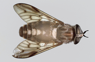
Figure 7-8 Tabanus species, the largest blood-feeding Dipteran. This tabanid is approximately 2.5 cm in length. (From Hendrix CM, Robinson E: Diagnostic parasitology for veterinary technicians, ed 3, St Louis, 2006, Mosby.)
Adult flies lay eggs in the vicinity of open water. Larval stages of these flies are found in aquatic to semiaquatic environments, often buried deep in mud at the bottom of lakes and ponds. Adults are seen in summer and are fond of sunlight. Female flies feed in the vicinity of open water and have reciprocating, scissorlike mouthparts. They use these sharp, bladelike mouthparts to lacerate tissues and lap up the oozing vertebrate blood. These flies feed primarily on large animals such as cattle and horses. Preferred feeding sites include the underside of the abdomen around the navel, the legs, or the neck and withers.
Horse flies and deer flies feed a number of times at multiple feeding sites before they stop feeding. When disturbed by the animal’s swatting tail or by the panniculus reflex (skin twitching), the flies leave the host, but the blood continues to ooze from the open wound. These fly bites are painful. Affected cattle and horses become restless. Because they often feed on multiple hosts, these flies may act as mechanical transmitters of anthrax, anaplasmosis, and the virus of equine infectious anemia.
Diagnosis.: These flies are most easily recognized by their large, robust size and lacerating, scissorlike mouthparts. Species identification of intact adult and larval horse and deer flies is probably best left to an entomologist.
In the second group of blood-feeding Dipterans, both male and female adult flies require a vertebrate blood meal. These species include the stable fly Stomoxys calcitrans, the horn fly Haematobia irritans, the wingless sheep ked Melophagus ovinus, and Lynchia and Pseudolynchia species, the louse flies of raptors and songbirds.
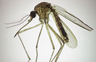
Stomoxys calcitrans (Stable Fly): The stable fly, Stomoxys calcitrans, is often called the “biting housefly.” It is approximately the size of Musca domestica, the common housefly. Rather than possessing sponging mouthparts, the stable fly has a bayonet-like proboscis that protrudes forward from the head (Fig. 7-9). These flies are found worldwide. In the United States, they are found in the central and southeastern states, where cattle are raised. Both male and female flies are avid blood feeders, feeding on any domestic animal. They usually attack the legs and ventral abdomen and may also bite the ears. These flies also feed on the tips of the ears of dogs with pointed ears, especially the German Shepherd. These dogs’ ears often demonstrate loss of hair and the presence of dried, crusty blood on the ear tips.
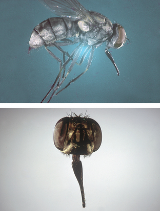
Figure 7-9 Stomoxys calcitrans, the stable fly. Note the bayonet-like proboscis that protrudes forward from the head. The stable fly is approximately the same size as a housefly. (From Hendrix CM, Robinson E: Diagnostic parasitology for veterinary technicians, ed 3, St Louis, 2006, Mosby.)
This fly feeds on both horses and cattle, with horses the preferred host. The fly usually lands on the host with its head pointed upward. It is a sedentary fly, not moving on the host. The fly inflicts painful bites that puncture the skin and bleed freely. The stable fly stays on the host for short periods, during which it obtains the blood meals. This is an outdoor fly; however, in the late fall and during rainy weather, it may enter barns.
Stable flies are mechanical vectors of anthrax in cattle and equine infectious anemia. They are the intermediate host for Habronema muscae, a nematode found in the stomach of horses. When large numbers of stable flies attack dairy cattle, milk production can fall. Beef cattle may refuse to graze in the daytime when they are attacked by large numbers of flies; as a result, these cattle do not gain the usual amount of weight.
Haematobia irritans (Horn Fly): Haematobia irritans is often called the horn fly. This dark-colored fly is approximately 3 to 6 mm in length, half the size of Stomoxys calcitrans, the biting housefly. Like the stable fly, the horn fly has a bayonet-like proboscis that protrudes forward from the head. These flies are found almost exclusively on cattle throughout North America.
When the air temperature is less than 70° F, horn flies cluster around the base of the horns; this is the origin of the name horn fly. In warmer climates, thousands of flies often cluster on the hosts’ shoulders, backs, and side; these areas are least disturbed by tail switching. On hot, sunny days, horn flies congregate on the ventral abdomen.
Adult horn flies spend most of their lives on cattle and leave the host only to deposit their eggs in fresh cow manure. Using their tiny bayonet mouthparts, they feed frequently, sucking blood and other fluids, and cause considerable irritation. Female flies are more aggressive than males. Harassment by the flies and loss of blood often reduce weight gains and milk production in cattle. Horn flies probably cause greater losses in cattle in the United States than any other blood-sucking fly. Adult horn flies also cause focal midline dermatitis on the ventral abdomen of horses. These flies also serve as the intermediate host for Stephanofilaria stilesi, a filarial parasite that produces plaquelike lesions on the ventral abdomen of cattle.
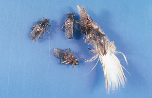
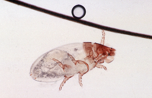
Melophagus ovinus (Sheep Ked): Melophagus ovinus is often called the sheep ked. Members of the order Diptera usually have one pair of wings (two wings). Keds are an exception to that rule; they are wingless Dipterans. Keds are hairy, leathery, and 4 to 7 mm in length. The head is short and broad, the thorax brown, and the abdomen broad and grayish-brown. The legs are strong and armed with stout claws (Fig. 7-10). Some say that keds have a “louselike” appearance, but they are not related to lice.
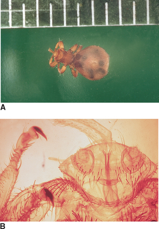
Figure 7-10 Melophagus ovinus, the sheep ked. Keds are hairy, leathery, and 4 to 7 mm in length. The head is short and broad. The thorax is brown, and the abdomen broad and grayish brown. The legs are strong and armed with stout claws. (From Hendrix CM, Robinson E: Diagnostic parasitology for veterinary technicians, ed 3, St Louis, 2006, Mosby.)
Keds are permanent ectoparasites of sheep and goats. Their pupal stages are often found attached to the wool or hair of the host. Keds are avid blood feeders. Heavy infestations can reduce the condition of the host considerably and even cause anemia. Their bites cause pruritus over much of the host’s body; infested sheep often bite, scratch, and rub themselves, damaging the wool. Ked feces often stain the wool and do not wash out readily. Keds are most numerous in the cold temperatures of the fall and winter months. Their numbers decline as temperatures warm in the spring and summer months.
Lynchia and Pseudolynchia species (Louse Flies): Lynchia and Pseudolynchia species are often called louse flies and, like the keds, they are not related to lice. These Dipterans are closely related to the wingless sheep keds, but they are found to parasitize a wide variety of birds—from songbirds to pigeons to raptors. They are found among the feathers of these birds; however, they enjoy sucking blood from areas of the skin that are sparsely covered with feathers. These winged Dipterans are dark brown in color, hairy, and leathery in appearance. They are from 4 to 6 mm in length with legs armed with stout, fierce claws (Fig. 7-11). They are swift fliers and move about quickly—even attempting to get into human hair.
Musca autumnalis (Face Fly): The final periodic parasite among the Dipteran flies is one that is not a blood feeder but instead feeds on mucus, tears, and saliva of large animals, particularly cattle. Face flies, Musca autumnalis, are so named because they gather around the eyes and muzzle of livestock, particularly cattle. They also may be found on the withers, neck, brisket, and sides. Face flies feed mostly on liquid media: saliva, tears, and mucus. They usually are not considered blood feeders because their mouthparts are not piercing nor bayonet-like. Instead, their mouthparts are adapted for sponging up saliva, tears, and mucus (Fig. 7-12). They follow blood-feeding flies, disturb them during their feeding process, and then lap up the blood and body fluids that accumulate on the host’s skin. Face flies are found on animals outdoors; they usually do not follow animals into barns.
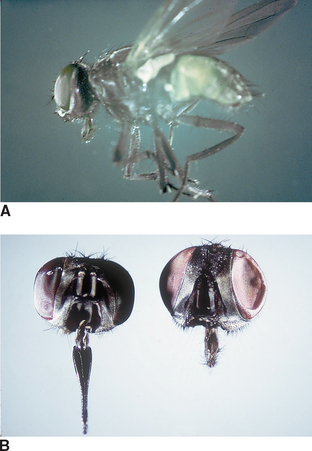
Figure 7-12 Musca autumnalis, the face fly. Its mouthparts are adapted for sponging saliva, tears, and mucus. (From Hendrix CM, Robinson E: Diagnostic parasitology for veterinary technicians, ed 3, St Louis, 2006, Mosby.)
Face flies produce considerable annoyance to the host. The irritation around the host’s eyes stimulates the flow of tears, which attracts even more flies. This harassment ultimately interferes with the host’s productivity. Face flies may be involved in the transmission of Moraxella bovis, a bacterium associated with infectious keratoconjunctivitis or pinkeye in cattle.
Diagnosis.: Face flies are morphologically similar to the housefly, Musca domestica. These two species may be differentiated by minor differences in eye position and color of the abdomen. The veterinary technician probably should not attempt to speciate this fly; speciation requires the skills of a trained entomologist. If flies are found around the face of cattle or horses, they are probably face flies.
Myiasis-Producing Flies: With regard to their roles as ectoparasites, larval Dipterans may develop in the subcutaneous tissues of many domestic animals. When Dipteran larvae develop in the tissue or organs of vertebrate hosts, they produce a condition known as myiasis. On the basis of the degree of host dependence, the myiases (plural of “myiasis”) are divided into two classifications. In facultative myiasis, the fly larvae are usually free living. Under certain circumstances, these normally free-living larvae can adapt to a parasitic dependence on a host. In obligatory myiasis, the fly larvae are completely parasitic; that is, they are completely dependent on the host to develop through the life cycle. In other words, without the host, the obligatory parasites will die.
Facultative Myiasis-Producing Flies: This group of flies includes Musca domestica (housefly) and Calliphora, Phaenicia, Lucilia, Phormia (blowfly, bottle flies), and Sarcophaga (flesh fly) species. The adults of these fly species are colloquially known as “filth flies” because of their propensity to fly from feces to food. Like the face fly, these flies are “vomit drop feeders,” disgorging their stomach contents with their associated liquefaction enzymes and then lapping up the resulting liquid food. These adult flies are frequently seen in kennel situations, alighting on feces that have not been removed from dog runs.
Dipteran larvae that produce facultative myiasis include the housefly M. domestica, the blowflies or bottle flies Calliphora, Phaenicia, Lucilia, and Phormia species, and the flesh fly Sarcophaga species. Larval stages of these flies usually are associated with skin wounds contaminated with bacteria or with a matted haircoat contaminated with feces or urine.
Under their “normal” living conditions, adult flies of these genera lay their eggs in decaying animal carcasses or in feces. In facultative myiasis, the adult flies are attracted to an animal’s moist wound, skin lesion, or soiled haircoat. These sites provide the adult fly with a moist medium on which it feeds. As adult female flies feed in these sites, they lay their eggs. These eggs hatch, producing larvae (maggots) that move independently about the wound surface, ingesting dead cells, exudate, secretions, and debris, but not live tissue. This condition is known as “fly strike” or “strike.” These larvae irritate, injure, and kill successive layers of skin and produce exudates.
Maggots can tunnel through the thin epidermal layer into the subcutis. This process produces tissue cavities in the skin that measure up to several centimeters in diameter. Unless the process is halted by appropriate therapy, the infested animal may die from shock, intoxication, histolysis, or infection. A peculiar, distinct, pungent odor permeates the infested tissues and the affected animal. Advanced lesions may contain thousands of maggots.
As adults, these flies can be pestiferous flies in a veterinary clinical setting. These flies are “vomit drop” feeders and fly from feces to food, spreading bacteria on their feet and their disgorged stomach contents. A veterinary clinic with outdoor kennels is especially susceptible to the assault of these pestiferous flies.
Diagnosis.: A tentative diagnosis of maggot infestation in any domestic animal can easily be made because maggots can be observed in an existing wound or among the soiled, matted haircoat. When facultative myiasis has been diagnosed, the veterinarian must rule out the possibility of obligatory myiasis caused by Cochliomyia hominivorax.
Cochilomyia hominivorax (Primary Screwworm): In obligatory myiasis, the Dipteran larvae must lead a parasitic existence. Only one fly in North America, Cochliomyia hominivorax, is a primary invader of fresh, uncontaminated skin wounds of domestic animals. These larvae must not be confused with the larvae of the more common facultative myiasis-producing flies previously described. C. hominivorax is often referred to as the screwworm fly. Economically it is the most important fly that may attack livestock in the southwestern and southern United States.
Adult female flies are attracted to fresh skin wounds on any warm-blooded animal, where they lay batches of 15 to 500 eggs in a shinglelike pattern at the edge of wounds. The female lays several thousand cream-colored, elongated eggs during her lifetime. They hatch within 24 hours. Larvae enter the wound, where they feed for 4 to 7 days before developing into third-stage (fully grown) larvae. They may be as long as 1.5 cm in length. At this stage they resemble a wood screw. These larvae also bore down into the tissues, much like a wood screw penetrating a piece of wood. When fully mature, the larvae drop from the host to the ground and pupate for approximately a week, after which the adult flies emerge. The adult male and female fly mate only once during their lifetime, a fact that is used to control these flies biologically, a program known as the sterile male release technique.
Diagnosis.: Adult flies are shiny and greenish blue, with a reddish-orange head and eyes, and are 8 to 15 mm long. Larvae often are identified by their wood screw shape and by the deeply pigmented tracheal tubes on the posterior third of the dorsal aspect of the caudal ends. Because of the obligatory nature of the screwworm with regard to breeding in the fresh wounds of any warm-blooded animal, the veterinarian must report any screwworm infestations to both state and federal authorities. C. hominivorax has been eradicated from the United States but occasionally surreptitiously enters the country in imported animals (Fig. 7-13).
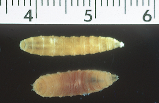
Figure 7-13 Larvae of Cochliomyia hominivorax can be identified by wood screw appearance and two deeply pigmented tracheal tubes on the dorsal aspect of the caudal ends of the third larval stage. (From Hendrix CM, Robinson E: Diagnostic parasitology for veterinary technicians, ed 3, St Louis, 2006, Mosby.)
Cuterebra species (Wolf Warble): Larvae of the genus Cuterebra infest the skin of rabbits, squirrels, mice, rats, chipmunks, and occasionally, dogs and cats. A large discrepancy exists concerning the descriptions of larval Cuterebra. Most of the specimens recovered in a veterinary setting are second- or third-stage larvae. Second-stage larvae are grublike, 5 to 10 mm long, and cream to grayish white in color. They are often sparsely covered with tiny, black, toothlike spines. Third-stage larvae are large, robust, black, and heavily spined (Fig. 7-14). Larval stages usually are found in swollen, cystlike subcutaneous sites, with a fistula or pore communicating to the outside environment. The larval Cuterebra species breathes through this pore (Fig. 7-15).
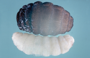
Figure 7-14 Different developmental stages of Cuterebra species. Larval Cuterebra are either sparsely or thickly covered with tiny black spines. (from Hendrix CM, Robinson E: Diagnostic parasitology for veterinary technicians, ed 3, St Louis, 2006, Mosby.)
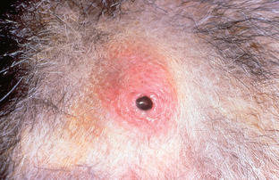
Figure 7-15 Larval Cuterebra species are usually found in swollen, cystlike subcutaneous sites, with a fistula (pore, or hole) communicating to the outside environment. (from Hendrix CM, Robinson E: Diagnostic parasitology for veterinary technicians, ed 3, St Louis, 2006, Mosby.)
Adult flies lay eggs near the entrance to rodent burrows. As a result of this site of egg deposition, the adult flies never annoy the host. Pets usually contract this parasite while investigating or hunting rodent prey. As a result, the most commonly affected sites in dogs and cats are the subcutaneous tissues of the neck and face. Most cases occur during the late summer and early fall. Among the myiasis-producing flies, this Dipteran larva is known for its aberrant or erratic migrations, having been found in a variety of extracutaneous sites, the cranial vault, the eye, and the pharyngeal regions. Clinical signs vary with the site of infection or infestation. Larvae often are discovered in subcutaneous sites during physical examination. The larva usually is removed by enlarging the breathing pore and extracting it with thumb forceps. Great care must be taken not to crush the larva during extraction because anaphylaxis might result.
Hypoderma Species (Ox Warble, Cattle Grub): Two larval stages of Hypoderma flies infect cattle: Hypoderma lineatum and Hypoderma bovis. H. lineatum is found in the southern United States; both species are found in the northern United States and Canada. The adult flies are heavy bodied and resemble honeybees; the adults often are called “heel flies.”
The life cycle is almost a year in duration. Adult flies are bothersome to cattle because they approach cattle to lay eggs. Animals often become apprehensive and disturbed and attempt to escape the fly by running away, an action called gadding. The eggs are approximately 1 mm long and attached to hairs on the legs of cattle. H. lineatum deposits a row of six or more eggs on an individual hair shaft, whereas H. bovis lays its eggs singly. The larvae hatch in approximately 4 days and crawl down the hair shaft to the skin, which they penetrate. They wander through the subcutaneous connective tissue in the leg, migrating to the esophagus (H. lineatum) or the region of the spinal canal and epidural fat (H. bovis), until they reach the subcutaneous tissues of the back. Here the larvae create breathing pores in the skin of the dorsum, through which they later exit and fall to the ground to pupate. The adult flies emerge from the pupae.
Diagnosis.: Adults are beelike and covered with yellow to orange hairs. Mature larvae are 25 to 30 mm long, cream to dark brown in color, and covered with small spines (Fig. 7-16). Lesions consist of large, cystlike or boil-like swellings on the back, with a central breathing pore. As with Cuterebra species, great care must be taken not to crush the larva during extraction because anaphylaxis might result.
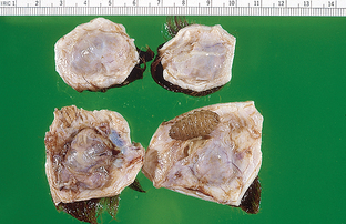
Figure 7-16 Mature larvae of Hypoderma species are 25 to 30 mm long, cream to dark brown in color, and covered with small spines. Lesions consist of large, cystlike swellings on the back, with a central breathing pore. (from Hendrix CM, Robinson E: Diagnostic parasitology for veterinary technicians, ed 3, St Louis, 2006, Mosby.)
Order: Siphonaptera (Fleas)
Of all the ectoparasites discussed thus far, the flea is perhaps the most economically important insect to the veterinarian; treating and preventing flea infestations can be a veterinary practice builder. Because of the extreme popularity of dogs and cats and the prolific nature of the flea (it is able to rebound after populations are exterminated on the animal and in the animal’s environment), special attention should be paid to detecting the various life cycle stages of fleas both on the pet and in the pet’s environment.
Fleas are Siphonapterans, small (4 to 5 mm in length), laterally compressed, wingless insects with powerful hind legs that are used for jumping. Figure 7-17 shows the life stages of Ctenocephalides felis, the cat flea. Adult fleas have piercing/sucking (siphonlike) mouthparts that are used to suck the blood of their host. Figure 7-18 shows the morphologic details of the adult female and male C. felis. More than 2000 species of fleas have been identified throughout the world. Adult fleas are always parasitic, feeding on both mammals and birds. Dogs and cats are host to comparatively few species of fleas.
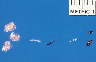
Figure 7-17 Life stages of Ctenocephalides felis, the cat flea. Left to right, Pupae, larvae, eggs, adult male, and female. (from Hendrix CM, Robinson E: Diagnostic parasitology for veterinary technicians, ed 3, St Louis, 2006, Mosby.)
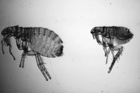
Figure 7-18 Morphologic details of the adult female and male Ctenocephalides felis, the cat flea. (from Hendrix CM, Robinson E: Diagnostic parasitology for veterinary technicians, ed 3, St Louis, 2006, Mosby.)
C. felis is the most common flea found on dogs and cats. The dog flea, C. canis, is uncommon and occurs far less frequently on dogs than does the cat flea.
Echidnophaga gallinacea is also known as the “stick-tight flea” of poultry (Fig. 7-19). A common flea of chickens and guinea fowl, it also feeds on dogs and cats. This flea has unique feeding habits. The female flea inserts its mouthparts into the skin of the host and remains attached at that site. These specimens resemble attached ticks; however, they are fleas.
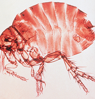
Figure 7-19 Adult Echidnophaga gallinacea, the stick-tight flea of poultry. (from Hendrix CM, Robinson E: Diagnostic parasitology for veterinary technicians, ed 3, St Louis, 2006, Mosby.)
Fleas are not commonly found on horses or ruminants. In barns where feral cats abound and where excessive bedding is used, fleas have been found on calves in large numbers and can produce significant anemia. Pulex irritans, the human flea, has been recovered from dogs and cats, especially in the southeastern United States.
Although the adult flea is the stage most commonly encountered, the veterinary technician also may be presented with flea eggs, larval fleas, or flea droppings from the pet’s environment. Flea eggs and larvae are commonly found in the owner’s bedclothes, the pet’s bedding, travel carriers, doghouses, or clinic cages. Flea eggs resemble tiny, smooth pearls; they are nonsticky, 0.5 mm long, white, oval, and rounded at both ends (Fig. 7-20). Flea larvae resemble tiny fly maggots. They are 2 to 5 mm long, white (after feeding they become brown), and sparsely covered with hairs (Fig. 7-21). Flea larvae spin a sticky, silky cocoon that often becomes covered with environmental debris. This is the pupal stage, a stage that is seldom detected in the pet’s environment (Fig. 7-22).
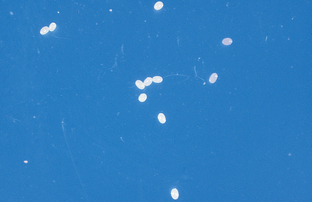
Figure 7-20 Eggs of Ctenocephalides felis, the cat flea. (from Hendrix CM, Robinson E: Diagnostic parasitology for veterinary technicians, ed 3, St Louis, 2006, Mosby.)
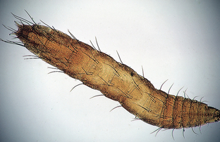
Figure 7-21 Larva of Ctenocephalides felis, the cat flea. (from Hendrix CM, Robinson E: Diagnostic parasitology for veterinary technicians, ed 3, St Louis, 2006, Mosby.)
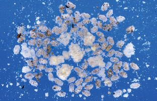
Figure 7-22 Sand-covered pupae of Ctenocephalides felis, the cat flea. (from Hendrix CM, Robinson E: Diagnostic parasitology for veterinary technicians, ed 3, St Louis, 2006, Mosby.)
Diagnosis
Because adult fleas spend most of their time on the host, flea infestation is usually obvious. However, in animals with flea-allergy dermatitis, fleas may be so few in number on the pet as to make diagnosis of flea infestation difficult.
Definitive diagnosis of flea infestation requires demonstration of the adult fleas and/or their droppings (flea dirt, flea feces, flea frass) (Fig. 7-23). Adult fleas defecate large quantities of partially digested blood, commonly called “flea dirt” or “flea frass.” These feces are reddish-black and can appear as fine pepper-like specks, comma-shape columns, or long coils.
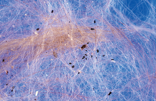
Figure 7-23 Flea dirt, flea feces, or flea frass of Ctenocephalides felis, the cat flea. (from Hendrix CM, Robinson E: Diagnostic parasitology for veterinary technicians, ed 3, St Louis, 2006, Mosby.)
Adult fleas usually are encountered on the animal but also may be collected in the pet’s environment. When recovered from the pet, the larger fleas with an orange to light brown abdomen are females; the smaller, darker specimens are males.
Flea collection is facilitated by spraying the pet with an insecticide. In a few minutes, dead fleas drop off the animal. Alternatively, fleas may be collected with a fine-tooth flea comb available at any veterinary supply store or pet store.
“Flea dirt” may be used to diagnose current or recent flea infestation. To collect a sample of flea dirt, the pet should be combed with a flea comb and the collected samples of detritus placed on a white paper towel moistened with water. Rubbing the flea dirt with a fingertip causes the flea dirt to dissolve, producing a characteristic blood red or rust-red color.
Control: Flea control is important because fleas cause discomfort and irritation to the pet and serve as intermediate hosts to certain helminth parasites. Fleas serve as intermediate host for Dipylidium caninum, the double-pore tapeworm of dogs and cats, and as intermediate host for Dipetalonema (Acanthocheilonema) reconditum, a filarial parasite that resides in the subcutaneous tissues of dogs. Some types of fleas also may transmit disease, such as bubonic plague and endemic typhus, to human beings. In many areas throughout the world, flea control on dogs and cats is one of the most economically important scenarios encountered in a small animal veterinary practice.
Class: Acarina (Mites and Ticks)
Mites and ticks of veterinary importance are members of the class Acarina.
Mites of Veterinary Importance
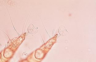
The first group of parasitic mites can be classified together as sarcoptiform mites. Sarcoptiform mites have several common key characteristics or features. These mites may produce severe dermatologic problems in a variety of domestic animals. This dermatitis usually is accompanied by a severe pruritus. Sarcoptiform mites are small, barely visible to the naked eye, and approximately the size of a grain of salt. Their bodies have a round to oval shape. Sarcoptiform mites have legs with pedicels or stalks at the tip. The pedicels may be long or short. If the pedicel is long, it may be straight (unjointed) or jointed. At the tip of each pedicel may be a tiny sucker. Veterinarians and veterinary technicians should use the description of the pedicel (long or short, jointed or unjointed) to identify these sarcoptiform mites.
Sarcoptiform mites are divided into two basic families: Sarcoptidae, which burrow or tunnel within the epidermis, and Psoroptidae, which reside on the surface of the skin or within the external ear canal. Species of Sarcoptidae includes Sarcoptes, Notoedres, and Cnemidocoptes; species of Psoroptidae include Psoroptes, Chorioptes, and Otodectes.
Family Sarcoptidae: Sarcoptic mites burrow or tunnel within the epidermis of the infested definitive host. The entire four-stage life cycle is spent on the host. Male and female mites breed on the skin surface. Female mites penetrate the keratinized layers of the skin and burrow or tunnel through the epidermis. Over a 10- to 15-day period, the female deposits 40 to 50 eggs within the tunnel. After egg deposition, the female dies. Larvae emerge from the eggs in 3 to 10 days and exit the tunnel to wander on the skin surface. These larvae molt to the nymphal stage within minute pockets in the epidermis. Nymphs become sexually active adults in 12 to 17 days and the life cycle begins again.
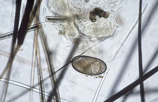
Sarcoptes scabei (Scabies Mites): The disease caused by Sarcoptes scabei is called sarcoptic acariasis. This condition is considered extremely pruritic.
Certain varieties of Sarcoptes mites infest specific hosts. For example, S. scabiei var. canis infests only dogs, and S. scabiei var. suis infests only pigs. Almost every domestic species has its own distinct variety of this mite.
Scabies in dogs is caused by S. scabiei var. canis, which produces an erythematous, papular rash. Scaling, crusting, and excoriation are common. The ears, lateral elbows, and ventral abdomen are most likely to harbor mites. The host’s entire body, however, may be infested. These mites are spread by direct contact and can affect all dogs in a household or kennel. Scabies is extremely contagious. Also, the dog owner can become infested with this mite, but the infestation in people is self-limiting. The mites burrow into human skin, producing a papulelike lesion, but they do not establish a full-blown infestation in people. This mite is considered to be zoonotic, that is, able to transmit disease from animals to human beings. Some dogs may be asymptomatic carriers of this mite. S. scabiei var. felis, which causes scabies in cats, is an extremely rare mite.
Among large animals, pigs most commonly are affected by scabies. Lesions caused by S. scabiei variety suis include small, red papules; alopecia; and crusts, most commonly on the trunk and ears. The mite is rare in cattle (S. scabiei var. bovis). The main infested areas are the head, neck, and shoulders. S. scabiei var. equi of horses is even rarer. The main infested area is the neck. S. scabiei var. ovis infests the face and muzzle of sheep and goats instead of the fleece.
Diagnosis.: Areas with an erythematous, papular rash and crust should be scraped, especially the areas most associated with sarcoptic infestation (ears, lateral elbows, and ventral abdomen of dogs). Repeated scrapings (as many as six scrapings) may be necessary to detect mites. Adult sarcoptic mites are oval and 200 to 400 μm in diameter and have eight legs. The key morphologic feature used to identify this species is the long, unjointed pedicel with a sucker on the end of some of the legs (Fig. 7-24). The anus is located on the caudal end of the body. The eggs of Sarcoptes mites are oval (Fig. 7-25).
Notoedres cati (Feline Scabies Mite): Notoedres cati infests mainly cats but on occasion parasitizes rabbits. This sarcoptiform mite is found chiefly on the ears, back of the neck, face, feet, and in extreme cases, on the entire body. The life cycle is like that of S. scabiei, with the mite burrowing or tunneling in the superficial layers of the epidermis. The characteristic lesion of notoedric acariasis is a yellowing crust in the region of the ears, face, or neck.
Diagnosis.: Notoedric mites are easier to demonstrate in cats than are sarcoptic mites in dogs. Likely infestation sites should be scraped. Like Sarcoptes species, Notoedres mites have a long, unjointed pedicel with sucker on the end of some of the legs. Adult notoedric mites are similar to sarcoptic mites but are smaller, with a dorsal anus. The eggs of notoedric mites are oval.
Cnemidocoptes pilae (Scaly Leg Mite of Budgerigars): Cnemidocoptes pilae causes scaly leg in budgerigars, or parakeets. This mite tunnels in the superficial layers of the epidermis of the pads and shanks of the feet. In severe cases the beak and cere also may be affected. The mite characteristically produces a yellow to gray-white mass that resembles a honeycomb. This condition may be disfiguring to the parakeet. The parasites pierce the skin underlying the scales, causing an inflammation with exudate that hardens on the surface and displaces the scales superficially. This process causes the thickened, scaly nature of the skin. A related species, Cnemidocoptes mutans, produces “scaly leg” in chickens and turkeys.
Diagnosis.: Infested sites should be scraped. Great care should be taken in handling infested birds because parakeets are fragile. The eight-legged, globular mites are approximately 500 μm in diameter. Adult female mites have very short legs and no suckers on the end of their legs (Fig. 7-26). Adult males have longer legs and a long, unjointed pedicel with suckers on the end of some of the legs.
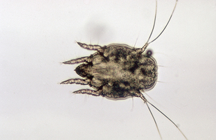
Family: Psoroptidae
Members of the family Psoroptidae reside on the surface of the skin or within the external ear canal. The entire five-stage life cycle (egg, larva, protonymph, deutonymph or pubescent female, and adult egg-laying female) is spent on the host. Adult male and female mites breed on the skin surface. The female produces 14 to 24 elliptic, opaque, shiny white eggs that hatch within 1 to 3 days. The six-legged nymphs are small, oval, soft, and grayish brown. The eight-legged nymphs are slightly larger than larvae. Larval and nymphal stages may last 7 to10 days. The life cycle is completed in 10 to 18 days. Under favorable conditions, psoroptic mites can live off the host for 2 to 3 weeks or longer. Under optimal conditions, mite eggs may remain viable for 2 to 4 weeks.
Psoroptes cuniculi (Ear Canker Mite of Rabbits): Psoroptes cuniculi occurs most commonly in the external ear canal of rabbits but also has been found in horses, goats, and sheep. These mites live on the surface of the skin and feed on the rabbit host by puncturing the epidermis to obtain tissue fluids. Within the external ear canal of the infested host are the characteristic dried crusts of coagulated serum. The rabbit’s ears appear to be packed with dried corn flakes. Affected animals shake the head and scratch their ears. Lesions sometimes occur on the head and legs. Severely infested animals may become debilitated. Loss of equilibrium may occur, with head tilt.
Diagnosis.: The mites within the crusty debris inside the ear can be easily isolated. The brownish-white female is large, up to 750 μm long. The mites exhibit characteristic long, jointed pedicels with suckers on the ends of some of the legs (Figs. 7-27 and 7-28]). The anus is in a terminal slit.
Psoroptes species (Scab Mite of Large Animals): Psoroptes ovis, Psoroptes bovis, and Psoroptes equi are the scab mites of large animals, residing on sheep, cattle, and horses, respectively. These mites are host specific and reside within the thick hair or long wool areas of the animal. They are surface dwellers and feed by puncturing the epidermis to feed on lymphatic fluid. Serum exudes through the puncture site. After the serum coagulates and forms a crust, wool is lost. The feeding site is extremely pruritic and the animal excoriates itself, producing further wool loss. The mites then migrate to adjacent undamaged skin. As the mites proliferate, tags of wool are pulled out and the fleece becomes matted. Finally, patches of skin are exposed and the skin becomes parchmentlike, thickened, and cracked. The skin may bleed easily. Infested sheep constantly rub against fences, posts, farm equipment, and anything else that might serve as a scratching post. The disease is spread by direct contact or infested premises.
P. bovis in cattle produces lesions on the withers, neck, and rump. These consist of papules; crusts; and wrinkled, thickened skin. P. equi in horses is rare and affects the base of the mane and the tail.
Because of the intense pruritus and the highly contagious nature of the infestation, the occurrence of Psoroptes species in large animals should be reported to state and federal authorities and the United States Department of Agriculture.
Chorioptes species (Foot and Tail Mite, Itchy Leg Mite): Chorioptes equi, Chorioptes bovis, Chorioptes caprae, and Chorioptes ovis are the foot and tail mites of large animals, residing on horses, cattle, goats, and sheep, respectively. These mites are found on the skin surface on the distal (lower) part of the hind legs but may spread to the flank and shoulder area. On cattle, they are frequently found in the tail region, especially in the area of the escutcheon. These mites do not spread rapidly or extensively. They puncture the skin, causing serum to exude. Thin crusts of coagulated serum form on the skin surface. The skin eventually wrinkles and thickens although pruritus is not severe.
Infested horses stamp, bite, and kick, especially at night. Mites typically infest the pasterns, especially those of the hind legs.
Diagnosis.: The characteristic mites can be identified from skin scrapings of infested areas. Chorioptic mites have short, unjointed pedicels, with suckers on the ends of some of the legs (Fig. 7-29). The female mites are approximately 400 μm long.
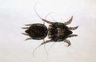
Otodectes cynotis (Ear Mite): Ear mites, Otodectes cynotis, are a common cause of otitis externa in dogs, cats, and ferrets. Although they occur primarily in the external ear canal, ear mites may be found on any area of the body. A common infestation site is the tail and head region. As dogs and cats curl up to sleep, their head (and ears) are often in close proximity to the base of the tail. These mites are spread by direct contact and are highly transmissible both among and between the canine and feline species.
Ear mites are found within the external ear canal, where they feed on epidermal debris and produce intense irritation. Infection is usually bilateral. The host responds to the mite infestation by shaking its head and scratching its ears. Severe infestations may cause otitis media, with head tilt, circling, and convulsions. Auricular hematomas may develop.
Diagnosis.: Mites usually are identified by an otoscope; through an otoscope the mites appear as white, motile objects. The brown exudate collected by swabbing the ear may be placed in mineral oil on a glass slide and the mites observed with a low power microscopic objective. These mites are fairly large, approximately 400 μm; they also may be easily seen with a magnifying glass or even the unaided eye. The mites exhibit characteristic short, unjointed pedicels, with suckers on the ends of some of the legs (Figs. 7-30 and 7-31]). The anus is terminal.
Miscellaneous Mites
Other parasitic mites can be grouped together because they are not sarcoptiform mites. They can, however, produce severe dermatologic problems in a variety of domestic animals. These mites lack pedicels or stalks on their legs that identify sarcoptiform mites.
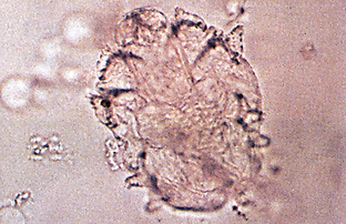
Demodex species: Mites of the genus Demodex reside in the hair follicles and sebaceous glands of most domesticated animals and human beings. In many species they are considered normal, nonpathogenic fauna of the skin. These mites are host specific and are not transmissible from one host species to another. The clinical disease, caused by an increased number of these mites, is called demodicosis.
Demodex mites resemble eight-legged alligators. They are elongated mites with short, stubby legs on the anterior half of the body. Adult and nymphal stages have eight legs, whereas the larvae have six. Adult Demodex mites are approximately 250 μm long (Fig. 7-32). The eggs are spindle shaped or tapered at each end (Fig. 7-33).
Figure 7-32 Adult Demodex canis mite. (from Hendrix CM, Robinson E: Diagnostic parasitology for veterinary technicians, ed 3, St Louis, 2006, Mosby.)
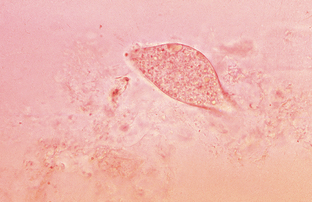
Figure 7-33 Egg of Demodex canis. (from Hendrix CM, Robinson E: Diagnostic parasitology for veterinary technicians, ed 3, St Louis, 2006, Mosby.)
The life cycle of Demodex species has five stages: egg, larva, protonymph, deutonymph, and adult. The developmental periods of these various life cycle stages are not well known.
Of all the domestic animals infested with Demodex species, the dog is the most commonly and most seriously infested. Small numbers of these mites are considered part of the normal skin flora of all dogs. In dogs with immunodeficiencies, however, these mites proliferate and cause skin disease.
Demodicosis occurs in two forms in dogs: localized demodicosis and generalized demodicosis. The predominant clinical sign of the localized form is patchy alopecia, especially of the muzzle, face, and forelimbs. The mites presumably are acquired during intimate contact when the puppy nurses the dam. As a result of that close contact, localized demodicosis often develops in the region of the face. Generalized demodicosis is characterized by diffuse alopecia, erythema, and secondary bacterial contamination over the entire body surface of the dog. An inherited defect in the dog’s immune system is thought to be an important factor in the development of generalized demodicosis.
Cats are infested by two species of demodectic mites: Demodex cati and an unnamed species. D. cati is an elongated mite similar to D. canis. The unnamed species has a broad, blunted abdomen compared with the elongated one of D. cati. The presence of either species on the skin of cats is rare. Localized feline demodicosis is characterized by patchy areas of alopecia and erythema and occasionally crusting on the head (especially around the eyes), ears, and neck. In generalized feline demodicosis, the alopecia, erythema, and crusting usually involve the entire body. Demodicosis also has been associated with ceruminous otitis externa.
Demodectic mites reside in the hair follicles of other species of domestic animals but rarely produce clinical disease. Cattle and goats are most commonly infested but only rarely.
In cattle, D. bovis causes large nodules (abscesses) on the shoulders, trunk, and lateral aspects of the neck. In goats, D. caprae occurs in small papular or nodular lesions on the shoulders, trunk, and lateral aspect of the neck. In sheep, D. ovis rarely causes pustules and crusting around the coronet, nose, ear tips, and periorbital areas. In pigs, D. phylloides rarely produces pustules and nodules on the face, abdomen, and ventral neck. In horses, D. equi occurs around the face and eyes and rarely produces clinical disease.
Diagnosis.: Skin areas with altered pigmentation, obstructed hair follicles, erythema, or alopecia should always be scraped. In localized demodicosis, the areas most commonly affected are the forelegs, perioral region, and periorbital regions. In generalized demodicosis, the entire body may be affected; however, the face and feet usually are the most severely involved. In dogs, areas of apparently normal skin also should be scraped to determine if the disease is generalized. The areas should be clipped and a fold of skin gently squeezed just before scraping. Gentle scraping of affected areas should be continued until capillary blood oozing is observed because these mites live deep in the hair follicles and sebaceous glands (Fig. 7-34).
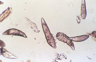
Figure 7-34 A deep skin scraping produces numerous mites of Demodex canis. (from Hendrix CM, Robinson E: Diagnostic parasitology for veterinary technicians, ed 3, St Louis, 2006, Mosby.)
Nodular lesions in large animals should be incised with a scalpel and the caseous material within smeared on a slide with mineral oil, covered with a cover slip, and examined microscopically for mites.
The mites on the slide should be counted and the live/dead ratio determined. The presence of any larval or nymphal stages or eggs should be noted. During therapy for Demodex species, a decrease in the number of eggs and live mites is a good prognostic indicator.
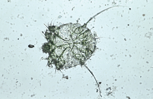
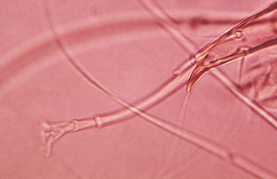
Cheyletiella species (Walking Dandruff): Mites of the genus Cheyletiella are surface dwelling (nonburrowing), residing in the keratin layer of the skin and in the haircoat of the definitive host, which may be a dog, cat, or rabbit. These mites ingest keratin debris and tissue fluids. Cheyletiella mites are sometimes referred to as “walking dandruff” because the mites resemble large, mobile flakes of dandruff. Cheylettid mites have distinct key morphologic features. They are large (386 μm by 266 μm) and visible to the unaided eye. With the compound microscope, their most characteristic key morphologic feature may easily be seen: their enormous hooklike accessory mouthparts (palpi). These palpi assist the mite in attaching to the host as it feeds on tissue fluids. The mite also has comblike structures at the tip of each leg. Members of the genus also are known for their characteristic body shape resembling a shield, a bell pepper, an acorn, or a western horse saddle when viewed from above (Fig. 7-35). Eggs are 235 to 245 μm long and 115 to 135 μm wide (smaller than louse nits) and are supported by cocoonlike structures bound to the host’s hair shaft by strands of fibers. Two or three eggs may be bound together on one hair shaft.
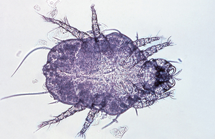
Figure 7-35 Adult Cheyletiella mite. (from Hendrix CM, Robinson E: Diagnostic parasitology for veterinary technicians, ed 3, St Louis, 2006, Mosby.)
Diagnosis.: The key feature of Cheyletiella species infestation is often the moving white “dandruff” flakes along the dorsal midline and head of the host. A handheld magnifying lens or binocular headband magnifier (e.g., OptiVISOR, Donegan Optical Company Inc., Lenexa, KS) often are used to view questionable dandruff flakes or hairs; these are perhaps the quickest methods of diagnosing cheyletiellosis. A fine-tooth flea comb may be used to collect mites; combing dandrufflike debris onto black paper often facilitates visualization of these highly mobile mites. The use of clear cellophane tape to entrap mites collected from the haircoat often simplifies viewing with the compound microscope.
Lynxacarus radovskyi (Feline Fur Mite): Lynxacarus radovskyi, the feline fur mite, is found attached to the shafts of individual hairs on the back, neck, thorax, and hind limbs of cats residing in tropical or warm areas of the United States, such as Florida, Puerto Rico, and Hawaii. These fur mites are laterally compressed. The adults are approximately 500 μm long (Fig. 7-36). Pruritus is not always associated with infestations of L. radovskyi in cats. This mite also may affect human beings who handle infested cats, producing a papular dermatitis.
Trombicula species (Chigger): The chigger (Trombicula species) is yellow to red, has six legs, and is 200 to 400 μm in diameter. The larval stage is the only developmental stage that parasitizes human beings, domestic animals, and wild animals. The larvae are most common in the late summer and early fall and are transmitted by direct contact with the ground or with foliage in fields or heavy underbrush. Nymphal and adult chiggers are nonparasitic and are free living in nature.
Chigger larvae do not burrow into the skin as commonly believed, and they do not feed primarily on host blood. Their food consists of the serous components of tissues. Chiggers attach firmly to the host and inject a digestive fluid that liquefies host cells. The host’s skin becomes hardened and a tube called a stylostome forms at the chigger’s attachment site. Chiggers suck the liquefied host tissues. When the mite has finished feeding, it loosens its grip and falls to the ground. The injected digestive fluid causes the attachment site to itch intensely. Cutaneous lesions tend to be restricted to areas of the body that come in contact with the ground, such as the limbs, interdigital areas, and ventrum, in addition to the head and ears.
The most common chigger mite affecting animals and people is Trombicula alfreddugesi (the North American chigger). Lesions consist of an erythematous, often pruritic papular rash on the ventrum, face, feet, and legs.
Diagnosis.: Chigger infestation (trombiculosis) is diagnosed by an orange, crusting dermatosis; a history of exposure (roaming the outdoors); and identification of the typical six-legged larvae in skin scrapings or on collection from the host. The larvae remain attached to the skin only for several hours. Consequently, trombiculosis may be difficult to diagnose because the pruritus persists after the larvae have dropped.
Pneumonyssoides (Pneumonyssus) caninum (Nasal Mite of Dogs): Pneumonyssoides (Pneumonyssus) caninum is a rare mite that lives in the nasal passages and associated sinuses of dogs. Nasal mites are generally considered to be nonpathogenic; however, reddening of the nasal mucosa, sneezing, head shaking, and rubbing of the nose often accompany infestation. Fainting, labored breathing, asthmalike attacks, and orbital disease have been associated with this mite. Sinusitis caused by these mites may lead to disorders of the central nervous system. Owners may observe these mites exiting the animal’s nostrils.
The life cycle is unknown, but it apparently takes place entirely on the host. Adult males, adult females, and larvae have been identified, but no nymphal stages have been observed. Transmission probably occurs by direct contact with an infested animal.
Ornithonyssus sylviarum (Northern Mite of Poultry) and Dermanyssus gallinae (Red Mite of Poultry): These two mites parasitize poultry, but they differ in the sites they tend to infest. Ornithonyssus sylviarum is a 1-mm, elongated to oval mite usually found on birds but also in their nests or houses. They feed intermittently on the birds, producing irritation, weight loss, decreased egg production, anemia, and even death. These mites have been known to bite human beings.
Dermanyssus gallinae is a 1-mm, elongated to oval, whitish, grayish, or black mite that feeds on birds. This mite has a distinct red color when it has recently fed on its host’s blood. D. gallinae lays its eggs in cracks and crevices in the walls of poultry houses. Nymphs and adults are periodic parasites, hiding in the crevices of the poultry houses and making frequent visits to the host to feed. Because of their blood-feeding activity, they may produce significant anemia and much irritation to the host. Infested birds are listless and egg production may drop. Loss of blood may result in death. These mites also occur in birds’ nests in the eaves of houses or in air conditioners. The mites can migrate into homes and infest human beings.
Diagnosis.: O. sylviarum usually is found on the avian host, whereas D. gallinae is a periodic parasite, usually found in the host’s environment. Specimens should be cleared in lactophenol and the anal plates examined with a compound microscope. The anus of O. sylviarum is on the cranial half of the anal plate, whereas the anus of D. gallinae is on the caudal half.
Ticks of Veterinary Importance
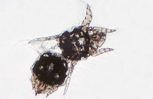
Ticks are small- to medium-sized acarines with dorsoventrally compressed, leathery bodies. The tick’s head, the capitulum, serves as an organ of cutting and attachment. It is made of a penetrating, anchorlike sucking organ, the hypostome; and four accessory appendages, two cutting chelicerae and two pedipalps, which act as sensors and supports when the tick fastens to the host’s body. The tick’s body may be partially or entirely covered by a hard, chitinous plate, the scutum. Mouthparts may be concealed under the tick’s body or may extend from the cranial border. Most ticks are inornate; that is, they are reddish or mahogany, without markings. Some species are ornate and have distinctive white patterns on the dark scutum background. Adult ticks have eight legs, with claws on the ends of the legs.
Ticks are important parasites because of their voracious blood-feeding activity. They are important also because they can transmit many parasitic, bacterial, viral, and other diseases, such as borreliosis (Lyme disease), among animals and from animals to human beings. These pathogenic organisms may be passively transmitted, or the tick may serve as an obligatory intermediate host for protozoan parasites.
The salivary secretions of some female ticks are toxic and can produce a syndrome known as “tick paralysis” in human beings and animals. Tick species commonly associated with tick paralysis are Dermacentor andersoni (the Rocky Mountain spotted fever tick), Dermacentor occidentalis (the Pacific Coast tick), Ixodes holocyclus (the Australian paralysis tick), and Dermacentor variabilis (the wood tick).
Ticks of veterinary importance are divided into two families: the Argasid, or soft ticks and the Ixodid, or hard ticks. Argasid ticks lack a scutum, or hard, chitinous plate. The mouthparts of the adults cannot be seen when viewed from the dorsal aspect. Ixodid ticks have a hard, chitinous scutum that covers all the male tick’s dorsum and approximately one third or less of the female’s dorsum. Depending on the degree of engorgement, male ticks are much smaller than female ticks.
Two species of Argasid ticks are important: Otobius megnini (the spinose ear tick) and Argas persicus (the fowl tick). Thirteen economically important tick species are in the Ixodid family. These include Rhipicephalus sanguineus, Ixodes scapularis, Dermacentor species, and Amblyomma species. Of these species, only R. sanguineus infests buildings; the remaining ticks attack their hosts outdoors.
Specific identification of ticks is difficult and should be performed by a veterinary parasitologist or a trained arthropodologist. Ticks usually are identified by the shape and length of the capitulum, the shape and color of the body, and the shape and markings on the scutum. Male and unengorged female ticks are easier to identify than engorged females. Determining the species of larval or nymphal ticks is difficult. Common species may be identified by their size, shape, color, body markings, host, and location on the host.
Four major stages exist in the life cycle of ticks: egg, larva, nymph, and adult. After engorgement on the host, female ticks drop off the host and seek protected places, such as within cracks and crevices or under leaves and branches to lay their eggs (Fig. 7-37). The six-legged larvae, or seed ticks, hatch from the eggs and feed on a host (Fig. 7-38). The larva molts to the eight-legged nymphal stage, which resembles the adult stage but lacks the functioning reproductive organs of the adult stage. After one or two blood meals, the nymph matures and molts to the adult stage. During the larval, nymphal, and adult stages, ticks may infest one to three or even many different host species. This ability to feed on several hosts during the life cycle plays an important role in the transmission of disease pathogens among hosts. Any infestation of domestic animals by mites or ticks is referred to as acariasis.
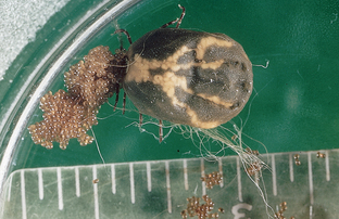
Figure 7-37 Adult female Dermacentor variabilis tick laying hundreds of eggs. (from Hendrix CM, Robinson E: Diagnostic parasitology for veterinary technicians, ed 3, St Louis, 2006, Mosby.)
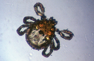
Figure 7-38 Six-legged larval Rhipicephalus sanguineus. (from Hendrix CM, Robinson E: Diagnostic parasitology for veterinary technicians, ed 3, St Louis, 2006, Mosby.)
Most ticks do not tolerate direct sunlight, dryness, or excessive rainfall. They can survive as long as 2 to 3 years without a blood meal, but females require blood before egg fertilization and deposition. Tick activity is restricted during the cold winter months.
Argasid (Soft) Ticks
Otobius megnini (Spinose Ear Tick): Otobius megnini, the spinose ear tick, is an unusual soft tick in that only the larval and nymphal stages are parasitic. The adult stages are not parasitic but are free living, found in the environment of the definitive host, usually in dry, protected places, in cracks and crevices, under logs, and on fence posts. The larval and nymphal stages feed on horses, cattle, sheep, goats, and dogs. These ticks usually are associated with the semiarid or arid areas of the southwestern United States. With widespread interstate movement of animals, this soft tick may occur throughout North America. As with most soft ticks, the mouthparts may not be visible when viewed from the dorsal aspect (Fig. 7-39). The nymphal stage is widest in the middle, almost violin shape. It is covered with tiny, backward-projecting spines, which is the origin of the name spinose. Larvae and nymphs usually are found within the ears of the definitive host.
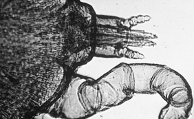
Figure 7-39 Otobius megnini, the spinose ear tick, is an unusual soft tick in that only the larval and nymphal stages are parasitic. The mouthparts may not be visible when viewed from the dorsal aspect. (from Hendrix CM, Robinson E: Diagnostic parasitology for veterinary technicians, ed 3, St Louis, 2006, Mosby.)
Spinose ear ticks are extremely irritating to the definitive host. They often occur in large numbers, deep within the external ear canal. These ticks imbibe large amounts of host blood; however, because they are soft ticks, they do not enlarge with feeding. Large numbers may produce ulceration deep within the ear. The ears become highly sensitive and the animals may shake their heads. The pinnae may become excoriated by the animal’s shaking and rubbing its head.
Argas persicus (Fowl Tick): Argas persicus, the fowl tick, is a soft tick of chickens, turkeys, and wild birds. These ticks are periodic parasites, hiding in cracks and crevices during the day and becoming active during the night, when they feed intermittently on the avian host. The adults are 7 mm long and 5 mm wide. In the unengorged state, they are reddish brown. After engorgement, they are slate blue. These ticks are flat and leathery, and the tegument is covered with tiny bumps. As soft ticks, they lack a scutum. The mouthparts are not visible when the adult tick is viewed from the dorsal aspect.
Heavily infested birds may develop anemia. These ticks are worrisome to birds, particularly at night. Egg production by hens may decrease or cease.
Ixodid (Soft) Ticks
Rhipicephalus sanguineus (Brown Dog Tick): Rhipicephalus sanguineus, the brown dog tick, is an unusual hard tick; it may invade both kennel and household environments. This tick is distributed throughout North America. It has an inornate, uniformly reddish-brown scutum, and it feeds almost exclusively on dogs. R. sanguineus also has a distinguishing morphologic feature. Its basis capitulum has prominent lateral extensions that give this structure a decidedly hexagonal appearance (Fig. 7-40). The engorged female is often slate gray. In southern climates, the tick is found outdoors but in northern climates it becomes a serious household pest, breeding indoors.
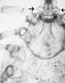
Figure 7-40 Lateral expansion of the basis capitulum (base of the head) (arrows) of an adult Rhipicephalus sanguineus. This key morphologic feature is used to identify this parasite, which can breed in the host’s environment. (from Hendrix CM, Robinson E: Diagnostic parasitology for veterinary technicians, ed 3, St Louis, 2006, Mosby.)
The bites of this tick can irritate dogs. Severe infestations may cause heavy blood loss. This tick is also an intermediate host for Babesia canis, the agent that causes piroplasmosis in dogs.
Dermacentor variabilis (American Dog Tick, Wood Tick): Dermacentor variabilis, the American dog tick or wood tick, is found primarily in the eastern two thirds of the United States; however, with increased mobility of American households, the tick may occur throughout the country. Unlike R. sanguineus, this tick inhabits only grassy, scrub brush areas, especially roadsides and pathways. This three-host tick initially feeds on small mammals and rodents; however, dogs (and human beings) can serve as hosts for this ubiquitous tick. This tick is a seasonal annoyance to human beings and domestic animals. It can serve as a vector of Rocky Mountain spotted fever, tularemia, and other diseases and may also produce tick paralysis in animals and human beings. This tick has a dark-brown, ornate scutum with white striping. Unfed adults are approximately 6 mm long, whereas adult engorged females are about 12 mm long and blue-gray (Fig. 7-41).
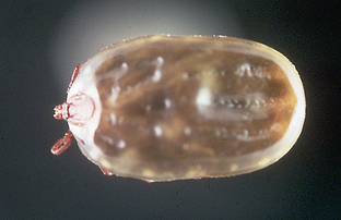
Amblyomma americanum (Lone Star Tick): Amblyomma americanum, the “lone star” tick, gets its common name from a characteristic white spot on the apex of its scutum. The spot is more conspicuous on male ticks than on female ticks. This tick is distributed throughout the southern United States but also is found in the Midwest and on the Atlantic coast.
This three-host tick is found most often in the spring and summer months, parasitizing the head, belly, and flanks of wild and domestic animal hosts. It also feeds on human beings and is said to have a painful bite. It can produce anemia and has been incriminated as a vector of tularemia and Rocky Mountain spotted fever.
Amblyomma maculatum (Gulf Coast Tick): Amblyomma maculatum, the Gulf Coast tick, is a three-host tick found in the ears of cattle, horses, sheep, dogs, and human beings. It occurs in areas of high humidity on the Atlantic and Gulf coasts. It produces severe bites and painful swellings and is associated with tick paralysis. This tick has silvery markings on its scutum. Larval and nymphal stages occur on ground birds throughout the year. The number of adult ticks on cattle decreases during the winter and spring and increases in the summer and fall. When the ear canals of cattle and horses are infested, the pinna may droop and become deformed.
Diagnosis.: A. maculatum is easily identified by the silvery markings on its scutum (Fig. 7-42).
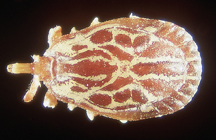
Boophilus annulatus (Texas Cattle Fever Tick): Boophilus annulatus is often called the Texas cattle fever tick or the North American tick. This one-host tick has historic significance in that it is the first arthropod shown to serve as an intermediate host for a protozoan parasite, Babesia bigemina of cattle, a milestone in veterinary parasitology. This tick has been completely eradicated from the United States; however, any Boophilus species infestation should be reported to the proper regulatory agencies. The tick should be identified by a specialist and control methods applied. B. annulatus frequently enters the United States from Mexico.
The engorged female is 10 to 12 mm long and the male 3 to 4 mm. The mouthparts are short and no festoons are on the caudal aspect of the abdomen.
Because this is a one-host tick, larvae, nymphs, and adult ticks may be found on the same animal. They do not have to leave the host to complete the life cycle. Animals with heavy infestations are restless and irritated. In an attempt to rid themselves of ticks, they rub, lick, bite, and scratch themselves. Irritated areas may become raw and secondarily infected. Heavily infested cattle may become anemic.
PHYLUM: NEMATODA (ROUNDWORMS)
The next part of this chapter discusses the superfamilies belonging to the class Secernentea and how these skin-dwelling parasites are relevant to veterinary practice.
Superfamily: Rhabditoidea
Pelodera strongyloides (Free-Living Saprophytic Nematode): Pelodera strongyloides is a free-living saprophytic nematode that normally lives in moist soil. These are facultative parasites. They are normally free living; however, under certain circumstances they can invade mammalian skin and develop to a parasitic existence. Male and female adult P. strongyloides nematodes are found in soil mixed with moist organic debris, such as straw. The females produce eggs that hatch into first-stage larvae. These larvae invade the superficial layers of damaged or scarified skin, producing mild dermatitis. The skin may become reddened, denuded, and covered with a crusty material. Occasionally a pustular dermatitis develops. Because dogs become infested by lying on contaminated soil, these lesions usually are observed on the ventrum or medial (inner) surface of the limbs. Larvae of P. strongyloides also may be recovered from the skin of cattle recumbent on soil containing these larvae.
Diagnosis.: These larvae (and possibly adults) can be identified in superficial skin scrapings. Larvae of P. strongyloides are 596 to 600 μm long. These larvae must be differentiated from the microfilariae of the canine heartworm, D. immitis; the microfilariae of D. reconditum; and the first-stage larvae of Dracunculus insignis.
Superfamily: Spiruroidea (Spirurid Nematodes)
Habronema and Draschia species: Adult Habronema nematodes are found on the stomach mucosa of horses under a thick layer of mucus. Adult Draschia species produce large, fibrous nodules in the equine stomach wall; the nematodes reside within the nodules. The adults do not parasitize the skin (see Chapter 6). However, the larval stages of these stomach parasites can be deposited by flies (Musca domestica, Stomoxys calcitrans) into skin wounds on horses. Here the larvae produce a condition known as “cutaneous habronemiasis,” “cutaneous draschiasis,” or “summer sores.”
The lesions vary in size and have an uneven surface consisting of a soft, reddish-brown material covering a mass of firmer granulations. These lesions are seen on body parts most susceptible to trauma, such as the legs, withers, male genitalia, and medial canthus of the eye. Infested wounds tend to increase in size and do not respond to ordinary treatment until the following winter, when they spontaneously heal.
Superfamily: Filarioidea (Filarial Nematodes)
Onchocerca cervicalis: Microfilariae of Onchocerca cervicalis, the filarial parasite of horses, produce recurrent dermatitis and periodic ophthalmia. The adults live in the ligamentum nuchae. Females produce microfilariae that migrate to the dermis by way of connective tissue. This parasite is spread by the biting midge Culicoides species. The midge feeds on a horse’s blood and ingests the microfilariae, which develop to the infective third stage within the fly. When the fly bites another horse, larvae are injected into the connective tissue and develop to adults during migration to the ligamentum nuchae. The characteristic signs of cutaneous onchocerciasis include patchy alopecia; scaling on the head, neck, shoulders, and ventral midline; and sometimes, intense pruritus.
Many infected horses are asymptomatic. Microfilariae of Onchocerca species concentrate in certain areas, with the ventral midline the most common area of concentration. Because more than 90% of normal hosts are probably infected with O. cervicalis, detection of microfilariae in the skin of the ventral midline is not diagnostic for cutaneous onchocerciasis. However, the presence of microfilariae in diseased skin is highly indicative, though not diagnostic, of cutaneous onchocerciasis.
Diagnosis.: Onchocerca microfilariae may be demonstrated by the following procedure. After clipping the skin site and performing a surgical scrub, a 6-mm punch biopsy is used to obtain a skin sample (see Chapter 9). With a single-edge razor blade or scalpel blade, half of the tissue is minced in a small amount of preservative-free physiologic saline on a glass slide and allowed to stand for 5 to 10 minutes. Drying of the specimen is prevented by placing the slide in a covered chamber with a small amount of saline. The slide is then examined with the low power (10×) objective. Because the translucent microfilariae are difficult to observe, low-intensity light and high contrast (achieved by lowering the condenser) are essential. Live microfilariae are identified by their vigorous swimming activity at the edge of the tissue. O. cervicalis microfilariae are slender and 207 to 240 μm long. The other half of the biopsy should be submitted for routine histopathologic examination.
Stephanofilaria stilesi: Stephanofilaria stilesi is a small filarial nematode found in the skin of ruminants such as cattle, goats, and buffalo and of wild mammals. It commonly causes dermatitis along the ventral midline of cattle in the United States. The infective larvae are transmitted by the bites of horn flies, Haematobia irritans. The skin lesions are thought to be caused by both adult and microfilarial stages.
The lesions caused by S. stilesi are located near the umbilicus and initially consist of small red papules. Later the lesions develop into large pruritic areas (up to 25 cm) of alopecia, with thick, moist crusts.
Dipetalonema (Acanthocheilonema) reconditum (Subcutaneous Filarial Parasite of Dogs): Adult Dipetalonema (Acanthocheilonema) reconditum is a nonpathogenic parasite that resides in the subcutaneous tissues of the dog. It also may be found within a body cavity. Occasional subcutaneous abscesses and ulcerated areas have been associated with this parasite. The intermediate host for this parasite is the flea, C. felis. Because this parasite is found in enzootic areas where D. immitis is present, differentiation of the microfilariae of these two filarial parasites is necessary (see Chapter 6).
Diagnosis.: Adults of D. (Acanthocheilonema) reconditum rarely are recovered from their subcutaneous sites. Microfilariae may be recovered rarely in deep skin scrapings that draw blood. When subjected to the modified Knott’s procedure, the microfilariae of D. reconditum average approximately 285 μm long, with a button-hook tail and a blunt (broom handle–shaped) cranial end. They must be differentiated from microfilariae of D. immitis, first-stage larvae of Pelodera strongyloides, and first-stage larvae of Dracunculus insignis.
Dirofilaria immitis (Canine Heartworm): Dirofilaria immitis, the canine heartworm, normally resides in the right ventricle and pulmonary arteries of its definitive host, the dog. Adult heartworms also may occur aberrantly and may be found in a variety of extravascular sites, including cystic spaces in the subcutaneous sites (Fig. 7-43). When adult heartworms are found aberrantly, they are usually single, immature, isolated worms. Any female heartworms found within the cyst have not been fertilized by a male heartworm. Therefore such females are not gravid and do not produce microfilariae.
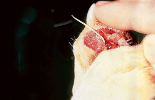
Figure 7-43 Aberrant adult Dirofilaria immitis heartworm in a subcutaneous interdigital cyst in a dog. (from Hendrix CM, Robinson E: Diagnostic parasitology for veterinary technicians, ed 3, St Louis, 2006, Mosby.)
Adult female heartworms in the right ventricle and pulmonary arteries may produce microfilariae after mating with adult males. These microfilariae may be recovered occasionally in deep skin scrapings that draw blood.
Diagnosis.: Aberrant adult D. immitis heartworms within cystic spaces in the skin can be surgically removed. When subjected to the modified Knott’s procedure, the microfilariae of D. immitis are 310 to 320 μm long, with a straight tail and tapering cranial end. They must be differentiated from the microfilariae of D. (Acanthocheilonema) reconditum, first-stage larvae of Pelodera strongyloides, and first-stage larvae of Dracunculus insignis.
Superfamily: Dracunculoidea
Dracunculus insignis (Guinea Worm): Dracunculus insignis, the Guinea worm, is a nematode found in the skin of dogs. The adult female nematode resides subcutaneously and produces a draining, ulcerous lesion in the skin, usually on the dog’s limb. The cranial end of the female worm extends from this ulcer. When the female worm within the lesion comes in contact with water, its uterus prolapses through the worm’s cranial end and ruptures, releasing a mass of first-stage larvae into the water. These larvae are 500 to 750 μm long and have a unique, long tail (Fig. 7-44). The larvae are ingested by tiny crustaceans in the water; within the crustaceans, the larvae develop to the infective third stage. Dogs become infected with D. insignis by drinking water containing the infected crustaceans.
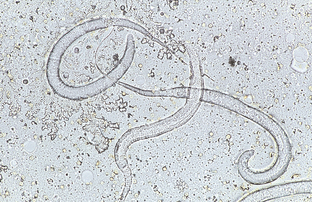
Figure 7-44 Unique first-stage larva of Dracunculus insignis. These larvae are 500 to 750 μm long and have a characteristic long tail. (from Hendrix CM, Robinson E: Diagnostic parasitology for veterinary technicians, ed 3, St Louis, 2006, Mosby.)
Diagnosis.: If D. insignis infection is suspected, the ulcer with its associated worm should be dipped in cool water. The cool water is a stimulus for the female worm to expel her larvae. The water containing expelled larvae should be centrifuged and the sediment examined for the characteristic first-stage larvae. Larvae of D. insignis must be differentiated from the microfilariae of D. immitis, microfilariae of D. reconditum, and first-stage larvae of P. strongyloides. Once identified, the adult female worm may be surgically removed.
PHYLUM: ANNELIDA (SEGMENTED WORMS)
Order: Gnathobdellida
Hirudo medicinalis (Medicinal Leech): Leeches are annelids; they are not considered true helminths but are often described as parasitic worms. As ectoparasites of human beings, domestic animals, and wild animals, leeches are members of the phylum Annelida and the class Hirudinea. Leeches may have a pathologic or beneficial role in veterinary medicine.
The term hirudiniasis is derived from the classic Linnaean nomenclature and is defined as invasion of the nose, mouth, pharynx, or larynx by leeches, or the attachment of leeches to the skin. Leeches are voracious blood feeders; depending on the number that attach to the host, the host may become anemic and die from blood loss. Leeches have recently gained favor as postsurgical tools in reconstructive and microvascular surgery. H. medicinalis, the medicinal leech, has been used in reconstructive and microvascular surgery in human beings; such use in veterinary medicine is forthcoming.
Diagnosis.: Leeches are segmented worms with slender, leaf-shaped bodies devoid of bristles. A typical leech has two suckers, a large and adhesive caudal sucker and a smaller cranial one that surrounds the mouth. Most leeches are found in fresh water and a few in salt water; some are terrestrial varieties.
Recommended Reading
Bowman, DD, Lynn, RC, Eberhard, ML. Georgi’s parasitology for veterinarians, ed 8. Philadelphia: WB Saunders, 2003.
Hendrix, CM, Robinson, E. Diagnostic parasitology for veterinary technicians, ed 3. St Louis: Mosby, 2006.
Zajac, AM, Conboy, GA. Veterinary clinical parasitology, ed 7. Ames, IA: Blackwell Publishing, 2006.