Page 47 

Chapter 3 Healing

Healing 48
Wound Healing 49–51
Healing – Fibrosis 52
Healing – Special Situations 53–57
Fracture Healing 58–60
  Page 48 

Healing

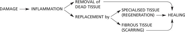
Healing is the final stage of the response of tissue to injury.

image

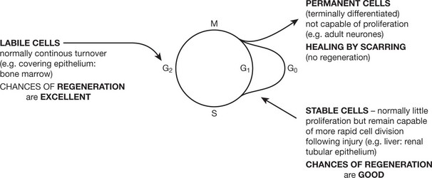
The capacity of a tissue for regeneration depends on its proliferative ability and on the type and severity of the damage. In particular, regeneration is not possible if the STEM CELLS are destroyed.

Three broad groups of cells are considered in the context of the cell cycle (p.3).

image

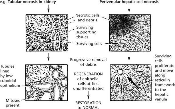
REGENERATION involves TWO PROCESSES:

1. proliferation of surviving cells to replace lost tissue.
2. migration of surviving cells into the vacant space.

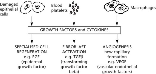
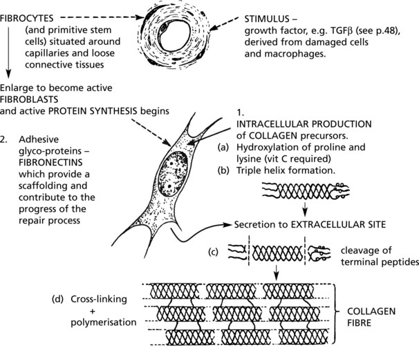
The FACTORS which CONTROL healing and repair are complex: they include the production of a large variety of growth factors.

  Page 49 
image

Wound Healing

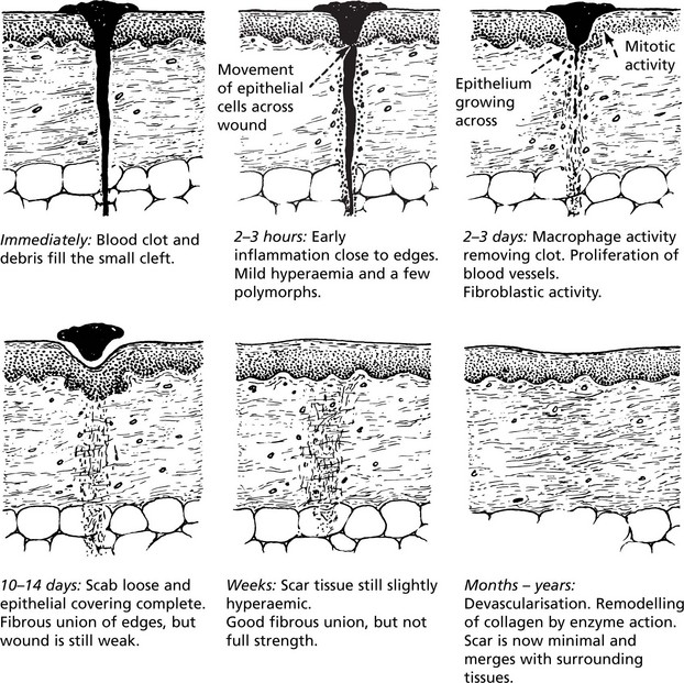
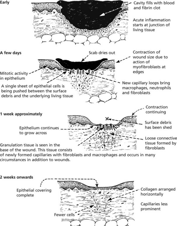
Healing of a wound shows both epithelial regeneration (healing of the epidermis) and repair by scarring (healing of the dermis).

Two patterns are described depending on the amount of tissue damage. These are the same process varying only in amount.

1. Healing by first intention (primary union)

This occurs in clean, incised wounds with good apposition of the edges – particularly planned surgical incisions.

  Page 50 
image
2. Healing by second intention (secondary union)

This occurs in open wounds, particularly when there has been significant loss of tissue, necrosis or infection.
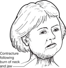
  Page 51 
image

Wound Contraction

Wound contraction, which is beneficial and begins early, is due mainly to the young, specialised ‘myofibroblasts’ in the granulation tissue exerting a traction effect at the wound edges. The exposed surface is reduced by gradual regeneration of the surface epithelium.

The remodelling of the collagen continues for many months.

Complications

1. Contracture

Later, contracture may cause serious cosmetic and functional disability, particularly in deep and extensive skin burns and around joints if muscles are badly damaged.

image
2. Keloid

The formation of excess collagen in the form of thick interlacing bundles which causes marked swelling at the site of the wound is known as a KELOID. The essential cause is unknown. It is particularly common in black people.

image
  Page 52 

Healing – Fibrosis

FIBROSIS is the end result of wound healing, chronic inflammation and organisation.

Formation of Fibrous Tissue

image

REMODELLING follows: Action of collagenasescar tissue + secretion of collagen

Factors Delaying Healing

1. Local

infection, a poor blood supply, excessive movement and presence of foreign material delay healing.

2. General

deficiency of vitamin c
deficiency of amino acids (in malnutrition)
deficiency of zinc
excess of adrenal glucocorticoids
debilitating chronic disease
image Failure of proper collagen synthesis with delayed healing and weak scars.
  Page 53 

Healing – Special Situations

Internal Surfaces

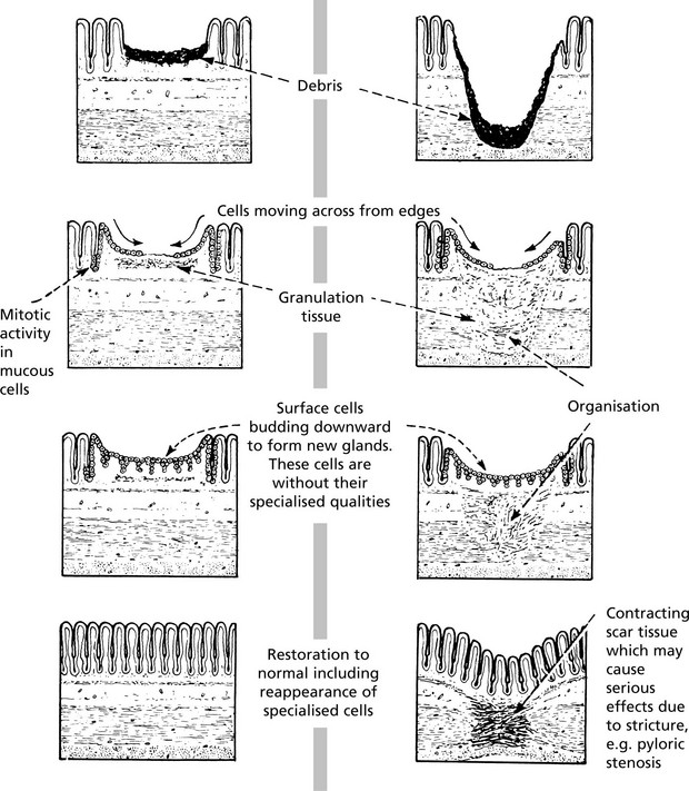
The epithelial lining of the gastrointestinal tract regenerates in a similar way to the skin.

image
  Page 54 

Solid Epithelial Organs

1. Following gross tissue damage – including supporting tissue (post-necrotic scarring)
image
2. Following cell damage with survival of the supporting (reticular) tissues
  Page 55 
image

Muscle

Muscle fibres of all 3 types – skeletal, cardiac and smooth – have only limited capacity to regenerate.

When a mass of muscle tissue is damaged, repair by scarring occurs. This is particularly important in the heart after infarction.

If the damage affects individual muscle fibres diffusely and with varying severity, then regeneration of the specialised fibres is possible (e.g. the myocardium may recover completely from the effects of diphtheria toxin and virus infection).

Nervous Tissue

Central Nervous System

Regeneration does not occur when a neurone is lost.

In cases of acute damage, the initial functional loss often exceeds the loss of actual nerve tissue because of the reactive changes in the surrounding tissue. As these changes diminish, some function may be restored.

image

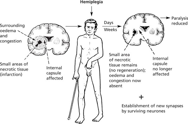
Scarring within the CNS is by proliferation of astrocytes and the production of fibrillary glial acidic protein – a process known as gliosis.
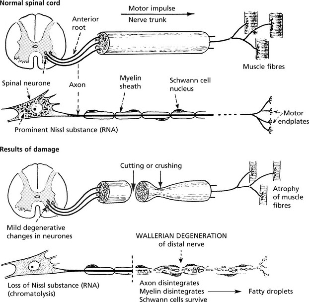
  Page 56 

Peripheral Nerves

When a peripheral nerve is damaged, the axon and its myelin sheath rapidly degenerate distally. The supporting tissues of the nerve (Schwann cells) degenerate slowly.

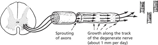
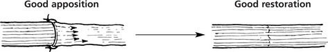
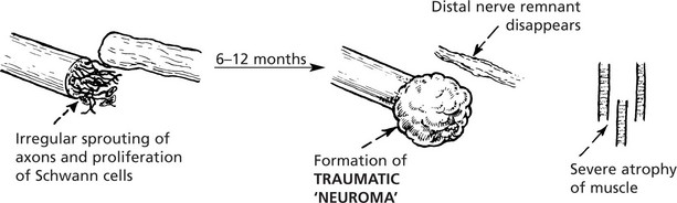
Regeneration can occur because the central neurone of which the axon is a peripheral extension is remote from the site of damage.

A spinal motor nerve is taken as an example.

  Page 57 

Peripheral Nerves

image

Regeneration takes the form of a sprouting of the cut ends of the axons.

image

The results depend on the apposition of the distal remnant with the sprouting axons.

image

The best results are seen in crushing injuries where the sheaths remain in continuity.

  Page 58 
image

Fracture Healing

Bone – Fracture Healing

1. Immediate effects
image
2. Early reaction-inflammatory First 4–5 days
image
3. Formation of callus (early bone regeneration) – after 1 week.
image
4. Mature callus – from 3 weeks onwards
image
5. Remodelling of callus Definitive – weeks into months
image
6. Final reconstruction Months later
image
  Page 59 

Complications

1. Fat embolism may occur in fracture of long bones due to entry of fat from the marrow cavity into the torn ends of veins.
image
2. Infection If the overlying skin is breached in any way, i.e. the fracture is ‘compound’, the risk of infection is greatly increased; this is an important adverse factor in the healing process.
image

Pathological Fracture

When the break occurs at the site of pre-existing disease of the bone, the term ‘pathological fracture’ is applied.

  Page 60 
image

Factors Influencing Healing of Fractures

image