CHAPTER 8 Meninges, ventricular system and blood supply
Meninges
Basic anatomy
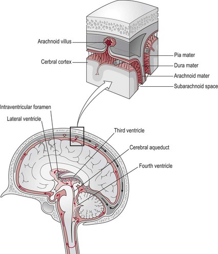
The brain and spinal cord are surrounded by three membranes termed ‘meninges’, which primarily protect and cushion the brain, brain stem and spinal cord.
Dura mater
This outer layer is conventionally described as two layers, however they are closely united except along certain lines where they part to surround and support large venous channels termed ‘the dural venous sinuses’ (Fig. 8.1). The two layers comprise:
Arachnoid mater
This middle layer is a delicate web-like structure lying between the dura mater and pia mater. The space between the dura mater externally and the arachnoid mater is called the ‘subdural space’. The space between the pia mater internally and the arachnoid mater is the ‘subarachnoid space’. The major arteries to the brain pass through the subarachnoid space and it is also filled with cerebrospinal fluid. This space extends down the spinal cord to the level of the second sacral vertebra.
In certain areas, the arachnoid mater projects into the dural venous sinuses to form arachnoid villi or granulations (Fig. 8.1) via which cerebrospinal fluid is recycled into the venous blood system.
Pia mater
This inner layer is a very delicate impermeable vascular membrane closely covering the brain surface (Fig. 8.1).
Function of the meninges
The outermost covering, the dura mater, serves to protect the underlying nervous tissue of the central nervous system and also has a role in restricting the displacement of the brain during head movement.
The primary function of the arachnoid mater and the space enclosing the cerebrospinal fluid is to further cushion the central nervous system. The pia mater supports capillaries that enter the tissue of the cerebral hemispheres.
The ventricular system
Basic anatomy
The ventricles are a system of spaces within the cerebral hemispheres, which produce and circulate cerebrospinal fluid around the central nervous system.
Ventricles
The ventricular system incorporates a series of interconnected spaces in the core of the brain. The largest spaces are the lateral ventricles, which are bilateral and are the site of the choroid plexus, which produce cerebrospinal fluid. The 3rd ventricle is linked to the lateral ventricles by the interventricular foramina and to the 4th ventricle by the cerebral aqueduct. The 3rd and 4th ventricles lie in midline (Fig. 8.1).
Cerebrospinal fluid
Cerebrospinal fluid (CSF) is a clear fluid produced by the choroid plexuses within the lateral ventricles. The CSF circulates through the ventricular system and ultimately enters the subarachnoid space which surrounds the cerebral cortex, cerebellum, brain stem and spinal cord down to the level of the second sacral vertebra. The CSF is recycled into the dural venous sinuses via the arachnoid villi (Fig. 8.1) and returned to the venous circulation.
Function of the ventricular system
The primary role of the CSF is the removal of waste products after neuronal activity and to provide basic mechanical and immunological protection to the nervous system. Along with the meninges, the CSF also cushions the delicate central nervous system from rubbing against the bony skull and vertebral column. The amount of CSF within the system can be regulated in order to maintain the correct intracranial pressure (ICP), so facilitating cerebral blood flow. Any disruption or blocking of the flow of CSF may result in cerebral oedema, reduced blood flow and brain damage.
Circulatory systems of the brain
Arterial supply
Basic anatomy
Normal function of the brain’s control centres is dependent upon an adequate supply of oxygen and nutrients through a dense network of blood vessels. The brain represents approximately 2% of the total body weight, however it receives 20% of the resting cardiac output. Blood is supplied by the two internal carotid arteries and the two vertebral arteries that join at the base of the brain to form the basilar artery.
Anterior circulation
The carotid arteries and their branches are referred to as the ‘anterior circulation’. The left common carotid artery arises from the aortic arch (Fig. 8.2). The right common carotid arises from the innominate artery. From each common carotid artery there are two branches:
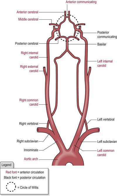
The left and right internal carotid arteries pierce the dura mater at the base of the brain and each bifurcates to form the anterior cerebral artery and the middle cerebral artery (Fig. 8.2).
Posterior circulation
The vertebrobasilar system is referred to as the ‘posterior circulation’ (Fig. 8.2).
The vertebral arteries arise from the subclavian artery and ascend through the transverse foramina of the cervical vertebrae entering the skull through the foramen magnum. The vertebral artery gives off branches to the spinal cord, the cerebellum and to the medulla.
The two vertebral arteries join at the junction of the pons and medulla oblongata (S2.10) to form the basilar artery, which then divides at the junction of the pons and mid-brain to form the two posterior cerebral arteries (Fig. 8.2). It also gives off branches to the cerebellum.
The circle of Willis
The circle of Willis (Fig. 8.2) is a polygon shaped network of blood vessels beneath the cerebral hemispheres. The two anterior cerebral arteries are connected by the anterior communicating artery and the posterior cerebral arteries are connected to the ipsilateral internal carotid arteries by the posterior communicating arteries. The six cerebral arteries, bilaterally the anterior, posterior and middle are termed end arteries.
Function of the arterial supply
The blood supply aims to meet the physiological demands of the brain cells, including neural and non-neural cells. Delivering oxygen, glucose and nutrients is a high priority as these tissues have a high metabolic rate and are highly sensitive to oxygen deprivation. A compromised blood supply lasting only seconds can cause neurological symptoms and after minutes, can lead to permanent damage.
The anatomical arrangement of the circle of Willis functions to provide the main vessels supplying the brain with a safety net. The circle design means that if there is damage to one of the main vessels, the distal smaller arteries can still be supplied from the other arteries in the circle (collateral circulation). However, if damage occurs to the end arteries, there is no such collateral circulation and consequently, brain damage cannot be avoided.
Understanding the area each main artery supplies and the function of that area allows the clinician to begin predicting the potential presentation of a patient prior to assessment:
The vertebral arteries
These supply the spinal cord via the medullary arteries which join to form the anterior and posterior spinal arteries. Based upon the general arrangement of the descending tracts anteriorly and the ascending tracts posteriorly, damage to these blood vessels could result in a motor loss or sensory loss, respectively.
The basilar artery
This is formed by the two vertebral arteries. Branches from this posterior circulation supply the brain stem and cerebellum.
The anterior cerebral arteries
Bilaterally, these supply the inferior parts of the frontal lobe, the medial surface of the frontal and parietal lobes and the anterior part of the corpus callosum. Branches from the anterior cerebral artery supply the limbic system, basal ganglia and anterior limb of the internal capsule.
The blood–brain barrier
A stable physiological environment within the brain is crucial for normal function. Neurons need to be protected from changes in this micro-environment. This is achieved by the blood–brain barrier, a specialized interface between the capillary walls and the surrounding neural tissues that restricts the passage of various chemical substances and microscopic objects such as bacteria between the blood and the neural tissue itself.
In other areas of the body, free movement of ions and molecules back and forth across the tissue boundary is permitted. However, in the brain, the endothelial cells of the capillary wall overlap and form tight junctions termed ‘end feet’ with astrocyte cells (S2.7) surrounding them. The astrocytes also provide them with biochemical support. Thus, there is little free movement from blood into the interstitial environment of the neural tissue. There are however, specific transporters across the barrier for critical ions and molecules such as glucose and specific amino acids.
Venous drainage
Basic anatomy
The venous drainage of the brain can be separated into two sub-divisions:
Superficial
The superficial system is composed of dural venous sinuses (Fig. 8.1) located on the surface of the cerebrum. The dural venous sinuses are situated between the two layers of the dura mater and lined with endothelium. They differ from other vessels in that their walls lack the characteristic layering, muscle and valves seen in other veins. Their function is to receive blood from the deep and superficial cerebral veins and cerebrospinal fluid from the subarachnoid space via the arachnoid villi and empty these components into the internal jugular vein to be returned to other organs for recycling.
Deep
The deep venous drainage is primarily composed of traditional veins inside the deep structures of the brain. However, the cerebral veins are thin walled and have no valves.
At the confluence of sinuses, the superficial and deep venous drainage join and form the two internal jugular veins. In the neck, the jugular veins run parallel to the upward course of the carotid arteries and ultimately drain blood into the superior vena cava.