Renal Osteodystrophy

Renal osteodystrophy is a general term that refers to the skeletal lesions that develop secondary to chronic, severe renal disease. In humans, this can include osteomalacia and fibrous osteodystrophy, either as separate diseases or in combination. Similarly, although fibrous osteodystrophy is the most common consequence of chronic renal disease, particularly in dogs, this sometimes is complicated by osteomalacia. Clinical signs of the disease include bone pain (lameness) and loss of teeth and deformity of the maxilla or mandible as the result of osteoclastic resorption of bone and replacement by fibro-osseous tissue. Renal osteodystrophy has a complex pathogenesis that likely varies, depending on the extent and nature of the renal disease and the availability of dietary vitamin D. Loss of glomerular function, inability to excrete phosphate, inadequate renal production of 1,25-dihydroxyvitamin D (calcitriol), and acidosis are central to its development. As the glomerular filtration rate falls in chronic renal disease, hyperphosphatemia develops, stimulating PTH synthesis and secretion. Hyperphosphatemia also suppresses the renal hydroxylation of inactive 25-hydroxyvitamin D to 1,25-dihydroxyvitamin D (calcitriol). Hypocalcemia develops primarily from decreased intestinal calcium absorption because of low serum calcitriol levels. Low serum calcitriol levels, hypocalcemia, and hyperphosphatemia have all been demonstrated to independently promote PTH synthesis and secretion. If serum levels of PTH remain elevated, fibrous osteodystrophy develops. The reduced production of 1,25-dihydroxyvitamin D by the diseased kidneys, together with impaired mineralization because of the acidosis of uremia, explain the development of osteomalacia.

Inflammation of Bone

Infectious Inflammation of Bone

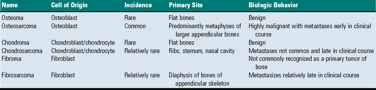
Inflammation of bone is termed osteitis. Periostitis occurs if the periosteum is involved, and osteomyelitis is the appropriate term if the medullary cavity of the bone is involved (Table 16-2). These conditions generally occur together and may be life threatening, requiring early diagnosis and vigorous treatment. They often involve the simultaneous necrosis and removal of bone and the compensatory production of new bone, occurring over a prolonged period. Infectious inflammation of bone in animals is usually caused by bacteria. Hematogenous bacterial osteomyelitis is uncommon in dogs and cats, but it is common in neonatal foals and animals used for food and fiber production. A wide range of Gram-positive and Gram-negative bacteria is responsible for hematogenous osteomyelitis in calves and foals. Arcanobacterium pyogenes and other pyogenic bacteria (e.g., Streptococcus spp., Staphylococcus spp.) and Salmonella spp., Escherichia coli, and other coliforms are among the most common microbes that cause hematogenous osteomyelitis. Staphylococcus intermedius is the most common cause of hematogenous osteomyelitis in dogs.

TABLE 16-2

Lesions of Inflammatory Joint Disease

image

Fungi, viruses, and protozoa also can cause bony lesions. Mycotic agents, such as Coccidioides immitis and Blastomyces dermatitidis, frequently spread hematogenously to bone to produce granulomatous or pyogranulomatous osteomyelitis, with bone lysis and irregular new bone formation. The viruses of hog cholera and infectious canine hepatitis can cause endothelial damage, resulting in metaphyseal hemorrhage, necrosis, and acute inflammation. Osseous localization of the distemper virus injures osteoclasts, disrupting metaphyseal modeling and producing a growth retardation lattice (described earlier), and a variant of the feline leukemia virus (FeLV) has been associated with myelosclerosis (increased density of medullary bone) in cats; however, these latter two viruses do not cause inflammation of bone.

Growth plate: Epiphyseal cartilage (Fig. 16-56) (cartilage of the epiphysis that has yet to undergo endochondral ossification) and physeal cartilage can be eroded by invasion of inflammation from adjacent bone or may undergo direct bacterial embolization via cartilage canal blood vessels or in the blind-loop vessels at sites of endochondral ossification (see the section on Portals of Entry of Bone). Growth cartilage may appear thickened secondary to osteomyelitis because of disruption of endochondral ossification by the inflammatory process and the resulting failure to replace cartilage with bone (see Fig. 16-35). In growing animals, articular cartilage can undergo lysis by extension of osteomyelitis from the subjacent AEC, a lesion that may be mistaken for primary arthritis (see Fig. 16-36).

image

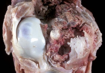
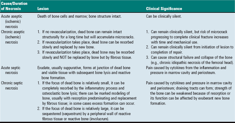
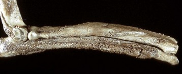
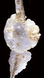
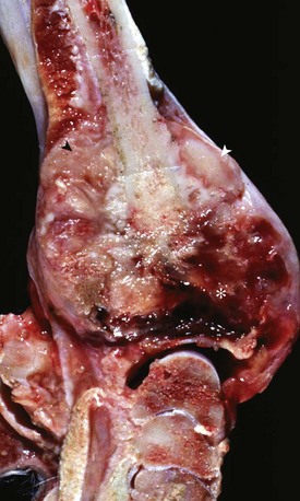
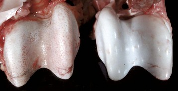
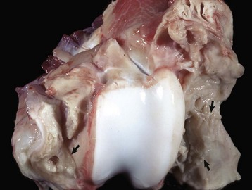
Fig. 16-56 Emboli (suppurative) epiphysitis, bone, distal femur, foal.
Bacterial emboli in the articular-epiphyseal complex (AEC) have produced suppurative inflammation that has destroyed both the subchondral bone and overlying articular cartilage of the condyle (right). (Courtesy Dr. S.E. Weisbrode, College of Veterinary Medicine, The Ohio State University.)

Trabecular bone: The composition of the exudate in metaphyseal osteomyelitis is determined by the infectious agent and typically is purulent in bacterial infections in domestic animals. Exudate in the medullary cavity increases the intramedullary pressure and can cause compression of vessels resulting in thrombosis and infarction of intramedullary fat, hematopoietic marrow, and bone. In areas of inflammation, bone resorption is mediated primarily by osteoclasts that are stimulated by prostaglandins and cytokines released by local tissue and inflammatory cells. Reduced blood flow through large vessels also promotes osteoclastic bone resorption, possibly by altering electrostatic charges in bone. Proteolytic enzymes released by inflammatory cells and activation of matrix metalloproteinases by the acid environment of inflammation also assist in resorbing matrix. Lack of drainage and persistence of the offending agent in areas of necrotic bone account for the chronicity of the process, which may continue for years. Inflammation in the medullary cavity also may penetrate into and through cortical bone and undermine the periosteum, where it can further disrupt the blood supply to the bone at the nutrient foramen and nutrient canal.

Cortical bone: The lesions involving cortical bone that occur with infectious osteomyelitis may vary based on the route of entry of the organism and the nature of the exudates (Fig. 16-57). Bone lysis is expected with suppurative inflammation and is subperiosteal for bacteria induced traumatically via the periosteum and endosteal in cases of embolic osteomyelitis. Lysis within the cortex begins within existing vascular channels and can occur with either route of entry. Periostitis can develop by direct inoculation from trauma (e.g., puncture wounds) or by centripetal spread of inflammation from the marrow cavity and through the cortex. Chronic bacterial periostitis is characterized by multiple coalescing pockets of exudate and areas of irregular periosteal new bone formation and cortical lysis (Fig. 16-58). Additional sequelae of osteomyelitis include extension of inflammation to adjacent bone, hematogenous spread to other bones and to soft tissues, pathologic fractures, and development of sinus tracts that penetrate cortical bone and drain to the exterior. Occasionally, fragments of dead bone become isolated from their blood supply and surrounded by exudate (bone sequestrum). Sequestra can form when bone fragments are contaminated at the site of a compound fracture, when the fragments at a fracture site become infected hematogenously, or when fragments of necrotic bone become isolated (and thus avascular) in osteomyelitis (Fig. 16-59). These sequestra and associated exudates can become surrounded by a dense collar of reactive bone, which is termed the involucrum. Extracellular matrix is not living tissue; therefore it cannot be resorbed in an area in which the cells (bone or marrow) are dead. For this reason, relatively large sequestra can persist for long periods and may interfere with repair. Grossly, they often become pale and chalky and lack the glistening appearance of normal bone.

image

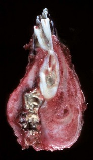
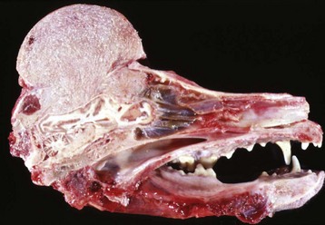
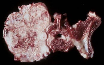
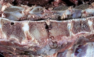
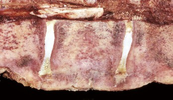
Fig. 16-57 Chronic (suppurative) osteomyelitis, bone, mandible, transverse section, sheep.
Chronic suppurative osteomyelitis has caused a fistulous tract that penetrates through the full dorsoventral thickness of the mandible. This lesion likely began as a periodontal bacterial infection. (Courtesy Dr. S.E. Weisbrode, College of Veterinary Medicine, The Ohio State University.)

image

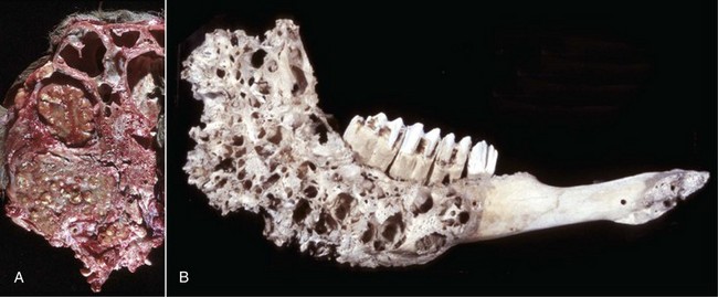
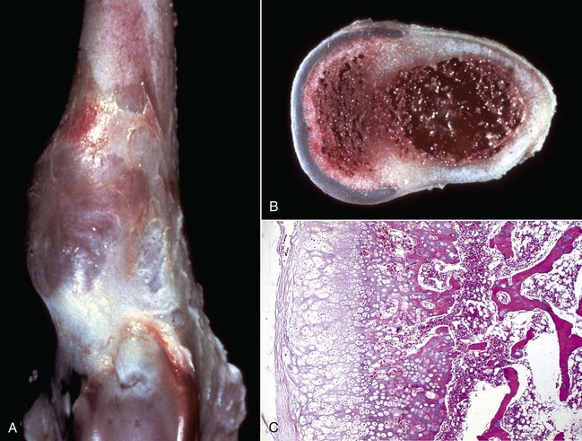
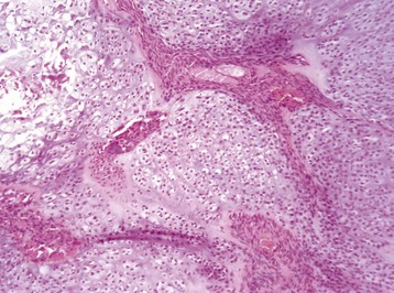
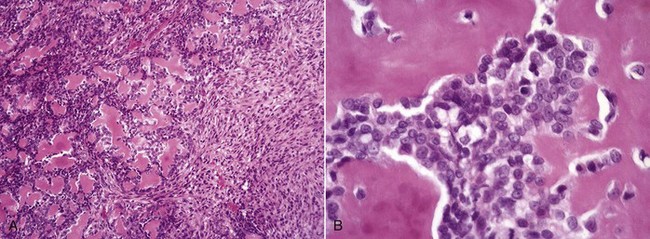
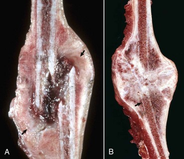
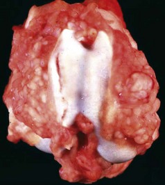
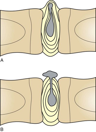
Fig. 16-58 Chronic (pyogranulomatous) osteomyelitis and sinusitis, actinomycosis (Actinomyces bovis), maxillary sinus, cow.
A, Transverse section of the maxillae. The nodules apparent within the masses in the sinuses and maxilla represent pockets of pyogranulomatous inflammation that are surrounded by fibrous tissue and woven bone. B, Macerated and bleached specimen of the mandible. Note the spicules of woven bone radiating from the mandible. Within the spaces formed by this reactive bone were nodules of pyogranulomatous inflammation and colonies of Actinomyces bovis. (Courtesy Dr. S.E. Weisbrode, College of Veterinary Medicine, The Ohio State University.)

image

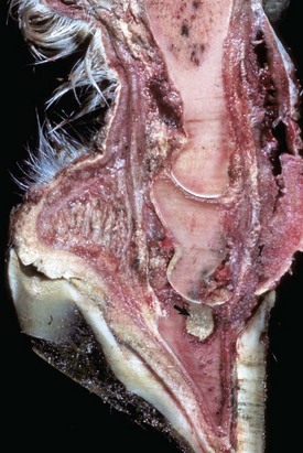
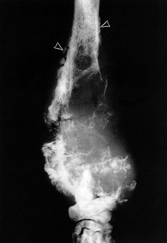
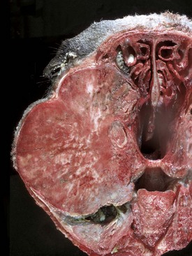
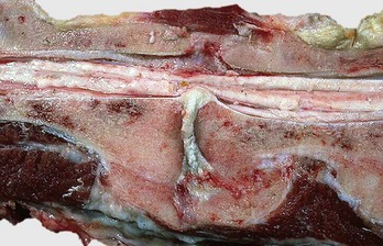
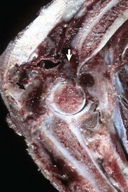
Fig. 16-59 Suppurative periostitis and osteomyelitis, phalanges, horse.
Trauma to the dorsal aspect of the hoof inoculated bacteria into the subcutis, causing suppurative cellulitis and periostitis and, subsequently, cortical osteolysis of the dorsal aspect of the distal first phalanx and the entire dorsal surface of the second phalanx. From there, the infection spread to the distal interphalangeal joint and then to the third phalanx, where it caused suppurative osteomyelitis, loss of articular cartilage, and formation of a sequestrum in the proximal portion of the third phalanx (arrow). The viable tissue immediately adjacent to the sequestrum is not different from that which is more distant, implying that in this case an involucrum (reactive bone surrounding the exudate around a sequestrum) was not formed. (Courtesy Dr. S.E. Weisbrode, College of Veterinary Medicine, The Ohio State University.)

Noninfectious Inflammation of Bone

Metaphyseal Osteopathy: Metaphyseal osteopathy, previously termed hypertrophic osteodystrophy, is a disease of young (usually 3 to 6 months of age), growing dogs of the large and giant breeds. Both names, unfortunately, are misleading, since the initial lesion is a suppurative and fibrinous osteomyelitis of the trabecular bone of the metaphysis, which is replaced in the chronic stages of the disease by the formation of abundant periosteal new bone. Remissions/exacerbations may occur over weeks to months, but most cases resolve completely. The cause and pathogenesis are unknown; infectious agents have not been isolated. There are reports of metaphyseal osteopathy in litters of Weimaraners, in which granulocytopathies have been suspected, and in littermates of Irish setters, in which canine leukocyte adhesion deficiency was confirmed. The lack of CD18 expression on the neutrophil surface in the affected Irish setters results in the failure of neutrophils to marginate or extravasate, as well as a failure of these cells to phagocytose by CD18. Affected dogs have an underlying genetic defect that is expressed clinically to various degrees, ranging from normal to the development of severe, repeated infections. Interestingly, 75% to 85% of these dogs develop metaphyseal osteopathy by 10 to 12 weeks of age, and hematopoietic stem cell transplants lead to resolution of all lesions. Based on these findings, it is possible that neutrophil entrapment at the chondro-osseous junction in the metaphysis (same location that is predisposed to the development of osteomyelitis in young animals; see section on Portals of Entry into Bone) results in autoinflammation and necrosis, with the periosteal proliferation occurring as a secondary event. Clinically, metaphyseal osteopathy is characterized by lameness, fever, and swollen, painful metaphyses in multiple long bones.

Growth plate: Lesions in the growth plate are not expected in metaphyseal osteopathy.

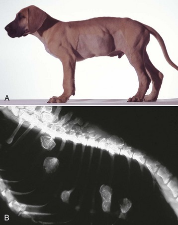
Trabecular bone: Lesions are usually bilaterally symmetric. Radiographically, alternating metaphyseal zones of increased lucency and increased density are present parallel to the physes (Fig. 16-60, A and B). Microscopically, the lucent areas represent suppurative and fibrinous inflammation and necrosis of the metaphyseal marrow and bone (Fig. 16-60, C). The death of osteoblasts results in primary trabeculae that are not reinforced by apposition of bone matrix. These trabeculae collapse and fracture without external distortion of the bone (infractions) and appear radiographically as relatively dense regions.

image

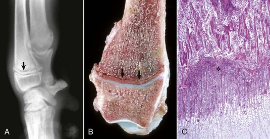
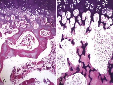
Fig. 16-60 Metaphyseal osteopathy, bone, distal radius, dog.
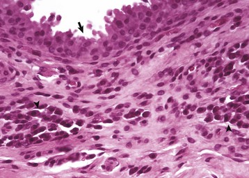
A, Radiograph. The radiolucent line in the metaphysis (arrow), parallel with growth plate, is characteristic of metaphyseal osteopathy. B, Grossly, this line appears to be a fracture (arrows) within the metaphysis. C, Histologically, this line is a hypercellular band (asterisk) of neutrophils between the primary and secondary trabeculae. (Courtesy Dr. S.E. Weisbrode, College of Veterinary Medicine, The Ohio State University.)

Cortical bone: The inflammation can extend from the medulla through the cutback zone into the periosteum. This periosteal inflammation, along with mechanical instability caused by the metaphyseal infractions, can cause notable new metaphyseal periosteal bone formation in chronic cases.

Eosinophilic Panosteitis: Eosinophilic panosteitis is another canine bone disease with an unfortunate name, since the lesion is neither inflammatory nor eosinophilic. The cause is unknown, and the disease is almost always self-limiting. It occurs in growing (commonly large-breed) dogs, usually at 5 to 12 months of age, and is painful. German shepherd dogs appear to be predisposed. Morphologic studies are few because the disease is easily recognized clinically and resolves spontaneously so that biopsy evaluation is rarely necessary. Radiographically, the lesions are recognized as increased densities in the medullary cavity in the diaphysis, usually beginning near the nutrient foramen. Increased densities can also be present in the periosteum. These densities are the result of proliferation of well-differentiated woven bone and fibrous tissue. No inflammation is present. The cause of the lameness is presumed to be pressure on nerves by the proliferating woven bone both within the medullary cavity and on the periosteum.

Aseptic Necrosis of Bone

Aseptic necrosis of bone in humans occurs in a variety of clinical conditions, including occlusive vascular disease (bone infarction), hyperadrenocorticism, fat embolism, nitrogenous embolism, sickle cell anemia, and intramedullary neoplasms, all of which likely result in arterial or venous infarction of the bone (Table 16-3). In domestic animals, aseptic necrosis of bone has been associated with intramedullary neoplasms and various nonneoplastic lesions, which likely result in decreased venous outflow from the bone and increased bone marrow pressure. Although the long-term use of steroids in humans has been associated with necrosis of the femoral head in adults, steroids do not appear to induce osteonecrosis in domestic animals.

TABLE 16-3

Necrosis of Bone

image

The gross appearance of necrotic bone varies with the extent of the affected area and the response of the body to it. Microscopically, the hallmark of bone necrosis is cell death and loss of osteocytes from their lacunae.

After an episode of ischemia leading to infarction, the cellular elements of the marrow lose their differential staining, and circular spaces (pooled lipid) develop within a few days. If the region of dead bone remains avascular, the coagulated tissue and mineralized matrix can persist for some time. Dead osteocytes elicit little reaction; their nuclei become pyknotic, but their disappearance from lacunae is slow and might not be complete for 2 to 4 weeks.

Reaction to and repair of necrotic bone requires revascularization that is associated with infiltration of macrophages and invasion by fibrous tissue that advances from the margins of the lesion. The bone marrow might eventually regenerate entirely, or a scar might form and remain. The necrotic matrix remains fully mineralized and might even “hypermineralize” because of calcification of the dead osteocytes and their lacunae. This mineralization is only possible if there is vascularization that brings additional calcium to the region. Dead bone is slowly removed by osteoclasts. The resorption of necrotic bone with simultaneous replacement by new bone is termed creeping substitution. The process is slow and often incomplete. Small areas of bone necrosis might not be detected clinically or radiographically.

Growth plate: Ischemic necrosis of metaphyseal bone and bone marrow can result in retained growth cartilage. The metaphyseal necrosis would not directly affect the zones of proliferating and hypertrophying chondrocytes; however, the physeal thickness would increase because the conversion of epiphyseal cartilage to bone by endochondral ossification at the chondro-osseous junction would be impaired. Ischemic necrosis of the epiphysis could result in premature closure of the growth plate because of the death of the proliferating chondrocytes, which depend on the epiphyseal blood vessels. Endochondral ossification would continue normally if the metaphyseal blood supply was not affected, and the plate would close because of failure to generate new proliferating zone chondrocytes.

Trabecular bone: In the femoral heads of young, small, and miniature breed dogs, aseptic necrosis of the femoral head is associated with clinical signs because of the collapse of the articular cartilage as a result of resorption of the necrotic subchondral bone (Legg-Calvé-Perthes disease) that occurs late in the course of the disease. Apparently the initial infarction is asymptomatic (Fig. 16-61). The cause of the infarction is usually not determined but might be caused by venous compression or increased pressure within the articular cavity. This pressure can result in increased intraosseous pressure and bone necrosis. In clinical cases of aseptic necrosis, the dead bone is not replaced by creeping substitution but is ultimately resorbed and replaced by fibrous tissue. The fibrous tissue does not provide adequate support for the articular cartilage, and the femoral head collapses. In stages preceding complete resorption, it is common, in revascularized necrotic medullary bone, for reactive new bone to be deposited on trabeculae of necrotic bone (Fig. 16-62). This sandwich of central dead bone covered by viable reactive woven bone can persist for months and may give (along with osteocyte mineralization described previously) the affected region a radiodense appearance. Ultimately, in clinical cases of aseptic necrosis of the femoral head, even these foci of new bone formed over dead bone are resorbed and replaced by fibrous tissue.

image

Fig. 16-61 Femoral head, necrosis (experimental), bone, femur, pig.
Necrosis was produced experimentally by a ligature placed around the femoral neck. Several days after the procedure, the only difference between the control (A) and the infarcted bones (B) is the coagulation necrosis of the marrow and bone cells. The hard tissue of the cartilage and bone remains unaffected. H&E stain. (Courtesy Dr. S.E. Weisbrode, College of Veterinary Medicine, The Ohio State University.)

image

Fig. 16-62 Revascularization, ischemic necrosis (experimentally induced), 1-month duration, femoral head, epiphyseal (cancellous) bone, pig.
Reactive woven bone (between arrows) has been deposited on the surface of the necrotic bone, which contains empty lacunae and karyolytic nuclei. Fibrovascular repair tissue in the marrow surrounds the bone. H&E stain. (Courtesy Dr. S.E. Weisbrode, College of Veterinary Medicine, The Ohio State University.)

Cortical bone: Large areas of necrotic cortical bone have a dry, chalky gross appearance and the periosteum can be removed easily. These areas can remain as a subclinical lesion for years. The formation of sequestra almost always requires inflammation; sequestra formation after sterile necrosis of bone is unlikely.

Proliferative and Neoplastic Lesions

Surprisingly, bone, as a tissue, offers little resistance to an expanding or invading neoplasm, and many skeletal neoplasms are accompanied by bone resorption as well as new bone formation (Fig. 16-63). Pain, hypercalcemia, increased serum alkaline phosphatase activity, pathologic fracture, and distant metastases are other possible manifestations of a skeletal neoplasm. New bone formation occurs, at least in part, in response to mechanical stress on a weakened cortex and is prominent in neoplasms that have a marked fibrous stroma. Neoplasms with little stroma, such as plasma cell myeloma and lymphosarcoma, have minimal reactive bone formation, even though bone may be destroyed by marked bone lysis. Tumor-associated bone destruction is largely accomplished by osteoclasts, but prostaglandins, cytokines, acid metabolic by-products, and lytic enzymes released by inflammatory or neoplastic cells can also be responsible for local bone resorption and formation. Hypercalcemia, a result in part of bone resorption induced by release of bone-resorbing factors from extraskeletal neoplasms, is well documented and is termed humoral hypercalcemia of malignancy. The best-known examples in animals occur in dogs secondary to adenocarcinoma of the apocrine glands of the anal sac and T lymphocyte lymphoma, in which the neoplastic cells produce PTH-related protein.

image

Fig. 16-63 Osteosarcoma, bone, distal radius, dog.
Radiograph of osteosarcoma with extensive destruction of preexisting bone; small areas of new bone formation on the periosteal surface (arrowheads). (Courtesy Dr. S.E. Weisbrode, College of Veterinary Medicine, The Ohio State University.)

Nonneoplastic Proliferative and Cystic Lesions

The nonneoplastic proliferative and cystic lesions considered here vary widely in their cause, structure, and ultimate effect on the host. Reactive bone formation, sometimes exuberant, can occur in fracture repair, chronic osteomyelitis, and degenerative joint disease in the form of periarticular osteophytes. An exostosis is a nodular, benign, bony growth projecting outward from the surface of a bone. An osteophyte is a similar growth that occurs at the margins of a diarthrodial (movable) joint. An enthesophyte is the calcification of a tendon or ligament at the point of its insertion into the bone. In addition to bone, these proliferations may include variable amounts of cartilage. The bone component can be woven and/or lamellar, depending on rate of growth and duration of lesion. Hyperostosis usually is used to indicate that the diameter of the bone has increased and implies more uniform thickening on the periosteal surface rather than the nodular appearance of an osteophyte or exostosis. An enostosis is a bony growth within the medullary cavity, usually originating from the cortical-endosteal surface, and can result in obliteration of the medullary cavity. These lesions are nonneoplastic proliferative lesions in which growth is seldom continuous. Some exostoses can remodel (e.g., an osteophyte may become indistinguishable from the preexisting bone over time, and its presence is only recognizable by a change in shape of the affected area) and some regress. Nonneoplastic proliferative lesions can be mistaken for skeletal neoplasia, particularly in small biopsy specimens. Conversely, a malignancy may be missed when small superficial biopsies contain only nonneoplastic reactive bone that is present adjacent to the tumor. These statements serve to highlight the problem of making a morphologic diagnosis from a small biopsy specimen, without the benefit of a clinical history, radiographic findings, and other laboratory data. One must also remember that more than one process might be active at any one site (e.g., osteosarcoma might be complicated by fracture repair or by osteomyelitis).

Hypertrophic Osteopathy (Hypertrophic Pulmonary Osteopathy)

Hypertrophic osteopathy (hypertrophic pulmonary osteopathy) occurs in humans and domestic animals; dogs are the most commonly affected species. The disease is characterized by progressive, bilateral, periosteal, new bone formation in the diaphyseal regions, particularly of the distal limbs, that occurs as a secondary reaction to a primary lesion (Fig. 16-64). The word “pulmonary” is sometimes included because most cases occur in association with intrathoracic neoplasms or inflammation. Other, less commonly associated lesions or agents include endocarditis, heartworms, rhabdomyosarcoma of the urinary bladder in young giant-breed dogs, and ovarian neoplasms in the horse. Although the association between the pulmonary lesions and the proliferation of new periosteal bone on the extremities is not clear, it has been postulated that pulmonary lesions lead to reflex vasomotor changes (mediated by the vagus nerve) and to increased blood flow to the extremities. Evidence to support this theory includes the observation that the bony lesions regress after the primary lesion is removed, as well as after vagotomy. In addition, lesions similar to hypertrophic osteopathy can be reproduced in dogs by creating shunts that allow blood to bypass the pulmonary circulation, thereby increasing the stroke volume of the left side of the heart, leading to increased blood flow to peripheral tissues. Increased arterial pressure, hyperemia, and edema of the periosteum lead to thickening first by fibrous tissue and later by new bone formation. Similar periosteal woven bone deposition can occur in the metaphysis, but in hypertrophic osteopathy (as opposed to metaphyseal osteopathy) the lesions are usually less severe and less frequent in the metaphysis than in the diaphysis.

image

Fig. 16-64 Hypertrophic osteopathy, bone, radius and ulna, dog.
Marked periosteal proliferation of woven bone has resulted in the surfaces of both bones, but particularly the ulna, being irregularly roughened/thickened. Macerated and bleached specimen. (Courtesy Dr. S.E. Weisbrode, College of Veterinary Medicine, The Ohio State University.)

Osteochondromas

Osteochondromas (multiple cartilaginous exostoses) reflect a defect in skeletal development (lesions appear shortly after birth) rather than being true neoplasms and are confirmed to have a hereditary etiology in humans and horses. A hereditary basis has been suggested in dogs as well. Osteochondromas project from bony surfaces as eccentric masses that are located adjacent to physes. They arise from long bones, ribs, vertebrae, scapulas, and bones of the pelvis, and they may be numerous (Figs. 16-65 and 16-66). Microscopically, they have an outer cap of hyaline cartilage that undergoes orderly endochondral ossification to give rise to trabecular bone that forms the base of the lesion (Fig. 16-66, C). The medullary cavity of the osteochondroma usually communicates with the medullary cavity of the underlying bone because the cortex of the underlying bone at this site has not completely developed. Normally, growth ceases at skeletal maturity when the cartilage cap is replaced by bone. Although the origin of osteochondromas is not clear, some arise secondary to a defect in the perichondral ring as peripheral areas of physeal cartilage that are separated and removed from the growth plate during longitudinal growth. Clinically, their importance is threefold: they might interfere mechanically with the action of tendons or ligaments; they can act as space-occupying masses that protrude into the vertebral canal and cause spinal cord compression; and they can undergo malignant transformation and give rise to chondrosarcomas. Osteochondromas in cats are different in that they develop in mature animals, less commonly affect long bones, do not have orderly endochondral ossification, and might be of viral origin; however, like those in horses and dogs, osteochondromas in cats may undergo malignant neoplastic transformation. The term osteochondroma is not recommended for cartilaginous osteophytes that have undergone central endochondral ossification. As will be discussed regarding callus formation in the section on Fracture Repair, the osteogenic tissue (cambium layer) of the periosteum can form hyaline cartilage instead of bone when the oxygen tension of the tissue is low.

image

Fig. 16-65 Osteochondromas, young dog.
A, Multiple osteochondromas (cartilaginous exostoses) are present as single and coalescing masses on the ribs. B, Radiograph. The cartilaginous exostoses have the density of bone and appear in this case as exophytic nodules on the ribs and the dorsal spinous processes of the vertebrae. (Courtesy Dr. S.E. Weisbrode, College of Veterinary Medicine, The Ohio State University.)

image

Fig. 16-66 Osteochondroma (cartilaginous exostosis), bone, distal femoral metaphysis, dog.
A, A plateaulike mass (left) protrudes from the metaphyseal cortex. B, On cross-section, the mass (left) has a cartilage cap. C, Histologically, the cartilage cap is undergoing endochondral ossification, similar to that which occurs in an articular-epiphyseal complex. H&E stain. (Courtesy Dr. S.E. Weisbrode, College of Veterinary Medicine, The Ohio State University.)

Fibrous Dysplasia

Fibrous dysplasia is an uncommon focal to multifocal, lytic, intraosseous lesion that has been found at various sites (skull, mandible, and long bones) in young animals and may be a developmental defect. Typically, preexisting bone is replaced by an expanding mass of fibro-osseous tissue that can weaken the cortex and enlarge the external contour of the bone. The lesion is firm, often contains mineralized areas, and may contain multiple cysts filled with sanguinous fluid. Microscopically, the lesion is composed of well-differentiated fibrous tissue containing trabeculae of woven bone that are relatively regularly spaced and sized. Osteoblasts are not recognizable on trabecular surfaces, which is a feature that helps to distinguish this lesion from ossifying fibroma.

Bone Cysts

Bone cysts are classified as subchondral, simple, or aneurysmal. Radiographically, all appear as well-demarcated lucent areas without evidence of aggressive growth. Subchondral cysts are sequelae to osteochondrosis and degenerative joint disease. Subchondral bone cysts caused by osteochondrosis represent failure of endochondral ossification with subsequent necrosis and cavitation of retained growth cartilage and are most common in the medial femoral condyle in young horses (1 to 3 years of age). Although less commonly, these lesions also may occur in older horses. Subchondral cysts secondary to degenerative joint disease represent herniation of synovial fluid into the subchondral bone through fissures in degenerated articular cartilage. These herniations become lined by a synovial–like membrane, and the lysis of bone occurs by osteoclasis, secondary either to pressure or to cytokines released from the expanding cyst. Trauma to intact articular cartilage also is proposed as an etiology because cystic lesions have been reproduced experimentally by lacerating the articular cartilage to the level of the subchondral bone.

Simple bone cysts can contain clear, colorless, serum-like fluid or serosanguineous fluid. The wall of the cyst is composed of variably dense fibrous tissue and woven to lamellar bone. The bone located peripheral to the cyst undergoes modeling to accommodate the expansile growth of the cyst. Simple bone cysts may be difficult to distinguish from fibrous dysplasias, depending on the biopsy sample and radiographic and clinical information that is available.

Aneurysmal bone cysts contain spaces, filled with blood or serosanguineous fluid, that are not usually lined by endothelium. Tissue adjacent to the spaces can vary from well-differentiated fibrous or fibro-osseous tissue to pronounced proliferation of undifferentiated mesenchymal cells admixed with osteoclast-like multinucleated giant cells. Hemorrhage and hemosiderosis are frequent. The cause of simple and aneurysmal bone cysts is unknown; however, they could be consequences of ischemic necrosis, hemorrhage, or congenital or acquired vascular malformations. Caution should be exercised in the interpretation of microscopic lesions in biopsy specimens of cysts, and these lesions should be correlated with the radiographic appearance to rule out cystic cavitation in a neoplasm.

Primary Neoplasms of Bone

The histopathologic diagnosis of skeletal neoplasia in domestic animals often involves evaluation of needle, trephine, or wedge biopsies (Table 16-4). Bone biopsy samples usually are small relative to the size of the neoplasm, and the most easily accessible tissue often is reactive periosteal tissue that is located exterior to the neoplasm, therefore it is important that the pathologist incorporate the radiographic and clinical findings in the interpretation of the sample. Cases in which there are radiographically aggressive lesions that have an effect on bone mass are usually associated with malignancy or inflammation; therefore, if the microscopic findings from these cases indicate the presence of a benign lesion and/or provide no explanation for the radiographic changes, it may be necessary to examine additional tissue to reconcile these differences.

TABLE 16-4

Primary Skeletal Neoplasms of the Dog

image

Ossifying fibromas are uncommon masses in the maxillae and mandibles of horses and cattle. In their early stages, these are intramedullary neoplasms; however, although considered benign, they destroy adjacent cortical and trabecular bone by expansile growth. Microscopically, they are composed of well-differentiated fibrous tissue with scattered spicules of woven bone covered by osteoblasts.

Fibrosarcomas are malignancies of fibroblasts that produce collagenous connective tissue but not bone or cartilage. Microscopically the matrix of fibrosarcomas should not mineralize or entrap cells in lacunae as occurs in normal and neoplastic bone and cartilage, and the cells may be arranged in a whirling or interlacing pattern. Central fibrosarcomas arise from fibrous tissue within the medullary cavity, whereas periosteal fibrosarcomas arise from periosteal connective tissue. Central fibrosarcomas must be distinguished grossly and microscopically from osteosarcoma. In general, central fibrosarcomas grow more slowly, are accompanied by less formation of reactive new bone, are slower to metastasize, and produce a smaller tissue mass than osteosarcomas. Grossly, fibrosarcomas are gray-white, fill part of the medullary cavity, and replace cancellous and cortical bone.

Chondromas are benign neoplasms of hyaline cartilage. They are very rare neoplasms of dogs, cats, and sheep and often arise from flat bones; those arising in the medullary cavity are termed enchondromas. Cartilaginous neoplasms in the skeleton do not arise from articular cartilage, most likely because of its low mitotic potential and avascularity. Because they usually occur in adult animals, which do not possess growth cartilage, the cell of origin is presumed to be a stromal cell with chondrogenic potential. Chondromas are multilobulated and have a blue-white appearance on cut surface. They tend to enlarge slowly, but progressively, and can cause thinning of underlying bone. Microscopically, they are composed of multiple lobules of well-differentiated hyaline cartilage that may include areas of endochondral ossification. Chondromas may be very difficult to distinguish from low-grade, well-differentiated chondrosarcomas; distinguishing between these two tumor types may require clinical information. Chondrosarcomas are malignant neoplasms in which the neoplastic cells produce cartilaginous matrix but not osteoid or bone. Chondrosarcomas arise most frequently in the flat bones of the skeleton and occur most commonly in mature, large-breed dogs and in sheep (Figs. 16-67 and 16-68). In dogs, the major sites of origin are the nasal bones, ribs, and pelvis; in sheep, they arise from the ribs and sternum. Most chondrosarcomas arise in the medullary cavity and destroy preexisting bone. Given time, they become large, lobulated neoplasms with a gray or blue-white cut surface. Grossly, some neoplasms are gelatinous, and some contain large areas of hemorrhage and necrosis. Microscopically (Fig. 16-69), the range of differentiation of neoplastic cells is wide: Some neoplasms are well differentiated, lack mitotic figures, and are difficult to distinguish from chondroma (grade I chondrosarcomas). Grade II chondrosarcomas are composed of pleomorphic chondrocytes, contain low-to-moderate numbers of mitotic figures, and contain no undifferentiated areas. Grade III chondrosarcomas exhibit marked nuclear pleomorphisms, numerous mitotic figures, and contain areas of undifferentiated sarcoma. Some chondrosarcomas are composed primarily of primitive mesenchyma tissue and contain abundant basophilic interstitial mucin and rare foci of chondroid differentiation (mesenchymal chondrosarcoma). Nonneoplastic bone can be present as the result of endochondral ossification of the malignant cartilage; however, the presence of malignant osteoblasts in close association with foci/areas of osteoid supports a diagnosis of osteosarcoma, even in tumors in which the majority of the tissue present is cartilaginous. Chondrosarcomas have a longer clinical course, grow more slowly, and develop metastases later than osteosarcomas. Metastases are usually pulmonary via the venous system, without metastasis to the regional or bronchial lymph nodes. Osteomas are uncommon benign neoplasms that usually arise from bones of the head as a single, dense mass that projects from the surface of the bone (Fig. 16-70). They do not invade or destroy adjacent bone; their growth is slow and progressive but not necessarily continuous. Microscopically, osteomas are covered by periosteum and are composed of cancellous bone; trabeculae are lined by well-differentiated osteoblasts and osteoclasts. The intertrabecular spaces contain delicate fibrous tissue, adipocytes, and hemopoietic tissue.

image

Fig. 16-67 Chondrosarcoma, bone, rib, cat.
A chondrosarcoma arising in a rib has destroyed and replaced the normal bone structure. (Courtesy Dr. S.E. Weisbrode, College of Veterinary Medicine, The Ohio State University.)

image

Fig. 16-68 Chondrosarcoma, bone, calvarium, dog.
A chondrosarcoma protrudes from the skull, compresses the underlying brain, and has invaded the frontal sinus. The widespread white foci within the mass represent areas of mineralization. (Courtesy Dr. K. Read, College of Veterinary Medicine, Texas A&M University; and Noah’s Arkive, College of Veterinary Medicine, The University of Georgia.)

image

Fig. 16-69 Chondrosarcoma, bone, dog.
Chondrocyte lacunae are prominent in well-differentiated regions (left) but are less apparent in the more poorly-differentiated regions (lower right). (Courtesy Dr. S.E. Weisbrode, College of Veterinary Medicine, The Ohio State University.)

image

Fig. 16-70 Osteoma, bone, maxilla and maxillary sinus, sheep.
The osteoma has proliferated and formed a dome-shaped mass above the normal contour of the maxilla and has compressed the maxillary sinus. Grossly, this mass was diffusely hard; histologically, it was composed of closely spaced trabeculae lined by well-differentiated osteoblasts. An Oestrus ovis larva is present in the nasal cavity. (Courtesy Dr. S.E. Weisbrode, College of Veterinary Medicine, The Ohio State University.)

Osteosarcomas are malignant neoplasms in which neoplastic cells form bone, osteoid, or both. These tumors are common neoplasms in dogs and cats, in which they comprise approximately 80% and 50%, respectively, of all the primary bone neoplasms; however, they are rare in other domestic animals. Osteosarcomas typically occur in mature dogs of the large and giant breeds and arise most commonly in the metaphyses. The distal radius, distal tibia, and proximal humerus are the usual sites (Fig. 16-71); however, osteosarcomas can occur in ribs, vertebrae, bones of the head, and various other parts of the skeleton (Fig. 16-72). Rarely, they arise in soft tissue (extraskeletal osteosarcoma). Growth of the neoplasm is often rapid, aggressively locally invasive, and painful. Except for those arising in the axial skeleton and in particular, the head, early hematogenous pulmonary metastasis is common. Metastasis also may be widespread, involving soft tissues, as well as other bones.

image

Fig. 16-71 Osteosarcoma, bone, distal radius, dog.
Osteosarcoma has lysed and replaced normal bone. There is reactive periosteal new bone formation (arrowheads) and a large area of hemorrhage and necrosis (asterisk). (Courtesy Department of Veterinary Biosciences, The Ohio State University; and Noah’s Arkive, College of Veterinary Medicine, The University of Georgia.)

image

Fig. 16-72 Osteosarcoma, bone, wing of ilium, dog.
Although the mass appears to be growing primarily by expansion into the adjacent soft tissues, there is evidence of invasion and lysis of the underlying ilium and focal invasion of the lumbar vertebra. (Courtesy Dr. S.E. Weisbrode, College of Veterinary Medicine, The Ohio State University.)

Osteosarcomas can be classified as simple (bone formed in a collagenous matrix), compound (both bone and cartilage are present) or pleomorphic (anaplastic, with only small islands of osteoid present). Classification has also been based on cell type and activity (osteoblastic, chondroblastic, or fibroblastic), radiographic appearance (lytic, sclerotic, or mixed), or site of origin (central/intraosseous, juxtacortical, or periosteal). An uncommon form of osteosarcoma is the telangiectatic type that grossly resembles hemangiosarcoma and is composed of osteoblasts, osteoid, and large cystic, blood-filled cavities lined by malignant osteoblasts. Because there may be a great deal of heterogeneity within an individual tumor, classifying osteosarcomas may be difficult and is not recommended when the diagnosis is based solely on a small biopsy sample.

Central (intraosseous) osteosarcomas have a gray-white gross appearance and contain variable amounts of mineralized bone. Large, pale areas surrounded by zones of hemorrhage (areas of infarction) and irregular, randomly located areas of hemorrhage are common in rapidly growing intramedullary neoplasms. Neoplastic tissue tends to fill the medullary cavity locally and can extend proximally and distally but typically does not penetrate articular cartilage and therefore does not invade the joint space. Cortical bone is usually destroyed, and neoplastic cells penetrate and undermine the periosteum and can extend outwardly as an irregular lobulated mass. Destruction of cortical bone is accompanied by varying amounts of reactive (nonneoplastic) periosteal bone, which may be differentiated from tumor bone microscopically by its regular appearance and well-differentiated lining osteoblasts. Variable amounts of woven bone or osteoid are produced by the neoplastic osteoblasts; in fact, bone/osteoid production by the tumor cells is the hallmark lesion of this neoplasm (Fig. 16-73). Bone formation by the malignant cells can be abundant and widespread, or it can be minimal, as in anaplastic or fibroblastic osteosarcomas that are composed of sheets of poorly differentiated mesenchymal cells or fibroblastic tissue.

image

Fig. 16-73 Osteosarcoma, bone, dog.
A, Islands of eosinophilic osteoid are being produced by sheets of malignant osteoblasts. B, At higher magnification, malignant osteoblasts surround and invest (malignant osteocyte formation) the osteoid. Mineralization is not apparent in this decalcified section. H&E stain. (Courtesy Dr. J. Sagartz, College of Veterinary Medicine, The Ohio State University; and Noah’s Arkive, College of Veterinary Medicine, The University of Georgia.)

Periosteal osteosarcomas also have an aggressive behavior and invade into the medullary cavity from the periphery. As with central or intraosseous osteosarcomas, periosteal osteosarcomas cause bone lysis and reactive bone formation in addition to production of neoplastic bone. At the time of presentation, it is often impossible to determine if the osteosarcoma originated from the periosteum or the medullary cavity. Rarely, osteosarcomas are also juxtacortical (parosteal) in origin. These neoplasms arise within the periosteum and form an expansive mass that adheres to and surrounds but does not invade the underlying cortex. Invasion of the shaft and metastasis can occur with parosteal osteosarcomas but is a late event; therefore early en bloc excision might effect a cure. Although parosteal osteosarcomas are very rare, it is important to distinguish them from periosteal osteosarcomas because of their more favorable prognosis.

Multiple skeletal osteosarcomas occur in humans and dogs, and these may represent a primary neoplasm that has metastasized to bone or may have a multicentric origin. The lesions have a random distribution, and pulmonary metastases are likely to be present. Although the cause of naturally occurring osteosarcomas is largely unknown, in both humans and domestic animals, it is known that osteosarcomas can develop in association with or subsequent to other conditions at the same bony site, including infarction, fracture, and the presence of metallic fixation devices. Osteosarcomas of viral origin are reported in mice.

A unique form of skeletal malignancy occurs in the skull of the dog and is awkwardly called a multilobular tumor of bone (referred to as chondroma rodens in the older literature). These tumors are single, nodular, smooth-contoured, immovable masses on the flat bones of the skull (bones of membranous origin) and the hard palate. Neoplastic tissue is firm and the cut surface is composed of multiple, gray, partially mineralized lobules separated by fibrous tissue. These neoplasms are slow growing and locally invasive and may compress and invade the brain. They metastasize to the lungs late in the clinical course; however, the metastases are frequently small and clinically silent. Histologically, these tumors consist of multiple lobules, each having centrally located cartilage or bone, that are surrounded by plump mesenchymal cells that blend into well-differentiated interlobular fibrous tissue.

Various other neoplasms, such as liposarcomas, giant cell tumors, and hemangiosarcomas, can arise in bone, and neoplasms, such as lymphosarcomas and plasma cell myeloma, can involve the bone marrow and surrounding bone. Most cases of tumor metastases to bone involve carcinomas, with the most common primary sites being mammary gland, liver, lung, and prostate gland.

Secondary Neoplasms of Bone

At autopsy, 60% of human cancer patients have skeletal metastases. These metastases are predominantly in red bone marrow, in which the vascular sinusoidal system is apparently predisposed to trap circulating malignant cells. The true incidence of skeletal metastasis in animals is unknown and estimates might be artificially low because early euthanasia shortens the course of the disease and because bone scanning and other types of clinical imaging techniques are done less often than in humans. Metastatic neoplasms in bone can be associated with pain, hypercalcemia, lysis of bone, pathologic fracture, and reactive new bone formation. Rib shafts, vertebral bodies, and humeral and femoral metaphyses (proximal appendicular skeleton) are common sites of metastatic neoplasms in dogs, and these most commonly involve carcinomas. The most common primary sites are the same as those in humans (mammary gland, liver, lung, and prostate gland). In cats, skeletal metastases are rare but, when present, appear to involve the distal appendicular skeleton. Clinically silent pulmonary carcinomas in cats are reported to metastasize to the digits, particularly the third phalanx, causing destruction of the nail bed epithelium and sloughing of the claw.

In about 50% of the carcinomas that are identified in the proximal appendicular skeleton in the dog, there is no clinical evidence of a primary tumor nor is one located at postmortem, raising the possibility that these are primary intraosseous carcinomas. Primary intraosseous carcinomas are reported in the mandible and maxilla of humans but are uncommon and are nearly always diagnosed as squamous cell carcinomas, some of which have features indicating an odontogenic origin.

Fracture Repair

Broken bones are a common occurrence, therefore it is important to understand how and why fractures heal and more importantly, why in some cases they do not heal. Fractures can be classified as traumatic (normal bone broken by excessive force) or pathologic (an abnormal bone broken by minimal trauma or by normal weight bearing). Osteoporosis, osteomyelitis, and bone neoplasia are examples of lesions that can weaken a bone and predispose it to pathologic fracture.

Growth plate: The Salter-Harris classification of growth plate fractures has been widely accepted and can be readily found in texts on clinical orthopedics. Growth plate fractures that involve only the hypertrophied layers of cartilage and/or the primary bone trabeculae (Salter-Harris I and II) usually heal with few or no complications. Fractures that cross the growth plate (III and IV) or crush the plate (V and VI) have the potential to heal with secondary growth abnormalities. Fractures that crush or cross the growth plate could irreversibly injure chondrocytes of the reserve (resting) cell layer of the growth plate or damage the branch of the epiphyseal artery that nourishes these cells. Loss of reserve cells can result in premature closure of the growth plate in these regions.

Trabecular bone: Fractures of trabeculae without external deformation of the cortex are called infractions. Inflammation and/or necrosis of bone often are predisposing factors.

Cortical bone: Fractures of cortical bone can be classified in many other ways: closed or simple, if the skin is unbroken; open or compound, if the skin is broken and the bone is exposed to the external environment; comminuted, if the bone has been shattered into several small fragments; avulsed, if the fracture was caused by the traction of a ligament at its insertion onto bone; greenstick, if one cortex of the bone is broken and the other cortex is only bent so that there is no separation or displacement of the fracture site; and transverse or spiral, depending on the orientation of the fracture line.

Stable fracture repair means that the fracture ends have been immobilized to give relative clinical stability (not necessarily weight-bearing ability) but have not been rigidly fixed surgically (Table 16-5). The events that normally occur in the healing of a closed stable fracture of cortical bone are summarized below; however, the reader should understand that this description represents a summary of a complex process that is subject to a great deal of variation. At the time of fracture, the periosteum is torn, bone fragments are displaced, soft tissue is traumatized, and bleeding occurs to form a hematoma (Fig. 16-74). Because of impaired blood flow and the presence of isolated bone fragments, bone and marrow tissue adjacent to the fracture sites can (and often does, at least to some extent) undergo necrosis. The hematoma and tissue necrosis can be important in subsequent callus formation. Growth factors are released by macrophages and platelets in the blood clot and the proliferating osteogenic tissue and even from the dead bone through the lysis and acidification of the matrix. These growth factors (bone morphogenetic proteins, TGF-β, and PDGFs among others) are important in stimulating proliferation of repair tissue (woven bone). Undifferentiated mesenchymal cells having osteogenic potential, along with proliferating blood vessels, begin to penetrate the hematoma from the periphery in 24 to 48 hours. The mesenchymal cells are derived from the periosteum, endosteum, stem cells in the medullary cavity, and possibly from metaplasia of endothelial cells. These mesenchymal cells proliferate in the hematoma to form a loose collagenous tissue that, combined with neovascularization, has been called granulation tissue; however, this is misleading because the ultimate outcome of granulation tissue is fibrous tissue, whereas the mesenchymal cells in the early stages of fracture healing have the potential to undergo metaplasia to cartilage and bone. Woven bone is visible microscopically as early as 36 hours, and regenerating nerve fibers are visible in the hematoma as early as 3 days, after the fracture has occurred.

TABLE 16-5

Stable Fracture Repair

Time Tissue at Repair Site Stability
Immediate Hematoma Unstable
24-48 hr Undifferentiated mesenchymal cells and neovascularization Unstable
36 hr Earliest woven bone Unstable
4-6 wk Primary callus of woven bone and possibly hyaline cartilage Stable
Months to years Modeling of woven bone into lamellar bone Stable

image

Fig. 16-74 Schematic diagram of the temporal course of callus formation and fracture repair. (Redrawn from Rubin E: Essential pathology, ed 3, Baltimore, 2001, Lippincott Williams & Wilkins.)

The term callus refers to a disorganized meshwork of woven bone that forms after a fracture. It can be external (formed by the periosteum) or internal (formed between the ends of the fragments and in the medullary cavity or endosteum). This “primary” callus should bridge the gap, encircle the fracture site, and stabilize the area (Fig. 16-75). In time, woven bone at the fracture site is replaced by stronger, mature lamellar bone (secondary callus). Depending on the mechanical forces acting at the site, the callus can eventually be reduced in size by osteoclasts until the normal shape of the bone is restored. This process, however, might take years to complete.

image

Fig. 16-75 Fractures, stable and unstable, bone, rib.
A, Unstable fracture. The fractured edges of the rib are improperly aligned, and there is abundant external callus composed of cartilage and bone (arrows); also present are areas of fibrous tissue admixed with hemorrhage. Fibrous tissue is produced in regions of tension in an unstable fracture. B, Stable fracture. The fractured edges of the rib are adequately aligned, and the fracture is stabilized by abundant cartilaginous callus that is being replaced by new bone. Note the location of the original cortex (arrow indicates original periosteal surface). (A courtesy College of Veterinary Medicine, University of Illinois. B courtesy Dr. F. A. Leighton, College of Veterinary Medicine, University of Saskatchewan; and Noah’s Arkive, College of Veterinary Medicine, The University of Georgia.)

A callus often contains hyaline cartilage, the amount of which reflects the adequacy of the blood supply, as less than optimal oxygen supply promotes mesenchymal stem cells to differentiate into chondroblasts rather than osteoblasts. Cartilage does not provide as strong a callus as woven bone; however, it will eventually undergo endochondral ossification and therefore ultimately contribute to the formation of the bony callus.

Rigid fracture repair is usually a result of surgical intervention that involves the application of devices to keep the bone ends in contact or in very close proximity for fracture stability during the repair process. Ideally (but rarely achieved across an entire fracture), contact healing, in which the fractured ends are touching each other and there is no instability, occurs. In such conditions, healing is by direct osteonal bridging of the fracture site (Web Fig. 16-6). Osteoclasts forming channels for new osteons will “jump” the fracture line and the new osteons will “knit” the bone ends together without formation of a callus. If a gap less than 1 mm is present between the bone ends, bone cells will migrate from the fracture ends and form lamellar bone at a right angle to the fracture line. This will eventually model into osteonal bone parallel to the long axis of the bone. In rigid fractures with gaps greater than 1 mm, woven bone fills the gap and must be modeled into osteonal bone (Web Fig. 16-7).

image

Web Fig. 16-6 Schematic diagram of “direct or contact” healing in bone.
This is an example of “first intention” wound healing of bone in stable fracture repair, which almost always involves the use of an orthopedic device. If fractured bone edges can be placed in direct contact, remodeling units of cortical bone (osteons/Haversian systems) can cross the fracture line and new osteons will unite the bone ends. A, Osteoclast; B, osteoblast; C, osteocyte; D, basic multicellular unit; E, Haversian canal; F, outline of Haversian system/osteon; G, lamellae; H, fracture line. (Courtesy Dr. J.F. Zachary, College of Veterinary Medicine, University of Illinois.)

image

Web Fig. 16-7 Schematic diagram of “small gap healing” in bone.
This example is included to represent what happens in more realistic situations of stable fracture repair, even with the use of orthopedic devices, where bone ends are close to each other but not in direct contact. The fracture gap (A) has largely been filled in with woven bone (C), in which the collagen fibers are irregularly oriented rather than being parallel, and the osteocytes are larger and more randomly oriented than in lamellar bone. The woven bone will model to lamellar bone over time. A, Gap between fracture margins, now largely filled with woven bone; B, osteoblasts; C, woven bone; D, osteocyte; E, lamellae; H, fracture line/margin. (Courtesy Dr. J.F. Zachary, College of Veterinary Medicine, University of Illinois.)

The most common complications of fracture healing are inadequate blood supply, instability, and infection. If the blood supply is less than optimal, hyaline cartilage will form; if blood supply is disrupted to the point of anoxia, necrosis will occur. Mechanical tension and compression at the fracture site also influence the reparative process because excessive movement and tension favor the development of fibrous tissue. Mature fibrous tissue is not desirable; it does not stabilize the fracture and unlike cartilage, does not act as a template for bone formation. Excessive fibrous tissue between bone ends in a fracture might result in a nonunion. With time, the bony ends of the nonunion can become smooth and move in a pocket of fibrous tissue and cartilage to form a false joint or pseudoarthrosis. Other factors that can interfere with the normal repair process include malnutrition and the interposition of large fragments of necrotic bone, muscle, or other soft tissue that might lead to delayed union or nonunion. Fractures heal more slowly in aged animals likely because of decreased red marrow and its constituent stromal stem cells.

There are several complications of fracture repair specifically associated with metallic implants used in fracture stabilization. Metallic devices that are too large deprive the bone of normal mechanical forces (stress shielding) and result in bone loss (disuse atrophy). Intramedullary fixation devices have the potential to damage the blood supply. Implanted material (metal, plastics, and bone cement) often is separated from the surrounding bone by a thin layer of fibrous tissue, sometimes with metaplastic cartilage that forms in response to operative trauma, implant mobility, or corrosion of the implant. In addition, the implant surface can be a nidus for bacterial growth, and the admixture of bacteria with amorphous host fluid can form a biofilm that is resistant to antibiotics and host inflammatory cells. Microscopic particulate debris from implanted fixation materials (“wear debris”) can elicit a macrophage or multinucleated giant cell response. These inflammatory cells can release cytokines and growth factors that result in bone resorption and deterioration at the bone-implant surface, causing loosening and failure of the implant. Neoplasia thought to be induced by metallic fracture fixation devices has been reported rarely in human and veterinary literature and is usually secondary to chronic osteomyelitis. The mechanism by which chronic osteomyelitis could be a predisposing factor for development of osteosarcoma is not known for certain. Generally, it is believed that any lesion that causes cell proliferation (as would be expected in chronic osteomyelitis) can increase the chance of cancer-causing spontaneous DNA damage by, for example, free radicals and solar radiation.

Pulmonary embolization of marrow fat from the trauma of the fracture or from trauma associated with repair of the fracture can cause severe clinical disease in humans. It appears that the frequency of fat embolization secondary to trauma to the bone marrow is relatively common in humans and dogs, but that clinical consequences of such embolization are relatively rare. Experimentally, fat embolization can be created readily in dogs by reaming the medullary cavity, followed by pressurization. Interestingly, fat released from the marrow cavity in dogs by reaming is greater from intact bones than from fractured bones because of decompression of the marrow cavity by the fracture in the latter.

Disorders of the Joints in Domestic Animals (Horses, Ruminants [Cattle, Sheep, And Goats], Pigs, Dogs, and Cats)

The joints, or junctions between the bones, can be classified as those that allow essentially no movement (fibrous joints or synarthrosis [e.g., sutures between the bones of the skull]), those that allow limited movement (cartilaginous joint or amphiarthrosis [e.g., intervertebral joints]), and joints that are freely movable (synovial joint or diarthrosis [e.g., stifle joint]). The guide for postmortem examination of the joints is presented in Web Appendix 16-1.

Abnormalities of Growth and Development

Arthrogryposis

Arthrogryposis refers to the congenital contracture of a joint, a condition that usually occurs with bilateral symmetry. The cause of arthrogryposis is often not established when it occurs sporadically. However, the pathogenesis is well established in outbreaks involving damage to the fetal central nervous system (CNS) with intrauterine viral infections (Akabane virus and bluetongue virus) in cattle and sheep. In some cases, the CNS lesions are clearly hereditary, with significant numbers of offspring affected after introduction of a new sire. The CNS lesions result in degeneration or atrophy of muscle groups with subsequent contraction of the distal limb. Alternatively, maternal intoxication with certain alkaloids (coniine in poison hemlock) and lupine plants (anagyrine) is believed to result in fetal paralysis. Lack of fetal motion during a critical window of development results in arthrogryposis, and this has been well documented experimentally. In arthrogryposis occurring secondary to lack of fetal motion in utero, there is no primary problem in the joint. Usually, on sectioning the tendons or the articular capsule, these contractures can be relieved and the joint can be straightened. In rare circumstances, there is congenital articular malformation resulting in incongruous joint surfaces.

Articular cartilage: Articular cartilage is usually normal, but subtle malformations can be present.

Articular capsule/synovium/synovial fluid: Articular capsule/synovium/synovial fluid is without gross lesions other than the lack of flexibility.

Subchondral bone: Subchondral bone is usually normal other than subtle malformation.

Hip Dysplasia

Hip dysplasia of the dog is a major orthopedic problem and is most common in large and giant breeds. It is inherited as a complex polygenic trait, and gene expression in individuals may be modified by environmental factors, including weight and exercise. Dogs on restricted caloric intake have a significantly delayed time of onset of both hip dysplasia and degenerative joint disease. Many different theories regarding the etiopathogenesis have been advanced, but most agree that it is a biomechanical disease in which joint laxity of the hip (instability) is one of the essential early findings, eventually resulting in chronic subluxation and severe secondary degenerative joint disease with marked modeling of the acetabulum and femoral head and neck. The lesions are not present at birth but can be well advanced by 1 year of age. The earliest radiographic lesion is delayed ossification of the craniodorsal acetabular rim, which may be identified as early as 7 weeks of age in severely affected individuals.

Hip dysplasia also occurs as an inherited disease (recessive, sex-limited) in bulls of certain beef breeds, including Herefords. Affected animals have shallow acetabula and joint laxity and instability, which lead to degenerative joint disease early in life.

Articular cartilage: In the advanced disease, there is notable erosion and ulceration of articular cartilage of both the femoral head and acetabulum.

Articular capsule/synovium/synovial fluid: In advanced disease, the articular capsule is distended and thickened, sometimes containing areas of osseous and cartilaginous metaplasia, and synovial fluid is increased in amount. The round ligament of the femoral head may be ruptured. It has been reported that the earliest detectable microscopic lesion in hip dysplasia is a mild lymphoplasmacytic synovitis, which probably occurs secondary to early cartilage breakdown.

Subchondral bone: In advanced disease, the dorsal rim of the acetabulum flattens and becomes shallow and wide. Subsequent to ulceration of cartilage of the femoral head, there is eburnation of underlying bone and formation of periarticular osteophytes on both the femur and the acetabulum.

Inflammatory Lesions

The term synovitis is restricted to inflammation of the synovium, whereas the term arthritis implies that lesions also are present in articular cartilage. Although arthritis is characterized by the presence of inflammatory cells in the synovial membrane, the nature of the inflammatory process is often reflected best in the volume and character of the exudate in the joint fluid. In general, it is useful to classify joint diseases as inflammatory (e.g., rheumatoid arthritis) or noninflammatory (e.g., osteoarthritis), although some degree of inflammation may be present in noninflammatory joint disease. Arthritis also can be classified by cause (bacterial, viral, sterile immune-mediated, or urate deposits of gout), duration (acute, subacute, or chronic), or the nature of the exudate produced (serous, fibrinous, purulent, or lymphoplasmacytic). The term arthropathy is all-encompassing and refers to any joint disease. Like osteomyelitis, arthritis can be a serious threat to the well-being of an animal by causing pain and leading to permanent deformity. Chronicity can be the result of an inability of the animal to remove the causative agent or substance, repeated trauma, persistence of bacterial cell wall material, or ongoing autoimmune-mediated inflammation. If there is irreversible damage to cartilage or synovium, even if the cause of the primary inflammation is cleared, the joint could progress to degenerative joint disease. In fact, end-stage rheumatoid arthritis in humans (the prototype of inflammatory arthritis) may be indistinguishable from end-stage osteoarthritis (considered by most to be a noninflammatory condition). Injury to intraarticular structures can be the result of the offending agent or substance, to the inflammatory process, to proteolytic enzymes released from cells of cartilage or synovial tissues, to activation of latent matrix metalloproteinases, or to failure of degenerating or necrotic chondrocytes to maintain the proteoglycan content of the matrix. Mediators of inflammation that contribute to joint injury include prostaglandins, cytokines, leukotrienes, lysosomal enzymes, free radicals, nitric oxide, neuropeptides, and products of the activated coagulation, kinin, complement, and fibrinolytic systems in synovial fluid.

Infectious Arthritis

Neonatal bacteremia secondary to omphalitis or oral-intestinal entry commonly leads to polyarthritis in lambs, calves, piglets, and foals. Bacteria can also reach the joint by direct inoculation, as in a puncture wound, by direct extension from periarticular soft tissue, or by extension from adjacent bone, although these routes are less common. Bacterial osteomyelitis can extend through the cortex at the metaphysis (see Figs. 16-37 to 16-39) (especially in young animals in which this cortex is thin or incomplete) into the joint, or epiphyseal osteomyelitis can lyse directly through articular cartilage (see Fig. 16-56). Bacterial arthritis is not common in dogs or cats. In a retrospective study in a university veterinary hospital, most cases of bacterial arthritis in dogs involved the stifle and occurred as complications from surgery on this joint.

The lesions of infectious arthritis may be very similar regardless of the agent; therefore the lesions in the next section are presented by time frame and whether the initial exudates are/were mostly neutrophilic (suppurative arthritis) or fibrinous (fibrinous arthritis) (see Table 16-2).

Articular cartilage: The response of cartilage to inflammation with time depends on the nature and severity of the exudates. In acute inflammation, independent of the nature of the initial exudate, the articular cartilage is normal grossly and microscopically. In subacute suppurative or fibrinous arthritis, there can be thinning of the cartilage because of lysis and erosion (see previous discussion) of collagenous matrix by the enzymes in the exudates, activation of matrix metalloproteinases, and collapse of the cartilage as a result of a loss and failure to replace the water-binding proteoglycans by the degenerate or necrotic chondrocytes. In chronic suppurative arthritis, extensive cartilage erosion and ulceration is expected (Fig. 16-76). In chronic fibrinous arthritis, cartilage ulceration may occur but not as consistently as with chronic suppurative arthritis. Pannus formation also may occur in chronic fibrinous arthritis but is unusual with chronic suppurative arthritis. As described previously, cartilage erosion and ulceration is expected subsequent to pannus formation. In chronic infectious arthritis in which there was no acute exudate in the joint, cartilage loss might be quite slow and occur secondary to low grade lymphoplasmacytic synovitis (see later discussion).

image

Fig. 16-76 Chronic suppurative (active) arthritis, tibial-tarsal bone, articular surface, hock, horse (left) and normal contralateral bone (right).
The suppurative inflammation has caused widespread erosions (lysis) of the articular cartilage that appear as darkened pitted areas on the articular surface (left). The reddish discoloration of these areas is due to the subchondral (vascularized) bone marrow, which is now visible subjacent to the thinned areas of cartilage. Normal left tibial-tarsal bone is on the right. (Courtesy Dr. S.E. Weisbrode, College of Veterinary Medicine, The Ohio State University.)

Articular capsule/synovium/synovial fluid: In acute suppurative and fibrinous arthritis, the synovial fluid is usually reduced in viscosity because of a combination of enzymatic digestion of the glycosaminoglycans and dilution of the synovial fluid with edema. The fluid may be turbid because of the presence of neutrophils and strands of fibrin and may be reddened because of mild hemorrhage. Exudate in the synovial fluid can be extensive in acute lesions (Fig. 16-77), whereas the synovial membrane can appear only slightly hyperemic and edematous even microscopically. Therefore, in acute arthritis, evaluation of synovial fluid may be much more informative than evaluation of synovial membrane. Reports in the horse suggest that culturing for bacteria in synovial fluid is more sensitive than either culturing synovial membrane or examining synovial membrane histologically using special stains to identify bacteria. In contrast, in the dog, culturing the synovial membrane has been reported to be more sensitive than culturing synovial fluid. Given these discrepancies, it may be prudent to culture both tissues. In acute fibrinous or suppurative bacterial arthritis that has been treated effectively with antibiotics, the lesions can resolve without residual defects.

image

Fig. 16-77 Acute fibrinous arthritis, bone, tibial-tarsal joint (hock), calf.
The joint space is distended by layers of yellowish-brown fibrin that coat the synovial surface (arrows) of the joint capsule. (Courtesy Dr. C.S. Patton, College of Veterinary Medicine, University of Tennessee.)

In subacute suppurative and fibrinous arthritis and subacute infectious arthritis in which there was no acute exudation into the joint, the synovium is expected to contain lymphoplasmacytic inflammation and exhibit variable hyperplasia of synovial lining cells, regardless of the cause (Fig. 16-78). The lymphoplasmacytic inflammation reflects the immunogenicity of the infectious agent. The synovial hyperplasia is a nonspecific response but presumably is an attempt to increase production of synovial fluid. In subacute (and chronic) suppurative and fibrinous arthritis, it is uncommon to find significant numbers of neutrophils and fibrin deposits, respectively, in the synovial membrane since they are expected to exude from the membrane and enter the joint space.

image

Fig. 16-78 Subacute synovitis, joint capsule, dog.
There is marked synovial cell hyperplasia (arrow) and infiltration of lymphocytes and plasma cells into the synovial subintima (arrowheads). H&E stain. (Courtesy Dr. S.E. Weisbrode, College of Veterinary Medicine, The Ohio State University.)

In chronic suppurative arthritis, granulation tissue with pronounced lymphoplasmacytic inflammation can replace the synovial membrane and there can be notable fibrosis of the articular capsule. In chronic fibrinous arthritis, if deposits of fibrin are extensive, they can be invaded and replaced by fibrous tissue, leading to restricted articular movement. Chronic fibrinous arthritis (e.g., erysipelas and mycoplasmosis [see later discussion]) of long duration is often accompanied by pronounced villous hypertrophy, lymphoplasmacytic synovitis and pannus formation, and progressive destruction of cartilage (Fig. 16-79). In both chronic fibrinous arthritis and chronic suppurative arthritis, fibrin and pus (suppurative exudate) continue to be produced and are present in the joint space in active (bacteria is still present) lesions. Fibrous ankylosis of joints can occur in extreme cases of either fibrinous or suppurative arthritis.

image

Fig. 16-79 Chronic fibrinous (active) synovitis, erysipelas, stifle joint, pig.
A chronic arthritis from Erysipelothrix rhusiopathiae has resulted in villous hypertrophy of the synovial membrane. The tips of some villi are hemorrhagic and necrotic. (Courtesy Dr. D. Harrington, College of Veterinary Medicine, Purdue University; and Noah’s Arkive, College of Veterinary Medicine, The University of Georgia.)

Subchondral bone: Subchondral bone is affected only secondarily in infectious arthritis. In chronic purulent arthritis, the exudates can erode the overlying cartilage and extend into the subchondral bone plate (Fig. 16-80). If there is severe chronic lameness, the subchondral bone can undergo disuse atrophy and appear osteopenic.

image

Fig. 16-80 Suppurative discospondylitis, joint, intervertebral disk, dog.
Chronic marked suppurative discospondylitis (inflammation of the intervertebral disc and adjacent vertebrae) with marked lysis of the disk and cortices, epiphyses, and metaphyses of the adjacent vertebrae. (Courtesy Dr. S.E. Weisbrode, College of Veterinary Medicine, The Ohio State University.)

Bacterial Arthritis: Many different bacteria cause arthritis in animals. The duration of bacterial arthritis is variable; some organisms are rapidly removed and synovitis is short-lived. In other instances, bacteria can persist, and the inflammatory process can become chronic but remain active. The extent and mechanism of cartilaginous destruction differ somewhat, depending on the nature of the exudates. In turn, the nature of the exudates can depend on the infectious agent involved. Generally, fibrinous inflammation is expected more often with Gram-negative bacteria, whereas suppurative arthritis is expected more often with Gram-positive bacteria. The exudates in the joint in acute stages of infection with Gram-positive bacteria, however, can be primarily fibrinous but become purulent with time. An example of chronic fibrinous arthritis is that caused in pigs by Erysipelothrix rhusiopathiae septicemia, which is an exception to the previous generality about fibrinous inflammation being caused by Gram-negative bacteria. Erysipelothrix rhusiopathiae, a Gram-positive bacteria, causes a fibrinous arthritis that does not become purulent with time. Survivors may have lesions secondary to localization of Erysipelothrix rhusiopathiae in the skin, synovial joints, valvular endocardium, or intervertebral disks. Chronic painful polyarthritis is a common sequela. Arcanobacterium pyogenes is a common cause of suppurative arthritis in cattle and pigs. Escherichia coli and streptococci initially cause septicemia in neonatal calves and piglets and then localize in joints, meninges, and, sometimes, serosal surfaces. The synovitis often is acutely serofibrinous, becoming more purulent with time. Haemophilus parasuis causes Glasser’s disease in pigs 8 to 16 weeks of age. Lesions consist of fibrinous polyserositis, polyarthritis, and meningitis. Acute serofibrinous polyarthritis is seen frequently in cattle dying of thrombotic meningoencephalitis caused by Haemophilus somnus.

Borrelia burgdorferi, a spirochete, is the tick-borne cause of Lyme disease (borreliosis). Arthritis occurs with Lyme disease in dogs, cattle, and horses and affects single or multiple joints. In experimental studies in dogs, lameness developed in about half of the infected animals 2 months or longer after infection. In the acute stages of the disease, the exudate is a combination of fibrinous and suppurative (fibrinopurulent) inflammation. In the chronic stages, pannus (usually a sequela to fibrinous arthritis) and chronic suppurative inflammation can be seen.

Mycoplasma Arthritis: Generally, the lesions of mycoplasma arthritis are similar to those described previously for bacteria causing fibrinous arthritis. Multiple joints usually are involved, indicative of a hematogenous route of infection. Mycoplasma hyorhinis causes fibrinous polyarthritis and polyserositis in weanling pigs, and Mycoplasma hyosynoviae causes fibrinous polyarthritis in pigs older than 3 months of age. It is not certain how these mycoplasma gain access to the circulation and ultimately, the joints; however, it is likely an oral-pharyngeal/pulmonary route secondary to stress or concurrent respiratory disease, as both agents are commonly isolated from nasal and pharyngeal regions in asymptomatic individuals.

Mycoplasma bovis causes fibrinous to pyogranulomatous polyarthritis in feedlot cattle, and the disease is characterized by lameness and swelling of the large synovial joints of the limbs, which can contain large volumes of serofibrinous to frankly suppurative exudate. Mycoplasma bovis likely gains access to joints through a hematogenous route, possibly secondary to mycoplasma pneumonia or mastitis.

Viral Arthritis: Reoviral arthritis in chickens was the first viral arthritis to be discovered. At the time, there was great hope that idiopathic arthritides (such as rheumatoid arthritis) would also be shown to have a viral etiology; however, other than in goats, viral arthritis does not appear to be a significant, or even recognized, disease in domestic mammals. The caprine arthritis-encephalitis virus (a retrovirus) causes chronic fibrinous arthritis in older goats. The disease is characterized by debilitating lameness, carpal hygromas, and distention of the larger synovial joints. Chronic cases exhibit lymphoplasmacytic synovitis, synovial villous hyperplasia, and pannus formation typical of chronic fibrinous arthritis. An additional lesion that is peculiar to chronic cases of this disease is necrosis and mineralization of synovial villi that may give the membrane a chalky white appearance.

Noninfectious Arthritis

Noninfectious arthritis includes specific joint diseases that have inflammation as the initiating event but are known to be sterile. These disorders are often classified as erosive or nonerosive, depending on the effect of the disease on articular cartilage. Three examples of sterile erosive arthritis follow. These diseases often are chronic, sometimes lasting for months, as they can be very difficult to control with medication.

Rheumatoid Arthritis: Rheumatoid arthritis in dogs is an uncommon, chronic, sterile, erosive polyarthritis that resembles the disease in humans. The cause is unknown in both species, although it is clear that the process is immune mediated (humoral and cell-mediated immunity). Antibodies (rheumatoid factor) of the immunoglobulin (Ig) G (IgG) or IgM classes are produced in response to an unknown stimulus. Factors that may be involved include alterations in the stearic configuration of IgG, persistent bacterial cell wall components that cross-react with normal proteoglycans, anticollagen antibodies, and defective suppressor T lymphocyte activity. Neutrophils that are activated by ingestion of immune complexes release lysosomal enzymes, which sustain the inflammatory reaction and injure intraarticular structures. In addition to inflammatory mediators and their effects on synovium and cartilage, rheumatoid arthritis characteristically includes exuberant pannus formation (see Figs. 16-31 and 16-32). Fibroblasts in pannus can enzymatically degrade cartilage, and pannus may additionally act as a physical barrier between the synovial fluid and the cartilage to prevent nutrition of the chondrocytes. Antibodies against normal and altered articular cartilage collagen are present in human cases of rheumatoid arthritis and might be important mediators of the ongoing joint inflammation and injury that occur in this disease. In dogs, rheumatoid arthritis is characterized clinically by progressive lameness involving primarily the distal joints of the limbs (carpal, tarsal, and phalangeal joints).

Reactive Arthritis: Reactive arthritis is the name given to a sterile erosive oligoarthritis of uncertain pathogenesis. This condition is rarely reported in domestic animals but is a recognized problem in primate research colonies as occurring subsequent to diarrheal disease and is likely to be underdiagnosed in other species. Reactive arthritis is defined clinically as sterile inflammation in joints that occurs subsequent to infectious inflammation in other organ systems—usually intestinal and urogenital in humans and usually caused by bacteria such as Yersinia, Salmonella, Campylobacter, and Shigella. Several hypotheses have been presented that are not mutually exclusive, including cross-reactivity (molecular mimicry) between bacterial heat-shock proteins and articular glycosaminoglycans, inexplicable homing of sensitized gut lymphocytes to joints, and inexplicable localization of antigenic bacterial peptidoglycans in joints.

Postinfectious Sterile Arthritis: Postinfectious sterile arthritis is suspected to represent the immune reaction to antigenic breakdown products of bacterial cell walls that can remain sequestered in a joint after a confirmed bacterial infection within the joint. A description of the lesions occurring in all three types of erosive, noninfectious arthritis follows.

Articular cartilage: Cartilage erosion that is clearly related to the presence of pannus is expected at the subacute phases of disease and is later followed by ulceration that may be extensive.

Articular capsule/synovium/synovial fluid: Grossly, the lesions in advanced cases consist of marked villous hypertrophy of the synovial membrane, pannus formation that may appear as a velvet-like layer overlying the subchondral bone, periarticular osteophytes, and in some cases, fibrous ankylosis of affected joints. Microscopically, the alterations in the joint include hyperplasia of synovial lining cells and infiltration of the synovium by large numbers of plasma cells and lymphocytes. Additionally, necrotic foci, fibrinous exudate, and infiltrating neutrophils may be present. The synovial fluid contains large numbers of neutrophils.

Subchondral bone: Particularly in cases of active disease in which there is pannus formation, subchondral bone exhibits lytic changes. Secondary osteoarthritis may be present in chronic disease and may result in subchondral bone sclerosis.

Nonerosive Noninfectious Arthritis: Nonerosive noninfectious arthritis has been best described in the dog. Most cases are idiopathic symmetric oligoarthritides, but they can be associated with concurrent sterile immune-mediated diseases such as steroid responsive meningitis/arteritis, neoplasia, infectious inflammation in other organ systems (nonerosive reactive arthritis), and systemic lupus erythematosus (SLE). Dogs with SLE may also have dermatitis, anemia, thrombocytopenia, polymyositis, and glomerulonephritis. It is not clear why nonerosive noninfectious arthritis, which is thought to be mediated by synoviotrophic immune complexes, does not result in articular destruction, as occurs in rheumatoid arthritis.

Articular cartilage: Articular cartilage lesions are not expected in nonerosive arthritis even in chronic cases.

Articular capsule/synovium/synovial fluid: Villous hypertrophy can be minimal to marked, with variable neutrophilic and lymphoplasmacytic synovitis. Pannus formation does not occur; the exudate in the synovial fluid in chronic nonerosive arthritis is neutrophilic.

Subchondral bone: Subchondral bone lesions are not expected in nonerosive arthritis.

In sterile inflammatory joint disease, a definitive diagnosis is often not possible. It should be remembered that response to antibiotics does not confirm that a process was a result of infectious agents. Antibiotics that reduce Gram-positive bacteria in the intestine may allow coliform overgrowth and increased lipopolysaccharide production. Increased intestinal absorption of lipopolysaccharide is associated with decreased clinical signs in autoimmune arthritis, possibly by downregulation of the immune system or by establishing a more rigorous recognition of self.

Crystal Deposition Disease: Crystal deposition disease is characterized by deposits of minerals, such as urates, calcium phosphates, and calcium pyrophosphates, in articular cartilage and/or the soft tissue of joints. Clinical disease caused by crystal deposition is rare in domestic mammals. Species that lack the enzyme uricase (primates, birds, and reptiles), an enzyme that promotes the oxidation of uric acid to allantoin, may experience excessive accumulation of uric acid in the bloodstream. Crystal-induced synovitis with secondary degeneration of articular cartilage occurs when urate crystals are deposited in and around joints, a condition known as gout. Deposits of urate, called tophi, incite an acute to chronic granulomatous inflammation and can appear grossly as white caseous material. Joint disease involving one or multiple joints also has been reported in young dogs resulting from deposition of calcium and phosphorus in different forms (calcium pyrophosphate deposition disease, pseudogout, and calcium phosphate deposition disease) in the soft tissue of the synovium, articular capsule, and adjacent ligaments. Underlying metabolic disease in this condition is not recognized. Intraarticular crystal deposition disease is perhaps the most common of the crystal deposition diseases but usually is clinical silent. This condition is usually caused by calcium pyrophosphate deposition and may occur in locations in horses and dogs that are subjected to increased mechanical use (scapulohumeral joint of racing dogs and metacarpophalangeal and metatarsophalangeal joints of horses). The deposition is initiated around the chondrocytes and can be seen grossly as bright white foci. The significance of the deposition is uncertain, but it might play a role in the progression of degenerative joint disease.

Degenerative Joint Disease

Degenerative joint disease (osteoarthritis, osteoarthrosis), recognized since antiquity, is a destructive disease of synovial joints that occurs in all animals with a bony skeleton (Table 16-6). It can be monoarticular or polyarticular, can occur in immature or mature animals, and can be symptomatic or clinically silent. Affected animals have variable degrees of joint enlargement and deformity, pain, and articular malfunction. The etiopathogenesis of degenerative joint disease is incompletely understood, and it is likely that the term encompasses a variety of diseases that have a common end-stage (Fig. 16-81). Initial changes can be the result of traumatic injury to articular cartilage; inflammation of the synovium; increased stiffness of the subchondral bone; or abnormalities in conformation, joint stability, and congruence of joint surfaces (Fig. 16-82). The number one risk factor for degenerative joint disease in humans is age, although the disease is not considered to be an inevitable consequence of aging. Most cases in humans are primary (no identifiable cause); however, most cases in animals appear to be secondary, with osteochondrosis being an important predisposing factor in those species having a high prevalence of this disease.

TABLE 16-6

Degenerative Joint Disease

Name of Stage Gross Appearance Microscopic Lesion
Early degeneration Normal; articular cartilage may be somewhat softened on palpation Mild increase in thickness of articular cartilage; decreased proteoglycan content apparent with histochemical stains for sulfated polysaccharides (e.g., toluidine blue, safranin O)
Mild May appear normal; articular cartilage may contain superficial erosions and may have normal or reduced thickness Degenerative changes involving superficial articular cartilage, including chondrocyte degeneration/death and superficial fibrillation
Moderate Articular cartilage contains locally extensive areas of fibrillation (frayed appearance) Loss of articular cartilage integrity, with fibrillation and loss of tissue extending into tangential and radial zones; this change may be accompanied by a mild synovitis, locally extensive increased thickness of subchondral bone, and the presence of periarticular osteophytes
Chronic Full thickness loss of articular cartilage with exposure of the subchondral bone, which is thickened; the surface of the thickened subchondral bone may be smooth (eburnation); osteophytes may be grossly visible and joint capsule is thickened Loss of cartilage to the level of the subchondral bone; subchondral bone may be severely thickened; periarticular osteophytes are often present; subchondral cysts may be present; synovium exhibits chronic inflammation and fibrosis

image

Fig. 16-81 Schematic diagram illustrating degenerative joint disease of the coxofemoral joint. (Redrawn from Damjanov I, Linder J: Anderson’s pathology, ed 10, St Louis, 1996, Mosby.)

image

Fig. 16-82 Ankylosing spondylosis, joints, intervertebral, bull.
The bony proliferation (ventrally) has bridged the intervertebral space between the adjacent vertebrae and caused fusion (ankylosis) of several joints. Periosteal new bone formation on the ventral and lateral periosteal surfaces of the vertebrae is called spondylosis and may result from mechanical instability or excess mechanical stress on the intervertebral joints. (Courtesy Dr. S.E. Weisbrode, College of Veterinary Medicine, The Ohio State University.)

The initial biochemical change in articular cartilage in degenerative joint disease is loss of proteoglycan aggregates. In the early stages of degeneration, this loss of proteoglycans is associated with an increase in water content in the cartilage matrix that causes swelling of the tissue. This may seem contradictory because proteoglycans in normal cartilage play a critical role in binding water. The explanation is not clear; however, the increased water in the matrix in early degenerative joint disease is not bound normally to the proteoglycans and does not contribute to the normal lubrication and flushing of nutrients and waste products. In addition, core proteins of proteoglycan aggregates are susceptible to the action of neutral proteoglycanases, which are increased in early degenerative joint disease. In electron micrographs, the findings in early degenerative joint disease include focal loss of the amorphous layer covering the surface of the articular cartilage and fraying of the superficial collagen fibers. Continued proteoglycan loss interferes with joint lubrication and allows collagen fibers to collapse along lines perpendicular to the joint surface because of the loss of the hydrated gel of proteoglycans and water that normally keeps the fibers separated.

Synovitis in degenerative joint disease is generally mild and occurs secondary to release of inflammatory mediators by injured chondrocytes and from synovial macrophages that have phagocytosed cartilaginous breakdown products.

Articular cartilage: Lesions often are topographically variable within a joint. The loss of proteoglycans and improper binding of water (actual increase in water content) causes the articular cartilage to become soft (chondromalacia). In hinge-type joints, linear grooves often are present, particularly in horses (see Fig. 16-17). Histologically, these represent linear depressions in the cartilage associated with scattered necrotic chondrocytes and localized loss of proteoglycan. The pathogenesis of these grooves is uncertain, but they might represent sequelae to jetties of synovial fluid secondary to incongruities of the joint surfaces. Alternatively, these may appear on an opposite joint surface to a lesion such as a chip fracture, in which case direct mechanical injury is more likely. Chondromalacia is followed by abnormal wearing of the cartilage and loss of superficial articular cartilage (early erosion). As the lesions progress, erosions becomes deeper (cartilage becomes thinner) and grossly apparent fraying of collagen fibers along their radial arrangement can be seen (fibrillation [see Figs. 16-27 and 16-28]). This outcome can be appreciated grossly as a roughened, opaque articular surface, often with discoloration of the cartilage to yellow or brown. Advanced lesions can have notable loss of cartilage down to the mineralized layer and subchondral bone (ulceration [see Fig. 16-29]).

Articular capsule/synovium/synovial fluid: Synovitis characterized by villous hypertrophy, hyperplasia of synoviocytes, and infiltration of lymphocytes, plasma cells, and macrophages is usually present in chronic cases. The synovial fluid does not contain exudates and is clear and colorless but might have reduced viscosity because of increased plasma filtrate relative to glycosaminoglycans in the synovial fluid and increased degradation of glycosaminoglycans by enzymes released by the inflamed synovium. Fibrosis of the articular capsule caused by instability or release of cytokines, such as TGF-β, might contribute, along with osteophytosis and joint incongruity, to the joint stiffness and limited range of motion seen in advanced degenerative joint disease.

Subchondral bone: In advanced disease, sclerosis of subchondral bone, which may be severe, is a consistent finding. Some investigators consider a mild increase in subchondral bone thickness to be an early lesion of degenerative joint disease, possibly preceding articular cartilage damage. If the articular cartilage is ulcerated and the joint remains in use, the exposed subchondral bone may develop a smooth, polished appearance (eburnation). Marginal (periarticular) osteophytes form, particularly with joint instability, and there can be pronounced modeling of the epiphyseal and metaphyseal bone because of altered mechanical use. Joint fusion, caused by a combination of bony or fibrous bridging (ankylosis) of the joint space may occur. Subchondral bone cysts, cavities in the subchondral bone with a synovial-like lining and peripheral osteoclastic bone lysis and fibrosis, may be present, particularly in severe cases. Presumably, these cysts arise secondary to fissures in the overlying cartilage or eburnated bone that allow synovial fluid to be forced into the subchondral bone. These cysts appear to be more common in humans than domestic animals, possibly the result of the longer duration of the disease in humans. The Hartley guinea pig spontaneously develops degenerative joint disease in the stifles before 1 year of age. Subchondral cysts are present in the proximal tibial articular surface and in this model, they appear to be invaginations of synovial membranes surrounding the cruciate ligaments as they insert into the subchondral bone.

Degeneration of Intervertebral Disks

Degeneration of the intervertebral disks is an age-related phenomenon in many species. In general, loss of water and proteoglycans, reduced cellularity, and an increase in collagen content of the nucleus pulposus occur, so that the distinction between the nucleus pulposus and the annulus fibrosus is obscured. Grossly, the central part of the degenerated disk is yellow-brown and is composed of friable fibrocartilaginous material (Fig. 16-83). These degenerative changes are likely caused by various metabolic and mechanical insults that lead to a breakdown of proteoglycan aggregates in the nucleus pulposus and to degenerative changes in the annulus fibrosus. Both rotational and compressive types of movement may further injure the annulus fibrosus. Changes in structure of the nucleus pulposus, together with a weakened annulus, often lead to concentric and radial tears or fissures in the annulus that allow bulging or herniation of the nucleus pulposus material (Fig. 16-84). Herniation usually occurs dorsally in domestic animals. In humans (rarely in domestic animals), disk material can be extruded through the end-plate into the vertebral body, producing a lesion known as Schmorl’s node.

image

Fig. 16-83 Intervertebral disc disease, degenerate intervertebral disc, prolapsed disc, ankylosing spondylosis, intervertebral joint, dog.
Dorsal protrusion of a degenerate nucleus pulposus has compressed the spinal cord. Marked ventral ankylosis (coalescing periarticular osteophyte formation) is secondary to the instability of the joint. (Courtesy J. King, College of Veterinary Medicine, Cornell University.)

image

Fig. 16-84 Schematic diagram illustrating stages of prolapse of the nucleus pulposus in intervertebral disc disease.
A, Prolapse of the nucleus pulposus may be secondary to partial rupture of the annulus fibrosus. B, Complete rupture of the annulus fibrosus allows extrusion of the nucleus pulposus into the vertebral canal.

In chondrodystrophic breeds of dogs, such as the dachshund, chondroid metaplasia of the nucleus pulposus is followed by calcification during the first year of life. These alterations can result in disk prolapse, with total rupture of the annulus fibrosus and extrusion of disk material into the vertebral canal at sites of mechanical stress such as the cervical and thoracolumbar vertebrae.

Senile degenerative disk disease is independent of breed in the dog and also occurs in humans, pigs, and horses. These lesions are characterized by progressive dehydration and collagenization of the nucleus pulposus and degeneration of the annulus fibrosus. The lesions develop slowly, and calcification is rare. Prolapse of the disk is secondary to partial rupture of the annulus fibrosus and is characterized by bulging of the dorsal surface of the disk into the vertebral canal. An important consequence of degeneration of intervertebral disks is prolapse of the disk. Prolapse or herniation can be dorsal (spinal cord compression) or lateral (spinal nerve compression and entrapment). Because each intervertebral joint is a three-joint complex (intervertebral joint and two facet joints), the reduced disk thickness that follows degeneration and dehydration allows overriding of articular facets and some degree of joint instability. These changes contribute to the development of degenerative disease and enlargement of articular facets, which may cause impingement on spinal nerves and even compression of the spinal canal because the medial aspect of these facets is adjacent to the intervertebral foramen. Degeneration of intervertebral disks and the ensuing intervertebral joint instability can result in the development of osteophytes at the margins of the vertebral bodies around or adjacent to the disk (spondylosis) in many species, including dogs, cattle, pigs, and horses (see Figs. 16-82 and 16-83). Vertebral osteophytes are usually located ventrally and laterally; if present dorsally, they may cause stenosis of the vertebral canal.

Neoplasms of Joints

Primary neoplasms within joints arise from the synovial membrane and are considered to be malignant, but vary notably in their metastatic potential. These tumors are uncommon in dogs and very rare in other species. Two different malignancies of the synovium are recognized. One is derived from histiocytes and is called a histiocytic sarcoma. As the name implies, these cells have a histiocytic phenotype; atypia is sometimes extreme with bizarre mitotic figures and pronounced pleomorphism. Histiocytic sarcomas have a high probability of distant metastasis.

Synovial cell sarcoma is the term given to a malignancy of synovial fibrocyte origin (Fig. 16-85). These tumors are more common in joints but may also occur in synovia of tendon sheaths. The malignant cells composing synovial cell sarcoma are negative for histiocytic cell markers, positive for mesenchymal markers (vimentin); inexplicably, a small percentage are positive for epithelial markers (cytokeratins). Such epithelial cell expression is not found in the synovia of normal joints. In humans, there is a characteristic gene translocation that occurs in synovial cell sarcomas. Synovial cell sarcomas have a moderate-to-low chance of distant metastasis. Some fibrocytic cell tumors in the synovium have notable myxomatous metaplasia and have been called myxomas. These tumors appear not to have metastatic potential. However, because of their ability to invade bone, these should also be considered low-grade malignancies. The following lesions are characteristic of fibrocytic synovial cell sarcoma and not histiocytic sarcoma.

image

Fig. 16-85 Synovial cell sarcoma, joint, elbow, dog.
Synovial cell sarcoma presenting as a tan and hemorrhagic mass within the joint has invaded into the distal humeral condyle (white arrow) and caused pronounced lysis of the proximal ulna (black arrow). (Courtesy Dr. S.E. Weisbrode, College of Veterinary Medicine, The Ohio State University.)

Articular cartilage: Articular cartilage is usually not affected other than secondarily as a result of a loss of subchondral bone support caused by invasion by the tumor.

Articular capsule/synovium/synovial fluid: Synovium can be markedly and asymmetrically thickened by the presence of a gray-to-tan mass that contains variable hemorrhage. The tumor tissue can be firm to gelatinous (myxoid matrix produced by the neoplastic cells) and usually is relatively confined to the region of the joint. Microscopically, the mass has the appearance of a moderate- to low-grade fibrosarcoma, with little collagen production. Myxomatous metaplasia of the malignant cells can be seen in some cases, causing the tumor to resemble a myxosarcoma.

Subchondral bone: Radiographically, there is evidence of invasion into subchondral and periosteal bone, usually on both sides of the joint space and ranging from minimal to extensive. However, this bone invasion can be subtle and often is not apparent on gross examination.

Disorders of Tendons and Ligaments in Domestic Animals (Horses, Ruminants [Cattle, Sheep, and Goats], Pigs, Dogs, and Cats)

Of the 33 million musculoskeletal injuries reported in the United States in humans each year, roughly 50% involve injuries to the soft tissue, including tendons and ligaments. These injuries also are common in veterinary medicine, particularly in dogs and horses. Traumatic injuries may result in either partial or complete ligament or tendon discontinuities. Aging changes that occur in tendons/ligaments, including cartilaginous metaplasia, ischemia, and local fibroblastic proliferations, may make them more susceptible to traumatic injury, although these changes often are present in animals with intact tendons/ligaments.

Degeneration and rupture of the cranial cruciate ligament is a common cause of lameness in some large breed dogs. The clinical disease is known as cranial cruciate ligament deficiency and can lead to severe degenerative joint disease. The pathogenesis is not well understood but appears to require the presence of synovial inflammation and is likely complicated by genetic and conformational factors. It is uncertain if the synovitis associated with the disease is the same or different from the pathogenesis of the synovitis seen in degenerative joint disease (see previous section) of other causes. The degenerative changes in the ruptured ligament consist of variable coagulation necrosis and loss of fibroblasts, chondroid metaplasia of some of the remaining fibroblasts, and loss of the normal crimp of the collagen fibers (see previous section).

Neoplasms of Tendons and Ligaments

A lesion termed localized nodular tenosynovitis has been diagnosed occasionally in dogs and may actually be better characterized as a benign neoplasm. Histologically, the lesion has cleft-like spaces lined by synoviocytes and proliferating fibrous connective tissue. Variable numbers of multinucleated giant cells, hemosiderin-laden macrophages, and mononuclear inflammatory cells also are present. Some nodules contain a spindle cell population, resembling fibroma. Lesions containing increased numbers of multinucleated giant cells may fall under the category of benign giant cell tumor but may just be a variant of localized nodular tenosynovitis. Similarly, fibromas arising from the mesenchyme of the paratenon have been described in horses, but these may actually be sclerotic variants of localized nodular tenosynovitis. Regardless of the histologic subtype, these lesions are slow growing, lack malignant change, and are not reported to metastasize. Malignant tumors originating in tendons and ligaments are rare.

Suggested Readings

Agerholm, JS, Arnbjerg, J, Andersen, O. Familial chondrodysplasia in Holstein calves. J Vet Diagn Invest. 2004;16:293–298.

Ahlen, J, Andersson, S, Mukohyama, H, et al. Characterization of the bone-resorptive effect of interleukin-11 in cultured mouse calvarial bones. Bone. 2002;31:242–251.

Ballanti, P, Minisola, S, Pcitti, MT, et al. Tartrate-resistant acid phosphate activity as osteoclastic marker: sensitivity of cytochemical assessment and serum assay in comparison with standardized osteoclast histomorphometry. Osteoporosis Int. 1997;7:39–43.

Benjamin, M, Toumi, H, Ralphs, JR, et al. Where tendons and ligaments meet bone: attachment sites (“entheses”) in relation to exercise and/or mechanical load. J Anat. 2006;208:471–490.

Bodine, PVN, Harris, HA, Komm, BS. Suppression of ligand-dependent estrogen receptor activity by bone-resorbing cytokines in human osteoblasts. Endocrinology. 1999;140:2439–2451.

Bonewald, LF. Osteocytes as dynamic multifunctional cells. Ann N Y Acad Sci. 2007;1116:281–290.

Braun, U, Ohlerth, S, Liesegang, A, et al. Osteoporosis in goats associated with phosphorus and calcium deficiency. Vet Rec. 2009;164:211–213.

Bullough, PG. Atlas of orthopedic pathology with clinical and radiographic correlations. Philadelphia: JB Lippincott; 1992.

Drogemuller, C, Becker, D, Brunner, A, et al. A missense mutation in the SERPINH1 gene in dachshunds with osteogenesis imperfecta. PLoS Genetics. 2009;5:1–9.

Eyre, D. Collagen of articular cartilage. Arthritis Res. 2002;4:30–35.

Frank, CB. Ligament structure, physiology and function. J Musculoskel Neuron Interact. 2004;4:199–201.

Havill, LM, Levine, SM, Newman, DE, et al. Osteopenia and osteoporosis in adult baboons (Papio hamadryas). J Med Primatol. 2007;37:146–153.

Hazenberg, JG, Taylor, D, Lee, TC. The role of osteocytes and bone microstructure in preventing osteoporotic fractures. Osteoporosis Int. 2007;18:1–8.

Hruska, KA, Teitelbaum, SL. Mechanisms of disease: renal osteodystrophy. N Engl J Med. 1995;333:166–174.

Huang J-W, Luo, H-Y, Li, Q, et al. Primary intraosseous squamous cell carcinoma of the jaws. Arch Pathol Lab Med. 2009;133:1834–1840.

James, R, Kesturu, G, Balian, G, et al. Tendon: biology, biomechanics, repair, growth factors, and evolving treatment options. J Hand Surg Am. 2008;33:102–112.

Jones, G. Pharmacokinetics of vitamin D toxicity. Am J Clin Nutr. 2008;88(2):582S–586S.

Mankin, HJ. Rickets, osteomalacia, and renal osteodystrophy. J Bone Joint Surg. 1974;56:352–386.

Menaa, C, Reddy, SV, Kurihara, N, et al. Enhanced RANK ligand expression and responsivity of bone marrow cells in Paget’s disease of bone. J Clin Invest. 2000;105:1833–1838.

Mohr, W, Hummler, N, Pelster, B, et al. Proliferation of pannus tissue cells in rheumatoid arthritis. Rhematol Int. 1986;6:127–132.

Mundy, GR. Role of cytokines in bone resorption. J Cell Biochem. 1993;53:296–300.

Olstad, K, Ytrehus, B, Ekman, S, et al. Epiphyseal cartilage canal blood supply to the metatarso-phalangeal joint of foals. Equine Vet J. 2009;41(9):865–871.

Olstad, K, Ytrehus, B, Ekman, S, et al. Epiphyseal cartilage canal blood supply to the distal femur of foals and relationship to osteochondrosis. Equine Vet J. 2008;40:433–439.

Pettifor, JM. Nutritional rickets: deficiency of vitamin D, calcium, or both? Am J Clin Nutr. 2004;80(suppl):1725S–1729S.

Pfeilschifter, J, Chenu, C, Bird, A, et al. Interleukin-1 and tumor necrosis factor stimulate the formation of human osteoclastlike cells in vitro. J Bone Miner Res. 1989;4:113–118.

Pool, RR, Thompson, KG. Tumors of joints. In Meuten DJ, ed.: Tumors of domestic animals, ed 4, Ames: Iowa State Press, 2002.

Popoff, SN, Schneider, GB. Animal models of osteopetrosis: the impact of recent molecular developments on novel strategies for therapeutic intervention. Mol Med Today. 1996;2(8):349–358.

Sawyer, A, Lott, P, Titrud, J, et al. Quantification of tartrate resistant acid phosphatase distribution in mouse tibiae using image analysis. Biotechnic Histochemistry. 2003;78:271–278.

Segovia-Silvestre, T, Nuetzsky-Wulff, AV, Sorensen, MG, et al. Advances in osteoclast biology resulting from the study of osteopetrotic mutations. Hum Genet. 2009;124:561–577.

Shapiro, JR, McBride, DJ, Fedarko, NS. OIM and related animal models of osteogenesis imperfecta. Connective Tissue Research. 1995;31:265–268.

Slauterbeck, JR, Hickox, JR, Beynnon, B, et al. Anterior cruciate ligament biology and its relationship to injury forces. Orthop Clin N Am. 2006;37:585–591.

Speltz, MC, Olson, EJ, Hunt, LM, et al. Equine intervertebral disk disease: a case report. J Eq Vet Sci. 2006;26(9):1–7.

Suttle, NF, Angus, KW, Nisbet, DI, et al. Osteoporosis in copper-depleted lambs. J Comp Path. 1972;82:93–97.

Thompson, K. Bones and joints. In Maxie MG, ed.: Jubb, Kennedy, and Palmer’s pathology of domestic animals, ed 5, Philadelphia: Elsevier Saunders, 2007.

Thompson, KG, Pool, RR. Tumors of bones. In Meuten DJ, ed.: Tumors of domestic animals, ed 4, Ames: Iowa State Press, 2002.

Tolar, J, Teitelbaum, SL, Orchard, PJ. Mechanisms of disease: osteopetrosis. N Engl J Med. 2004;351:2839–2849.

Vigorita, VJ. Orthopedic pathology. Philadelphia: Lippincott Williams & Wilkins; 1999.

Waddington, RJ, Roberts, HC, Sugars, RV, et al. Differential roles for small leucine-rich proteoglycans in bone formation. Eur Cell Mater. 2003;6:12–21.

Woodard, JC. Skeletal system. In Jones TC, Hunt RD, King NW, eds.: Veterinary pathology, ed 6, Baltimore: Williams & Wilkins, 1997.

Ytrehus, B, Carlson, CS, Ekman, W. Etiology and pathogenesis of osteochondrosis. Vet Pathol. 2007;44:429–448.

Web Appendix 16-1

Postmortem Examination and Evaluation of Bones

The entire skeleton is rarely examined at necropsy; instead, a focused examination usually is dictated by the clinical history. Antemortem clinical and radiographic findings are valuable and should be in hand before the postmortem examination is begun, especially for cases suspected of having relatively small localized lesions. Because of the variability in bone and joint tissues among sites, species, and ages of individuals, it is extremely useful to compare suspected lesions with age-matched control tissue, although, for obvious reasons, this is not always possible. In addition, it should be remembered that a lesion responsible for lameness may involve the skeletal, muscular, or nervous systems.

Regardless of whether lesions specific to the skeletal system are suspected, certain areas of the skeleton should be examined in every necropsy for completeness and to provide familiarity with normal osseous structures. This includes examination of marrow for fat cell stores and hemopoietic activity, thickness of cortical bone, amount and distribution of cancellous bone, thickness and uniformity of metaphyseal growth plates, articular surfaces, and tendon insertions in at least one long bone that is cut longitudinally. In small animals, testing bone strength by breaking a rib can be informative, although these results are relative because of the marked variation in size among species and breeds. Also, determining the degree to which the rib bends before breaking is important because increased pliability might indicate the presence of a fibrous osteodystrophy lesion. Bony tissues are more readily visualized if bone marrow contents are flushed out with a jet of water, and some lesions can be best visualized radiographically. Postmortem radiographs of slabs of bones or entire bones with much of the soft tissue removed can provide information that may not be visible in routine radiographs that are taken in vivo. Postmortem autolytic changes do not usually pose major problems in the evaluation of the skeleton at necropsy, since postmortem bacterial invasion is less rapid than in most other tissue. Bone marrow cultures taken postmortem can be useful in detecting bacteremia (e.g., salmonellosis). Sometimes, bones are fractured at euthanasia and/or by postmortem transport and handling, and these fractures are distinguished from those occurring in vivo by the absence of hemorrhage in bone or adjacent soft tissue. Fracture of bones during euthanasia could be difficult to distinguish from very recent fracture, especially if recent antemortem trauma was reported in the history. However, as in a postmortem bone fracture, less blood from hard and soft tissue is expected in agonal fracturing of bones.

Postmortem Examination and Evaluation of Joints

The routine necropsy examination also should include opening and examining several large synovial joints, such as the shoulder and hip, to become familiar with the expected age-related changes in joint tissues. In cases in which septicemic joint disease is suspected, many joints should be examined, including the carpal and tarsal joints, since the larger joints may not be affected. Joints should be disarticulated so that articular surfaces, synovial fluid, and all associated structures are clearly visible. Consideration should be given to aspirating synovial fluid before disarticulation to obtain a sample free of contamination and suitable for culture and analysis that includes viscosity (mucin precipitation), cell count, and cytology. The best time to retrieve a sample of synovial membrane is while the joint is being opened because this tissue retracts rapidly. If lesions are mild, it may be difficult to locate a membrane in an opened joint. Articular cartilage also should be examined as soon as the joint is opened because dehydration of cartilage occurs rapidly on exposure to air. If the site of interest cannot be immersed in fixative immediately, keep it covered with a moist paper towel. Fine fingerlike proliferations of synovium (villous hypertrophy) are best evaluated grossly when the specimen is submerged in water, saline, or formalin. Microscopic examination of synovium is required to further characterize the lesions.